directing cancer cells to self-destruct with pro-apoptotic ...dipbsf.uninsubria.it/monti/bfpn...
TRANSCRIPT

Cancer accounts for more deaths than heart disease in those under the age of 85. New biological therapies are beginning to extend patient survival and reduce mor-tality. Nevertheless, it is predicted that about 1.5 million new cases of cancer and approximately 565,000 cancer-associated deaths will occur in the United States in 2008 (REF. 1). In Europe, one in four deaths is attributable to cancer, and in those aged 45 to 64 that rate is almost dou-bled2. Innovative therapies that help eradicate tumours or stop them in their tracks, together with molecular diagnostic strategies to define the optimal therapeutic approach for each individual patient, are likely to bring tremendous benefit.
Cancer is caused by the uncontrolled proliferation of aberrant cells that harbour multiple oncogenic muta-tions. Apoptosis (type 1 programmed cell death) has evolved in multicellular animals as a means of elimi-nating abnormal cells that pose a serious threat to the organism’s life. Indeed, apoptosis kills most tumorigenic cells; however, occasionally a cell can acquire additional mutations that allow it to avoid apoptotic death, thus enabling its malignant progression. Traditional cancer therapies can activate apoptosis, but they do so indirectly and often encounter tumour resistance. Advances in our knowledge about how apoptosis is activated and how cancer cells avoid an apoptotic fate have opened exciting new avenues for the development of pro-apoptotic thera-pies. One class of agents — the pro-apoptotic receptor agonists (PARAs) — holds remarkable promise. By target-ing the extrinsic apoptosis pathway, these protein-based agents might help circumvent some of the most common
anti-apoptotic mutations in cancer cells. Moreover, by taking advantage of the inherent ‘priming’ of tumour cells for apoptosis, PARAs could drive this cell-suicide programme selectively in malignant tissue.
This Review considers the scientific rationale for targeting the extrinsic apoptosis pathway in cancer. I describe the molecular components and signalling events of the extrinsic pathway and highlight the two major types of PARA that have been developed to date, focusing on agents that activate the pro-apoptotic recep-tors DR4 (TRAILR1) and DR5 (TRAILR2). I review the published preclinical data and early clinical results with PARAs, discuss new discoveries in the area of diagnostic biomarkers for these agents, explain key similarities and differences between PARAs, and explore future direc-tions for translating this class of molecules into a new cornerstone of cancer therapy.
The apoptotic cell-suicide programmeMulticellular animals depend on apoptosis for proper tissue development and maintenance: they use apop-tosis to eliminate cells that are misplaced, no longer needed or irreparably damaged3,4. Conditions that signal cell suicide include certain developmental cues, such as a decline in the local concentration of a criti-cal tissue morphogen or growth factor. Other stimuli include severe stress or damage to vital cellular compo-nents such as DNA or the cytoskeleton, often caused by ionizing radiation, heat shock, toxins, cell detachment from surrounding tissue, bacterial or viral infection and oncogenic transformation5.
Department of Molecular Oncology, Genentech Inc., 1 DNA Way, South San Francisco, California 94080-4918, USA. e-mail: [email protected]:10.1038/nrd2637 Published online 7 November 2008
ApoptosisA form of programmed cell death that serves to eliminate cells that are misplaced, no longer needed, or irreparably damaged.
Directing cancer cells to self-destruct with pro-apoptotic receptor agonistsAvi Ashkenazi
Abstract | Each day, the human body eliminates billions of unwanted cells by apoptotic suicide. Apoptosis provides an important barrier against cancer; however, specific mutations enable some tumour cells to escape apoptotic death and become more malignant. Two signalling pathways initiate apoptosis: one acts through intracellular Bcl‑2 proteins, the other through cell‑surface pro‑apoptotic receptors. New molecular insights have inspired the development of pro‑apoptotic receptor agonists (PARAs), including the recombinant human protein apoptosis ligand 2/TNF‑related apoptosis‑inducing ligand (Apo2L/TRAIL) and agonistic monoclonal antibodies to its signalling receptors. Acting alone, or in concert with other agents, PARAs may overcome key apoptosis blocks and direct cancer cells to self‑destruct.
R E V I E W S
NATURE REvIEwS | drug discovery vOLUME 7 | DECEMBER 2008 | 1001
R E V I E W S
NATURE REvIEwS | drug discovery ADvANCE ONLINE PUBLICATION | 1001
f o c u S o n a p o p to S I S
© 2008 Macmillan Publishers Limited. All rights reserved.
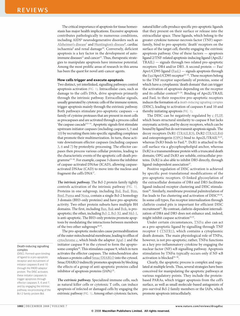
Death-inducing signalling complex (DISC). Formed upon binding of ligand to a pro-apoptotic receptor and recruitment of initiator caspases 8 and 10 through the FADD adaptor protein. The DISC activates these initiator caspases to trigger apoptosis through effector caspases 3, 6 and 7, and by engaging the intrinsic pathway via processing of the Bcl-2 family protein Bid.
The critical importance of apoptosis for tissue homeo-stasis has major health implications. Excessive apoptosis contributes pathologically to numerous conditions, including AIDS6 neurodegenerative disorders such as Alzheimer’s disease7 and Huntington’s disease8, cardiac ischaemia9 and renal damage10. Conversely, deficient apoptosis is a key factor in the development of auto-immune diseases11 and cancer12. Thus, therapeutic strate-gies to manipulate apoptosis have immense potential. Among the most prolific areas of research in this arena has been the quest for novel anti-cancer agents.
How cells trigger and execute apoptosisTwo distinct, yet interlinked, signalling pathways control apoptosis activation (FIG. 1). Intracellular cues, such as damage to the cell’s DNA, drive apoptosis primarily through the intrinsic pathway. Extracellular signals, usually generated by cytotoxic cells of the immune system, trigger apoptosis mainly through the extrinsic pathway. Both pathways stimulate pro-apoptotic caspases —a family of cysteine proteases that are present in most cells as procaspases and are activated through a process called ‘the caspase cascade’13–15. Apoptotic signals first stimulate upstream initiator caspases (including caspases 8, 9 and 10) by recruiting them into specific signalling complexes that promote their multimerization. In turn, these acti-vate downstream effector caspases (including caspases 3, 6 and 7) by proteolytic processing. The effector cas-pases then process various cellular proteins, leading to the characteristic events of the apoptotic cell-death pro-gramme13–16. For example, caspase 3 cleaves the inhibitor of caspase-activated DNAse (ICAD), allowing caspase-activated DNAse (CAD) to move into the nucleus and fragment the cell’s DNA17.
The intrinsic pathway. The Bcl-2 protein family tightly controls activation of the intrinsic pathway (FIG. 1). Proteins in one subgroup, including Bid, Bad, Bim, Bmf, Puma and Noxa, contain a single Bcl-2 homology 3 domain (BH3-only proteins) and have pro-apoptotic activity. Two other protein subsets have multiple BH domains. The first, including Bax, Bak and Bok, is pro-apoptotic; the other, including Bcl-2, Bcl-XL and Mcl-1, is anti-apoptotic. The BH3-only proteins promote apop-tosis by modulating the interactions between members of the two other subgroups18,19.
The pro-apoptotic molecules cause permeabilization of the outer mitochondrial membrane, leading to efflux of cytochrome c, which binds the adaptor Apaf-1 and the initiator caspase 9 in the cytosol to form the apopto-some complex20. This stimulates caspase 9, which in turn activates the effector caspases. The mitochondrion also releases a protein called Smac/DIABLO into the cytosol. Smac/DIABLO indirectly promotes apoptosis by blocking the effects of a group of anti-apoptotic proteins called inhibitor of apoptosis proteins (IAPs) 21.
The extrinsic pathway. Specialized immune cells, such as natural killer cells or cytotoxic T cells, can induce apoptosis of infected or damaged cells by engaging the extrinsic pathway (FIG. 1). Among other cytotoxic factors,
natural killer cells produce specific pro-apoptotic ligands that they present on their surface or release into the extra cell ular space. These ligands, which belong to the greater cytokine tumour-necrosis factor (TNF) super-family, bind to pro-apoptotic ‘death’ receptors on the surface of the target cell, thereby engaging the extrinsic apoptosis pathway. One of these factors — apoptosis ligand 2/TNF-related apoptosis-inducing ligand (Apo2L/TRAIL) — signals through two related pro-apoptotic receptors: DR4 and/or DR5. A second protein — Fas/Apo1/CD95 ligand (FasL) — signals apoptosis through the Fas/Apo1/CD95 receptor22–24. These receptors belong to the TNF receptor superfamily of proteins, some of which have a cytoplasmic ‘death domain’ that can trigger the activation of apoptosis depending on the receptor and its cellular context22–24. Binding of Apo2L/TRAIL and FasL to their respective pro-apoptotic receptors induces the formation of a death-inducing signalling complex (DISC), leading to activation of caspases 8 and 10 and thereby initiating apoptosis (FIG. 1).
The DISC can be negatively regulated by c-FLIP, which bears structural similarity to caspase 8 but lacks enzymatic activity, and by decoy receptors, which can be bound by ligand but do not transmit apoptosis signals. The decoy receptors DcR1 (TRAILR3), DcR2 (TRAILR4) and osteoprotegerin (OPG) bind to Apo2L/TRAIL22,25, whereas DcR3 binds to FasL26. DcR1 is attached to the cell surface via a glycophospholipid anchor, whereas DcR2 is a transmembrane protein with a truncated death domain; OPG and DcR3 are soluble, extracellular pro-teins. DcR2 is also able to inhibit DR5 directly, through ligand-independent association27.
Positive regulation of DISC activation is achieved by specific post-translational modifications of the pro-apoptotic receptors. O-linked glycosylation of the extracellular domains of DR4 and DR5 facilitates ligand-induced receptor clustering and DISC stimula-tion28. Similarly, membrane proximal palmitoylation of Fas leads to Fas clustering and activation of DISC29,30. In some cell types, Fas receptor internalization through clathrin-coated pits is important for efficient DISC recruitment31. By contrast, clathrin-dependent internali-zation of DR4 and DR5 does not enhance and, indeed, might inhibit caspase activation32,33.
Under certain circumstances, TNFa also can act as a pro-apoptotic ligand by signalling through TNF receptor 1 (TNFR1), which contains a cytoplasmic death domain. The main physiological role of TNFa, however, is not pro-apoptotic; rather, TNFa functions as a key pro-inflammatory cytokine by engaging the nuclear factor (NF)-kB signalling pathway. Apoptosis stimulation by TNFa typically occurs only if NF-kB activation is blocked34–36.
Clearly, the apoptotic process is complex and regu-lated at multiple levels. Thus, several strategies have been conceived for manipulating the apoptotic pathways at various regulatory points. They include the protein-based PARAs, which trigger apoptosis from the cell’s surface, as well as small molecule-based antagonists of pro-survival Bcl-2 family members or the IAPs, which promote apoptosis intracellularly.
R E V I E W S
1002 | DECEMBER 2008 | vOLUME 7 www.nature.com/reviews/drugdisc
R E V I E W S
1002 | ADvANCE ONLINE PUBLICATION www.nature.com/reviews/drugdisc
R E V I E W S
© 2008 Macmillan Publishers Limited. All rights reserved.

Nature Reviews | Drug Discovery
Extrinsicpathway
DNA damage
Intrinsicpathway
Apo2L/TRAIL
Puma, Noxa
Bax, Bak
Cytochrome c
Apaf-1
Smac/DIABLO
IAP
Bid
Caspase 8,10
Bcl-2, Bcl-XL, Mcl-1
Caspase 3,6,7
Apoptosis
Mitochondria
p53
Bad
PI3KAkt
Growth factorreceptorsChemotherapy
Radiotherapy
Caspase 9Apoptosome
FADD
DR4/DR5
Procaspase 8,10
DISCc-FLIP
Apoptosis activation as a cancer therapyOne of the most attractive features of apoptosis activa-tion as an anti-cancer approach is its potential to induce tumour regression rather than simply to stop tumour growth. On the flip side, pro-apoptotic agents that cannot discriminate between malignant and normal cells carry a significant risk of side effects. This is an important issue with traditional cancer treatments: radiotherapy and chemotherapeutics induce apoptosis only as a sec-ondary effect of the damage they cause to vital cellular components. These treatments affect most proliferating cells without distinction between malignant or normal types.
Oncogenic transformation drives uncontrolled cell proliferation, leading to misalignment of cell-cycle checkpoints, DNA damage and metabolic stress12,37,38. These aberrations veer tumour cells towards an apoptotic path; indeed, most transformed cells die by apoptosis. However, rare cells with additional mutations that enable apoptosis evasion can survive and progress to give rise to malignant tumours. Although such cells might man-age to escape the apoptosis signals that are activated by their aberrant phenotype, they remain ‘primed’ to die. These aberrant cells often retain the capacity to execute apoptosis if triggered through mechanisms that can overcome their anti-apoptotic mutations. Recent studies suggest that it might be possible to leverage the apoptotic priming of cancer cells by targeting certain key compo-nents of the apoptosis pathways directly and selectively (see below).
How do malignant cells usually evade apoptosis? One of the most widely mutated genes in human cancers is p53 — a pivotal stimulator of the intrinsic apoptosis pathway in response to severe cellular stresses such as DNA damage and hypoxia39–41 (FIG. 1). Inactivating p53 mutations occur in over 50% of cancers and further p53 inhibition in some cancers is mediated by genomic amplification of the E3 ubiquitin ligase HDM2, which promotes ubiquitination and degradation of p53.
Although less frequent, direct impairment of the apoptosis signalling cascades also occurs in cancer25,42–45. Rare mutations in components of the extrinsic path-way, such as DR5 (REF. 46), and epigenetic repression of caspase 8 have been reported47. Inactivation of the intrinsic pathway occurs more often, possibly because it has greater involvement in the initial elimination of oncogene-transformed cells4,12,48. Examples include a chromosome translocation that drives Bcl-2 overexpres-sion in follicular lymphomas49, inactivating mutations in phosphatidylinositide-3-kinase (PI3K) or Akt in sev-eral malignancies50, and mutational silencing of Bax in tumours with DNA mismatch repair deficiency51.
Targeting pro-apoptotic receptors in cancerSeveral considerations make the extrinsic pathway uniquely attractive as a drug-development target. The pro-apoptotic receptors are widely expressed in cancers, so PARAs might have a broad spectrum of anti-cancer activity. Furthermore, prevalent oncogenes such as Myc or Ras appear to increase tumour sensitivity to the extrinsic pathway, in part by facilitating its crosstalk with
Figure 1 | Key steps in apoptotic signalling pathways. Intrinsic pathway: cellular stress activates the p53 tumour‑suppressor protein. p53 initiates the intrinsic pathway by upregulating Puma and Noxa, which in turn activate Bax and Bak169,170. By contrast, the kinase Akt, which acts downstream of many growth factors, inhibits the intrinsic pathway by phosphorylating Bad171. Bax and Bak permeabilize the outer mitochondrial membrane, resulting in efflux of cytochrome c, which binds to the adaptor Apaf‑1 to recruit the initiator procaspase 9 into a signalling complex termed the apoptosome. Activated caspase 9 then cleaves and activates the effector caspases 3, 6 and 7 to trigger apoptosis16. The mitochondrial protein Smac/DIABLO augments apoptosis by binding inhibitor of apoptosis proteins (IAP) and reversing their grip on several caspases17. Extrinsic pathway: cytotoxic immune cells produce pro‑apoptotic ligands such as Apo2L/TRAIL. The homotrimeric ligand binds to the pro‑apoptotic receptors DR4 and/or DR5 on the surface of a target cell. Ligand binding induces receptor clustering and recruitment of the adaptor protein Fas‑associated death domain (FADD) and the initiator caspases 8 and 10 as pro‑caspases, forming a death‑inducing signalling complex (DISC). This triggers activation of the apical caspases, driving their autocatalytic processing and release into the cytoplasm, where they activate the effector caspases 3, 6 and 7. DISC formation is modulated by several inhibitory mechanisms, including c‑FLIP, which associates with the DISC by interacting with FADD to block initiator caspase activation172,173 and decoy receptors, which can block ligand binding or directly interfere with receptor activation (see text). Cross‑talk between the pathways: although the extrinsic and intrinsic pathways can function separately, they often interact. p53 mainly stimulates the intrinsic pathway, but it also upregulates some of the pro‑apoptotic receptors such as DR5 and augments extrinsic signalling174. Extrinsic‑pathway activation leads to caspase 8‑mediated processing of Bid; truncated Bid subsequently stimulates Bax and Bak to engage the intrinsic pathway175,176. The effector caspases can then feed back to promote further caspase 8 processing. In so‑called Type I cells stimulation of the extrinsic pathway is sufficient to drive apoptosis, whereas in Type II cells, further amplification by Bid and the intrinsic pathway is required for commitment to apoptosis39,176.
R E V I E W S
NATURE REvIEwS | drug discovery vOLUME 7 | DECEMBER 2008 | 1003
R E V I E W S
NATURE REvIEwS | drug discovery ADvANCE ONLINE PUBLICATION | 1003
f o c u S o n a p o p to S I S
© 2008 Macmillan Publishers Limited. All rights reserved.

AnoikisA type of apoptosis induced by cell detachment.
the intrinsic pathway12,38,52. Importantly, pro-apoptotic receptors activate caspases independently of p53, pro-viding a strategy to kill tumour cells regardless of their p53 status. By contrast, traditional therapies depend on p53 for effective apoptosis induction, and the frequent loss of p53 function causes significant resistance to such treatments39,40,53.
Indeed, PARAs could be useful not only for mono-therapy against tumours that are particularly sensitive to their action, but also more broadly, in combinatorial regimens with traditional or other therapies. Such com-binations might help overcome tumour resistance caused by p53 mutations (and possibly by other mechanisms). PARAs may cooperate with targeted agents that inhibit growth factor receptors or their downstream signalling pathways by simultaneously activating pro-apoptotic receptors and inhibiting pro-survival mechanisms54–57. PARAs might also interact positively with anti-angiogenic therapies and with therapeutic antibodies that work by recruiting immune effector cells against tumours, such as rituximab58. Finally, PARAs combined with other kinds of pro-apoptotic agents could prove to be particu-larly effective. For example, Bcl-2 family antagonists can cooperate with PARAs by enhancing apoptosis-pathway crosstalk, whereas IAP antagonists might synergize by augmenting caspase activation58–61.
Although these considerations could apply in general to all pro-apoptotic receptors, the receptors do have dif-ferent properties. One key difference relates to the degree of selectivity for tumour versus normal cells associated with activation of different pro-apoptotic receptors. For instance, the pro-inflammatory effects of TNFa on normal tissues have significantly hampered the clinical development of TNFa-based treatments for systemic therapy62. Nevertheless, TNFa has been used success-fully to treat soft tissue sarcomas of the extremities by isolated limb perfusion63. Agonistic antibodies to Fas23,64
or leucine-zipper-tagged FasL65 were deemed unsuitable for clinical investigation because they caused massive hepatocyte apoptosis and lethal liver damage in animal models. However, a novel FasL fusion protein based on a collagen trimerization platform might be better tolerated and is currently in early clinical safety evaluation.
Despite some initial concerns about potential for hepatotoxicity, PARAs targeting DR4 or DR5 have been well tolerated in preclinical safety models and in Phase I clinical safety trials (see below). Indeed, although DR4 and DR5 are expressed both in malignant and in nor-mal tissues, PARAs targeting these receptors appear to induce apoptosis selectively in tumour cells while sparing most normal cells4,38,66,67. This selectivity is not yet fully understood, although it might relate to multiple fac-tors. These include apoptotic priming of cancer cells by oncogenes, tumour overexpression of specific O-glycosyl transferase enzymes that hyperglycosylate DR4 and DR5 and thereby enhance their pro-apoptotic activity30, and differential decoy receptor expression in tumours versus normal tissues. It has been suggested that DR4 and DR5 are expressed at higher levels in tumours compared with normal tissues68, but this requires further quantitative examination.
Approaches to activate pro-apoptotic receptorsThe endogenous Apo2L/TRAIL molecule was dis-covered independently in the mid-1990s in my labora-tory at Genentech69 and in Ray Goodwin’s laboratory at Immunex70. Subsequently, a series of receptors that bind to this ligand, namely, DR4, DR5, DcR1 and DcR2, were identified22. Loss-of-function studies in mice sug-gest that endogenous Apo2L/TRAIL is important for the ability of certain types of natural killer cells to block tumour growth and metastasis71–73. More recent findings suggest that Apo2L/TRAIL also has a role in modulating memory T-cells74–76 and in anoikis77–79. Mice have only one pro-apoptotic receptor for Apo2L/TRAIL (mouse DR5 or TRAILR2). In DR5-knockout mice certain immune functions are enhanced and the sensitivity to radiation-induced apoptosis is diminished80,81. Transcription of the Apo2L/TRAIL gene is upregulated by interferons, which is consistent with its apparent involvement in innate immunity82,83, and p53 increases DR5 expres-sion, which is in keeping with DR5’s contribution to radiosensitivity84.
Since the discovery of Apo2L/TRAIL and its recep-tors, two classes of PARAs that target DR4 and/or DR5 have been developed: recombinant human (rh)Apo2L/TRAIL25,67,85, which activates both receptors, and mono-clonal agonistic antibodies, which activate either DR4 or DR5 (REF. 86–94) (FIG. 2).
Immune cells express endogenous Apo2L/TRAIL as a type II transmembrane polypeptide of 281 amino acids; the extracellular region of the protein is sometimes shed by immune cells to release a soluble ligand69,70,76,95. The first PARAs to target DR4 and DR5 were based on recom-binant versions of the soluble ligand. Several variants were generated for preclinical studies, including a non-tagged protein85, and versions with exogenous polypep-tide tags65,69,96–98. The tags are typically added to enable artificial crosslinking of the protein with antibodies, to aid with purification, or to stabilize the homotrimeric structure. Our group has developed an optimized, clinical-grade version of the protein called recombinant human Apo2L/TRAIL (rhApo2L/TRAIL)85.
RhApo2L/TRAIL comprises amino acids 114–281 of the endogenous molecule, without any added tag. It is produced in Escherichia coli. Optimization of rhApo2L/TRAIL was facilitated by X-ray crystallographic studies, which revealed a homotrimeric structure99,100 (FIG. 3A). A crucial discovery was the observation that a zinc mol ecule coordinates the sulphydryl groups of three unpaired cysteines, located at position 230 of each sub-unit. Addition of zinc to the bacterial cell culture media and to purification buffers during recombinant protein production enabled nearly stoichiometric zinc coordina-tion, thereby stabilizing the trimeric protein’s structure and maintaining its solubility 85,100. Certain normal cell types, including hepatocytes and keratinocytes, can dis-play apoptotic sensitivity to the tagged, non-optimized versions of this ligand, but not to the non-tagged, zinc-optimized rhApo2L/TRAIL molecule101,102. Further studies comparing tagged and non-tagged variants have verified this observation103,104. Normal cells might require a higher degree of receptor crosslinking to trigger
R E V I E W S
1004 | DECEMBER 2008 | vOLUME 7 www.nature.com/reviews/drugdisc
R E V I E W S
1004 | ADvANCE ONLINE PUBLICATION www.nature.com/reviews/drugdisc
R E V I E W S
© 2008 Macmillan Publishers Limited. All rights reserved.

Nature Reviews | Drug Discovery
Caspases
Apoptosis
DR4DR4DR5 DR5
DR4 agonistantibodyMapatumumab
Recombinant human ligandrhApo2L/TRAIL
DR5 agonistantibodiesLexatumumabApomabAMG655CS-1008LBY-135
Tumour cell
EpitopeThe site on a large molecule to which an antibody binds.
apoptosis, and such effects may occur with the non-optimized variants, which tend to aggregate25,101. X-ray crystallographic analysis of the complex between rhApo2L/TRAIL and the extracellular portion of DR5 revealed that the ligand binds three receptor molecules, with each ligand subunit contacting two receptors, through cysteine-rich domain (CRD) 2 and CRD3 of DR5 (FIG. 3A).
The second PARA approach has been based on mono-clonal antibodies that display agonistic activity towards DR4 or DR5. Detailed information was recently pub-lished for one of the DR5 antibodies, Apomab, which was developed by our group105. An antibody was isolated from a human antibody phage-display library, as a rare single-chain Fv fragment that exhibited selective pro-apoptotic activity against DR5-expressing cancer cells; it was further engineered to optimize affinity, potency and structural stability, resulting in the antibody dubbed Apomab. Crystallographic analysis of the complex between Apomab and DR5 revealed that Apomab’s binding site has significant overlap with that of rhApo2L/TRAIL, con-tacting CRD2 and CRD3 on DR5 (REF. 105,106) (FIG. 3b). However, the ligand-binding site of rhApo2L/TRAIL con-sists of two separate patches, whereas the Apomab epitope is more continuous. In contrast to Apomab, another DR5 antibody, BDF1, which has little or no agonist activity, interacts with CRD2 but not CRD3 (REF. 105).
The rhApo2L/TRAIL molecule has the same trimeric subunit structure as the endogenous ligand and therefore probably mimics the natural mode of receptor engage-ment. Precisely how homodimeric agonist antibodies
activate DR4 or DR5 is not yet fully understood. Because Apomab’s single-chain Fv fragment has agonistic activity, it is possible that its epitope, which overlaps the ligand-binding site, enables the antibody to confer or stabilize a DR5 conformation that is more conducive to activation (FIG. 4a). Comparisons of crystallographic structures of DR5 in complex with ligand or Fab fragments of Apomab or other DR5 antibodies indicate that the orientation of CRD3 with respect to CRD1 and CRD2 has significant conformational diversity (FIG. 3c). Thus, the membrane proximal CRD3 region might be dynamic in vivo and could be stabilized by some agonists in a specific arrange-ment that promotes DR5 activation105. Moreover, a bivalent antibody might bring together two or more pre-associated receptor trimers, augmenting DISC recruitment (FIG. 4b). Crosslinking of the Fc region of DR4 or DR5 agonistic antibodies usually enhances apoptotic signalling (as does ligand crosslinking), possibly by promoting further receptor clustering and DISC activation105 (FIG. 4c).
Like rhApo2L/TRAIL, certain optimized agonistic DR4 and DR5 antibody preparations, provided they are stable and do not contain significant levels of aggregates, have been shown to induce apoptosis in various cancer cell lines but not in most normal cells. Several anti-bodies are currently in Phase II clinical trials, including mapatumumab (Human Genome Sciences; HGS), which targets DR4 (REF. 92), as well as lexatumumab (HGS)107, Apomab (Genentech)105, AMG655 (Amgen)106, CS-1008 (Daiichi) 107 and LBY-135 (Novartis)108, all of which target DR5 (TABLE 1).
Ligand-based pro-apoptotic receptor agonismPreclinical data with rhApo2L/TRAIL. The first pre-clinical data for the soluble, non-tagged rhApo2L/TRAIL were published in 1999 (REFS 85,107). Anti-tumour activity of the recombinant ligand was evaluated in vitro using multiple cell lines derived from human colon, lung, breast, central nervous system, kidney and skin cancer. Exposure to rhApo2L/TRAIL induced apoptosis in 16 of 39 cell lines, but not in several normal cell types. Subsequently, rhApo2L/TRAIL or other variants were shown to induce apoptosis in malignant cell lines from the prostate106,109, thyroid110 and pancreas111, as well as in leukaemia112, mul-tiple myeloma113 and non-Hodgkin’s lymphoma (NHL) cell lines114. In vivo, rhApo2L/TRAIL has been shown to exert marked anti-tumour activity in mouse xenograft models of human cancer derived from colon22,115, lung116 and pancreatic117 carcinomas, multiple myeloma113, NHL114 and glioma118,119. Similar activity was observed with a leucine-zipper-tagged version in a breast cancer xenograft model65.
Numerous preclinical experiments in various cell lines and xenograft models have shown that co-administration of rhApo2L/TRAIL and various chemotherapeutics, including anti-metabolites, topoisomerase inhibitors and other DNA damaging agents, as well as microtubule targeting drugs, often leads to additive or synergistic activity85,86,116–138. Similarly, positive inter actions have been observed upon co-administration of rhApo2L/TRAIL with agents that target other points in the apoptosis pathway; for example, Bcl-2 antagonists61
Figure 2 | Pro-apoptotic receptor agonists. The two types of pro‑apoptotic receptor agonists discussed in this Review include ligand‑based and antibody‑based protein agents, which induce apoptosis via receptor‑mediated activation of the caspase cascade. Recombinant human (rh) Apo2L/TRAIL interacts with the death‑inducing receptors DR4 and/or DR5, whereas the monoclonal agonist antibodies are monospecific for either DR4 or DR5.
R E V I E W S
NATURE REvIEwS | drug discovery vOLUME 7 | DECEMBER 2008 | 1005
R E V I E W S
NATURE REvIEwS | drug discovery ADvANCE ONLINE PUBLICATION | 1005
f o c u S o n a p o p to S I S
© 2008 Macmillan Publishers Limited. All rights reserved.

Nature Reviews | Drug Discovery
a b c
DR5rhApo2L/TRAIL
Apomab
CRD1
CRD2
CRD3
rhApo2L/TRAILDR5
DR5DR5
DR5DR5
Zn2+
CRD1
CRD2
CRD3
Antibody-dependent cell-mediated cytotoxicityRefers to the lysis of antibody-coated target cells by immune cells.
Complement activationRefers to the sequential activation of serum proteins, resulting in an inflammatory response.
and IAP-blocking Smac/DIABLO mimetics58,59,139. Cooperation was noted between rhApo2L/TRAIL and inhibitors of the proteasome140–142, and between rhApo2L/TRAIL and histone deacetylase inhibitors143. Although the mechanisms underlying such interactions are diverse, it is likely that combinatorial stress triggers apoptosis more effectively than singular insults and that this outcome is uniquely facilitated by the underlying aberrant pheno-type of malignant cells. Another promising approach is the combination of rhApo2L/TRAIL with the anti-CD20 antibody rituximab. Rituximab kills NHL B cells prima-rily via antibody-dependent cell-mediated cytotoxicity and complement activation; thus, its anti-tumour synergy with rhApo2L/TRAIL is achieved through integration of non-apoptotic and pro-apoptotic cell death mechanisms114.
The cynomolgus monkey equivalent of rhApo2L/TRAIL has been tested extensively in this nonhuman pri-mate. There was no detectable toxicity to the liver or any other major organs and tissues in these studies68,85,101.
Predicting tumour sensitivity to rhApo2L/TRAIL. Because cancer cells are not universally sensitive to rhApo2L/TRAIL, there has been extensive research to identify tumour biomarkers that might predict sensitivity to this PARA. Although at least some expression of DR4 or DR5 is necessary for responsiveness, sensitivity can not be predicted from the levels of these receptors or the decoy receptors25,28,97,144. However, a number of potential biomar-kers have been identified. Of these, the most robust predic-tors of sensitivity across different cancer types are certain O-glycosylation enzymes that were identified by testing a panel of 119 cancer cell lines28 (FIG. 5). In 83 pancre-atic, non-small cell lung cancer (NSCLC) and malignant melanoma cell lines, high mRNA expression of GALNT14 strongly correlated with sensitivity to rhApo2L/TRAIL (p = 9×10⁻⁶). In 36 colorectal cancer cell (CRC) lines, high GALNT3, FUT3 and FUT6 mRNA levels signifi-cantly correlated with sensitivity (p = 0.026, 0.0013 and
0.01, respectively). A direct functional link between the activity of these enzymes, O-glycosylation of DR4 and DR5 and responsiveness to rhApo2L/TRAIL pro-vided further validation. Other parameters that might affect tumour responsiveness to PARAs include muta-tions in caspase 8 (REF. 145) or Bax146 and altered expres-sion of certain anti-apoptotic proteins147–149, pro-apoptotic proteins150, Myc151, or Raf-1 kinase inhibitor protein (RKIP)152. Recent data suggest that breast-cancer cell lines lacking expression of oestrogen, progesterone and HER2 receptors, and displaying a mesenchymal phenotype, are particularly sensitive to rhApo2L/TRAIL153.
Early clinical data with rhApo2L/TRAIL. Preliminary results from Phase I studies with rhApo2L/TRAIL have been reported154,155 (TABLE 1). The single-agent Phase Ia study enrolled patients with advanced solid tumours or NHL and was designed to evaluate the safety and pharmaco kinetics of rhApo2L/TRAIL154. It comprised a stepwise, staggered dose-escalation phase in two patient groups (those without or with hepatic metastases), fol-lowed by an expansion phase in which additional subjects were treated at a dose of 8 mg per kg. A total of 71 patients were treated with rhApo2L/TRAIL at doses ranging from 0.5 to 30 mg per kg per day. Each treatment cycle com-prised consecutive, daily infusions of rhApo2L/TRAIL for 5 days and cycles were repeated every 21 days. No drug-related dose-limiting toxicity (DLT) was observed and a maximum tolerated dose (MTD) was not reached during the dose escalation phase. RhApo2L/TRAIL dem-onstrated linear pharmacokinetics, with an estimated effective half-life of 0.6 hours. There were no pharmaco-kinetic differences in patients with or without hepatic metastases. Notably, one patient with chondrosarcoma exhibited a confirmed, partial response (PR) by Response Evaluation Criteria in Solid Tumours (RECIST) with > 80% tumour regression and has continued on rhApo2L/TRAIL therapy for over 2.5 years.
Figure 3 | crystal structures of pro-apoptotic receptor agonists and dr5. a | The complex between rhApo2L/TRAIL and the extracellular domains of three DR5 molecules. The ligand is shown as a ribbon diagram with connecting loops as lines; each colour represents one of the three subunits. The zinc atom that coordinates the sulphydryl groups of three unpaired cysteines, located at position 230 of each subunit, is shown in purple. Each receptor molecule is depicted as a space‑filling model (white), with blue lines indicating the polypeptide backbone and blue and yellow balls indicating disulphide linkages of the cysteine‑rich domains (CRDs). There is one disulphide bond in CRD1 and three in CRD2 and CRD3 (Protein Data Bank identifier code 1D0G). b | Comparison of the interaction sites between rhApo2L/TRAIL (blue) or the Fab fragment of Apomab (red) and DR5 (yellow). c | Conformational orientation of the DR5 CRDs in complexes with ligand or various antibodies.
R E V I E W S
1006 | DECEMBER 2008 | vOLUME 7 www.nature.com/reviews/drugdisc
R E V I E W S
1006 | ADvANCE ONLINE PUBLICATION www.nature.com/reviews/drugdisc
R E V I E W S
© 2008 Macmillan Publishers Limited. All rights reserved.

Nature Reviews | Drug Discovery
a b c
DISC DISCDISC DISC DISCDISC DISC DISC
DR4DR5
Agonistantibody
Crosslinkingmolecule
Phase Ib studies of rhApo2L/TRAIL in relapsed, low-grade NHL and in NSCLC have been completed. In the NHL study, the combination of rituximab and rhApo2L/TRAIL seemed to be well tolerated; with few exceptions, adverse events were mild to moderate and no maximally tolerated dose of rhApo2L/TRAIL with rituximab was reached. The treatment yielded a radiographic response rate of 55%, with three complete responses (CRs) and three PRs in 11 of the evaluable patients that were treated155.
In the NSCLC study, the combination of rhApo2L/TRAIL with carboplatin, paclitaxel and bevacizumab seemed to be well tolerated and was associated with a safety profile that is consistent with the latter three agents; no DLTs were observed156. The overall tumour response rate was 56% (1 CR and 9 PRs in 18 patients in the first three cohorts). Randomized Phase II trials based on these Phase Ib studies are in progress.
Pro-apoptotic receptor agonism by antibodiesPreclinical data. Preclinical efficacy data for DR4 and DR5 antibody-based PARAs were published in 2001 (REFS 86,89). Studies with other antibodies were sub-sequently reported87,88,90–94. The DR4 agonist antibody mapatumumab was effective in preclinical tests and interacted positively with chemotherapeutic agents in vitro and in vivo92. A detailed structural and func-tional characterization of the DR5 agonist antibody Apomab has been published recently104. Apomab was shown to induce apoptosis in various cancer cell lines. It showed intrinsic pro-apoptotic activity, which could be further augmented by Fc crosslinking and displayed potent anti-tumour activity in xenograft models of lung, colon and pancreatic cancer. Apomab cooperated with camptothecin or gemcitabine against colon or pancrea-tic tumour xenografts. It had no effect on cultured hepatocytes, even upon Fc crosslinking, and was well tolerated in cynomolgus monkeys. Recent data have demonstrated significant cooperation of Apomab with carboplatin–paclitaxel in an orthotopic xenograft model of NSCLC157.
Clinical data. Phase I studies with mapatumumab (sponsored by Human Genome Sciences) were under-taken in patients with advanced solid tumours . Stable disease was reported in 19 (39%) patients in one Phase Ia study93, and in 12 (29%) patients in another158. In the former trial, two patients had elevated liver func-tion tests that were probably related to mapatumumab93. In other Phase I studies in patients with advanced solid tumours, mapatumumab was given in combination with chemotherapy (paclitaxel plus carboplatin, and gemcitabine plus cisplatin), yielding a PR in six (21%) patients159 and nine (20%) patients160, respectively (L. Q. Chow et al. personal communication), and stable dis-ease (SD) in 13 (46%) patients and 13 (29%) patients, respectively. In Phase II studies involving patients with relapsed/refractory NHL, CRC or NSCLC, SD was achieved in 9–12 patients (29–34%) receiving mapatu-mumab161–163. One NHL patient had a CR and two other NHL patients had a PR161. Mapatumumab mono-therapy was generally well tolerated in the Phase II set-ting, with only one drug-related serious adverse event (vomiting).
Two Phase Ia monotherapy studies91,164 and one combination study165 have been reported for lexatumu-mab. Several patients experienced SD with single-agent lexatumumab91,164 and a patient with chemotherapy-refractive Hodgkin’s disease had abdominal tumour regression164. In one study, there was DLT, including asymptomatic elevations of serum amylase, transaminases and bilirubin91. In the Phase Ib study, lexatumumab was given with gemcitabine–FOLFIRI (folinic acid, fluoro-u racil and irinotecan) or pemetrexed–doxorubicin. Three of the 41 participating patients achieved a PR165. Data on serious adverse events in this study have yet to be published.
Preliminary Phase Ia data are available for Apomab166.
In this study, patients with advanced, treatment-refractory solid tumours received up to eight cycles of intravenous Apomab. A dose-escalation phase ranging from 0.5 to 20 mg per kg was followed by an expansion phase in which additional subjects were treated at 10 mg per kg every 2 weeks. Preliminary safety data from 37 of the 50 enrolled patients suggested that Apomab was gener-ally safe, with no MTD reached; there was one report of reversible, dose-limiting transaminitis and one report of pulmonary embolism. Of the 23 subjects evaluated at the time of reporting, one patient with ovarian can-cer metastatic to the liver had 23% tumour shrinkage at cycle 8 and remained stable 10 months after treatment completion, and one patient with CRC metastatic to the lungs had 28% tumour shrinkage at cycle 8.
Preliminary results of a Phase Ia study with AMG655 in patients with advanced solid tumours have indicated that one (5%) patient with NSCLC achieved a PR and 10 (48%) patients had stable disease (SD)108.
The first clinical trial data for the DR5 antibodies, LBY-135 and CS-1008, were reported recently167,168. For LBY-135, which is the only antibody PARA that is chimeric rather than fully human, monotherapy was compared with combination treatment with capecitabine in patients with advanced solid tumours167. In patients
Figure 4 | Potential modes of pro-apoptotic receptor activation by agonistic antibodies. a | Depending on the epitope, an agonistic antibody might mimic the stimulatory effect of Apo2L/TRAIL on DR4 or DR5 by inducing or stabilizing an active conformation, thereby recruiting the death‑inducing signalling complex (DISC). b | As a bivalent molecule, an agonistic antibody that binds to a suitable epitope also may bring together pre‑associated receptors productively to promote DISC assembly. c | Crosslinking of the antibody by its Fc portion may further augment DISC activation.
R E V I E W S
NATURE REvIEwS | drug discovery vOLUME 7 | DECEMBER 2008 | 1007
R E V I E W S
NATURE REvIEwS | drug discovery ADvANCE ONLINE PUBLICATION | 1007
f o c u S o n a p o p to S I S
© 2008 Macmillan Publishers Limited. All rights reserved.

treated with monotherapy, there was one minor response (3%), and in those on combination therapy, there was one PR (4%) and one metabolic PR (4%). There were no DLTs in the monotherapy group, two DLTs in the combination group and immunogenicity was detected in 13 out of 51 (25%) patients. In the CS-1008 study, 17 patients with relapsed/refractory solid tumours or lym-phomas were treated with monotherapy, and 7 (41%) patients achieved SD. No DLTs were observed168.
Conclusions and future directionsTargeted pro-apoptotic cancer therapies have tremen-dous potential to help regress tumours more safely and effectively than conventional treatments. The main rationale for this approach is based on the concept that although cancer cells acquire specific anti-apoptotic mutations in the course of their malignant transforma-tion, they remain primed for apoptosis because of their underlying aberrant phenotype. Agents that can bypass
Table 1 | Clinical studies with pro-apoptotic receptor agonists
Phase Treatment Patients references
rhApo2L/TRAIL (target receptors: DR4 and DR5)
Phase Ia Monotherapy Advanced solid tumours or NHL (n = 51) 154
Phase Ib/II + R Relapsed low‑grade NHL after previous R‑containing therapy (n = 105)
155 Ongoing
Phase Ib + IRI and CET Metastatic CRC (n = 12) Ongoing
Phase Ib + CAR, PAC and BEV Stage IIIb/IV NSCLC (n = 24) 156
Phase Ib/II + CAR, PAC and BEV Stage IIIb/IV NSCLC (n = 200) Ongoing
Mapatumumab (target receptor: DR4)
Phase Ia Monotherapy Advanced solid tumours (n = 49) 93
Phase Ia Monotherapy Advanced solid tumours (n = 41) 158
Phase Ib + PAC and CAR Advanced solid tumours (n = 28) 159
Phase Ib + GEM and CIS Advanced solid tumours (n = 49) 160
Phase II Monotherapy Relapsed/refractory NHL (n = 40) 161
Phase II Monotherapy Relapsed/refractory stage IIIb/IV or recurrent CRC (n = 38)
162
Phase II Monotherapy Relapsed/refractory stage IIIb/IV or recurrent NSCLC (n = 32)
163
Phase II + BOR Relapsed/refractory multiple myeloma (n = 100) Ongoing
Phase II + PAC and CAR Advanced NSCLC (n = 105) Ongoing
Apomab (target receptor: DR5)
Phase Ia Monotherapy Advanced treatment‑refractory solid tumours (n = 50) 165
Phase Ib + CET and IRI Metastatic CRC in patients who have progressed following, or cannot tolerate, first‑line therapy
Ongoing
Phase II + R NHL that has progressed after treatment with R (n = 80) Ongoing
Phase II + PAC, CAR and BEV Patients with previously untreated stage IIIb, IV or recurrent NSCLC (n = 120)
Ongoing
Lexatumumab (target receptor: DR5)
Phase Ia Monotherapy Advanced solid tumours (n = 37) 91
Phase Ia Monotherapy Advanced solid tumours and lymphoma (n = 31) 164
Phase Ib + GEM–FOLFIRI or pemetrexed–DOX
Range of cancer types (n = 41) 165
AMG655 (target receptor: DR5)
Phase Ia Monotherapy Advanced solid tumours (n = 22) 106
LBY-135 (target receptor: DR5)
Phase I Monotherapy versus + CAP
Advanced solid tumours (n = 56) 167
CS-1008 (target receptor: DR5)
Phase I Monotherapy Relapsed/refractory solid tumours or lymphomas 168
BEV, bevacizumab; BOR, bortezomib; CAP, capecitabine; CAR, carboplatin; CET, cetuximab; CIS, cisplatin; CRC, colorectal cancer; DOX, doxorubicin; FOLFIRI, folinic acid, fluorouracil and irinotecan; GEM, gemcitabine; IRI, irinotecan; NHL, non‑Hodgkin’s lymphoma; NSCLC, non‑small cell lung cancer; PAC, paclitaxel; rhApo2L/TRAIL, recombinant human Apo2L/TRAIL; R, rituximab.
R E V I E W S
1008 | DECEMBER 2008 | vOLUME 7 www.nature.com/reviews/drugdisc
R E V I E W S
1008 | ADvANCE ONLINE PUBLICATION www.nature.com/reviews/drugdisc
R E V I E W S
© 2008 Macmillan Publishers Limited. All rights reserved.

O-linkedsugars
Receptorclustering
Apo2L/TRAIL
DR4DR5 FADD
Nature Reviews | Drug Discovery
Caspase 8,10
Apoptosis
Caspase 3,6,7
Red = High expression
Blue = Low expression
Gene-expressionprofiling of cancer cell lines
SensitiveO-glycosylationenzymesGALNT14GALNT3FUT3FUT6
Resistant
Procaspase 8,10
a b
some of the more frequent anti-apoptotic mechanisms in cancers — most importantly, p53 inactivation — might lead to in major therapeutic advances. PARAs fit this pro-file well because the extrinsic pathway activates apoptosis independently of p53. Of the pro-apoptotic receptors that control this signalling pathway, DR4 and DR5 are particularly attractive targets for agonistic drugs, because they are expressed in multiple cancers and appear to be better primed for stimulation in malignant versus nor-mal cells. As discussed here, PARAs targeting DR4 and/or DR5 have shown promising safety and efficacy in diverse preclinical cancer models. Moreover, several of these agents have successfully met the rigorous safety cri-teria of Phase I clinical trials. Early-phase clinical studies in cancer are designed primarily to evaluate drug safety and pharmacokinetics, and are usually conducted with patients who have advanced disease that has become refractory to multiple prior therapies. Nonetheless, Phase I data for anti-cancer activity of PARAs have been encouraging. On that basis, randomized and controlled Phase II studies in multiple cancer types have been ini-tiated. Many of the Phase II trials are designed to assess the safety and efficacy of specific PARAs in combina-tion with various standard-of-care treatments, including several chemotherapeutics and certain targeted agents such as bortezomib in multiple myeloma, bevacizumab in NSCLC and rituximab in NHL.
How do PARAs compare to other pro-apoptotic agents? One key difference is that they are proteins as opposed to small molecule compounds. Furthermore, PARAs work from outside the cell, whereas the small
molecules work from within the cell. Finally, PARAs directly trigger apoptosis through receptor agonism, whereas the small molecules do so less directly, by antago nizing apoptosis inhibitors such as certain Bcl-2 proteins or IAPs. As such, PARAs probably have different pharmacokinetic, pharmacodynamic and mechanistic properties compared with other pro-apoptotic agents.
Another important, frequently asked question is how the PARAs compare with one another. Although there are several differences between the two PARA classes, it is difficult to predict what impact such differences might have on the tolerability and activity of these agents; this will doubtlessly require empirical testing and further clinical experience. Because of the distinct molecular size and composition, there is likely to be significant variation in pharmacokinetics and pharmacodynamics between rhApo2L/TRAIL (a 60 kDa homotrimer) and agonistic antibodies to DR4 and DR5 (~150 kDa homodimers). This could impact on the duration and frequency of tumour exposure, tumour penetration and effects on normal tissue. The recombinant ligand activates both DR4 and DR5, whereas the antibodies are monospecific. In certain tumours, either DR4 or DR5 might be rela-tively more responsive, so in some cases stimulation of both receptors might be beneficial. RhApo2L/TRAIL mimics more faithfully the endogenous mode of recep-tor engagement, whereas the antibodies possibly use a different molecular mechanism of agonism. whether interactions of rhApo2L/TRAIL, or pro-apoptotic recep-tors, with decoy receptors have significant therapeutic implications remains to be determined.
Figure 5 | identification of biomarkers that might predict tumour sensitivity to rhApo2L/TrAiL. a | O‑glycosylation of the extracellular domains of DR4 and DR5 facilitates ligand‑induced receptor clustering, death‑inducing signalling complex (DISC) recruitment and apoptosis initiation. b | Gene expression profiling of multiple cancer cell lines reveals that the levels of specific O‑glycosylation enzymes in various cancers strongly correlate with sensitivity to rhApo2L/TRAIL (representative panel shown). O‑glycosylation enzymes may be useful biomarkers to help predict tumour responsiveness to rhApo2L/TRAIL in different cancers and in individual patients. See also REF. 28.
R E V I E W S
NATURE REvIEwS | drug discovery vOLUME 7 | DECEMBER 2008 | 1009
R E V I E W S
NATURE REvIEwS | drug discovery ADvANCE ONLINE PUBLICATION | 1009
f o c u S o n a p o p to S I S
© 2008 Macmillan Publishers Limited. All rights reserved.

1. Jemal, A. et al. Cancer statistics, 2008. CA Cancer J. Clin. 58, 71–96 (2008).
2. Albreht, T., McKee, M., Alexe, D. M., Coleman, M. P. & Martin-Moreno, J. M. Making progress against cancer in Europe in 2008. Eur. J. Cancer 44, 1451–1456 (2008).
3. Chowdhury, I., Tharakan, B. & Bhat, G. K. Current concepts in apoptosis: the physiological suicide program revisited. Cell. Mol. Biol. Lett. 11, 506–525 (2006).
4. Elmore, S. Apoptosis: a review of programmed cell death. Toxicol. Pathol. 35, 495–516 (2007).
5. Gulbins, E., Jekle, A., Ferlinz, K., Grassme, H. & Lang, F. Physiology of apoptosis. Am. J. Physiol. Renal. Physiol. 279, F605–F615 (2000).
6. Casella, C. R. & Finkel, T. H. Mechanisms of lymphocyte killing by HIV. Curr. Opin. Hematol. 4, 24–31 (1997).
7. Rohn, T. T., Head, E., Nesse, W. H., Cotman, C. W. & Cribbs, D. H. Activation of caspase-8 in the Alzheimer’s disease brain. Neurobiol. Dis. 8, 1006–1016 (2001).
8. Sanchez Mejia, R. O. & Friedlander, R. M. Caspases in Huntington’s disease. Neuroscientist 7, 480–489 (2001).
9. Hayakawa, K. et al. Sensitivity to apoptosis signal, clearance rate, and ultrastructure of fas ligand-induced apoptosis in in vivo adult cardiac cells. Circulation 105, 3039–3045 (2002).
10. Singh, A. B., Kaushal, V., Megyesi, J. K., Shah, S. V. & Kaushal, G. P. Cloning and expression of rat caspase-6 and its localization in renal ischemia/reperfusion injury. Kidney Int. 62, 106–115 (2002).
11. Prasad, K. V. & Prabhakar, B. S. Apoptosis and autoimmune disorders. Autoimmunity 36, 323–330 (2003).
12. Gerl, R. & Vaux, D. L. Apoptosis in the development and treatment of cancer. Carcinogenesis 26, 263–270 (2005).
13. Fan, T. J., Han, L. H., Cong, R. S. & Liang, J. Caspase family proteases and apoptosis. Acta Biochim. Biophys. Sin. (Shanghai) 37, 719–727 (2005).
14. Lavrik, I. N., Golks, A. & Krammer, P. H. Caspases: pharmacological manipulation of cell death. J. Clin. Invest. 115, 2665–2672 (2005).
15. Thornberry, N. A. Caspases: a decade of death research. Cell Death Differ. 6, 1023–1027 (1999).
16. Boatright, K. M. et al. A unified model for apical caspase activation. Mol. Cell 11, 529–541 (2003).
17. Nagata, S. Apoptotic DNA fragmentation. Exp. Cell Res. 256, 12–18 (2000).
18. Coultas, L. & Strasser, A. The role of the Bcl-2 protein family in cancer. Semin. Cancer Biol. 13, 115–123 (2003).
19. Letai, A. Pharmacological manipulation of Bcl-2 family members to control cell death. J. Clin. Invest. 115, 2648–2655 (2005).
20. Chinnaiyan, A. M. The apoptosome: heart and soul of the cell death machine. Neoplasia 1, 5–15 (1999).
21. van Loo, G. et al. The role of mitochondrial factors in apoptosis: a Russian roulette with more than one bullet. Cell Death Differ. 9, 1031–1042 (2002).
22. Ashkenazi, A. & Dixit, V. M. Death receptors: signaling and modulation. Science 281, 1305–1308 (1998).
23. Nagata, S. Apoptosis by death factor. Cell 88, 355–365 (1997).
24. Peter, M. E. & Krammer, P. H. The CD95(APO-1/Fas) DISC and beyond. Cell Death Differ. 10, 26–35 (2003).
25. Ashkenazi, A. Targeting death and decoy receptors of the tumour-necrosis factor superfamily. Nature Rev. Cancer 2, 420–430 (2002).
26. Pitti, R. M. et al. Genomic amplification of a decoy receptor for Fas ligand in lung and colon cancer. Nature 396, 699–703 (1998).
27. Clancy, L. et al. Preligand assembly domain-mediated ligand-independent association between TRAIL receptor 4 (TR4) and TR2 regulates TRAIL-induced apoptosis. Proc. Natl Acad. Sci. USA 102, 18099–18104 (2005).
28. Wagner, K. W. et al. Death-receptor O-glycosylation controls tumor-cell sensitivity to the proapoptotic ligand Apo2L/TRAIL. Nature Med. 13, 1070–1077 (2007). This study identifies specific biomarkers that robustly predict sensitivity to rhApo2L/TRAIL across numerous and diverse cancer cell lines.
29. Feig, C., Tchikov, V., Schutze, S. & Peter, M. E. Palmitoylation of CD95 facilitates formation of SDS-stable receptor aggregates that initiate apoptosis signaling. EMBO J. 26, 221–231 (2007).
30. Muppidi, J. R. & Siegel, R. M. Ligand-independent redistribution of Fas (CD95) into lipid rafts mediates clonotypic T cell death. Nature Immunol. 5, 182–189 (2004).
31. Lee, K. H. et al. The role of receptor internalization in CD95 signaling. EMBO J. 25, 1009–1023 (2006).
32. Austin, C. D. et al. Death-receptor activation halts clathrin-dependent endocytosis. Proc. Natl Acad. Sci. USA 103, 10283–10288 (2006).
33. Kohlhaas, S. L., Craxton, A., Sun, X. M., Pinkoski, M. J. & Cohen, G. M. Receptor-mediated endocytosis is not required for tumor necrosis factor-related apoptosis-inducing ligand (TRAIL)-induced apoptosis. J. Biol. Chem. 282, 12831–12841 (2007).
34. Karin, M. & Lin, A. NF-kB at the crossroads of life and death. Nature Immunol. 3, 221–227 (2002).
35. Micheau, O. & Tschopp, J. Induction of TNF receptor I-mediated apoptosis via two sequential signaling complexes. Cell 114, 181–190 (2003).
36. Varfolomeev, E. E. & Ashkenazi, A. Tumor necrosis factor: an apoptosis JuNKie? Cell 116, 491–497 (2004).
37. Letai, A. G. Diagnosing and exploiting cancer’s addiction to blocks in apoptosis. Nature Rev. Cancer 8, 121–132 (2008).
38. Nieminen, A. I., Partanen, J. I., Hau, A. & Klefstrom, J. c-Myc primed mitochondria determine cellular sensitivity to TRAIL-induced apoptosis. EMBO J. 26, 1055–1067 (2007).
39. Igney, F. H. & Krammer, P. H. Death and anti-death: tumour resistance to apoptosis. Nature Rev. Cancer 2, 277–288 (2002).
40. Hollstein, M., et al. Database of p53 gene somatic mutations in human tumors and cell lines. Nucleic Acids Res. 22, 3551–3555 (1994).
41. Levine, A. J. p53, the cellular gatekeeper for growth and division. Cell 88, 323–331 (1997).
42. Ghobrial, I. M., Witzig, T. E. & Adjei, A. A. Targeting apoptosis pathways in cancer therapy. CA Cancer J. Clin. 55, 178–194 (2005).
43. Hanahan, D. & Weinberg, R. A. The hallmarks of cancer. Cell 100, 57–70 (2000).
44. Kaufmann, S. H. & Vaux, D. L. Alterations in the apoptotic machinery and their potential role in anticancer drug resistance. Oncogene 22, 7414–7430 (2003).
45. Lowe, S. W., Cepero, E. & Evan, G. Intrinsic tumour suppression. Nature 432, 307–315 (2004).
46. Bin, L. et al. Tumor-derived mutations in the TRAIL receptor DR5 inhibit TRAIL signaling through the DR4 receptor by competing for ligand binding. J. Biol. Chem. 282, 28189–28194 (2007).
47. Harada, K. et al. Deregulation of caspase 8 and 10 expression in pediatric tumors and cell lines. Cancer Res. 62, 5897–5901 (2002).
48. Russo, A., Terrasi, M., Agnese, V., Santini, D. & Bazan, V. Apoptosis: a relevant tool for anticancer therapy. Ann. Oncol. 17 (Suppl. 7), 115–123 (2006).
49. Viardot, A., Barth, T. F., Moller, P., Dohner, H. & Bentz, M. Cytogenetic evolution of follicular lymphoma. Semin. Cancer Biol. 13, 183–190 (2003).
50. Kim, M. S., Jeong, E. G., Yoo, N. J. & Lee, S. H. Mutational analysis of oncogenic AKT E17K mutation in common solid cancers and acute leukaemias. Br. J. Cancer 98, 1533–1535 (2008).
51. Rampino, N. et al. Somatic frameshift mutations in the BAX gene in colon cancers of the microsatellite mutator phenotype. Science 275, 967–969 (1997).
52. Wang, Y., Quon, K. C., Knee, D. A., Nesterov, A. & Kraft, A. S. RAS, MYC, and sensitivity to tumor necrosis factor-a-related apoptosis-inducing ligand-induced apoptosis. Cancer Res. 65, 1615–1616 (2005). This paper provides direct evidence that oncogenes can sensitize cells to apoptosis stimulation by a PARA.
53. Lee, J. M. & Bernstein, A. Apoptosis, cancer and the p53 tumour suppressor gene. Cancer Metastasis Rev. 14, 149–161 (1995).
54. Cuello, M. et al. Down-regulation of the erbB-2 receptor by trastuzumab (herceptin) enhances tumor necrosis factor-related apoptosis-inducing ligand-mediated apoptosis in breast and ovarian cancer cell lines that overexpress erbB-2. Cancer Res. 61, 4892–4900 (2001).
55. Kim, S. H., Ricci, M. S. & El Deiry, W. S. Mcl-1: a gateway to TRAIL sensitization. Cancer Res. 68, 2062–2064 (2008).
56. Panner, A., Parsa, A. T. & Pieper, R. O. Use of APO2L/TRAIL with mTOR inhibitors in the treatment of glioblastoma multiforme. Expert Rev. Anticancer Ther. 6, 1313–1322 (2006).
57. Poh, T. W., Huang, S., Hirpara, J. L. & Pervaiz, S. LY303511 amplifies TRAIL-induced apoptosis in tumor cells by enhancing DR5 oligomerization, DISC assembly, and mitochondrial permeabilization. Cell Death Differ. 14, 1813–1825 (2007).
58. Guo, F. et al. Ectopic overexpression of second mitochondria-derived activator of caspases (Smac/DIABLO) or cotreatment with N-terminus of Smac/DIABLO peptide potentiates epothilone B derivative-(BMS 247550) and Apo-2L/TRAIL-induced apoptosis. Blood 99, 3419–3426 (2002).
59. Li, L. et al. A small molecule Smac mimic potentiates T. Science 305, 1471–1474 (2004).
60. Ou, D. et al. Synergistic inhibition of tumor necrosis factor-related apoptosis-inducing ligand-induced apoptosis in human pancreatic beta cells by Bcl-2 and X-linked inhibitor of apoptosis. Hum. Immunol. 66, 274–284 (2005).
61. Ray, S., Bucur, O. & Almasan, A. Sensitization of prostate carcinoma cells to Apo2L/TRAIL by a Bcl-2 family protein inhibitor. Apoptosis 10, 1411–1418 (2005).
62. Hersh, E. M. et al. Phase II studies of recombinant human tumor necrosis factor-a in patients with malignant disease: a summary of the Southwest Oncology Group experience. J. Immunother. 10, 426–431 (1991).
63. Grunhagen, D. J., De Wilt, J. H., Graveland, W. J., van Geel, A. N. & Eggermont, A. M. The palliative value of tumor necrosis factor-a-based isolated limb perfusion in patients with metastatic sarcoma and melanoma. Cancer 106, 156–162 (2006).
As clinical trials with PARAs progress, it will be important to investigate what determines tumour sen-sitivity to these agents, with the hope of optimizing treat-ment for different cancers and for individual patients. The discovery that O-glycosylation of DR4 and DR5 promotes activation by rhApo2L/TRAIL and that the level of the O-glycosylation enzymes that are responsible for receptor modification can robustly predict tumour sensitivity in preclinical models will be further examined by way of clinical investigation. Greater understanding of how the aberrant phenotype of cancer cells and their
interactions with their microenvironment may affect extrinsic-pathway signalling will be useful for maximiz-ing the effectiveness of PARA-containing combinatorial treatment regimens. It will also be crucial to explore what resistance mechanisms may arise during PARA therapy and how best to overcome these.
Apoptosis deregulation is a key hallmark of cancer, and the PARA class of drugs provides an exciting oppor-tunity to attack tumour cells on the basis of their inherent apoptotic vulnerability. Over the next few years, PARAs may become a new cornerstone of cancer therapy.
R E V I E W S
1010 | DECEMBER 2008 | vOLUME 7 www.nature.com/reviews/drugdisc
R E V I E W S
1010 | ADvANCE ONLINE PUBLICATION www.nature.com/reviews/drugdisc
R E V I E W S
© 2008 Macmillan Publishers Limited. All rights reserved.

64. Ogasawara, J. et al. Lethal effect of the anti-Fas antibody in mice. Nature 364, 806–809 (1993).
65. Walczak, H. et al. Tumoricidal activity of tumor necrosis factor-related apoptosis-inducing ligand in vivo. Nature Med. 5, 157–163 (1999).
66. Bouralexis, S., Findlay, D. M. & Evdokiou, A. Death to the bad guys: targeting cancer via Apo2L/TRAIL. Apoptosis 10, 35–51 (2005).
67. Kelley, S. K. & Ashkenazi, A. Targeting death receptors in cancer with Apo2L/TRAIL. Curr. Opin. Pharmacol. 4, 333–339 (2004).
68. Rowinsky, E. K. Curtailing the high rate of late-stage attrition of investigational therapeutics against unprecedented targets in patients with lung and other malignancies. Clin. Cancer Res. 10, 4220s–4226s (2004).
69. Pitti, R. M. et al. Induction of apoptosis by Apo-2 ligand, a new member of the tumor necrosis factor cytokine family. J. Biol. Chem. 271, 12687–12690 (1996).
70. Wiley, S. R. et al. Identification and characterization of a new member of the TNF family that induces apoptosis. Immunity 3, 673–682 (1995).
71. Sedger, L. M. et al. IFNg mediates a novel antiviral activity through dynamic modulation of TRAIL and TRAIL receptor expression. J. Immunol. 163, 920–926 (1999).
72. Takeda, K. et al. Involvement of tumor necrosis factor-related apoptosis-inducing ligand in surveillance of tumor metastasis by liver natural killer cells. Nature Med. 7, 94–100 (2001).
73. Wang, S. & El Deiry, W. S. TRAIL and apoptosis induction by TNF-family death receptors. Oncogene 22, 8628–8633 (2003).
74. Hamilton, S. E., Wolkers, M. C., Schoenberger, S. P. & Jameson, S. C. The generation of protective memory-like CD8+ T cells during homeostatic proliferation requires CD4+ T cells. Nature Immunol. 7, 475–481 (2006).
75. Huntington, N. D. et al. Interleukin 15-mediated survival of natural killer cells is determined by interactions among Bim, Noxa and Mcl-1. Nature Immunol. 8, 856–863 (2007).
76. Janssen, E. M. et al. CD4+ T-cell help controls CD8+ T-cell memory via TRAIL-mediated activation-induced cell death. Nature 434, 88–93 (2005).
77. Finnberg, N., Klein-Szanto, A. J. & El Deiry, W. S. TRAIL-R deficiency in mice promotes susceptibility to chronic inflammation and tumorigenesis. J. Clin. Invest. 118, 111–123 (2008).
78. Grosse-Wilde, A. et al. TRAIL-R deficiency in mice enhances lymph node metastasis without affecting primary tumor development. J. Clin. Invest. 118, 100–110 (2008).
79. Laguinge, L. M. et al. DR5 receptor mediates anoikis in human colorectal carcinoma cell lines. Cancer Res. 68, 909–917 (2008).
80. Diehl, G. E. et al. TRAIL-R as a negative regulator of innate immune cell responses. Immunity 21, 877–889 (2004).
81. Wang, S. & El Deiry, W. S. Inducible silencing of KILLER/DR5 in vivo promotes bioluminescent colon tumor xenograft growth and confers resistance to chemotherapeutic agent 5-fluorouracil. Cancer Res. 64, 6666–6672 (2004).
82. Almasan, A. & Ashkenazi, A. Apo2L/TRAIL: apoptosis signaling, biology, and potential for cancer therapy. Cytokine Growth Factor Rev. 14, 337–348 (2003).
83. Smyth, M. J. et al. Tumor necrosis factor-related apoptosis-inducing ligand (TRAIL) contributes to interferon g-dependent natural killer cell protection from tumor metastasis. J. Exp. Med. 193, 661–670 (2001).
84. Wu, G. S. et al. KILLER/DR5 is a DNA damage-inducible p53-regulated death receptor gene. Nature Genet. 17, 141–143 (1997).
85. Ashkenazi, A. et al. Safety and antitumor activity of recombinant soluble Apo2 ligand. J. Clin. Invest. 104, 155–162 (1999).
86. Ashkenazi, A., Holland, P. & Eckhardt, S. G. Ligand-based targeting of apoptosis in cancer: the potential of recombinant human apoptosis ligand 2/Tumor necrosis factor-related apoptosis-inducing ligand (rhApo2L/TRAIL). J. Clin. Oncol. 26, 3621–3630 (2008).
87. Georgakis, G. V. et al. Activity of selective fully human agonistic antibodies to the TRAIL death receptors TRAIL-R1 and TRAIL-R2 in primary and cultured lymphoma cells: induction of apoptosis and enhancement of doxorubicin- and bortezomib-induced cell death. Br. J. Haematol. 130, 501–510 (2005).
88. Guo, Y. et al. A novel anti-human DR5 monoclonal antibody with tumoricidal activity induces caspase-dependent and caspase-independent cell death. J. Biol. Chem. 280, 41940–41952 (2005).
89. Ichikawa, K. et al. Tumoricidal activity of a novel anti-human DR5 monoclonal antibody without hepatocyte cytotoxicity. Nature Med. 7, 954–960 (2001).
90. Motoki, K. et al. Enhanced apoptosis and tumor regression induced by a direct agonist antibody to tumor necrosis factor-related apoptosis-inducing ligand receptor 2. Clin. Cancer. Res. 11, 3126–3135 (2005). Refs. 87–90 demonstrate in vivo anti-tumour activity of agonist antibodies to DR4 or DR5 in xenograft models.
91. Plummer, R. et al. Phase 1 and pharmacokinetic study of lexatumumab in patients with advanced cancers. Clin. Cancer. Res. 13, 6187–6194 (2007).
92. Pukac, L. et al. HGS-ETR1, a fully human TRAIL-receptor 1 monoclonal antibody, induces cell death in multiple tumour types in vitro and in vivo. Br. J. Cancer 92, 1430–1441 (2005).
93. Tolcher, A. W. et al. Phase I pharmacokinetic and biologic correlative study of mapatumumab, a fully human monoclonal antibody with agonist activity to tumor necrosis factor-related apoptosis-inducing ligand receptor-1. J. Clin. Oncol. 25, 1390–1395 (2007).
94. Zhang, L., Zhang, X., Barrisford, G. W. & Olumi, A. F. Lexatumumab (TRAIL-receptor 2 mAb) induces expression of DR5 and promotes apoptosis in primary and metastatic renal cell carcinoma in a mouse orthotopic model. Cancer Lett. 251, 146–157 (2007).
95. Krammer, P. H., Behrmann, I., Daniel, P., Dhein, J. & Debatin, K. M. Regulation of apoptosis in the immune system. Curr. Opin. Immunol. 6, 279–289 (1994).
96. Bodmer, J. L., Meier, P., Tschopp, J. & Schneider, P. Cysteine 230 is essential for the structure and activity of the cytotoxic ligand TRAIL. J. Biol. Chem. 275, 20632–20637 (2000).
97. Keane, M. M., Ettenberg, S. A., Nau, M. M., Russell, E. K. & Lipkowitz, S. Chemotherapy augments TRAIL-induced apoptosis in breast cell lines. Cancer Res. 59, 734–741 (1999).
98. Ashkenazi, A., Herbst, R. S. To kill a tumor cell: the potential of proapoptotic receptor agonists. J. Clin. Invest. 118, 1979–1990 (2008).
99. Hymowitz, S. G. et al. Triggering cell death: the crystal structure of Apo2L/TRAIL in a complex with death receptor 5. Mol. Cell 4, 563–571 (1999).
100. Lawrence, D. et al. Differential hepatocyte toxicity of recombinant Apo2L/TRAIL versions. Nature Med. 7, 383–385 (2001).
101. LoRusso, P. et al. First-in-human study of AMG 655, a pro-apoptotic TRAIL receptor-2 agonist, in adult patients with advanced solid tumors. J. Clin. Oncol. Abstr. 25, 3534 (2007).
102. Jo, M. et al. Apoptosis induced in normal human hepatocytes by tumor necrosis factor-related apoptosis-inducing ligand. Nature Med. 6, 564–567 (2000).
103. Ganten, T. M. et al. Preclinical differentiation between apparently safe and potentially hepatotoxic applications of TRAIL either alone or in combination with chemotherapeutic drugs. Clin. Cancer. Res. 12, 2640–2646 (2006).
104. Hao, C., et al. TRAIL inhibits tumor growth but is nontoxic to human hepatocytes in chimeric mice. Cancer Res. 64, 8502–8506 (2004).
105. Adams, C. et al. Structural and functional analysis of the interaction between the agonistic monoclonal antibody Apomab and the proapoptotic receptor DR5. Cell Death Differ. 15, 751–761 (2008). This paper reports the X-ray crystal structure of an agonist antibody in complex with a pro-apoptotic receptor, providing insights into the potential mechanisms of apoptosis activation.
106. Sheridan, J. P. et al. Control of TRAIL-induced apoptosis by a family of signaling and decoy receptors. Science 277, 818–821 (1997).
107. Marini, P. Drug evaluation: lexatumumab, an intravenous human agonistic mAb targeting TRAIL receptor 2. Curr. Opin. Mol. Ther. 8, 539–546 (2006).
108. Hymowitz, S. G. et al. A unique zinc-binding site revealed by a high-resolution X-ray structure of homotrimeric Apo2L/TRAIL. Biochemistry 39, 633–640 (2000).
109. Yu, R., Mandlekar, S., Ruben, S., Ni, J. & Kong, A. N. Tumor necrosis factor-related apoptosis-inducing ligand-mediated apoptosis in androgen-independent prostate cancer cells. Cancer Res. 60, 2384–2389 (2000).
110. Mitsiades, N., Poulaki, V., Tseleni-Balafouta, S., Koutras, D. A. & Stamenkovic, I. Thyroid carcinoma cells are resistant to FAS-mediated apoptosis but sensitive to tumor necrosis factor-related apoptosis-inducing ligand. Cancer Res. 60, 4122–4129 (2000).
111. Xia, X. X., Shen, Y. L. & Wei, D. Z. Purification and characterization of recombinant sTRAIL expressed in Escherichia coli. Acta Biochim. Biophys. Sin. (Shanghai) 36, 118–122 (2004).
112. Yao, G. H. et al. Induction of apoptosis by recombinant soluble human TRAIL in Jurkat cells. Biomed. Environ. Sci. 20, 470–477 (2007).
113. Mitsiades, C. S. et al. TRAIL/Apo2L ligand selectively induces apoptosis and overcomes drug resistance in multiple myeloma: therapeutic applications. Blood 98, 795–804 (2001).
114. Daniel, D. et al. Cooperation of the proapoptotic receptor agonist rhApo2L/TRAIL with the CD20 antibody rituximab against non-Hodgkin lymphoma xenografts. Blood 110, 4037–4046 (2007). This study demonstrated in vivo synergy between rhApo2L/TRAIL and rituximab against non-Hodgkin’s lymphoma xenografts and provided insight into the underlying mechanism.
115. Kelley, S. K. et al. Preclinical studies to predict the disposition of Apo2L/tumor necrosis factor-related apoptosis-inducing ligand in humans: characterization of in vivo efficacy, pharmacokinetics, and safety. J. Pharmacol. Exp. Ther. 299, 31–38 (2001).
116. Jin, H. et al. Apo2 ligand/tumor necrosis factor-related apoptosis-inducing ligand cooperates with chemotherapy to inhibit orthotopic lung tumor growth and improve survival. Cancer Res. 64, 4900–4905 (2004).
117. Hylander, B. L. et al. The anti-tumor effect of Apo2L/TRAIL on patient pancreatic adenocarcinomas grown as xenografts in SCID mice. J. Transl. Med. 3, 22 (2005).
118. Pollack, I. F., Erff, M. & Ashkenazi, A. Direct stimulation of apoptotic signaling by soluble Apo2l/tumor necrosis factor-related apoptosis-inducing ligand leads to selective killing of glioma cells. Clin. Cancer. Res. 7, 1362–1369 (2001).
119. Roth, W. et al. Locoregional Apo2L/TRAIL eradicates intracranial human malignant glioma xenografts in athymic mice in the absence of neurotoxicity. Biochem. Biophys. Res. Commun. 265, 479–483 (1999).
120. Cuello, M., Ettenberg, S. A., Nau, M. M. & Lipkowitz, S. Synergistic induction of apoptosis by the combination of trail and chemotherapy in chemoresistant ovarian cancer cells. Gynecol. Oncol. 81, 380–390 (2001).
121. El Zawahry, A., McKillop, J. & Voelkel-Johnson, C. Doxorubicin increases the effectiveness of Apo2L/TRAIL for tumor growth inhibition of prostate cancer xenografts. BMC Cancer 5, 2 (2005).
122. Frese, S., Brunner, T., Gugger, M., Uduehi, A. & Schmid, R. A. Enhancement of Apo2L/TRAIL (tumor necrosis factor-related apoptosis-inducing ligand)-induced apoptosis in non-small cell lung cancer cell lines by chemotherapeutic agents without correlation to the expression level of cellular protease caspase-8 inhibitory protein. J. Thorac. Cardiovasc. Surg. 123, 168–174 (2002).
123. Gliniak, B. & Le, T. Tumor necrosis factor-related apoptosis-inducing ligand’s antitumor activity in vivo is enhanced by the chemotherapeutic agent CPT-11. Cancer Res. 59, 6153–6158 (1999).
124. Lacour, S. et al. Anticancer agents sensitize tumor cells to tumor necrosis factor-related apoptosis-inducing ligand-mediated caspase-8 activation and apoptosis. Cancer Res. 61, 1645–1651 (2001).
125. Mizutani, Y., Yoshida, O., Miki, T. & Bonavida, B. Synergistic cytotoxicity and apoptosis by Apo-2 ligand and adriamycin against bladder cancer cells. Clin. Cancer. Res. 5, 2605–2612 (1999).
126. Xiang, H. et al. Enhanced tumor killing by Apo2L/TRAIL and CPT-11 co-treatment is associated with p21 cleavage and differential regulation of Apo2L/TRAIL ligand and its receptors. Oncogene 21, 3611–3619 (2002).
127. Nimmanapalli, R. et al. Pretreatment with paclitaxel enhances Apo-2 ligand/tumor necrosis factor-related apoptosis-inducing ligand-induced apoptosis of prostate cancer cells by inducing death receptors 4 and 5 protein levels. Cancer Res. 61, 759–763 (2001).
128. Odoux, C. & Albers, A. Additive effects of TRAIL and paclitaxel on cancer cells: implications for advances in cancer therapy. Vitam. Horm. 67, 385–407 (2004).
129. Ravi, R. et al. Elimination of hepatic metastases of colon cancer cells via p53-independent cross-talk between irinotecan and Apo2 ligand/TRAIL. Cancer Res. 64, 9105–9114 (2004).
R E V I E W S
NATURE REvIEwS | drug discovery vOLUME 7 | DECEMBER 2008 | 1011
R E V I E W S
NATURE REvIEwS | drug discovery ADvANCE ONLINE PUBLICATION | 1011
f o c u S o n a p o p to S I S
© 2008 Macmillan Publishers Limited. All rights reserved.

130. Ray, S. & Almasan, A. Apoptosis induction in prostate cancer cells and xenografts by combined treatment with Apo2 ligand/tumor necrosis factor-related apoptosis-inducing ligand and CPT-11. Cancer Res. 63, 4713–4723 (2003).
131. Vignati, S., Codegoni, A., Polato, F. & Broggini, M. Trail activity in human ovarian cancer cells: potentiation of the action of cytotoxic drugs. Eur. J. Cancer 38, 177–183 (2002).
132. Ricci, M. S. et al. Reduction of TRAIL-induced Mcl-1 and cIAP2 by c-Myc or sorafenib sensitizes resistant human cancer cells to TRAIL-induced death. Cancer Cell 12, 66–80 (2007).
133. Hyer, M. L. et al. Synthetic triterpenoids cooperate with tumor necrosis factor related apoptosis inducing ligand to induce apoptosis of breast cancer cells. Cancer Res. 65, 4799–4808 (2005).
134. Naka, T. et al. Effects of tumor necrosis factor-related apoptosis-inducing ligand alone and in combination with chemotherapeutic agents on patients’ colon tumors grown in SCID mice. Cancer Res. 62, 5800–5806 (2002).
135. Meng, X. W. et al. Mcl-1 as a buffer for proapoptotic Bcl-2 family members during TRAIL-induced apoptosis: a mechanistic basis for sorafenib (Bay 43–9006)-induced TRAIL sensitization. J. Biol. Chem. 82, 29831–29846 (2007).
136. Rosato, R. R., Almenara, J. A., Coe, S. & Grant, S. The multikinase inhibitor sorafenib potentiates TRAIL lethality in human leukemia cells in association with Mcl-1 and cFLIPL down-regulation. Cancer Res. 67, 9490–9500 (2007).
137. Shankar, S. et al. The sequential treatment with ionizing radiation followed by TRAIL/Apo-2L reduces tumor growth and induces apoptosis of breast tumor xenografts in nude mice. Int. J. Oncol. 24, 1133–1114 (2004).
138. Shankar, S., Singh, T. R. and Srivastava, R. K. Ionizing radiation enhances the therapeutic potential of TRAIL in prostate cancer in vitro and in vivo: Intracellular mechanisms. Prostate 61, 35–49 (2004).
139. Fulda, S., Wick, W., Weller, M. & Debatin, K. M. Smac agonists sensitize for Apo2L/T. Nature Med. 8, 808–815 (2002).
140. Brooks, A. D. et al. The proteasome inhibitor bortezomib (Velcade) sensitizes some human tumor cells to Apo2L/TRAIL-mediated apoptosis. Ann. NY Acad. Sci. 1059, 160–167 (2005).
141. Johnson, T. R. et al. The proteasome inhibitor PS-341 overcomes TRAIL resistance in Bax and caspase 9-negative or Bcl-xL overexpressing cells. Oncogene 22, 4953–4963 (2003).
142. Zhu, H. et al. Proteasome inhibitors-mediated TRAIL resensitization and Bik accumulation. Cancer Biol. Ther. 4, 781–786 (2005).
143. Nakata, S. et al. Histone deacetylase inhibitors upregulate death receptor 5/TRAIL-R2 and sensitize apoptosis induced by TRAIL/APO2-L in human malignant tumor cells. Oncogene 23, 6261–6271 (2004).
144. Kelley, R. F. et al. Receptor-selective mutants of apoptosis-inducing ligand 2/tumor necrosis factor-related apoptosis-inducing ligand reveal a greater contribution of death receptor (DR) 5 than DR4 to apoptosis signaling. J. Biol. Chem. 280, 2205–2212 (2005).
145. Eggert, A. et al. Resistance to TRAIL-induced apoptosis in neuroblastoma cells correlates with a loss of caspase-8 expression. Med. Pediatr. Oncol. 35, 603–607 (2000).
146. LeBlanc, H. N. & Ashkenazi, A. Apo2L/TRAIL and its death and decoy receptors. Cell Death Differ. 10, 66–75 (2003).
147. Fulda, S., Meyer, E. & Debatin, K. M. Inhibition of TRAIL-induced apoptosis by Bcl-2 overexpression. Oncogene 21, 2283–2294 (2002).
148. Jonsson, G., Paulie, S. & Grandien, A. High level of cFLIP correlates with resistance to death receptor-induced apoptosis in bladder carcinoma cells. Anticancer Res. 23, 1213–1218 (2003).
149. Ullenhag, G. J. et al. Overexpression of FLIPL is an independent marker of poor prognosis in colorectal cancer patients. Clin. Cancer. Res. 13, 5070–5075 (2007).
150. Zhang, L. & Fang, B. Mechanisms of resistance to TRAIL-induced apoptosis in cancer. Cancer Gene Ther. 12, 228–237 (2005).
151. Ricci, M. S. et al. Direct repression of FLIP expression by c-myc is a major determinant of TRAIL sensitivity. Mol. Cell Biol. 24, 8541–8555 (2004).
152. Baritaki, S. et al. Regulation of tumor cell sensitivity to TRAIL-induced apoptosis by the metastatic suppressor Raf kinase inhibitor protein via Yin Yang 1 inhibition and death receptor 5 up-regulation. J. Immunol. 179, 5441–5453 (2007).
153. Rahman, M., et al. TRAIL induces apoptosis in triple-negative breast cancer cells with a mesenchymal phenotype. Breast Cancer Res. Treat 12 Feb 2008 [Epub ahead of print].
154. Herbst, R. S. et al. A Phase I safety and pharmacokinetic study in patients with advanced cancer treated with recombinant Apo2L/TRAIL, an apoptosis-inducing protein. J. Clin. Oncol. Abstr. 24, 3013 (2006).
155. Fanale, M., et al. Results of a phase Ib study of recombinant human Apo2L/TRAIL with rituximab in patients with relapsed, low-grade NHL. Ann. Oncol. Abstr. 19 (Suppl 4), iv161 (2008).
156. Soria, J. et al. Phase Ib study of recombinant human (rh)Apo2L/TRAIL in combination with paclitaxel, carboplatin, and bevacizumab (PCB) in patients (pts) with advanced non-small cell lung cancer (NSCLC). J. Clin. Oncol. Abstr. 26 (Suppl.), 3539 (2008).
157. Jin, H. et al. Cooperation of the agonistic DR5 antibody Apomab with chemotherapy to inhibit orthotopic lung tumor growth and improve survival. Clin. Canc. Res. (in the press)
158. Hotte, S. J. et al. A Phase 1 study of mapatumumab (fully human monoclonal antibody to TRAIL-R1) in patients with advanced solid malignancies. Clin. Cancer. Res. 14, 3450–3455 (2008).
159. Chow, L. Q. et al. HGS-ETR1, an antibody targeting TRAIL-R1, in combination with paclitaxel and carboplatin in patients with advanced solid malignancies: Results of a phase 1 and PK study. J. Clin. Oncol. Abstr. 24 (Suppl. 18), 2515 (2006).
160. Oldenhuis, C., et al. A phase I study with the agonistic TRAIL-R1 antibody, mapatumumab, in combination with gemcitabine and cisplatin. J. Clin. Oncol. Abstr. 26 (Suppl.), 3540 (2008).
161. Younes, A., Vose, J. M. & Zelenetz, A. D. Results of a Phase 2 trial of HGS-ETR1 (agonistic human monoclonal antibody to TRAIL receptor 1) in subjects with relapsed/refractory non-Hodgkin’s lymphoma (ETR1-HM01). Blood Abstr. 106, 489 (2005).
162. Kanzler, S., Trarbach, T., Heinemann, V., Koehne, C. H. & Seeber, S. Results of a Phase 2 trial of HGS-ETR1 (agonistic human monoclonal antibody to TRAIL receptor 1) in subjects with relapsed or refractory colorectal cancer (CRC). Abstract 630. 13th European Cancer Conference, Paris, France October 30–November 3, 2005.
163. Bonomi, P. et al. Results of a Phase 2 trial of HGS-ETR1 (agonistic human monoclonal antibody to TRAIL receptor 1) in subjects with relapsed/recurrent non-small cell lung cancer. Abstract 1851. 11th World Conference on Lung Cancer, Barcelona, Spain July 3–6. 2005.
164. Patnaik, A. et al. HGS-ETR2 - A fully human monoclonal antibody to TRAIL-R2: Results of a phase I trial in patients with advanced solid tumors. J. Clin. Oncol. 24 (Suppl.18), 3012 (2006).
165. Sikic, B. I. et al. A Phase Ib study to assess the safety of lexatumumab, a human monoclonal antibody that activates TRAIL-R2, in combination with gemcitabine, pemetrexed, doxorubicin or FOLFIRI. J. Clin. Oncol. 25 (Suppl. 18), 14006 (2007).
166. Camidge, D. R. et al. A phase I safety and pharmacokinetic study of apomab, a human DR5 agonist antibody, in patients with advanced cancer. J. Clin. Oncol. Abstr. 25 (Suppl Suppl. 18), 3582 (2008).
167. Sharma, S. et al. Phase I trial of LBY135, a monoclonal antibody agonist to DR5, alone and in combination with capecitabine in advanced solid tumors. J. Clin. Oncol. Abstr. 26 (Suppl.), 3538 (2008).
168. Saleh, M. N. et al. A phase I study of CS-1008 (humanized monoclonal antibody targeting death receptor 5 or DR5), administered weekly to patients with advanced solid tumors or lymphomas. J. Clin. Oncol. Abstr. 26 (Suppl.), 3537 (2008).
169. Levine, A. J. et al. The 1993 Walter Hubert Lecture: the role of the p53 tumour-suppressor gene in tumorigenesis. Br. J. Cancer 69, 409–416 (1994).
170. Vousden, K. H. & Lu, X. Live or let die: the cell’s response to p53. Nature Rev. Cancer 2, 594–604 (2002).
171. Datta, S. R. et al. Akt phosphorylation of BAD couples survival signals to the cell-intrinsic death machinery. Cell 91, 231–241 (1997).
172. Sharp, D. A., Lawrence, D. A. & Ashkenazi, A. Selective knockdown of the long variant of cellular FLICE inhibitory protein augments death receptor-mediated caspase-8 activation and apoptosis. J. Biol. Chem. 280, 19401–19409 (2005).
173. Thome, M. & Tschopp, J. Regulation of lymphocyte proliferation and death by FLIP. Nature Rev. Immunol. 1, 50–58 (2001).
174. El Deiry, W. S. Insights into cancer therapeutic design based on p53 and TRAIL receptor signaling. Cell Death Differ. 8, 1066–1075 (2001).
175. Eskes, R., Desagher, S., Antonsson, B. & Martinou, J. C. Bid induces the oligomerization and insertion of Bax into the outer mitochondrial membrane. Mol. Cell Biol. 20, 929–935 (2000).
176. Wei, M. C. et al. tBID, a membrane-targeted death ligand, oligomerizes BAK to release cytochrome c. Genes Dev. 14, 2060–2071 (2000).
Competing interests statementThe authors declare competing financial interests: see web version for details.
DATABASESEntrez Gene: http://www.ncbi.nlm.nih.gov/entrez/query.fcgi?db=geneAkt | Bad | Bak | Bax | Bcl‑2 | Bcl‑XL | Bid | Bim | Bmf | Bok | caspase 3 | caspase 6 | caspase 7 | caspase 8 | caspase 9 | caspase 10 | c‑FLIP | cytochrome c | Fas | FasL | FUT3 | GALNT3 | GALNT14 | HDM2 | Mcl‑1 | Myc | Puma | p53 | RKIP | Smac/DIABLO | TNFa | TNFR1 | TRAILR1 | TRAILR2 | TRAILR3 oMIM: http://www.ncbi.nlm.nih.gov/entrez/query.fcgi?db=OMIMAlzheimer’s disease | Huntington’s disease uniprotKB: http://ca.expasy.org/sprot
FURTHER INFORMATIONashkenazi’s homepage: http://www.gene.com/gene/research/sci‑profiles/rsrchonc/moleconc/ashkenazi/profile.htmlthe unpublished data referred to within this manuscript can be accessed at: http://media.asco.org/player/default.aspx?LectureID=3,587&conferenceFolder=vm2006&SessionFolder=PD10D&slideonly=yes&TrackID=N929&LectureTitle=HGS‑ETR1%2c%20an%20antibody%20targeting%20TRAIL‑R1%2c%20in%20combination%20with%20paclitaxel%20and%20carboplatin%20in%20patients%20with%20advanced%20solid%20malignancies%3a%20Results%20of%20a%20phase%201%20and%20PK%20study.&Key=vm_40_103_365_3,587&SpeakerName=&mediaURL=%2fmedia&ServerName=media.asco.org&max=26&ext=jpg&useASX=false&playtype=&playtype=&playtype=
ALL LinKs Are AcTive in The onLine Pdf
R E V I E W S
1012 | DECEMBER 2008 | vOLUME 7 www.nature.com/reviews/drugdisc
R E V I E W S
1012 | ADvANCE ONLINE PUBLICATION www.nature.com/reviews/drugdisc
R E V I E W S
© 2008 Macmillan Publishers Limited. All rights reserved.