direct role of bardet–biedl syndrome proteins in...
TRANSCRIPT

Direct role of Bardet–Biedl syndrome proteins intranscriptional regulation
Cecilia Gascue1, Perciliz L. Tan2, Magdalena Cardenas-Rodriguez1, Gabriela Libisch3,Tamara Fernandez-Calero4, Yangfan P. Liu2, Soledad Astrada5, Carlos Robello3,6, Hugo Naya4,Nicholas Katsanis2 and Jose L. Badano1,*1Human Molecular Genetics Laboratory, Institut Pasteur de Montevideo, Montevideo, CP 11400, Uruguay2Center for Human Disease Modeling, Duke University, Durham, NC 27710, USA3Molecular Biology Unit, 4Bioinformatics Unit, and 5Cell Biology Unit, Institut Pasteur de Montevideo, Montevideo, CP 11400, Uruguay6Biochemistry Department, School of Medicine, Universidad de la Republica, Montevideo, CP 11800, Uruguay
*Author for correspondence ([email protected])
Accepted 19 September 2011Journal of Cell Science 125, 362–375� 2012. Published by The Company of Biologists Ltddoi: 10.1242/jcs.089375
SummaryPrimary cilia are conserved organelles that play crucial roles as mechano- and chemosensors, as well as transducing signaling cascades.
Consequently, ciliary dysfunction results in a broad range of phenotypes: the ciliopathies. Bardet–Biedl syndrome (BBS), a modelciliopathy, is caused by mutations in 16 known genes. However, the biochemical functions of the BBS proteins are not fully understood.Here we show that the BBS7 protein (localized in the centrosomes, basal bodies and cilia) probably has a nuclear role by virtue of thepresence of a biologically confirmed nuclear export signal. Consistent with this observation, we show that BBS7 interacts physically
with the polycomb group (PcG) member RNF2 and regulate its protein levels, probably through a proteasome-mediated mechanism. Inaddition, our data supports a similar role for other BBS proteins. Importantly, the interaction with this PcG member is biologicallyrelevant because loss of BBS proteins leads to the aberrant expression of endogenous RNF2 targets in vivo, including several genes that
are crucial for development and for cellular and tissue homeostasis. Our data indicate a hitherto unappreciated, direct role for the BBSproteins in transcriptional regulation and potentially expand the mechanistic spectrum that underpins the development of ciliaryphenotypes in patients.
Key words: BBS, Cilia, Polycomb group
IntroductionPrimary cilia are antennae-like extensions that emanate from the
cellular membrane and are typically present in most phyla and, in
vertebrates, in most cell types. Recent data have linked primary
cilia to diverse sensory processes, including chemo- and mechano-
sensation as well as the transduction and/or interpretation of
different paracrine signaling cascades (Cardenas-Rodriguez and
Badano, 2009; Christensen et al., 2008; Gerdes et al., 2009; Goetz
and Anderson, 2010; Wallingford and Mitchell, 2011). Consistent
with their broad incidence and diversity of function, ciliary defects
can result in a range of clinical manifestations that are shared, to a
variable extent, among clinically distinct human genetic disorders,
known collectively as ciliopathies (Badano et al., 2006b; Fliegauf
et al., 2007). One such ciliopathy is Bardet–Biedl syndrome
(BBS; OMIM 209900), a pleiotropic disorder characterized by
retinal degeneration, obesity, learning difficulties, polydactyly
and gonadal and renal malformations. BBS is a genetically
heterogeneous disorder for which 16 genes have been identified
to date: BBS1–BBS12, MKS1, CEP290, FRITZ/C2ORF86 and
SDCCAG8 [Kim et al., 2010; Leitch et al., 2008; Otto et al., 2010;
Stoetzel et al., 2007 (and references within)].
All BBS proteins tested to date localize to cilia, basal bodies
and centrosomes (Ansley et al., 2003; Badano et al., 2006a; Fan
et al., 2004; Kim et al., 2004; Kim et al., 2005; Li et al., 2004;
Marion et al., 2009; May-Simera et al., 2009). In the context of
ciliary biology, the BBS proteins appear to have both structural
and functional roles. Studies in Caenorhabditis elegans have
demonstrated that bbs7 and bbs8 are necessary for intraflagellar
transport (IFT), a mechanism that enables and regulates the
trafficking of proteins along the ciliary axoneme (Blacque et al.,
2004). Moreover, in cultured cells, several of the BBS proteins
can form a complex, the BBSome, that plays a role during
ciliogenesis (Jin et al., 2010; Nachury et al., 2007). More
recently, it has been shown that the BBS complex is able to
recognize sorting signals in a number of ciliary proteins and plays
a role transporting this cargo into the ciliary compartment (Jin
et al., 2010). In addition to their participation in the formation
and maintenance of cilia, several BBS proteins have also been
shown to modulate paracrine signals. In zebrafish embryos, loss
of BBS proteins leads to Shh-dependent migration phenotypes;
similar genetic manipulations in zebrafish, mouse and cultured
mammalian cells also cause Wnt signaling defects by altering the
balance between the different outcomes of the pathway (Gerdes
et al., 2007). Depletion of the BBS proteins leads to defective
planar cell polarity (PCP) signaling and the concomitant
upregulation of canonical signaling, possibly through the
stabilization of b-catenin, the main effector of the pathway
(Gerdes et al., 2007; Ross et al., 2005).
To gain further insight into the biological role of this group of
proteins, we have initiated a detailed characterization of the
362 Research Article
Journ
alof
Cell
Scie
nce

sequence and binding partners of the BBS proteins, with primaryemphasis on BBS1, BBS2 and BBS7, which together account for
35–40% of the genetic load in the disorder. This is in contrast tothe second-most frequently mutated group of the three type IIchaperonins (BBS6, BBS10 and BBS12), whose primary
sequence exhibits similarity over loosely defined domains ofunknown function (Badano et al., 2003). Here we show that notonly BBS1, BBS2 and BBS7, but also the plurality of bona fideBBS proteins are predicted to possess nuclear export signals
with concomitant detection of BBS proteins in the nucleusof mammalian cells. This subcellular distribution is probablyimportant to the pathomechanism of BBS because mutations
found in BBS patients can alter this nuclear localization pattern.Consistent with a nuclear role for these proteins, we also showthat BBS7 physically interacts with the polycomb group (PcG)
member Ring Finger Protein 2 (RNF2) and controls its proteinlevel, probably by mediating the rate of its degradation by theproteasome. Moreover, we show that other BBS proteins alsoparticipate in the process. Depletion of BBS7 leads to increased
RNF2 protein levels and results in the transcriptionalmisregulation of a number of RNF2 target genes, both incultured cells as well as in vivo in Danio rerio (zebrafish).
Finally, our data indicate that the role of the BBS proteins in generegulation might not be restricted to RNF2 but probably extendsto other PcG members. These studies point to a hitherto unknown
facet of BBS protein activity and lead to the surprisingobservation that this group of proteins could have a directrole in transcriptional regulation that might contribute to the
pathogenesis of BBS in humans.
ResultsIn silico analysis of BBS proteins reveals nuclearexport signals
We have shown recently that whereas overexpression of wild-typeBBS7 in HeLa and NIH3T3 cells results in a general cytoplasmicstaining and a general exclusion from the nuclear compartment,expression of two BBS7 mutants (H323R and T211I) reported in
BBS patients results in ubiquitous cellular staining, including thenucleus (Badano et al., 2003; Zaghloul et al., 2010). Given that theBBS proteins characterized to date localize to the centrosome,
basal body and the cilium, this observation raised the possibilitythat at least BBS7 might also localize to the nucleus. As a first testof this hypothesis, we performed an in silico analysis of the amino
acid sequence of the 16 BBS proteins to identify possible nuclearlocalization signals (NLS) or nuclear export signals (NES) usingprediction software for both NLS (NLStradamus; http://www.
moseslab.csb.utoronto.ca/NLStradamus/) and NES (NetNES 1.1;http://www.cbs.dtu.dk/services/NetNES/) motifs (la Cour et al.,2004; Nguyen Ba et al., 2009). Interestingly, although 15 out of the16 BBS proteins, CEP290 being the exception, seem to lack an
NLS (data not shown), the analysis for NES predicts the presenceof a NES motif in each of the BBS proteins tested, with theexception of BBS8, suggesting that these proteins might localize to
the nucleus at least transiently (supplementary material Figs S1and S2).
BBS7 has a dynamic cellular localization pattern and hasthe capacity to enter the nucleus
Given our previous results with the BBS7 mutants, wedetermined the localization pattern of endogenous BBS7 inmammalian cells by performing immunocytochemistry in
NIH3T3 cells. Co-staining of BBS7 with c- and acetylatedtubulin showed that BBS7 not only localizes to centrosomes and
basal bodies in a pattern that was indistinguishable from that ofother BBS proteins reported to date, but it also colocalized withthe ciliary axoneme (Fig. 1). Interestingly, not all ciliated cellsshowed positive staining for BBS7 in the cilium (Fig. 1, compare
B with C), suggesting that the exact localization of BBS7 in thecontext of this organelle might be regulated.
We reasoned that, if BBS7 is also found in the nuclear
compartment, it might be localized there transiently or atlow levels. Consistent with that notion, when we usedhigher concentrations of our primary anti-BBS7 antibody in
immunocytochemistry assays we observed nuclear staining(Fig. 2A, upper panels). BBS7 is predicted to have a NESmotif centered on amino acid residues leucine 625 and isoleucine634 (supplementary material Fig. S2, upper panel) and therefore
the fact that it appears to be present in the nucleus at low levelsmight be the result of its active export from this cellularcompartment. We reasoned that if this is the case, inhibiting
nuclear export might result in increased BBS7 nuclear signal.We therefore treated cells with leptomycin B (LMB) and N-ethylmaleimide (NEM), chemicals that block nuclear export
mediated by CRM1 through NES motifs, and assessed thelocalization of BBS7 (Fornerod et al., 1997; Holaska and Paschal,1998; Kudo et al., 1999). Treatment with LMB resulted inincreased BBS7 nuclear signal (Fig. 2A, lower panels). To better
visualize the effect of LMB, we processed the microscopy imagesusing pseudocolor to highlight the relative density of the BBS7signal. This analysis showed that cells treated with LMB
presented more BBS7 signal (increased green coloring) in thenucleus that correlated with a reduction in the cytoplasmic signal(supplementary material Fig. S3). Similarly, treatment with NEM
resulted in nuclear BBS7-positive staining, even when usingnormal concentrations of the primary antibody (Fig. 2B). Toconfirm these observations, we tested the subcellular localization
of BBS7 by cell fractionation, in which we isolated thecytoplasmic and nuclear fractions from NIH3T3 cells andassessed the presence of BBS7 by western blot. Consistent withour immunofluorescence data, although most BBS7 appeared in
the cytoplasmic fraction, the protein was also observed in thenucleus (Fig. 2C).
We next tested whether the predicted NES in BBS7 is
functional. We performed site-directed mutagenesis, exchangingan isoleucine residue with alanine at position 634 (I634A) in ourMyc-tagged mammalian BBS7 expression construct (Myc–BBS7
NES), a change that was predicted to disrupt the NES completely(supplementary material Fig. S2, lower panel). We transientlytransfected NIH3T3 cells with both the Myc–BBS7 and theMyc–BBS7 NES expression constructs and performed
immunocytochemistry using the monoclonal anti-Myc antibody.As reported previously, overexpression of wild-type BBS7 doesnot recapitulate the localization pattern of the endogenous protein
because high levels of signal are observed along the entirecytoplasm (Zaghloul et al., 2010). However, the nuclear signal inMyc–BBS7-expressing cells was low in the majority of cases
(Fig. 2D). By contrast, Myc–BBS7 NES was readily observedboth in the cytoplasm and the nuclear compartment (Fig. 2D). Toquantify this effect, we performed transfections in triplicate of
both wild-type and mutant constructs and assessed this nuclearlocalization pattern by counting at least 70 cells per experiment.Our data indicates that at least 50% of cells transfected with the
BBS proteins and transcriptional regulation 363
Journ
alof
Cell
Scie
nce

NES mutant construct showed positive nuclear staining compared
with 8% of wild-type Myc–BBS7-transfected cells (Fig. 2C;
x2595.9, P,0.001). We quantified the effect reported previously
for the BBS7 missense changes H323R and T211I (Zaghloul
et al., 2010) and found an intermediate phenotype, with 20–25%
of cells presenting nuclear staining (Fig. 2D). Importantly, the
nuclear accumulation of the NES mutant compared with wild-
type BBS7 could also be observed by using cell fractionation
assays (Fig. 2E).
Collectively, our data indicate that BBS7 localizes to
centrosomes, basal bodies and the ciliary axoneme, at least in
some cases, and can also be found in the nucleus from where it
appears to be actively exported. Moreover, a NES motif located
between residues 625 and 634 is likely to be functional and might
mediate this process, at least in part, given that neither the H323R
nor the T211I mutations are predicted to disrupt the motif but do
affect the rate of BBS7 nuclear export.
BBS proteins interact with the nuclear protein RNF2
In parallel to characterizing the cellular distribution of BBS
proteins, we have been working towards dissecting the protein
complex in which these proteins participate. Focusing on BBS7,
we cloned its full-length open reading frame (ORF) into the
pSOS construct (pSOS-BBS7) and used this fusion protein as bait
in a cytoplasmic yeast two-hybrid screen using a fetal brain
cDNA library as prey (Cytotrap assay). From the initial 337
putative positives obtained in the screen, we selected 13 clones
that fulfilled our validation criteria: the ability to grow at 37 C
only in galactose (Fig. 3). Sequencing of the pMyr (library)
plasmids revealed that 11 of these clones corresponded to RNF2
(also known as RING1B, BAP1, DING, BAP-1, HIPI3 and
RING2), a PcG member with a role in chromatin remodeling and
gene regulation. The PcG proteins were described originally
in Drosophila as transcriptional repressors of homeotic genes
during development (Lewis, 1978). RNF2 is a nuclear protein,
Fig. 1. BBS7 has a dynamic localization pattern. (A–C) Immunocytochemistry assays in NIH3T3 cells using antibodies against endogenous BBS7
(red, left column) and c- and acetylated tubulin to label centrosomes, basal bodies and the ciliary axoneme (green, middle column). BBS7 localizes to the
pericentriolar region (A), to the basal body (B), and to the basal body and the primary cilium (C). DNA was stained with DAPI (blue).
Journal of Cell Science 125 (2)364
Journ
alof
Cell
Scie
nce

Fig. 2. BBS7 can enter the nuclear compartment. (A) Immunofluorescence staining of NIH3T3 with a high concentration of the BBS7 antibody in untreated
cells (upper panel) and in cells treated with LMB (bottom panel). In both cases, BBS7 staining is shown in green in the left column, the merge with c- and
acetylated tubulin and DAPI is shown in the right column. (B) Immunofluorescence staining of NIH3T3 cells showing the formation of nuclear aggregates that are
evident at normal concentrations of BBS7 antibody after treatment with NEM (red). The nucleus is stained with DAPI (blue) and the c- and acetylated tubulin are
shown in green. (C) Cell fractionation showing the presence of endogenous BBS7 in both cytoplasmic and nuclear compartments; antibodies against c-tubulin and
histone H3 were used as controls for each fraction. (D) Epifluorescence images of NIH3T3 cells expressing Myc–BBS7 wild-type (upper panel) and Myc–BBS7
NES (bottom panel) using an anti-Myc antibody (green). The graph shows the percentage of cells transfected with Myc–BBS7 wild-type, Myc–BBS7 NES and
the BBS7 mutants H323R and T211I, showing either a cytoplasmic distribution with nuclear exclusion (categorized as cytoplasmic) versus cytoplasmic and
nuclear staining. Values shown means ± s.d. (E) Cell fractionation after transfection with Myc–EV, Myc–BBS7 wild-type and Myc–BBS7 NES showing
that Myc–BBS7 NES is enriched in the nucleus; antibodies against c-tubulin and histone H3 were used as controls.
BBS proteins and transcriptional regulation 365
Journ
alof
Cell
Scie
nce

member of the polycomb repressive complex PRC1, that is
thought to act as an E3 ubiquitin ligase that ubiquitylates lysine
119 of histone H2A (Schwartz and Pirrotta, 2007; Wang et al.,
2004).
We first confirmed the BBS7–RNF2 interaction by testing for
bait–prey dependency in yeast, showing that only cells containing
both pSOS-BBS7 and pMyr-RNF2 were able to grow at the
restrictive temperature of 37 C (Fig. 3A). To test whether
the BBS7–RNF2 interaction can occur in mammalian cells,
we performed co-immunoprecipitation assays (CoIP). We
coexpressed epitope-tagged BBS7 (Myc–BBS7) and RNF2
(HA–RNF2) in cells and performed CoIPs. We were able to
detect a single band of the expected size for RNF2 only in the
samples where HA–RNF2 was coexpressed with Myc–BBS7 but
not with the empty vector (Myc–EV; Fig. 3B, left panel). In
addition, we immunoprecipitated BBS7 using our polyclonal
anti-BBS7 antibody and tested for the presence of RNF2 with a
monoclonal antibody. We detected the RNF2 band only in the
BBS7 immunoprecipitate but not when we used a purified rabbit
IgG to perform the immunoprecipitation (Fig. 3B, right panel).
This result supports the notion that BBS7 might exert at least part
of its biological role in the nucleus. As mentioned earlier, up to
16 proteins have been causally linked to BBS. We therefore
performed CoIPs using other Myc-tagged BBS proteins (BBS1,
BBS2, BBS4, BBS5, BBS6, BBS8 and BBS10) and both
the empty vector and a non-related Myc-tagged protein as
controls (Myc–EV and Myc–Control). HA–RNF2 was co-
immunoprecipitated when coexpressed with all the BBS
proteins tested but not with the controls (Fig. 3C). Thus, our
data from both yeast and mammalian cells indicate that the
nuclear protein RNF2 can interact with BBS7 and a number of
other BBS proteins.
Fig. 3. BBS proteins interact with RNF2. (A) Cytoplasmic yeast two-hybrid screen using BBS7 as bait resulted in the identification of RNF2. True positives,
MAFB-MAFB (positive control) and BBS7-RNF2 are shown growing at the restrictive temperature of 37 C only in galactose (condition in which the prey is
expressed). Negative controls include MAFB-Lamin C, Coll-MAFB and both empty vectors, EV-EV. (B) Co-immunoprecipitation assays showing that BBS7
interacts with RNF2 in mammalian cells. Left: Cell lysates of HEK293 cells transiently expressing constructs (as indicated) were used to immunoprecipitate with
an anti-Myc antibody. Both immunoprecipitates and cell lysates were analyzed by SDS-PAGE with an anti-HA antibody. Right: Endogenous BBS7 interacts with
endogenous RNF2. Lysates of untreated HeLa cells were immunoprecipitated with an anti-BBS7 antibody and purified rabbit IgGs as control, and western-blotted
with a mouse monoclonal anti-RNF2 antibody. (C) BBS1, BBS2, BBS4, BBS5, BBS6, BBS8 and BBS10, but not an unrelated protein (Myc–Control), can
also interact with RNF2 in mammalian cells.
Journal of Cell Science 125 (2)366
Journ
alof
Cell
Scie
nce

Several BBS proteins regulate the protein levels of RNF2
To understand the biological relevance of the BBS–RNF2
interaction, we analyzed the effect of depleting the protein
level of several BBS proteins on basic parameters addressing the
functionality of RNF2, such as its localization and expression
both at the RNA and protein level. We first assessed the
efficiency of our short hairpin (shRNA) constructs (pSUPER) by
comparing BBS1, BBS2, BBS4 and BBS7 mRNA levels between
cells transfected with pSUPER EV (control empty vector)
and the corresponding targeting constructs (Fig. 4A; Fig. 5A;
supplementary material Fig. S4). Upon evaluating endogenous
RNF2 localization by immunocytochemistry in cells transfected
with pSUPER BBS7, we did not observe any changes in the
pattern of subcellular distribution of RNF2, which retained its
nuclear localization (data not shown). Thus, BBS7 does not
appear to be required for RNF2 to enter the nucleus.
Next, we looked at whether the BBS proteins might affect the
protein levels of RNF2 by comparing the total amount of RNF2
in cells depleted not only of BBS7 but also of the other two
members of this BBS sub-group of proteins, BBS1 and BBS2, as
well as BBS4. We transfected HeLa cells (in triplicate) with
either pSUPER EV or pSUPER BBS1, BBS2, BBS4 and BBS7,
and analyzed RNF2 by western blot. We observed a marked
increase in the protein levels of RNF2 upon depletion of each of
the four BBS proteins (Fig. 4A,B and data not shown). Moreover,
although the effects of depleting BBS1, BBS4 and BBS7 were
comparable (approximately 50% increase in RNF2 levels
with respect to control cells), inhibition of BBS2 resulted in
significantly higher amounts of RNF2 (.200% increase; see
Fig. 4B). Although this result might reflect differences in
the efficiency of the corresponding shRNAs (supplementary
material Fig. S4), it is interesting to note that BBS2 is readily
observable in the nuclear compartment by immunocytochemistry
(supplementary material Fig. S5).
We then assessed whether the changes in RNF2 levels were
due to altered gene expression by performing both semi-
quantitative reverse transcription PCR (RT-PCR) and real time
PCR. We observed that in cells depleted for BBS7, the amounts
of RNF2 mRNA were decreased (Fig. 5A,B), suggesting that
RNF2 might regulate its own transcription through a negative
feedback loop. We have shown recently that depletion of BBS4
results in defective proteasome-mediated protein clearance,
leading to the accumulation of b-catenin in BBS4 knockdown
cells (Gerdes et al., 2007). Therefore, we tested whether
depletion of BBS7 resulted in a similar proteasomal phenotype
using the HEK293 ZsGreen proteasome sensor cell line in
which the green fluorescent protein (GFP) is targeted
constitutively for degradation (see Materials and Methods).
Fig. 4. Depletion of BBS proteins affects proteasome efficiency and RNF2 protein levels. (A) Western blots of cell extracts after transient inhibition of BBS7
and RNF2 with pSUPER vectors (using the empty vector, pSUPER EV, as control) and blotted with an anti-RNF2 antibody (upper panel) and an anti-BBS7 (lower
panel). An anti-GAPDH antibody was used as a loading control. (B) RNF2 average band density quantification of the western blot (considering the three
biological replicates), including the effect of the inhibition of BBS1, BBS2 and BBS4 (blots not shown). (C) Immunofluorescence image of HEK293 proteasome
sensor line cells transfected with empty vector (left) and pSUPER BBS7 (right), showing the increased GFP signal in BBS7-depleted cells. (D) Quantification of
the GFP signal measured by flow cytometry and expressed as fold change. Values shown means ± s.d.
BBS proteins and transcriptional regulation 367
Journ
alof
Cell
Scie
nce
Cells transfected with our pSUPER BBS7 construct showed an
increase in GFP signal compared with pSUPER EV control
cells (Fig. 4C). A quantification of total luminescence by flow
cytometry showed a threefold increase in GFP signal in
pSUPER BBS7 cells (Fig. 4D), indicating that the efficiency
of proteasome-mediated protein degradation is compromised in
BBS7 knockdown cells. Cumulatively, our results show
that BBS1, BBS2, BBS4 and BBS7 have the capacity to
downregulate RNF2 at the protein level, probably by affecting
its rate of degradation.
Alterations in BBS7 protein levels result in the
transcriptional misregulation of RNF2 target genes
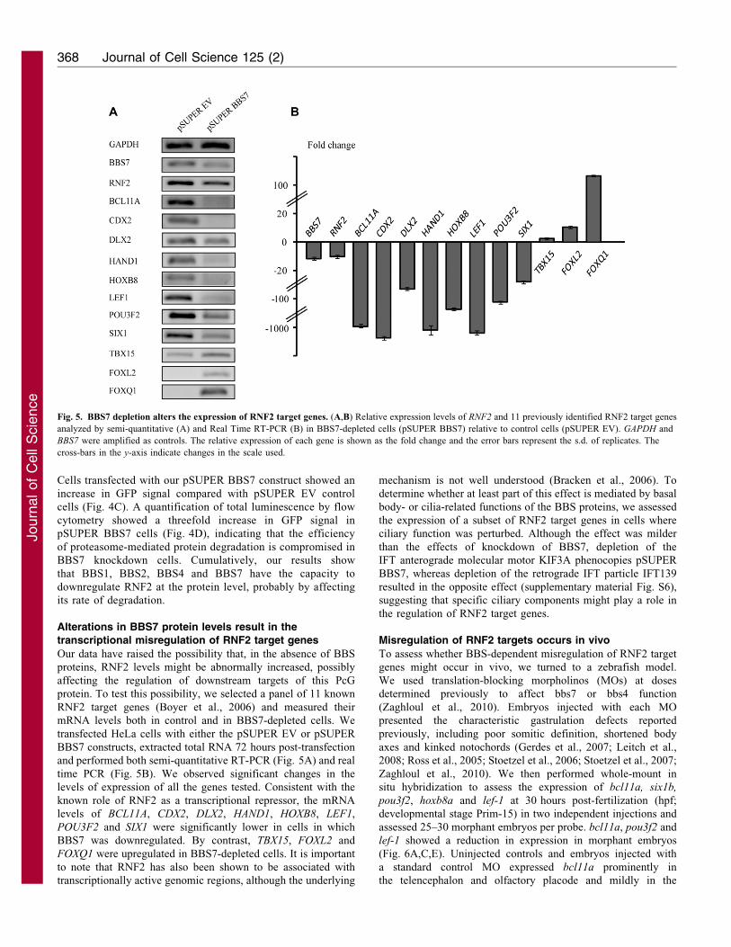
Our data have raised the possibility that, in the absence of BBS
proteins, RNF2 levels might be abnormally increased, possibly
affecting the regulation of downstream targets of this PcG
protein. To test this possibility, we selected a panel of 11 known
RNF2 target genes (Boyer et al., 2006) and measured their
mRNA levels both in control and in BBS7-depleted cells. We
transfected HeLa cells with either the pSUPER EV or pSUPER
BBS7 constructs, extracted total RNA 72 hours post-transfection
and performed both semi-quantitative RT-PCR (Fig. 5A) and real
time PCR (Fig. 5B). We observed significant changes in the
levels of expression of all the genes tested. Consistent with the
known role of RNF2 as a transcriptional repressor, the mRNA
levels of BCL11A, CDX2, DLX2, HAND1, HOXB8, LEF1,
POU3F2 and SIX1 were significantly lower in cells in which
BBS7 was downregulated. By contrast, TBX15, FOXL2 and
FOXQ1 were upregulated in BBS7-depleted cells. It is important
to note that RNF2 has also been shown to be associated with
transcriptionally active genomic regions, although the underlying
mechanism is not well understood (Bracken et al., 2006). To
determine whether at least part of this effect is mediated by basal
body- or cilia-related functions of the BBS proteins, we assessed
the expression of a subset of RNF2 target genes in cells where
ciliary function was perturbed. Although the effect was milder
than the effects of knockdown of BBS7, depletion of the
IFT anterograde molecular motor KIF3A phenocopies pSUPER
BBS7, whereas depletion of the retrograde IFT particle IFT139
resulted in the opposite effect (supplementary material Fig. S6),
suggesting that specific ciliary components might play a role in
the regulation of RNF2 target genes.
Misregulation of RNF2 targets occurs in vivo
To assess whether BBS-dependent misregulation of RNF2 target
genes might occur in vivo, we turned to a zebrafish model.
We used translation-blocking morpholinos (MOs) at doses
determined previously to affect bbs7 or bbs4 function
(Zaghloul et al., 2010). Embryos injected with each MO
presented the characteristic gastrulation defects reported
previously, including poor somitic definition, shortened body
axes and kinked notochords (Gerdes et al., 2007; Leitch et al.,
2008; Ross et al., 2005; Stoetzel et al., 2006; Stoetzel et al., 2007;
Zaghloul et al., 2010). We then performed whole-mount in
situ hybridization to assess the expression of bcl11a, six1b,
pou3f2, hoxb8a and lef-1 at 30 hours post-fertilization (hpf;
developmental stage Prim-15) in two independent injections and
assessed 25–30 morphant embryos per probe. bcl11a, pou3f2 and
lef-1 showed a reduction in expression in morphant embryos
(Fig. 6A,C,E). Uninjected controls and embryos injected with
a standard control MO expressed bcl11a prominently in
the telencephalon and olfactory placode and mildly in the
Fig. 5. BBS7 depletion alters the expression of RNF2 target genes. (A,B) Relative expression levels of RNF2 and 11 previously identified RNF2 target genes
analyzed by semi-quantitative (A) and Real Time RT-PCR (B) in BBS7-depleted cells (pSUPER BBS7) relative to control cells (pSUPER EV). GAPDH and
BBS7 were amplified as controls. The relative expression of each gene is shown as the fold change and the error bars represent the s.d. of replicates. The
cross-bars in the y-axis indicate changes in the scale used.
Journal of Cell Science 125 (2)368
Journ
alof
Cell
Scie
nce

mesencephalon and hindbrain, whereas the bbs7 and bbs4
morphants exhibited significant reduction of expression in all
structures, consistent with our cell-based predictions (Fig. 6A;
supplementary material Fig. S7). Similarly, both pou3f2
expression (Fig. 6C; supplementary material Fig. S7), normally
observed in all structures of the brain and spinal cord, and
lef1 expression (Fig. 6E), which is typically expressed in the
hypothalamus, diencephalon, tectum, midbrain hind–brain
boundary, pectoral fin and branchial arches, were reduced in
morphant embryos. Levels of expression for six1b and hoxb8a did
not appear to differ significantly between morphants and control
embryos (Fig. 6B,D). However, the pattern of six1b expression
was altered; whereas control embryos displayed expression in the
branchial arches and otic vesicles, additional expression in the
diencephalon of bbs7 morphants and in the pectoral fin bud and
hindbrain of bbs4 morphants could be observed (Fig. 6B). hoxb8a
expression patterns remained unchanged in morphants as
compared with controls (Fig. 6D). Importantly, we did not
Fig. 6. In vivo misregulation of RNF2 target genes
in zebrafish BBS morphants. (A–E) Embryos
injected with bbs7, bbs4 or rnf2 MOs and stained by
in situ hybridization at 30 hours post-fertilization
using probes for bcl11a, six1b, pou3f2, hoxb8a and
lef1. (A) Embryos injected with bbs7 and bbs4 MOs
had significantly less staining of bcl11a than
uninjected control embryos whereas co-injection of
bbs7/4 with rnf2 rescued and even expanded the field
of expression. (B) six1b show an altered pattern of
expression with staining in the diencephalon in bbs
morphants. (C) pou3f2 staining showed lower levels
of expression in bbs7 and bbs4 morphants, with
rescue of expression in double morphants of bbs7/rnf2
and bbs4/rnf2. (D) hoxb8a expression in bbs7, bbs4
and double morphants with rnf2 showed no
significant difference in expression. (E) lef1
expression is reduced in morphant embryos.
BBS proteins and transcriptional regulation 369
Journ
alof
Cell
Scie
nce

observe gross morphological defects that could be causing these
changes in expression patterns (Fig. 6D; supplementary material
Fig. S8). To confirm that these changes in gene expression are
specific effects of depleting the bbs proteins, we used the standard
control MO as an additional control and rescued the phenotype by
co-injecting human BBS4 and BBS7 mRNA together with the
corresponding MO. Importantly, whereas injecting the standard
control MO neither affected the expression of bcl11a nor pou3f2,
co-injection of BBS4 and BBS7 mRNAs with their corresponding
MOs fully rescued the expression of both genes (supplementary
material Fig. S7).
To determine whether the reduction in expression of bcl11a,
pou3f2 and lef-1 in the bbs MOs was dependent on Rnf2, we
investigated possible genetic interactions by using double
injections of bbs7 and rnf2 or of bbs4 and rnf2. The expression
of lef1 in the double morphants of bbs7 and rnf2 or bbs4 and rnf2
was partially rescued in all regions compared with bbs7 and bbs4
single morphants. Importantly, the expression of both bcl11a and
pouf32 was not only rescued but was also expanded compared
with controls. Expression of bcl11a, for example, was increased
in the midbrain and expanded to the hindbrain, probably as a
consequence of reducing rnf2 to lower levels than in controls
(Fig. 6A). In summary, our data indicate that rnf2 target genes
are misregulated in vivo in the absence of the bbs proteins,
corroborating and assigning physiological relevance to our in
vitro data.
Global analysis of BBS7 in gene regulation
Finally, we evaluated whether the role of the BBS proteins in
gene regulation is restricted to their interaction with RNF2 or
whether it extends beyond this particular PcG protein. We
therefore performed a microarray-based analysis to visualize, in a
global fashion, the changes in gene expression associated with
the loss of BBS7 function. We transfected HeLa cells (in
triplicate) with either pSUPER EV or pSUPER BBS7, extracted
RNA and hybridized our samples onto a 4644K Human Genome
Oligo Microarray interrogating §41,000 unique human genes
and transcripts arrayed in four replicas. This allowed us to
compare our three biological samples as well as a technical dye
swap control in a single experiment. After stringent quality
control steps, we retained 18,872 probes corresponding to 13,420
unique features.
Our initial analysis showed a significant number of affected
genes (for a complete list of genes with their corresponding
P-values and log2 fold change, see supplementary material
Table S1). We ordered putative altered genes by their adjusted P-
values and set an arbitrary cut-off at P,161025 and a log2 fold
change of ±2 to define a set of differentially expressed (DE)
transcripts for subsequent analyses (supplementary material
Fig. S9 and Table S1). Because the decision of a cut-off value
is arbitrary, we did not try to estimate the number or identity
of genes affected by BBS7 function. Rather, we focused on
confirming and possibly expanding on our previous results and
determined whether genes known to be targeted by RNF2, or the
other PcG proteins (SUZ12, EED and PHC1) for which lists of
putative target genes have been reported (Boyer et al., 2006), are
enriched in our DE dataset. From 7,275 analyzable genes that are
not present in any of the PcG lists (NO group) (Table 1), 503
(6.9%) are present in our DE list of genes. However, 17% of
targets of RNF2, 22% of targets of SUZ12, 25% of targets of
EED, 20% of targets of PHC1 and 21% of genes with a PcG-
dependent histone H3 trimethylation modification (H3K27) are
DE (Table 1). Thus, our data show a significant enrichment of
targets of RNF2 and all other PcG proteins tested in our dataset
of genes affected by BBS7 depletion (Pearsons Chi-squared
test, x25457, df56, P,2.2610216). Importantly, an equivalent
analysis using a PcG-independent group of developmental genes
reportedly targeted by Cyclin D1 (Ctrl-neg group) (Bienvenu
et al., 2010), showed that they were not significantly different to
the NO group (Table 1).
Next, we performed an unbiased analysis of our data to avoid
the need of establishing arbitrary cut-offs. We hypothesized that
if a group of genes is preferentially altered in the absence of
BBS7, these should present lower P-values than those genes that
are a priori not linked to BBS7. We first tested whether genes
included in the ciliary proteome (Gherman et al., 2006)
(www.ciliaproteome.org) are preferentially affected in BBS7
knockdown cells and did not find a significant difference in the
mean and distribution of their P-values compared with the genes
in the array that are not present in the database (Fig. 7A, NO
group). We then performed a similar analysis with the target
genes of RNF2, SUZ12, EED and PHC1 and the H3K27 list, and
compared their P-value distributions with those of the NO and
Ctrl-neg groups. Importantly, our analysis revealed that the
groups of genes associated previously with the PcG proteins
presented significantly lower P-values than our two controls
(Kruskal-Wallis Chi-squared test, x25461.1358, df56,
P,2.2610216; see Fig. 7B). Collectively, our data show that
BBS7 probably participates in the regulation of target genes of
RNF2 and other PcG proteins such as SUZ12, EED and PHC1.
Finally, we performed an ontology analysis using the list of
altered genes with P-values ,161025 in an effort to delineate
common biological processes or pathways that appear to be
altered in the absence of BBS7. This analysis revealed an
Table 1. Targets of different PcG proteins are significantly affected in the absence of BBS7
RNF2 SUZ12 EED PHC1 H3K27 NO* Ctrl-neg{
DE 97 105 82 103 241 503 51Not DE 464 370 242 419 896 6772 679Total 561 475 324 522 1137 7275 730% DE 17 22 25 20 21 6.9 7
Analysis of genes reported to be targeted by the PcG proteins RNF2, SUZ12, EED and PHC1 and of genes with a histone PcG-dependent trimethylationmodification (H3K27). The numbers and percentage of genes differentially expressed (DE) in the absence of BBS7 are given. We show a statistically significantenrichment of target of the different PcG proteins tested in our dataset (Pearsons Chi-squared test, x25457, df56, P,2.2610216).
*NO corresponds to all the analyzable genes that are not present in any of the PcG lists.{Ctrl-neg corresponds to reported targets of Cyclin D1 (Bienvenu et al., 2010).
Journal of Cell Science 125 (2)370
Journ
alof
Cell
Scie
nce

over-representation of genes involved in maintaining cellular
homeostasis, cell organization, cellular interaction with the
extracellular environment and, in general, genes playing a role
in developmental processes, a function that is consistent
with the commonly associated roles of PcG target genes
(Fig. 7C).
Fig. 7. Microarray analysis of BBS7-depleted cells. Transiently transfected HeLa cells were used to compare the transcriptional profile of control and BBS7-
depleted cells. (A) Boxplot of the log P-values obtained in the microarray experiment for the genes included in the cilia proteome compared with the genes in the
array that are not present in that database (NO). (B) Similarly, target genes for different PcG proteins (SUZ12, EED, PHC1 and RNF2) and for genes with the
trimethylated H3K27 marker (H3K27me3) compared with the genes not present in those groups (NO) and also compared with the CCND1 target genes used as a
negative control (Ctrl-neg). (C) Upregulated and downregulated genes (showing P,161025) in the absence of BBS7 were used in an ontology analysis.
Ontology classes with an over-representation in our dataset are shown.
BBS proteins and transcriptional regulation 371
Journ
alof
Cell
Scie
nce

DiscussionOur deeper knowledge of ciliary biology has been instrumental inbeginning to understand the phenotypic consequences of ciliary
dysfunction. However, and despite these advances, little isknown regarding the exact biological role of a number ofciliary proteins, both in the context of ciliary-dependent and
ciliary-independent functions. Here, we increase the complexityassociated with the BBS proteins by showing that these proteinsare probably found in the nuclear compartment, albeit at reduced
levels, where they are involved in the regulation of geneexpression. Our data indicate that several BBS proteins are ableto modulate RNF2 protein levels. Importantly, it has recently
been shown that cilia are involved in the regulation of the mTORpathway, a key regulator of protein synthesis in response todifferent extracellular stimuli (Boehlke et al., 2010; DiBella et al.,2009). As such, although we cannot discard the possibility that
cilia-dependent perturbation of mTOR signaling might affect theprotein levels of RNF2, our proteasome data indicate that theeffect of the BBS proteins on RNF2 is probably achieved
by affecting its turnover. Given that all the BBS proteins testedin this study are able to interact with RNF2, at least inoverexpressing conditions, it is possible that this regulatory
activity over RNF2 occurs in the context of a complex, either theBBSome or a different BBS complex. Supporting this possibility,whereas depletion of either BBS7, BBS1, BBS2 or BBS4 resultedin a marked increase in RNF2, overexpression of a single BBS
protein was not sufficient to reduce the levels of the PcG protein(data not shown).
The physiological relevance of the nuclear localization of the
BBS proteins is supported by several observations. First, the vastmajority of BBS proteins present NES motifs, and in the case ofBBS7 this signal appears to be functional. Second, whereas
depleting BBS1, BBS2, BBS4 and BBS7 results in increasedRNF2 levels, inhibition of BBS2, which appears to be abundantlypresent in the nuclear compartment, provoked the most
prominent effect on RNF2 abundance. Third, two BBS7
mutations found in patients, H323R and T211I (Badano et al.,2003), result in increased nuclear localization of the protein.Those mutants retain the ability to bind RNF2 (data not shown)
and behave as dominant negatives in a zebrafish-based in vivorescue assay (Zaghloul et al., 2010). Therefore, our data mightprove relevant to the understanding of this in vivo effect.
However, we note that these mutations are not predicted to affectthe NES motif tested in this work (located around residues 625–634). One possibility is that these mutations affect protein–
protein interactions required for efficient nuclear export. Proteinsthat are subjected to NES-mediated nuclear export physicallyinteract not only with the karyopherin receptor CRM1 (alsoknown as exportin 1 or XPO1) but also might need adaptor
proteins mediating the interaction and a number of othermoieties, such as RanGTP (Fornerod et al., 1997; la Cour et al.,2004; Sorokin et al., 2007). In addition, the activity of a NES
signal might be regulated by binding of additional proteins andpost-translational modifications (la Cour et al., 2004), aspectsthat could also be affected by these mutations. Finally, there are
examples of proteins, such as actin from S. cerevisiae, thatcontain more than one functional NES signal (Wada et al., 1998).Interestingly, one of the scores provided by the NetNES server
presents a peak centered on residue 218 of BBS7 that isdiminished by the T211I mutation (supplementary materialFig. S10).
In this scenario, elucidating what regulates the activity orsubcellular localization of proteins such as BBS7 will be crucial
to completely understand their function. In addition, and giventhat BBS7 and RNF2 can interact in the cytoplasm at least inyeast, understanding whether the interaction of BBS7 with RNF2is restricted to the nuclear compartment or can also occur outside
of it is also important. Our data suggest that the localization ofBBS7 appears to be variable in terms of its presence in the ciliaryaxoneme and possibly in terms of its ability to enter the nucleus.
The localization of the BBS proteins might be altered in responseto specific cellular or ciliation stages or upon the activation ofsignaling cascades that operate through the cilium. Moreover, our
data indicate that the presence of cilia might mediate the role ofBBS7 in the regulation of RNF2, given that knockdown of theanterograde motor KIF3A results in a mild misregulation ofRNF2 target genes that resembles the effect of depleting BBS7.
Consistent with this notion, depletion of other ciliary componentssuch as IFT139, a protein that has been causally linked to thepathogenesis of several ciliopathies (Davis et al., 2011), also
participates in the regulation of different RNF2 targets althoughthe exact mechanism remains to be elucidated.
Importantly, the ability of ciliary proteins to enter the nucleus
is not restricted to the BBS proteins. Inversin, mutated innephronophthisis (NPHP2); polycystin 1, altered in autosomaldominant polycystic kidney disease (ADPKD); and NEK1, acentrosomal protein mutated in mouse models of cystic kidney
disease, have also been reported in the nucleus (Hilton et al.,2009; Nurnberger et al., 2002; Yoder et al., 2002). Likewise,OFD1, the protein mutated in the ciliopathy orofaciodigital
syndrome type I (OMIM 311200), has also been shown to havethis capacity and has been linked to gene regulation by itsassociation with a histone acetyltransferase (Giorgio et al., 2007).
Interestingly, recent data highlights striking similarities betweenthe process of nuclear import and that of targeting to the ciliarycompartment. It has been shown that the molecular motor KIF17
possesses a ciliary localization signal that is similar to classicNLS motifs. Furthermore, Dishinger and colleagues have shownthat importin-b2 and RanGTP, key proteins in nucleartranslocation, are present in the cilium and are required for
KIF17 ciliary targeting (Dishinger et al., 2010). In this context,we speculate that mechanisms similar to nuclear export couldalso function to actively regulate the composition of the ciliary
compartment.
Lastly, our data both provide an extensive list of genes whoseexpression is potentially affected in the absence of the BBS
proteins and also show that not all the genes identified asdifferentially expressed in our cell-based assays will beaberrantly expressed in vivo. Nevertheless, the fact that theBBS proteins affect the expression of key developmental
regulators, such as targets of the PcG, might provide animportant clue to the cellular basis of the different clinicalmanifestations that characterize BBS and other ciliopathies.
For example, we show that depletion of BBS7 results indownregulation of SIX1 in our cell-based assays and probablyresults in ectopic expression in zebrafish bbs morphants at the
Prim-15 stage. Murine Six1 is implicated in the formationof specialized sensory structures, placodes, as well as otherdevelopmental aspects of the sensory system, findings that might
be relevant to the thermosensory defects recently reported in BBSanimals and patients (Christophorou et al., 2009; Tan et al.,2007). Likewise, SIX1 has been also implicated in processes such
Journal of Cell Science 125 (2)372
Journ
alof
Cell
Scie
nce

as epithelial–mesenchymal transitions (EMT) (Micalizzi et al.,2009), potentially providing clues to the increased fibrosisobserved in the kidney phenotype of BBS and other ciliopathies.
In summary, moieties such as the BBS proteins might provide
a link between the cilium and the interior of the cell, relayingdifferent types of signals and, finally, exerting their role throughthe regulation of gene transcription. Further studies will be
needed to understand the nature of the signal(s) that modulate theactivity of the BBS proteins, their capacity to enter the nucleus,their association with RNF2 and possibly other PcG proteins, andthe extent of their involvement in gene regulation. Understanding
the type of genes that are ultimately affected by the function ofproteins such as the BBSs will provide significant insight into thepathogenesis and cellular basis of the ciliopathies.
Materials and MethodsYeast two-hybrid screen
We performed the Cytotrap yeast two-hybrid screen following the manufacturer’sinstructions (Stratagene). We amplified the complete ORF of BBS7 from HeLacDNA by PCR using the Pfu high fidelity DNA polymerase (Stratagene) and usingprimers that were tagged with SalI and NotI restriction sites. We cloned the ORF ofBBS7 into the pSOS vector (bait) and used a fetal brain library cloned in the pMyrvector (prey; Stratagene) for the screen.
Mammalian expression constructs and mutagenesis
The complete ORF of BBS1, BBS2, BBS4, BBS5, BBS6, BBS7, BBS8, BBS10,RNF2 and an unrelated control gene (Myc–Control in Fig. 1) were cloned intopCMV-Myc and HA mammalian expression plasmids (Clontech). To generate theI634A mutant, we designed primers introducing the change and performed site-directed mutagenesis using the QuickChange Site-Directed Mutagenesis Kitfollowing the manufacturer’s instructions (Stratagene). Both the H323R and T211Imutants were previously generated (Zaghloul et al., 2010).
Cell culture, transfections and LMB treatment
HeLa, Hek293 and NIH3T3 cells were maintained in Dulbecco’s modified Eaglemedium (at PAA or Sigma) supplemented with 10% fetal bovine serum (FBS;Invitrogen) at 37 C in 5% CO2. Transfections were performed using the CalciumPhosphate Transfection Kit (Invitrogen). For nuclear export inhibition, cells wereincubated with LMB at a concentration of 50 ng/ml.
Co-immunoprecipitation assays
For co-immunoprecipitations (CoIP) we grew HEK293 cells in 10-cm dishes andco-transfected them with the appropriate pCMV-Myc construct and the pCMV-HARNF2 plasmid when needed. We harvested cells 48 hours after transfection (or atdesired confluency for endogenous CoIPs) by incubating them for 15 minutes at4 C in CoIP buffer (150 mM NaCl, 50 mM TRIS-HCl pH 7.5 and 1% Nonidet P-40) supplemented with a protease inhibitor cocktail (Sigma) and 1 mM sodiumorthovanadate. Cell lysates were further incubated at 4 C for 15 minutes withvortexing and then centrifuged for 15 minutes at 13,000 g at 4 C. For CoIP, weincubated cell lysates overnight and under rotation at 4 C with either 8 mg ofmonoclonal anti-Myc antibody or our rabbit polyclonal anti-BBS7 antibodyimmobilized onto a mix of protein A and protein G Sepharose beads (Invitrogen).We washed the beads using CoIp supplemented buffer, resuspended the samples inloading buffer, boiled them for 5 minutes and analyzed them by western blot usinga monoclonal anti-HA (Sigma) or a mouse monoclonal anti-RNF2 antibody(Abnova).
Immunocytochemistry
For immunocytochemistry, either HeLa or NIH3T3 cells were grown on glasscoverslips. At 24 hours post-transfection when appropriate or at desiredconfluency, cells were fixed in 220 C methanol for 10 minutes, blocked with5.5% FBS for 1 hour and incubated with the appropriate primary antibody at roomtemperature for 2 hours. We then used secondary antibodies coupled to eitherTetramethylrhodamine or Alexa Fluor 488 (Invitrogen) for visualization by bothfluorescent and confocal microscopy. We used 4,6-diamidino-2-phenylindole(DAPI) to stain DNA.
Antibodies
We used rabbit polyclonal antibodies raised against specific synthetic peptidesfor BBS7 and BBS2 for immunocytochemistry assays. We used monoclonalantibodies to Myc, HA, c-tubulin and acetylated tubulin (Sigma), a rabbitpolyclonal antibody to GAPDH (Abcam) as western blot control, a mouse
monoclonal antibody against RNF2 (H00006045-M01, Abnova) and a donkey
polyclonal antibody against BBS7 (Santa Cruz Biotechnology) for western blot.
RNA interference
For the expression of short interfering RNAs (siRNAs) we selected our 19-
nucleotide target sequences in the genes of interest using the siDESIGN center
(Dharmacon). We cloned the hairpins into the pSUPER vector by designing
oligonucleotides containing our 19-nucleotide target sequences according to themanufacturer’s instructions (OligoEngine). Positive clones were confirmed by
restriction enzyme digestion and direct sequencing. We transfected cell cultures
with either pSUPER EV (control empty vector), pSUPER BBS7, BBS1, BBS2,
BBS4 or pSUPER RNF2 and harvested cells after 72 hours for both protein and
RNA extraction. The IFT139 targeting sequence was cloned into the pCAGMir30vector (Addgene) and the hairpins for KIF3A and IFT88 were expressed using the
pSHAG Magic construct (Open Biosystems).
Cell fractionation and western blot analysis
We obtained cytoplasmic and nuclear fractions using the Qproteome Cell
Compartment Kit (Qiagen). For the gene inhibition experiments, we harvested
cells 72 hours after transfection with the pSUPER constructs, prepared cell lysates
as described for CoIP, quantified protein using the BCA method (Sigma), loaded
equal amounts on SDS-PAGE and analyzed the samples by western blot. Thequantification of western blot bands was performed using the ImageJ software
(NIH).
RT-PCR and real time PCR
To test both the efficiency of knockdown of our constructs as well as the effect ofdepleting BBS7 on different target genes (primers are available from the authors
upon request), we harvested cells 72 hours after transfection and extracted total
RNA with Trizol (Invitrogen) following the provider’s instructions, prepared
cDNA using the SuperScript First-Strand Synthesis System for RT-PCR
(Invitrogen) and used this cDNA as template in either limited-cycle PCR or realtime PCR. For real time PCR we followed SYBR green incorporation using the
Quantimix Easy SYG KIT (BioTools) in a Rotor-Gene 6000 (Corbett Research).
All real time PCR reactions were performed in biological and PCR duplicates. The
threshold cycle (CT) value for each gene in the panel was normalized to GAPDH,
calculating the DCt for each gene in all samples (replicates of pSUPER EV andpSUPER BBS7). The comparative CT method (DDCt method) was used to
determine the relative quantity of the target genes, and the fold change in
expression was calculated as 2-DDCt.
In vivo analysis of gene expression
Morpholinos (MOs) against zebrafish bbs4 (1.5 ng), bbs7 (6 ng), rnf2 (0.5 ng) anda standard control (5 ng) were obtained from Gene Tools. The standard control
targets a human b-globin intronic mutation and does not have a target in zebrafish.
Wild-type zebrafish embryos at a one- to two-cell stage were injected with 1 nl of
each solution. Zebrafish were collected at the Prim15 stage. Whole-mount in situ
hybridizations were carried out using antisense probes for bcl11a, six1b, pou3f2,hoxb8a and lef-1 made from clones (Openbiosystems) and following a published
protocol (Thisse and Thisse, 2008). Further processing included clearing in methyl
salicylate, flat mounting when needed, and photographing with a Nikon
SMZ-745T Zoom Stereo Photo Microscope at a 56 magnification using a
5.0-megapixel DS-Fi1 color digital camera head. Rescues were performed by co-injecting the MOs with the corresponding human wild-type mRNA that was
transcribed in vitro using the SP6 Message Machine Kit (Ambion).
Microarray: sample preparation and chip hybridization
Total RNA from pSUPER EV control and pSUPER BBS7 cells was isolated by
Trizol (Invitrogen). The concentration and integrity of RNA was determined using
a NanoDrop-1000 Spectrophotometer (NanoDrop Technologies) and a 2100
Bioanalyzer (Agilent) with an RNA 6000 Nano LabChip Kit, using the EukaryoteTotal RNA Nano assay according to the manufacturer’s instructions. RNAs with
an RNA integrity number (RIN) greater than 8 were used. Microarray analysis was
performed using a 4644K Human Genome Oligo Microarray (G4112F, Agilent),
in a two-color design. The Low RNA Input Linear Amplification Kit (Agilent) was
used to generate fluorescent complementary RNA (cRNA) for the microarrayhybridizations. Briefly, we amplified and labeled 1 mg of total RNA using the Cy5-
CTP or Cy3-CTP dyes. Equal amounts of labeled samples (pSUPER EV and
pSUPER BBS7) were hybridized to the arrays at 65 C for 17 hours in a rotating
oven (Agilent). Arrays were washed with the wash buffers 1 and 2 and the
stabilization and drying solutions from Agilent. Slides were scanned on an AgilentDNA microarray scanner, and microarray data were extracted with Agilent’s
Feature Extraction software (v9.5). Three biological and one technical dye swap
replicates were performed and a total of four arrays were analyzed.
BBS proteins and transcriptional regulation 373
Journ
alof
Cell
Scie
nce

Microarray data analysis
Data analysis was accomplished in ‘R’ (R Foundation for Statistical Computing,Vienna, Austria; http://www.R-project.org), mainly through packages in theBioconductor suite (Gentleman et al., 2004). Differential expression was assayedusing the limma software package (Smyth, 2005) and the ontology analysisconducted with GOstats (Falcon and Gentleman, 2007). Probes were flagged forfiltering considering saturation, signal above background and uniformity. Probes thathad any of the replicates flagged were eliminated. Absolute values of correlationsbetween the M-values exceeded 0.83 and displayed expected bivariate distributions(data not shown). Genes were considered differentially expressed when the multipletesting adjusted P-value was ,161025 and the absolute value of log2 of fold changegreater than 2 (supplementary material Fig. S5).
AcknowledgementsWe thank Florencia Irigoın and Pablo Aguilar for their criticalcomments on the manuscript and Norann Zaghloul for her commentsand experimental advice.
FundingThis study was supported by the Agencia Nacional de Investigacion eInnovacion (ANII-Innova) and Fondo Clemente Estable (FCE) [grantnumber PR_FCE_2009_1_2382 to J.L.B.]; by the National Instituteof Child Health and Development [grant number R01HD04260to J.K.]; and by the National Institute of Diabetes, Digestive andKidney disorders, National Institutes of Health [grant numbersR01DK072301, R01DK075972 to N.K.]. J.L.B. is supported by theGenzyme Renal Innovations Program (GRIP). J.L.B., C.G., G.L.,M.C.-R., S.A., C.R. and H.N. are supported by the ‘Programa deDesarrollo de las Ciencias Basicas’ (PEDECIBA), and by ANII-Innova, Uruguay. N.K. is a Distinguished Brumley Professor.Deposited in PMC for release after 12 months.
Supplementary material available online at
http://jcs.biologists.org/lookup/suppl/doi:10.1242/jcs.089375/-/DC1
ReferencesAnsley, S. J., Badano, J. L., Blacque, O. E., Hill, J., Hoskins, B. E., Leitch, C. C.,
Kim, J. C., Ross, A. J., Eichers, E. R., Teslovich, T. M. et al. (2003). Basal body
dysfunction is a likely cause of pleiotropic Bardet-Biedl syndrome. Nature 425, 628-
633.
Badano, J. L., Ansley, S. J., Leitch, C. C., Lewis, R. A., Lupski, J. R. and Katsanis,
N. (2003). Identification of a novel Bardet-Biedl syndrome protein, BBS7, that sharesstructural features with BBS1 and BBS2. Am. J. Hum. Genet. 72, 650-658.
Badano, J. L., Leitch, C. C., Ansley, S. J., May-Simera, H., Lawson, S., Lewis, R. A.,
Beales, P. L., Dietz, H. C., Fisher, S. and Katsanis, N. (2006a). Dissection of
epistasis in oligogenic Bardet-Biedl syndrome. Nature 439, 326-330.
Badano, J. L., Mitsuma, N., Beales, P. L. and Katsanis, N. (2006b). The ciliopathies:
an emerging class of human genetic disorders. Annu. Rev. Genomics Hum. Genet. 22,
125-148.
Bienvenu, F., Jirawatnotai, S., Elias, J. E., Meyer, C. A., Mizeracka, K., Marson, A.,
Frampton, G. M., Cole, M. F., Odom, D. T., Odajima, J. et al. (2010).
Transcriptional role of cyclin D1 in development revealed by a genetic-proteomicscreen. Nature 463, 374-378.
Blacque, O. E., Reardon, M. J., Li, C., McCarthy, J., Mahjoub, M. R., Ansley, S. J.,
Badano, J. L., Mah, A. K., Beales, P. L., Davidson, W. S. et al. (2004). Loss of C.
elegans BBS-7 and BBS-8 protein function results in cilia defects and compromised
intraflagellar transport. Genes Dev. 18, 1630-1642.
Boehlke, C., Kotsis, F., Patel, V., Braeg, S., Voelker, H., Bredt, S., Beyer, T.,
Janusch, H., Hamann, C., Godel, M. et al. (2010). Primary cilia regulate mTORC1
activity and cell size through Lkb1. Nat. Cell Biol. 12, 1115-1122.
Boyer, L. A., Plath, K., Zeitlinger, J., Brambrink, T., Medeiros, L. A., Lee, T. I.,
Levine, S. S., Wernig, M., Tajonar, A., Ray, M. K. et al. (2006). Polycomb
complexes repress developmental regulators in murine embryonic stem cells. Nature
441, 349-353.
Bracken, A. P., Dietrich, N., Pasini, D., Hansen, K. H. and Helin, K. (2006).
Genome-wide mapping of Polycomb target genes unravels their roles in cell fate
transitions. Genes Dev. 20, 1123-1136.
Cardenas-Rodriguez, M. and Badano, J. L. (2009). Ciliary biology: understanding the
cellular and genetic basis of human ciliopathies. Am. J. Med. Genet. C Semin. Med.
Genet. 151C, 263-280.
Christensen, S. T., Pedersen, S. F., Satir, P., Veland, I. R. and Schneider, L. (2008).
The primary cilium coordinates signaling pathways in cell cycle control and
migration during development and tissue repair. Curr. Top. Dev. Biol. 85, 261-301.
Christophorou, N. A., Bailey, A. P., Hanson, S. and Streit, A. (2009). Activation of
Six1 target genes is required for sensory placode formation. Dev. Biol. 336, 327-336.
Davis, E. E., Zhang, Q., Liu, Q., Diplas, B. H., Davey, L. M., Hartley, J., Stoetzel, C.,
Szymanska, K., Ramaswami, G., Logan, C. V. et al. (2011). TTC21B contributesboth causal and modifying alleles across the ciliopathy spectrum. Nat. Genet. 43, 189-196.
DiBella, L. M., Park, A. and Sun, Z. (2009). Zebrafish Tsc1 reveals functionalinteractions between the cilium and the TOR pathway. Hum. Mol. Genet. 18, 595-606.
Dishinger, J. F., Kee, H. L., Jenkins, P. M., Fan, S., Hurd, T. W., Hammond, J. W.,
Truong, Y. N., Margolis, B., Martens, J. R. and Verhey, K. J. (2010). Ciliary entryof the kinesin-2 motor KIF17 is regulated by importin-beta2 and RanGTP. Nat. Cell
Biol. 12, 703-710.
Falcon, S. and Gentleman, R. (2007). Using GOstats to test gene lists for GO termassociation. Bioinformatics 23, 257-258.
Fan, Y., Esmail, M. A., Ansley, S. J., Blacque, O. E., Boroevich, K., Ross, A. J.,
Moore, S. J., Badano, J. L., May-Simera, H., Compton, D. S. et al. (2004).Mutations in a member of the Ras superfamily of small GTP-binding proteins causesBardet-Biedl syndrome. Nat. Genet. 36, 989-993.
Fliegauf, M., Benzing, T. and Omran, H. (2007). When cilia go bad: cilia defects andciliopathies. Nat. Rev. Mol. Cell. Biol. 8, 880-893.
Fornerod, M., Ohno, M., Yoshida, M. and Mattaj, I. W. (1997). CRM1 is an exportreceptor for leucine-rich nuclear export signals. Cell 90, 1051-1060.
Gentleman, R. C., Carey, V. J., Bates, D. M., Bolstad, B., Dettling, M., Dudoit, S.,Ellis, B., Gautier, L., Ge, Y., Gentry, J. et al. (2004). Bioconductor: open softwaredevelopment for computational biology and bioinformatics. Genome Biol. 5, R80.
Gerdes, J. M., Liu, Y., Zaghloul, N. A., Leitch, C. C., Lawson, S. S., Kato, M.,Beachy, P. A., Beales, P. L., Demartino, G. N., Fisher, S. et al. (2007). Disruptionof the basal body compromises proteasomal function and perturbs intracellular Wntresponse. Nat. Genet. 39, 1350-1360.
Gerdes, J. M., Davis, E. E. and Katsanis, N. (2009). The vertebrate primary cilium indevelopment, homeostasis, and disease. Cell 137, 32-45.
Gherman, A., Davis, E. E. and Katsanis, N. (2006). The ciliary proteome database: anintegrated community resource for the genetic and functional dissection of cilia. Nat.
Genet. 38, 961-962.
Giorgio, G., Alfieri, M., Prattichizzo, C., Zullo, A., Cairo, S. and Franco, B. (2007).Functional characterization of the OFD1 protein reveals a nuclear localization andphysical interaction with subunits of a chromatin remodeling complex. Mol. Biol. Cell
18, 4397-4404.
Goetz, S. C. and Anderson, K. V. (2010). The primary cilium: a signalling centreduring vertebrate development. Nat. Rev. Genet. 11, 331-344.
Hilton, L. K., White, M. C. and Quarmby, L. M. (2009). The NIMA-related kinaseNEK1 cycles through the nucleus. Biochem. Biophys. Res. Commun. 389, 52-56.
Holaska, J. M. and Paschal, B. M. (1998). A cytosolic activity distinct from Crm1mediates nuclear export of protein kinase inhibitor in permeabilized cells. Proc. Natl.
Acad. Sci. USA 95, 14739-14744.
Jin, H., White, S. R., Shida, T., Schulz, S., Aguiar, M., Gygi, S. P., Bazan, J. F. and
Nachury, M. V. (2010). The conserved Bardet-Biedl syndrome proteins assemble acoat that traffics membrane proteins to cilia. Cell 141, 1208-1219.
Kim, J. C., Badano, J. L., Sibold, S., Esmail, M. A., Hill, J., Hoskins, B. E., Leitch,
C. C., Venner, K., Ansley, S. J., Ross, A. J. et al. (2004). The Bardet-Biedl proteinBBS4 targets cargo to the pericentriolar region and is required for microtubuleanchoring and cell cycle progression. Nat. Genet. 36, 462-470.
Kim, J. C., Ou, Y. Y., Badano, J. L., Esmail, M. A., Leitch, C. C., Fiedrich, E.,
Beales, P. L., Archibald, J. M., Katsanis, N., Rattner, J. B. et al. (2005). MKKS/BBS6, a divergent chaperonin-like protein linked to the obesity disorder Bardet-Biedlsyndrome, is a novel centrosomal component required for cytokinesis. J. Cell Sci.
118, 1007-1020.
Kim, S. K., Shindo, A., Park, T. J., Oh, E. C., Ghosh, S., Gray, R. S., Lewis, R. A.,Johnson, C. A., Attie-Bittach, T., Katsanis, N. et al. (2010). Planar cell polarity actsthrough septins to control collective cell movement and ciliogenesis. Science 329,1337-1340.
Kudo, N., Matsumori, N., Taoka, H., Fujiwara, D., Schreiner, E. P., Wolff, B.,
Yoshida, M. and Horinouchi, S. (1999). Leptomycin B inactivates CRM1/exportin 1by covalent modification at a cysteine residue in the central conserved region. Proc.
Natl. Acad. Sci. USA 96, 9112-9117.
la Cour, T., Kiemer, L., Mølgaard, A., Gupta, R., Skriver, K. and Brunak, S. (2004).Analysis and prediction of leucine-rich nuclear export signals. Protein Eng. Des. Sel.
17, 527-536.
Leitch, C. C., Zaghloul, N. A., Davis, E. E., Stoetzel, C., Diaz-Font, A., Rix, S., Al-Fadhel, M., Lewis, R. A., Eyaid, W., Banin, E. et al. (2008). Hypomorphicmutations in syndromic encephalocele genes are associated with Bardet-Biedlsyndrome. Nat. Genet. 40, 443-448.
Lewis, E. B. (1978). A gene complex controlling segmentation in Drosophila. Nature
276, 565-570.
Li, J. B., Gerdes, J. M., Haycraft, C. J., Fan, Y., Teslovich, T. M., May-Simera, H.,
Li, H., Blacque, O. E., Li, L., Leitch, C. C. et al. (2004). Comparative genomicidentification of conserved flagellar and basal body proteins that includes a novelgene for Bardet-Biedl syndrome. Cell 117, 541-552.
Marion, V., Stoetzel, C., Schlicht, D., Messaddeq, N., Koch, M., Flori, E., Danse,J. M., Mandel, J. L. and Dollfus, H. (2009). Transient ciliogenesis involving Bardet-Biedl syndrome proteins is a fundamental characteristic of adipogenic differentiation.Proc. Natl. Acad. Sci. USA 10, 1820-1825.
May-Simera, H. L., Ross, A., Rix, S., Forge, A., Beales, P. L. and Jagger, D. J.(2009). Patterns of expression of Bardet-Biedl syndrome proteins in the mammaliancochlea suggest noncentrosomal functions. J. Comp. Neurol. 514, 174-188.
Journal of Cell Science 125 (2)374
Journ
alof
Cell
Scie
nce

Micalizzi, D. S., Christensen, K. L., Jedlicka, P., Coletta, R. D., Baron, A. E.,Harrell, J. C., Horwitz, K. B., Billheimer, D., Heichman, K. A., Welm, A. L. et al.
(2009). The Six1 homeoprotein induces human mammary carcinoma cells to undergoepithelial-mesenchymal transition and metastasis in mice through increasing TGF-beta signaling. J. Clin. Invest. 119, 2678-2690.
Nachury, M. V., Loktev, A. V., Zhang, Q., Westlake, C. J., Peranen, J., Merdes, A.,Slusarski, D. C., Scheller, R. H., Bazan, J. F., Sheffield, V. C. et al. (2007). A corecomplex of bbs proteins cooperates with the GTPase Rab8 to promote ciliarymembrane biogenesis. Cell 129, 1201-1213.
Nguyen Ba, A. N., Pogoutse, A., Provart, N. and Moses, A. M. (2009). NLStradamus:a simple Hidden Markov Model for nuclear localization signal prediction. BMC
Bioinformatics 10, 202.Nurnberger, J., Bacallao, R. L. and Phillips, C. L. (2002). Inversin forms a complex
with catenins and N-cadherin in polarized epithelial cells. Mol. Biol. Cell 13, 3096-3106.
Otto, E. A., Hurd, T. W., Airik, R., Chaki, M., Zhou, W., Stoetzel, C., Patil, S. B.,
Levy, S., Ghosh, A. K., Murga-Zamalloa, C. A. et al. (2010). Candidate exomecapture identifies mutation of SDCCAG8 as the cause of a retinal-renal ciliopathy.Nat. Genet. 42, 840-850.
Ross, A. J., May-Simera, H., Eichers, E. R., Kai, M., Hill, J., Jagger, D. J., Leitch,C. C., Chapple, J. P., Munro, P. M., Fisher, S. et al. (2005). Disruption of Bardet-Biedl syndrome ciliary proteins perturbs planar cell polarity in vertebrates. Nat.
Genet. 37, 1135-1140.Schwartz, Y. B. and Pirrotta, V. (2007). Polycomb silencing mechanisms and the
management of genomic programmes. Nat. Rev. Genet. 8, 9-22.Smyth, G. K. (2005). Limma: linear models for microarray data. In Bioinformatics and
Computational Biology Solutions using R and Bioconductor (ed. R. Gentleman, V.Carey, S. Dudoit, R. Irizarry and W. Huber), pp. 397-420. New York: Springer.
Sorokin, A. V., Kim, E. R. and Ovchinnikov, L. P. (2007). Nucleocytoplasmictransport of proteins. Biochemistry (Mosc.) 72, 1439-1457.
Stoetzel, C., Laurier, V., Davis, E. E., Muller, J., Rix, S., Badano, J. L., Leitch,C. C., Salem, N., Chouery, E., Corbani, S. et al. (2006). BBS10 encodes avertebrate-specific chaperonin-like protein and is a major BBS locus. Nat. Genet. 38,521-524.
Stoetzel, C., Muller, J., Laurier, V., Davis, E. E., Zaghloul, N. A., Vicaire, S.,
Jacquelin, C., Plewniak, F., Leitch, C. C., Sarda, P. et al. (2007). Identification of anovel BBS gene (BBS12) highlights the major role of a vertebrate-specific branch ofchaperonin-related proteins in Bardet-Biedl syndrome. Am. J. Hum. Genet. 80, 1-11.
Tan, P. L., Barr, T., Inglis, P. N., Mitsuma, N., Huang, S. M., Garcia-Gonzalez,
M. A., Bradley, B. A., Coforio, S., Albrecht, P. J., Watnick, T. et al. (2007). Lossof Bardet Biedl syndrome proteins causes defects in peripheral sensory innervationand function. Proc. Natl. Acad. Sci. USA 104, 17524-17529.
Thisse, C. and Thisse, B. (2008). High-resolution in situ hybridization to whole-mountzebrafish embryos. Nat. Protoc. 3, 59-69.
Wada, A., Fukuda, M., Mishima, M. and Nishida, E. (1998). Nuclear export of actin:a novel mechanism regulating the subcellular localization of a major cytoskeletalprotein. EMBO J. 17, 1635-1641.
Wallingford, J. B. and Mitchell, B. (2011). Strange as it may seem: the many linksbetween Wnt signaling, planar cell polarity, and cilia. Genes Dev. 25, 201-213.
Wang, H., Wang, L., Erdjument-Bromage, H., Vidal, M., Tempst, P., Jones, R. S.and Zhang, Y. (2004). Role of histone H2A ubiquitination in Polycomb silencing.Nature 431, 873-878.
Yoder, B. K., Hou, X. and Guay-Woodford, L. M. (2002). The polycystic kidneydisease proteins, polycystin-1, polycystin-2, polaris, and cystin, are co-localized inrenal cilia. J. Am. Soc. Nephrol. 13, 2508-2516.
Zaghloul, N. A., Liu, Y., Gerdes, J. M., Gascue, C., Oh, E. C., Leitch, C. C.,
Bromberg, Y., Binkley, J., Leibel, R. L., Sidow, A. et al. (2010). Functionalanalyses of variants reveal a significant role for dominant negative and commonalleles in oligogenic Bardet-Biedl syndrome. Proc. Natl. Acad. Sci. USA 107, 10602-10607.
BBS proteins and transcriptional regulation 375
Journ
alof
Cell
Scie
nce