direct, electronic microrna detection for the rapid determination of differential expression...
TRANSCRIPT

Nanobiosensing (2)DOI: 10.1002/anie.200902577
Direct, Electronic MicroRNA Detection for the Rapid Determinationof Differential Expression Profiles**Hong Yang, Angela Hui, George Pampalakis, Leyla Soleymani, Fei-Fei Liu, Edward H. Sargent,and Shana O. Kelley*
MicroRNAs (miRNAs) are an emerging class of diagnosticmarkers that can signify the presence of disease and be usedto predict its course.[1, 2] Indeed, miRNAs are now known to beinvolved in tumor metastasis,[3] stem-cell differentiation andrenewal,[4] and viral replication.[5] The analysis of the intra-cellular levels of miRNAs is challenging, however, becausetheir short lengths, low abundances, and high levels ofsequence similarity present obstacles in the use of conven-tional analytical methods. Hybridization-based approaches(e.g. microarray analyses) are attractive for microRNAprofiling because of the potential for extensive multiplexingand the discrimination of closely related sequences; however,such methodology requires large quantities (micrograms) ofstarting material.[6–8] The lack of sensitivity of existing array-based methods is related to the type of readout used: typicallyfluorescence signals emitted from an RNA-conjugated fluo-rophore. Very low levels of signal derived from low-abun-dance sequences are extremely difficult to detect withoutsophisticated optics. Impressive progress in this area has beenmade with the development of novel methods for theultrasensitive detection of miRNA hybridization on arraysurfaces;[9, 10] however, the methods available involve manysteps and have not yet been validated with biological samples.We describe herein a new approach to ultrasensitive, direct,hybridization-based microRNA profiling using a multiplexedelectronic chip and electrocatalytic readout. The very highsensitivity of this method enables the direct analysis of smallsamples (nanograms of total RNA) within 30 minutes. Thepower of this method is demonstrated by the identification ofspecific microRNA sequences that are overexpressed inhuman head and neck cancer cells relative to normalepithelial cells.
We endeavored to develop a new method for microRNAprofiling that would feature the convenience of array-basedanalysis, but would augment the power of such multiplexingwith the exceptional sensitivity required to assay smallbiological samples for low-abundance microRNAs. We pur-sued an approach based on electronic readout and prepared amultiplexed chip that featured an electrode pattern generatedby photolithography (Figure 1a). The chip was made bydepositing a pattern of gold on the surface of a silicon wafer toprovide a multiplexed set of leads and external contacts. Alayer of SiO2 was deposited on top of the gold to passivate themetal; then, in the final fabrication step, apertures of 500 nmin diameter were opened at the end of each lead to exposegold. To generate protruding microelectrodes, palladium waselectrodeposited in the apertures (Figure 1b). The electro-deposition step can be engineered to produce highly nano-structured microelectrodes (NMEs; Figure 1b). Previousstudies have indicated that nanostructured sensing elements
Figure 1. Electronic microRNA detection with nanostructured micro-electrode (NME) chips. a) Photograph (left) showing microfabricatedchips that feature 500 nm openings for the electrochemical depositionof NMEs, and illustration (right) of the chip structure. b) Schematicillustration of the generation of sensing elements by palladium electro-deposition. A scanning electron microscope image of a depositednanostructured microelectrode is shown on the right. c) Hybridizationof unlabeled microRNA in samples containing 10 ng of total RNA to aprobe-modified chip. After 30 min, hybridization can be read outelectrochemically by using an electrocatalytic RuIII/FeIII reporter system.
[*] H. Yang, G. Pampalakis, S. O. KelleyDepartment of Pharmaceutical Sciences and Department ofBiochemistry, University of TorontoToronto, ON M5S3M2 (Canada)E-mail: [email protected]
L. Soleymani, E. H. SargentDepartment of Electrical and Computer EngineeringUniversity of Toronto, Toronto, ON M5S3G4 (Canada)
A. Hui, F. F. LiuDivision of Applied Molecular OncologyOntario Cancer Institute, University Health NetworkToronto, ON (Canada)
[**] We acknowledge the support of the Ontario Ministry of Researchand Innovation, Genome Canada through the Ontario GenomicsInstitute, and the NSERC.
Supporting information for this article is available on the WWWunder http://dx.doi.org/10.1002/anie.200902577.
AngewandteChemie
8461Angew. Chem. Int. Ed. 2009, 48, 8461 –8464 � 2009 Wiley-VCH Verlag GmbH & Co. KGaA, Weinheim

are more sensitive than bulk materials towards biomolecularanalytes and that they facilitate surface-complexation reac-tions;[11, 12] thus, the introduction of nanoroughness should beadvantageous.
To test the electronic chip for sensitivity and specificity inmicroRNA detection, Pd NMEs modified with PNA probeswere exposed to total RNA for hybridization (Figure 1c).Complexation was assayed with a redox reporter system[13–15]
previously shown to exhibit femtomolar sensitivity when usedin conjunction with nanostructured electrodes and PNAprobes.[13] This reporter system relies on the accumulationof RuIII when nucleic acids hybridize at an electrode surface.The signals observed with this reporter are amplified by theinclusion of ferricyanide, which can regenerate RuIII chemi-cally after its electrochemical reduction. Titrations of themicroRNA sequence miR-21 showed detectable signalchanges relative to noncomplementary control sequenceswhen solutions containing as little as 10 am of the target wereexposed to the chip-based NMEs (Figure 2). This concen-tration corresponds to approximately 10 molecules per micro-liter of sample. The very high level of sensitivity is accom-panied by a limited dynamic range of only 102; however, forthe detection of microRNAs this tradeoff is merited given thelow abundance of these sequences. A small dynamic rangemay necessitate multiple analyses of unknown samples atvaried dilutions. Nonetheless, 5–10-fold changes in the con-centration of sequences in the regime in which the sensorresponse is linear can be detected readily.
Two crucial additional sensing criteria are specificallyrequired for microRNA detection. First, closely relatedsequences—different by as few as one base—must beaccurately distinguished. Second, sequence appendages,such as those found in mature and precursor microRNAs,must be discriminated. We sought to challenge our systemwith each of these requirements. We investigated first thespecificity of the assay for mature microRNA sequences byanalyzing signal changes observed when the chip was exposedto solutions containing either the full-length, double-stranded, precursor form of miR-21, or the significantlyshorter, single-stranded, mature miR-21 sequence. The signalfor the hairpin precursor structure approached backgroundlevels, whereas a robust signal change was observed formature miR-21 (Figure 3a, right).
We evaluated the sensitivity of the detection approach topoint mutations by monitoring the response of probe-modified sensing elements to two closely related sequences,miR-26a and miR-26b. Probes complementary to eachsequence were arrayed on the chip, and the responses to thecomplementary sequences were monitored (Figure 3a, left).The signal observed when miR-26a was introduced wasapproximately four times as high for the fully matched miR-26a probe as for the mismatched miR-26b probe; similarly,the signal observed when miR-26b was introduced wasapproximately 4.5 times as high for the fully matched miR-26b probe as for its mismatched counterpart. These resultsindicate that this multiplexed chip can successfully discrim-inate closely related microRNA sequences, but also thatparallel interrogation of arrayed probes would be necessary todistinguish closely related sequences.
The derivation of a “fingerprint” of microRNA expres-sion from cell lines representing a particular tumor typerelative to microRNA expression in normal cells has pre-viously been shown to be a powerful approach for theidentification of microRNAs that can serve as biomarkers ofdisease in patients.[16,17] Having confirmed the specificity andsensitivity of the chip towards microRNA targets, we thentested it by using RNA samples extracted from normal humancells and RNA samples derived from human head and necksquamous cancer cells grown in culture. For example, totalRNA extracted from the human hypopharyngeal squamouscancer FaDu cell line and a normal oral epithelial cell line wastitrated onto a nanostructured microelectrode displaying aprobe complementary to miR-205 (Figure 3b). A positivesignal was observed with as little as 5 ng of RNA derived fromthe FaDu cells, whereas no signal change was observed fornormal epithelial cells with up to 20 ng of RNA. This resultindicates that the signal response corresponds to a uniquemarker present at significantly higher levels in the cancer celllines.
We profiled two different microRNAs, miR-21 and miR-205, in a panel of total RNA samples. We also monitored a
Figure 2. Electrocatalytic microRNA detection: readout and sensitivity.a) Differential pulse voltammetry (DPV) showing the signal increaseobserved after incubation of the complementary target (miR-21,100 am) with a probe-modified NME for 30 min. The signal observedwith a noncomplementary target at a 100 times higher concentrationis shown in the inset. b) Determination of the sensitivity and detectionlimit for electronic miR-21 detection.
Communications
8462 www.angewandte.org � 2009 Wiley-VCH Verlag GmbH & Co. KGaA, Weinheim Angew. Chem. Int. Ed. 2009, 48, 8461 –8464
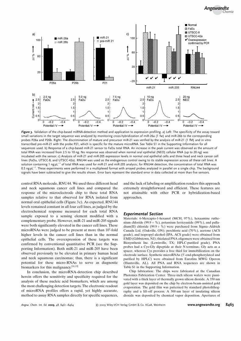
control RNA molecule, RNU44. We used three different headand neck squamous cancer cell lines and compared theresponse of the microelectrode chip to these total RNAsamples relative to that observed for RNA isolated fromnormal oral epithelial cells (Figure 3c). As expected, RNU44levels remained constant in all four cell lines, as judged by theelectrochemical response measured for each total RNAsample exposed to a sensing element modified with acomplementary probe. However, miR-21 and miR-205 signalswere both significantly elevated in the cancer cell lines. ThesemicroRNAs were judged to be present at more than 102-foldhigher levels in the cancer cell lines than in the normalepithelial cells. The overexpression of these targets wasconfirmed by conventional quantitative PCR (see the Sup-porting Information). Both miR-21 and miR-205 have beenobserved previously to be elevated in primary human headand neck squamous carcinomas; thus, there is a significantpotential for these micro-RNAs to serve as diagnosticbiomarkers for this malignancy.[18, 19]
In conclusion, the microRNA-detection chip describedherein offers the sensitivity and specificity required for theanalysis of these nucleic acid biomarkers, which are amongthe most challenging detection targets. The electronic readoutof microRNA profiles offers a rapid, yet highly accurate,method to assay RNA samples directly for specific sequences,
and the lack of labeling or amplification renders this approachextremely straightforward and efficient. These features arenot attainable with other PCR or hybridization-basedapproaches.
Experimental SectionMaterials: 6-Mercapto-1-hexanol (MCH, 97%), hexaamine ruthe-nium chloride (99.9 + %), potassium ferricyanide (99%), and palla-dium(II) chloride (99.9 + %) were purchased from Sigma–AldrichCanada Ltd. (Oakville, ON); perchloric acid (70%), acetone (ACSgrade), and isopropyl alcohol (IPA, ACS grade) were obtained fromEMD (Gibbstown, NJ); thiolated PNA oligomers were obtained fromBiosynthesis Inc. (Lewisville, TX; HPLC-purified grade). PNAprobes had a Cys-Gly dipeptide at their N terminus; Gly acts as aspacer, whereas Cys provides a free thiol for immobilization on theelectrode surface. Synthetic microRNAs (5’-end-phosphorylated andpurified by HPLC) were obtained from Eurofins MWG Operon(Huntsville, AL). All PNA and RNA sequences are shown inTable S1 in the Supporting Information.
Chip fabrication: The chips were fabricated at the CanadianPhotonics Fabrication Center. Three-inch silicon wafers were passi-vated with a thick layer of thermally grown silicon dioxide. A 350 nmgold layer was deposited on the chip by electron-beam-assisted goldevaporation. The gold film was patterned by standard photolithog-raphy and a lift-off process. A 500 nm layer of insulating silicondioxide was deposited by chemical vapor deposition. Apertures of
Figure 3. Validation of the chip-based miRNA-detection method and application to expression profiling. a) Left: The specificity of the assay towardsmall variations in the target sequence was analyzed by monitoring cross-hybridization of miR-26a (1 fm) and miR-26b to the correspondingprobes P26a and P26b. Right: The discrimination of mature and precursor miR-21 was verified by the analysis of miR-21 (1 fM) and in vitrotranscribed pre-miR-21 with the probe P21, which is specific for the mature microRNA. See Table S1 in the Supporting Information for allsequences used. b) Response of a chip-based miR-21 sensor to FaDu total RNA. An increase in the peak current was observed as the amount oftotal RNA was increased from 2.5 to 10 ng. No response was observed when normal oral epithelial (NEO) cellular RNA (up to 20 ng) wasincubated with the sensor. c) Analysis of miR-21 and miR-205 expression levels in normal oral epithelial cells and three head and neck cancer celllines (FaDu, UTSCC-8, and UTSCC-42a). RNU44 was used as the endogenous control owing to its stable expression across all these cell lines. Asolution containing 1 ngmL�1 of total RNA was used for miR-21 and miR-205 analysis; for RNU44 detection, the concentration of total RNA was0.5 ng mL�1. These experiments were performed in a multiplexed format with arrayed probes analyzed in parallel on a single chip. The backgroundsignals have been subtracted to give the results shown. Error bars represent the standard error in data collected at more than five sensors.
AngewandteChemie
8463Angew. Chem. Int. Ed. 2009, 48, 8461 –8464 � 2009 Wiley-VCH Verlag GmbH & Co. KGaA, Weinheim www.angewandte.org

500 nm in diameter were imprinted on the electrodes by standardphotolithography, and 2 � 2 mm2 bond pads were exposed by standardphotolithography.
Fabrication of nanostructured microelectrodes: Chips werecleaned by rinsing sequentially with acetone, IPA, and deionizedwater for at least 30 s and dried with a flow of nitrogen forapproximately 2 min. Electrodeposition was performed at roomtemperature with a Bioanalytical Systems Epsilon potentiostat byusing a three-electrode system with an Ag/AgCl reference electrodeand a platinum-wire auxiliary electrode. Apertures (500 nm indiameter) on the fabricated electrodes were used as the workingelectrode and were contacted by using the exposed bond pads. A2 mm portion of the chip was immersed in the plating bath containingpalladium(II) chloride (5 mm) and perchloric acid (0.5m) andincubated for approximately 5 min prior to electroplating. PdNMEs were fabricated by using DC potential amperometry at anapplied potential of �100 mV for 6 s.
Modification of NMEs with PNA probes: Single-strandedthiolated PNA probes were dissolved in a buffer solution (pH 7)containing sodium phosphate (25 mm) and sodium chloride (25 mm).MCH (10 mm) was then added to a final MCH concentration of100 nm. This PNA probe solution was then deposited on the chip in adark humidity chamber overnight at 4 8C. The probe-modified PdNMEs were rinsed thoroughly with the above buffer solution beforemeasurements. For multiplexed experiments, chips with eight indi-vidually addressable leads were used.
Target hybridization: Hybridization solutions were solutions ofthe target at various concentrations in sodium phosphate buffer(pH 7.0, 25 mm) containing NaCl (25 mm). Pd NMEs were incubatedwith 10 mL of the hybridization solution in a humidity chamber at37 8C for 30 min. The chip was then cooled and washed thoroughlywith the buffer before electrochemical analysis.
Electrochemical measurements: An electrochemical analyzer(BASi, West Lafayette, IN) was used for electrochemical measure-ments in an aqueous solution containing [Ru(NH3)6]
3+ (10 mm),[Fe(CN)6]
3� (4 mm), sodium phosphate (pH 7.0, 25 mm), and NaCl(25 mm). Cyclic voltammetry (CV) was conducted before and afterthe addition of the solution of the target at a scan rate of 100 mVs�1.Differential pulse voltammetry (DPV) was performed with apotential step of 5 mV, a pulse amplitude of 50 mV, a pulse width of50 ms, and a pulse period of 100 ms. Cyclic voltammetry signalsbefore and after hybridization were collected with a scan rate of100 mVs�1. The limiting reductive current (I) was quantified bysubtracting the background current at 0 mV from the cathodic currentat �300 mV. Signal changes corresponding to hybridization werecalculated according to the following equation: DI = 100(Ids�Iss)/Iss
(ss = before hybridization, ds = after hybridization). The detectionlimit was calculated by determining the first concentration at whichthe signal, after subtraction of the background signal (noncomple-mentary DI), was two times higher than the standard deviation of anoncomplementary control sample at a concentration of 10 fm.
RNA extraction for PCR analyses and amplification protocol:Total RNA was extracted from cell lines with the mirVana kit(Ambion). The quality of samples was assessed by reverse tran-scription–PCR analysis of the endogenous control RNU44 by usingthe Applied Biosystems TaqMan microRNA Assay. This assayincludes a reverse-transcription (RT) step with the TaqMan Micro-RNA Reverse Transcription Kit (Applied Biosystems, CA) in which astem-loop RT primer hybridizes specifically to an miR molecule andis then reverse transcribed with a MultiScribe reverse transcriptase.The reverse-transcription mix included stem–loop RT primers(50 nm), 1 � RT buffer, deoxynucleoside triphosphates (0.25 mm
each), MultiScribe reverse transcriptase (3.33 UmL�1), and anRNase inhibitor (0.25 Uml�1). The reaction mixture (7.5 mL) wasincubated in an Applied Biosystems 7900 Thermocycler for 30 min at16 8C, 30 min at 42 8C, and 5 min at 85 8C, and was then held at 4 8C.The RT products were subsequently amplified with sequence-specific
primers (hsa-miR-21 primer 4373090 and hsa-miR-205 primer4373093 from Applied Biosystems) by using the Applied Biosystems7900HT Real-Time PCR system. The PCR mix (10 mL) contained theRT product (0.67 mL), 1 � TaqMan Universal PCR Master Mix, theTaqMan probe (0.2 mm), the forward primer (1.5 mm), and the reverseprimer (0.7 mm). The reaction mixtures were incubated in a 384-wellplate at 95 8C for 10 min, and then subjected to 40 cycles of treatmentat 95 8C for 15 s and 60 8C for 1 min.
Received: May 14, 2009Revised: August 17, 2009Published online: October 6, 2009
.Keywords: biosensors · microchips · microelectrodes ·microRNA · nucleic acids
[1] V. Ambros, Nature 2004, 431, 350.[2] L. Soleymani, Z. Fang, E. H. Sargent, S. O. Kelley, Nat. Nano-
technol. 2009, DOI: 10.1038/NNANO.2009.276.[3] M. S. Nicoloso, R. Spizzo, M. Shimizu, S. Rossi, G. A. Calin, Nat.
Rev. Cancer 2009, 9, 293.[4] V. K. Gangaraju, H. Lin, Nat. Rev. Mol. Cell Biol. 2009, 10, 116.[5] B. R. Cullen, Nature 2009, 457, 421.[6] C. G. Liu, G. A. Calin, B. Meloon, N. Gamliel, C. Sevignani, M.
Ferracin, C. D. Dumitru, M. Shimizu, S. Zupo, M. Dono, H.Alder, F. Bullrich, M. Negrini, C. M. Croce, Proc. Natl. Acad.Sci. USA 2004, 101, 9740.
[7] N. Rossenfeld, R. Aharonov, E. Meiri, S. Rosenwald, Y. Spector,M. Zepeniuk, H. Benjamin, N. Shabes, S. Tabak, A. Levy, D.Lebanony, Y. Goren, E. Silberschein, N. Targan, A. Ben-Ari, S.Gilad, N. Sion-Vardy, A. Tobar, M. Feinmesser, O. Kharenko, O.Nativ, D. Nass, M. Perelman, A. Yosepovich, B. Shalmon, S.Polak-Charcon, E. Fridman, A. Avniel, I. Bentwich, Z. Bent-wich, D. Cohen, A. Chajut, I. Barshack, Nat. Biotechnol. 2008,26, 462.
[8] M. Selbach, B. Schwanhausser, N. Thierfelder, Z. Fang, R.Khanin, N. Rajewsky, Nature 2008, 455, 58.
[9] S. Fang, H. J. Lee, A. W. Wark, R. M. Corn, J. Am. Chem. Soc.2006, 128, 14044.
[10] A. W. Wark, H. J. Lee, R. M. Corn, Angew. Chem. 2008, 120,654; Angew. Chem. Int. Ed. 2008, 47, 644.
[11] L. Soleymani, Z. Fang, X. Sun, H. Yang, B. J. Taft, E. H. Sargent,S. O. Kelley, Angew. Chem. 2009, DOI: 10.1002/ange.200902439;Angew. Chem. Int. Ed. Engl. 2009, DOI: 10.1002/anie.200902439.
[12] R. Gasparac, B. J. Taft, M. A. Lapierre-Devlin, A. D. Lazareck,J. M. Xu, S. O. Kelley, J. Am. Chem. Soc. 2004, 126, 12270.
[13] Z. Fang, S. O. Kelley, Anal. Chem. 2009, 81, 612.[14] M. A. Lapierre, M. O�Keefe, B. J. Taft, S. O. Kelley, Anal. Chem.
2003, 75, 6327.[15] M. A. Lapierre-Devlin, C. L. Asher, B. J. Taft, R. Gasparac,
M. A. Roberts, S. O. Kelley, Nano Lett. 2005, 5, 1051.[16] J. Lu, G. Getz, E. A. Miska, E. Alvarez-Saavedra, J. Lamb, D.
Peck, A. Sweet-Cordero, B. L. Ebert, R. H. Mak, A. A. Fer-rando, J. R. Downing, T. Jacks, H. R. Horvitz, T. R. Golub,Nature 2005, 435, 834.
[17] N. Yanaihara, N. Caplen, E. Bowman, M. Seike, K. Kumamoto,M. Yi, R. M. Stephens, A. Okamoto, J. Yokota, T. Tanaka, G. A.Calin, C. G. Liu, C. M. Croce, C. C. Harris, Cancer Cell 2006, 9,189.
[18] S. S. Chang, W. W. Jiang, I. Smith, L. M. Poeta, S. Bequm, C.Glazer, S. Shan, W. Westra, D. Sidransky, J. A. Califano, Int. J.Cancer 2008, 123, 2791.
[19] N. Tran, T. McLean, X. Zhang, C. J. Zhao, J. M. Thomson, C.O�Brien, B. Rose, Biochem. Biophys. Res. Commun. 2007, 358,12.
Communications
8464 www.angewandte.org � 2009 Wiley-VCH Verlag GmbH & Co. KGaA, Weinheim Angew. Chem. Int. Ed. 2009, 48, 8461 –8464