direct agglutination test for serologic diagnosis of neospora caninum infection
TRANSCRIPT

ORIGINAL PAPER
Ste phane Romand á Philippe Thulliez á J.P. Dubey
Direct agglutination test for serologic diagnosisof Neospora caninum infection
Received: 26 June 1997 /Accepted: 5 August 1997
Abstract A direct agglutination test was evaluated forthe detection and quantitation of IgG antibodies toNeospora caninum in both experimental and naturalinfections in various animal species. As compared withresults obtained by the indirect ¯uorescent antibodytest, the direct agglutination test appeared reliable forthe serologic diagnosis of neosporosis in a variety ofanimal species. The direct agglutination test shouldprovide easily available and inexpensive tools for sero-logic testing for antibodies to N. caninum in many hostspecies.
Introduction
Neospora caninum is a recently recognized protozoanparasite, which until 1988 had been misidenti®ed withToxoplasma gondii due to close structural similarities(Dubey et al. 1988a). Its life cycle and sources of infec-tion remain unknown, and the transplacental route is theonly known mode of natural infection (Dubey andLindsay 1996). This parasite a�ects a wide range ofmammalian species and is distributed worldwide. Severeneonatal infection or death occurs in dogs, sheep, goats,and horses (Dubey and Lindsay 1996). N. caninum hasalso been identi®ed as an important cause of abortion incattle in North America, Europe, and several othercountries (Anderson et al. 1991, 1995; Dubey and
Lindsay 1996). De®nitive diagnosis is based on histo-logic examination of infected tissues using speci®cimmunohistochemical staining (Lindsay and Dubey1989a). However, this method is time-consuming andthe results are occasionally di�cult to interpret.
Serodiagnosis for the detection of speci®c antibodiesto N. caninum can be performed using an indirect ¯uo-rescent antibody test (IFAT) on tachyzoites or enzyme-linked immunosorbent assays (ELISA) using di�erentantigens (BjoÈ rkman et al. 1994; Dubey et al. 1996; Lallyet al. 1996). These tests have displayed high degrees ofsensitivity and speci®city in experimental and naturalN. caninum infections. However, several factors, such asthe nature and quality of antigens or conjugates used inthe di�erent tests, may result in poorly correlated ordiscrepant results (Dubey et al. 1997). Furthermore, theinterpretation of tachyzoite ¯uorescence on IFAT slidesis subjective and requires trained personnel. We evalu-ated a serologic test for the detection of anti-N. caninumIgG antibodies based on the agglutination of wholeformalin-®xed N. caninum tachyzoites according to amodi®ed technique used for the serodiagnosis of toxo-plasmosis (Desmonts and Remington 1980). The resultsof the new N. caninum agglutination test (NAT) werecompared with those of the IFAT for experimental andnatural infections in di�erent animal species.
Materials and methods
Antigen
Tachyzoites of the NC-1 strain of Neospora caninum were inocu-lated onto con¯uent monolayers of human ®broblasts as previouslydescribed (Dubey et al. 1988b). After 10 days, tachyzoites wereharvested from infected monolayers and washed in phosphate-bu�ered saline solution (PBS, pH 7.2) for mouse inoculation.Adult male Swiss-Webster mice (20 g) were given a 10-lg/ml con-centration of dexamethasone phosphate (Merck Sharpe andDohme, Whitehouse, N.J. USA) ad libitum in drinking water toachieve progressive, nonlethal, and constant immune suppressionthroughout the experiment as previously described for C57B/6Nmice (Yang and Healey 1993).
Parasitol Res (1998) 84: 50±53 Ó Springer-Verlag 1998
S. Romand á P. ThulliezLaboratoire de la Toxoplasmose, Institut de Pue riculture,26 Boulevard Brune, F-75014, Paris, France
J.P. Dubey (&)U.S. Department of Agriculture, Agricultural Research Service,Livestock and Poultry Sciences Institute,Parasite Biology and Epidemiology Laboratory,BARC-East, Building 1001,10300 Baltimore Avenue, Beltsville,MD 20705-2350, USAFax: 301-504-9222; e-mail: [email protected]
After 10 days of treatment with dexamethasone, mice were in-jected i.p. with 5 ´ 104 tachyzoites. On day 4 postinfection (p.i.),tachyzoites were harvested from the peritoneal cavities. Peritoneal¯uids containing free tachyzoites were then centrifuged along withTG 180 mouse sarcoma cells (two tachyzoites per cell), and aportion of the pellet was inoculated i.p. into immunosuppressedmice according to a method previously described using the RHstrain of Toxoplasma gondii for subsequent enhancement of intra-cellular development of the parasite (Desmonts and Remington1980). On day 4 p.i., peritoneal lavages collected from the micerevealed large numbers of extra- and intracellular N. caninum ta-chyzoites. The TG 180 mouse sarcoma cells have been maintainedfor approximately 20 years at the Paris laboratory by serial i.p.passage every 15 days since they were used by Desmonts andRemington (1980).
For disruption of cells harboring tachyzoites, pooled peritoneal¯uids in PBS were incubated at 37 °C for 45 min with 1% trypsin(bio-Me rieux, Lyon, France) under continuous agitation as previ-ously described for T. gondii (Desmonts and Remington 1980).After trypsin digestion, suspensions of parasites were washed threetimes in PBS (pH 7.2), and pellets were resuspended in a 6% so-lution of 100% formaldehyde.
Parasites were kept overnight in formalin, centrifuged, andwashed three times in PBS to remove the formaldehyde. Tachy-zoites were preserved at 4 °C in PBS supplemented with 0.1% (w/v)sodium azide until used for the NAT.
N. caninum agglutination test
The NAT was performed in 96 round-bottom-well microplatesaccording to the method previously described for toxoplasmosis(Desmonts and Remington 1980). In brief, 50 ll of 0.2 M 2-mer-captoethanol in PBS was distributed in each well and 2-fold serialdilutions of sera were performed, starting from either 1:20, 1:200,or 1:500. Parasites were resuspended in alkaline bu�er [7.02 gNaCl, 3.09 g H3BO3, 24 ml of 1 N NaOH, 4 g bovine plasma al-bumin (fraction V), and enough distilled water to bring the volumeto 1 l; pH 8.7] and their concentration was adjusted at 2 ´ 104/ll.After the sera had been diluted, 50 ll N. caninum antigen suspen-sion was distributed in each well. Plates were gently agitated toallow for complete mixing and were then incubated overnight at30 °C. A clear-cut button-shaped deposit of parasite suspension atthe bottom of the well was interpreted as a negative reaction, and acomplete carpet of agglutinated organisms was considered positive.Each assay included two negative controls and one positive control.The negative controls comprised antigen alone and one serumsample with a high titer of anti-T. gondii antibodies. A serumsample obtained from a rat with an experimental N. caninum in-fection was selected as the positive control.
Serum samples
All sera had previously been tested by the IFAT at the UnitedStates Department of Agriculture (Beltsville, Md., USA) and keptfrozen at )20 °C until used for the blind NAT at the Institut dePue riculture de Paris. Sera were obtained from di�erent animalspecies with either experimental or natural infections. Sera fromexperimental infections were obtained from animals inoculated ei-ther parenterally with N. caninum tachyzoites or orally with oocystsor sporocysts of the closely related apicomplexan protozoan par-asites T. gondii, Sarcocystis spp., and Hammondia hammondi.Blood samples were collected on the day of inoculation and atvarious time points after inoculation (see Tables 1, 2). Serologicresponses of these experimental infections have previously beenpublished elsewhere (Dubey et al. 1996).
Serum samples from natural infections were obtained from 70dairy cows from a herd in California that had experienced anoutbreak of N. caninum-induced abortion. A description of theoutbreak has been presented by McAllister et al. (1996) and Dubeyet al. (1997). All sera were collected on the 18th day of the out-break. Additionally, paired sera were obtained from a bitch and her
pup, which presented signs of congenital infection and in whichcongenital neosporosis was immunohistochemically con®rmed.Another litter-mate pup was also studied.
Statistical analysis
A chi-square test was used for comparisons of titers recorded fornaturally infected cows. A correlation coe�cient between the IFATand the NAT was calculated by analysis of variance.
Results
Experimental infections
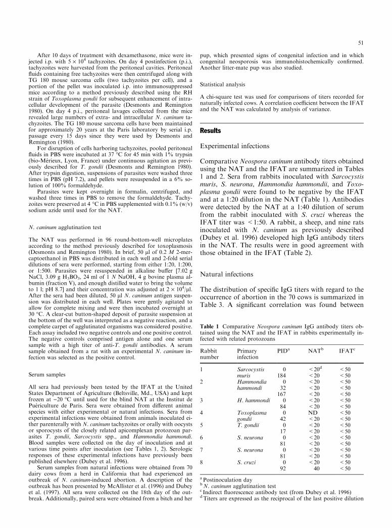
Comparative Neospora caninum antibody titers obtainedusing the NAT and the IFAT are summarized in Tables1 and 2. Sera from rabbits inoculated with Sarcocystismuris, S. neurona, Hammondia hammondii, and Toxo-plasma gondii were found to be negative by the IFATand at a 1:20 dilution in the NAT (Table 1). Antibodieswere detected by the NAT at a 1:40 dilution of serumfrom the rabbit inoculated with S. cruzi whereas theIFAT titer was <1:50. A rabbit, a sheep, and nine ratsinoculated with N. caninum as previously described(Dubey et al. 1996) developed high IgG antibody titersin the NAT. The results were in good agreement withthose obtained in the IFAT (Table 2).
Natural infections
The distribution of speci®c IgG titers with regard to theoccurrence of abortion in the 70 cows is summarized inTable 3. A signi®cant correlation was found between
Table 1 Comparative Neospora caninum IgG antibody titers ob-tained using the NAT and the IFAT in rabbits experimentally in-fected with related protozoans
Rabbitnumber
Primaryinfection
PIDa NATb IFATc
1 Sarcocystis 0 <20d <50muris 184 <20 <50
2 Hammondia 0 <20 <50hammondi 32 <20 <50
167 <20 <503 H. hammondi 0 <20 <50
84 <20 <504 Toxoplasma 0 ND <50
gondii 42 <20 <505 T. gondii 0 <20 <50
17 <20 <506 S. neurona 0 <20 <50
81 <20 <507 S. neurona 0 <20 <50
81 <20 <508 S. cruzi 0 <20 <50
92 40 <50
a Postinoculation daybN. caninum agglutination testc Indirect ¯uorescence antibody test (from Dubey et al. 1996)d Titers are expressed as the reciprocal of the last positive dilution
51
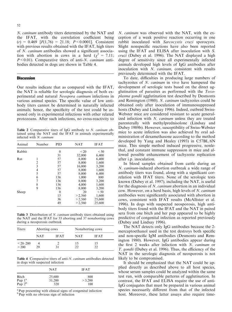
N. caninum antibody titers determined by the NAT andthe IFAT, with the correlation coe�cient being(r) = 0.469 [F(1,76) = 21.18; P<0.0001]. Consistentwith previous results obtained with the IFAT, high titersof N. caninum antibodies showed a signi®cant associa-tion with abortion in cows in a herd (v2 = 7.11;P<0.01). Comparative titers of anti-N. caninum anti-bodies detected in dogs are shown in Table 4.
Discussion
Our results indicate that as compared with the IFAT,the NAT is reliable for serologic diagnosis of both ex-perimental and natural Neospora caninum infections invarious animal species. The speci®c value of low anti-body titers cannot be determined in naturally infectedanimals; hence, the speci®city of the test could be as-sessed only in experimental infections with other relatedprotozoans. After such infections, no cross-reactivity to
N. caninum was observed with the NAT, with the ex-ception of a weak positive reaction occurring in onerabbit inoculated with Sarcocystis cruzi sporocysts.Slight nonspeci®c reactions have also been reportedusing the IFAT and ELISA after inoculation with S.cruzi (Dubey et al. 1996). The NAT displayed a highdegree of sensitivity since all experimentally infectedanimals developed high levels of IgG antibodies afterinoculation with N. caninum, consistent with resultspreviously determined with the IFAT.
To date, di�culties in producing large numbers oftachyzoites of N. caninum in vivo have hampered thedevelopment of serologic tests based on the direct ag-glutination of parasites as performed with the Toxo-plasma gondii agglutination test described by Desmontsand Remington (1980). N. caninum tachyzoites could beobtained only after inoculation of immunosuppressedhosts (Dubey and Lindsay 1996). Indeed, outbred Swiss-Webster mice are considered resistant to acute general-ized infection with N. caninum unless they are treatedparenterally with methylprednisolone (Lindsay andDubey 1989b). However, susceptibility of Swiss-Webstermice to acute infection was also achieved by oral ad-ministration of dexamethasone according to the methoddeveloped by Yang and Healey (1993) in C57BL/6Nmice. This simple method induced progressive, nonle-thal, and constant immune suppression in mice and al-lowed possible enhancement of tachyzoite replicationafter i.p. inoculation.
In blood samples obtained from cattle during anN. caninum-induced abortion outbreak a wide range ofantibody titers was found, along with a signi®cant cor-relation with IFAT titers. None of the serologic testsknown (Dubey et al. 1997), including the NAT, is usefulfor the diagnosis of N. caninum abortion in an individualcow. However, on a herd basis, high levels of N. caninumantibodies were signi®cantly associated with abortion incows, consistent with IFAT results (McAllister et al.1996). In dogs with suspected neosporosis, high anti-body titers found with the IFAT and the NAT in pairedsera from one bitch and her pup appeared to be highlypredictive of congenital infection as reported previously(Dubey and Lindsay 1996).
The NAT detects only IgG antibodies because the 2-mercaptoethanol used in the test destroys both speci®cand non-speci®c IgM antibodies (Desmonts and Rem-ington 1980). However, IgG antibodies appear duringthe ®rst 2 weeks after infection with N. caninum orT. gondii (Dubey et al. 1996). Thus, the e�ciency of theNAT in the serologic diagnosis of neosporosis is notlikely to be compromised.
It should be emphasized that the NAT could be ap-plied directly as described above to all host species,whose serum samples could be analyzed within the sametest run, with comparable patterns of agglutination. Incontrast, the IFAT and ELISA require the use of anti-IgG conjugates that must be prepared in various animalspecies necessarily di�erent from that of the infectedhost. Moreover, these latter assays also require time-
Table 2 Comparative titers of IgG antibody to N. caninum ob-tained using the NAT and the IFAT in animals experimentallyinfected with N. caninum
Animal Number PID NAT IFAT
Rabbit 0 <20 <5031 32,000 6,400
Rat 1 57 8,000 6,4002 57 8,000 1,6003 57 16,000 3,2004 57 8,000 1,6005 57 8,000 6,4006 136 1,000 8007 136 8,000 3,2008 136 4,000 1,6009 136 8,000 3,200
Sheep 0 20 <5015 >2,560 1,60036 >2,560 25,60049 >2,560 25,600
Table 3 Distribution of N. caninum antibody titers obtained usingthe NAT and the IFAT for 33 aborting and 37 nonaborting cowsduring a neosporosis outbreak
Titers Aborting cows Nonaborting cows
NAT IFAT NAT IFAT
<20±200 4 2 15 15>200 29 31 22 22
Table 4 Comparative titers of anti-N. caninum antibodies detectedin dogs with suspected infection
NAT IFAT
Bitch 25,600 800Pup 1a 51,200 >3,200Pup 2b 320 100
a Pup presenting with clinical signs of congenital infectionb Pup with no obvious sign of infection
52

consuming standardization of conjugates, depending onthe species of the infected host. Other drawbacks asso-ciated with the IFAT may be a rather subjective inter-pretation of some patterns of ¯uorescence oftachyzoites, resulting in poor reproducibility betweenlaboratories (Dubey et al. 1997). Furthermore, the widespectrum of N. caninum antigens used in the di�erentELISAs currently available accounts for importantvariations between the results obtained, thus resulting indiscrepant results for low antibody titers (Dubey et al.1997).
In conclusion, as compared with the reference IFAT,the NAT appears reliable for the detection and quanti-tation of IgG antibodies to N. caninum in various hostspecies. In contrast to other serologic tests currentlyavailable, the NAT is less expensive, easy to read and toperform, and requires a minimum of laboratory equip-ment and materials. Therefore, this test could be suitablefor large epidemiologic animal screenings in ®eld studiesas well as for evaluation of humoral responses toN. caninum.
Acknowledgements The authors are indebted to Jean Michelin andChristiane LePrince for their skillful technical assistance.
References
Anderson ML, Blanchard PC, Barr BC, Dubey JP, Ho�man RL,Conrad PA (1991) Neospora-like protozoan infection as a majorcause of abortion in California dairy cattle. J Am Vet MedAssoc 198: 241±244
Anderson ML, Palmer CW, Thurmond MC, Picanso JP,Blanchard PC, Breitmeyer RE, Layton AW, McAllisterMM, Daft B, Kinde H, Read DH, Dubey JP, Conrad PA, BarrBC (1995) Evaluation of abortions in cattle attributable to ne-
osporosis in selected dairy herds in California. J Am Vet MedAssoc 207: 1206±1210
BjoÈ rkman C, Lunde n A, Holmdahl OJM, Barber J, Trees AJ,Uggla A (1994) Neospora caninum in dogs: detection of anti-bodies by ELISA using an iscom antigen. Parasite Immunol 16:643±648
Desmonts G, Remington JS (1980) Direct agglutination test fordiagnosis of Toxoplasma infection: method for increasing sen-sitivity and speci®city. J Clin Microbiol 11: 562±568
Dubey JP, Lindsay DS (1996) A review of Neospora caninum andneosporosis. Vet Parasitol 67: 1±59
Dubey JP, Carpenter JL, Speer CA, Topper MJ, Uggla A (1988a)Newly recognized fatal protozoan disease of dogs. J Am VetMed Assoc 192: 1269±1285
Dubey JP, Hattel AL, Lindsay DS, Topper MJ (1988b) NeonatalNeospora caninum infection in dogs: isolation of the causativeagent and experimental transmission. J Am Vet Med Assoc 193:1259±1263
Dubey JP, Lindsay DS, Adams DS, Gay JM, Baszler TV, BlagburnBL, Thulliez P (1996) Serologic responses of cattle and other an-imals infected withNeospora caninum. Am J Vet Res 57: 329±336
Dubey JP, Jenkins MC, Adams DS, McAllister MM, Anderson-Sprecher R, Baszler TV, Kwok OCH, Lally NC, BjoÈ rkman C,Uggla A (1997) Neospora caninum serology in cattle: antibodyresponses of cows during an outbreak of neosporosis evaluatedby indirect ¯uorescent antibody test and di�erent enzyme-linked immunosorbent assays. J Parasitol 83 (in press)
Lally NC, Jenkins MC, Dubey JP (1996) Evaluation of twoNeospora caninum recombinant antigens for use in an enzyme-linked immunosorbent assay for the diagnosis of bovineneosporosis. Clin Diagn Lab Immunol 3: 275±279
Lindsay DS, Dubey JP (1989a) Immunohistochemical diagnosis ofNeospora caninum in tissue sections. Am J Vet Res 50: 1981±1983
Lindsay DS, Dubey JP (1989b) Neospora caninum (Protozoa:Apicomplexa) infections in mice. J Parasitol 75: 772±779
McAllister MM, Hu�man EM, Hietala SK, Conrad PA, AndersonML, Salman MO (1996) Evidence suggesting a point sourceexposure in an outbreak of bovine abortion due to neosporosis.J Vet Diagn Invest 8: 355±357
Yang S, Healey MC (1993) The immunosuppressive e�ects of dex-amethasone administered in drinking water to C57BL/6N miceinfected with Cryptosporidium parvum. J Parasitol 79: 626±630
53