dihydrotestosterone sensitises lncap cells to death induced by epigallocatechin-3-gallate (egcg) or...
TRANSCRIPT
The Prostate 69:219 ^224 (2009)
Dihydrotestosterone Sensitises LNCaPCells toDeath Inducedby Epigallocatechin-3-Gallate
(EGCG) oran IGF-IReceptor Inhibitor
Francis Thomas,1 Suril Patel,1 Jeff M.P. Holly,1 Raj Persad,2
Amit Bahl,3 and Claire M. Perks1*1IGF&Metabolic EndocrinologyGroup,Departmentof Clinical SciencesNorth Bristol,
TheMedical School Unit, SouthmeadHospital, Bristol,UK2Bristol Royal Inf|rmary,DepartmentofUrology, Bristol,UK
3Departmentof Clinical Oncology, Bristol Oncology Centre, Bristol,UK
BACKGROUND. Compelling evidence has accumulated for chemopreventive effects for theactive component of green tea Epigallocatechin-3-Gallate (EGCG) particularly for prostatecancer (CaP).METHODS. We have assessed interactions between the effects of EGCG and two mainregulators of prostate cell function, dihydrotestosterone (DHT) and insulin-like growth factor-1(IGF-I). Using LNCaP (androgen-sensitive), PC3 and DU145 (androgen-resistant) CaP cell lines,we assessed the effect of EGCG alone on growth (0–200 mM) and on cell death (0–50 mM).RESULTS. EGCG decreased the proliferation of all the CaP cancer cells in a dose-dependentmanner with an increase in apoptosis from 30 to 50 mM. With DU145 cells, a sub-apoptotic doseof EGCG (10–20 mM) reduced IGF-induced growth. With LNCaP cells, a sub-apoptotic dose ofEGCG (8 mM) switched DHT from a growth promoter to a growth inhibitor. A similar reversalof DHT effect was seen in the presence of an IGF-I receptor inhibitor, AG1024 (1 mM). Theseresponses appeared to be due to DHT sensitizing the cells to apoptosis by EGCG and AG1024(P< 0.01 and P< 0.001 respectively).CONCLUSIONS. Our data suggests that both green tea and AG1024 are effective in inhibitingcell growth and inducing death in CaP cells but the effects of both are more effective in thepresence of androgen. Prostate 69: 219–224, 2009. # 2008 Wiley-Liss, Inc.
KEY WORDS: EGCG; CaP cells; androgen; IGF-IR
INTRODUCTION
Prostate cancer (CaP) is the second leading causeof cancer-related deaths among men in Westerncountries, thus representing a major and growinghealth problem.
Prostate cancer represents an ideal disease forchemoprevention because it is typically a slowlydeveloping cancer diagnosed in elderly men; thereforeeven a modest slowing in the neoplastic processachieved through pharmacological or nutritional inter-vention could result in a substantial reduction in theincidence of the clinically detectable disease. Life-stylerelated factors, particularly diet, are considered to bethe major contributors to CaP promotion. Of all dietary
components the most compelling evidence for chemo-preventive effects has accumulated for the activecomponent of green tea Epigallocatechin-3-Gallate(EGCG) [1,2]. Several epidemiological studies havefound a lower incidence of CaP in Asian countries(where green tea is consumed regularly) compared to
Francis Thomas and Suril Patel contributed equally to this work.
*Correspondence to: Claire M. Perks, IGF & Metabolic Endocrino-logy Group, Department of Clinical Sciences North Bristol, TheMedical School Unit, Southmead Hospital, Bristol BS10 5NB, UK.E-mail: [email protected] 28 August 2008; Accepted 9 September 2008DOI 10.1002/pros.20873Published online 21 October 2008 in Wiley InterScience(www.interscience.wiley.com).
, 2008 Wiley-Liss, Inc.
Western populations [3]. In addition, Asian immigrantsto the US who have abandoned original dietary habitssoon develop the Western risk of CaP, which isassociated with the adoption of a Western lifestyle [3].A case-control study conducted in China showed thatgreen tea consumption was protective against thisdisease [4]. Bettuzzi et al. [2] performed a double-blinded and placebo-controlled study with sixty malevolunteers with high grade prostate intraepithelialneoplasia, (HGPIN), a premalignant condition of pro-state growth [5] that is known to result in a substantialnumber of cancers within 1 year after repeated biopsy[6,7]. Half of the men consumed three 200 mg decaf-feinated green tea catechin preparations orally, dailyfor 1 year, which resulted in a 90% reduction in the rateof HG-PIN positive men developing prostate cancerwith no significant side effects [2].
The data from the epidemiological studies andclinical trial by Bettuzzi et al. [2] provide convincingevidence that EGCG has a protective role againstthe progression of CaP. However, the mechanismsof action of EGCG are not clearly understood andtheir delineation will be critical to understandingthe effectiveness of EGCG and for optimizingfuture intervention studies for the prevention of CaPprogression.
Androgens are an essential element in regulatingprostate growth largely by driving proliferation [8] andso androgen withdrawal is often used as a treatment forprostate cancer [9]. However, the failure of anti-androgen therapy is common with progression to anandrogen independent state occurring within 3 years ofstarting treatment in one third of patients [10]. Thistreatment failure illustrates the development of alter-native growth pathways to drive CaP progression, ofwhich insulin-like growth factor-I (IGF-I) is known tobe a key player. Prospective studies have demonstratedan association between serum IGF levels with both theinitiation and progression of CaP [11–13]. The IGF axishas a critical role in the establishment and maintenanceof the transformed phenotype in numerous malignan-cies (reviewed in Ref. [14]). Their action is modulatedby a complex network of molecules consisting of2 ligands (IGF-I and IGF-II), 6 IGF binding proteins(IGFBP-1-6), and types I and II IGF receptors (IGF-IRand IGF-IIR). IGFs have potent mitogenic and anti-apoptotic effects on prostate tissue (reviewed inRef. [15]). Expression and activity of components ofthis pathway are altered in many human malignancies,including CaP [16] and therefore it has been suggestedthat treatments (such as tyrosine kinase inhibitorsand monoclonal antibodies) targeting the IGF-IR couldenhance current therapeutics for CaP [17]. Manycomponents of the IGF axis are regulated by androgens,such as IGF-I, the IGF-IR and IGFBPs [18–20].
MATERIALSANDMETHODS
Materials
All chemicals and inhibitors were purchased fromSigma-Aldrich (Dorset, UK) unless stated otherwise.
Cell Culture
DU145, LNCaP, and PC-3 cells were maintained inRPMI 1640 cell culture media (Lonza, Basel, Switzer-land) supplemented with 10% foetal bovine serum(FBS, Gibco, Paisley, UK), 1% penicillin/streptomycinsolution and 1% L-glutamine solution (200 mM). Cellgrowth and death were examined after the cells weregrown in serum-free conditions for 24 hr. Serumfree media for these cells was identical but with bovineserum albumin (BSA, 1 mg/ml) instead of FBS.
Tritiated Thymidine Incorporation (TTI)
DNA synthesis was measured using TTI asdescribed previously [21,22]. In brief, the cells wereincubated with 0.1 mCi [3H]thymidine per well forthe final 4 hr of the dosing time period. After theremoval of the supernatant, cells were then washedwith 500 ml of 5% trichloroacetic acid (Merck Ltd.,Middlesex, UK) at 48C for 10 min followed by solu-bilization of DNA incorporated thymidine with 400 mlof 1 M NaOH (Fisher Scientific Ltd., Leicestershire, UK)for 1 hr at room temperature. The resulting suspensionwas placed into individual scintillation vials, and 3 mlof scintillation fluid was added. Samples were ana-lyzed using a Beckman Scintillation Counter LS6500.Data were recorded as disintegrations per minute.
Cell Counting
Cell death was assessed using trypan blue cellcounting as described previously [21].
Western Immunoblotting
Cells were lysed, loaded according to proteinconcentration of lysates and separated on an 8%SDS–PAGE gel and transferred to Hybond Nþ nylonmembranes (Amersham, Bucks, UK) as outlinedpreviously [21]. Nonspecific binding sites on thenitrocellulose membranes were blocked overnightwith 5% milk in Tris-buffered saline (TBS)/2% Tweenfor probing with anti-p85 cleaved subunit of PARP(Promega, Southampton, UK), anti-Glyceraldehyde-3-Phosphate dehydrogenase (GAPDH; Chemicon,Hampshire, UK) anti-p-IGF-IR (Cell Signalling, Dan-vers, MA), anti-IGF-IR (Santa Cruz, CA) (all at 1:1,000)or blocked with 3% BSA for probing with phospho-MAPK (1:5,000). After the removal of excess unboundantibody appropriate secondary antibodies conjugated
The Prostate
220 Thomas et al.
to peroxidase were added for 1 hr. Binding of theperoxidase was visualized by enhanced chemilumi-nescence according to the manufacturer’s instructions.Chemiluminescence was detected using the Chemi-Doc-It Imaging System (UVP) (Biorad, .Hertfordshire,UK) and analysed using Vision WorksTH ls AnalysisSoftware (UVP, Inc., Upland, CA).
Statistics
The data were analyzed with the Microsoft Excelversion 5.0a software package using ANOVA followedby least-significant difference post hoc test. A statisti-cally significant difference was considered to bepresent at P< 0.05.
RESULTSANDDISCUSSION
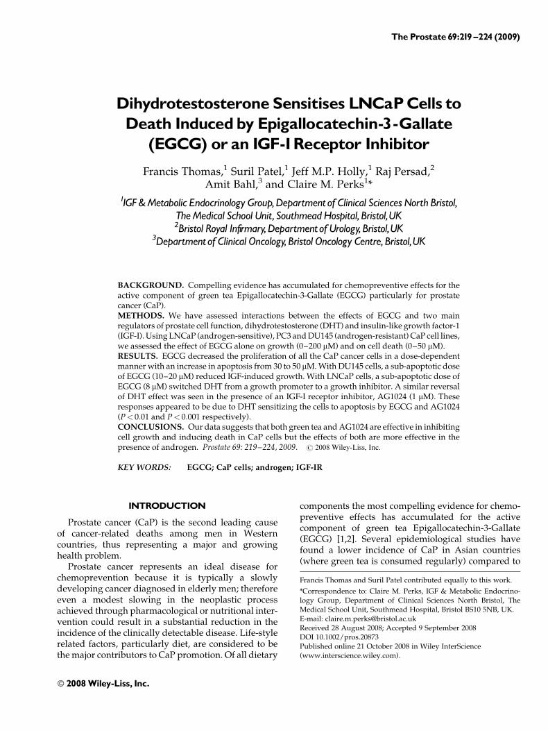
In order to understand the mechanisms by whichEGCG could protect against CaP, our goal was to assessits effects on the growth induced by the two maindrivers of CaP progression, dihydrotestosterone (DHT)and IGF-I. To achieve this we initially characterizedand compared the effects of EGCG on growth andthe induction of cell death in three CaP cell lines:the androgen-responsive LNCaP cells and androgen-insensitive DU145 and PC3 cells. Figure 1A shows thatEGCG decreased proliferation of all the prostate cancercell lines in a dose-dependent manner. A significantdecrease was first observed at 25 mM for DU145 cells(P< 0.001) and at 50 mM for both PC3 and LNCaP cells(P< 0.001 and P< 0.05 respectively). At the highestdose of 200 mM, there was a comparable reduction intritiated thymidine incorporation for DU145 and PC3cells (by 88.2% and 87.5% respectively; both P< 0.001),whereas the LNCaP cells seemed more resistant(decreased by 59.7%; P< 0.001). Figure 1B, shows thatsignificant cell death was induced from 30 mM (DU145;P< 0.01, PC3; P< 0.001 and LNCaP, P< 0.05) andincreased in a dose-dependent manner, with similarresponses in all of the cells lines examined. Theinduction of cleaved PARP (Fig. 1B, insert) confirmedthat this cell death was apoptotic. Doses of EGCGbelow 30 mM were anti-proliferative and sub-apoptotic,whereas doses above 30 mM decreased cell growth andwere associated with the induction of apoptosis. Theseresults are in keeping with previous studies [23,24].
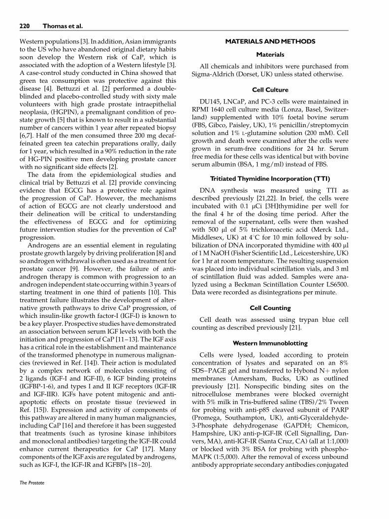
Initial studies with LNCaP cells revealed thatincreasing doses of DHT stimulated DNA synthesisup to a dose of 0.01 mM (P< 0.001), but at higher dosesthe responses were reduced (Fig. 2A). In addition, wefound that addition of exogenous IGF-I (0–200 ng/ml)to these cells had no effect on basal DNA synthesis (datanot shown). However, we did observe in Figure 2B,that proliferation of these cells was dose dependentlyinhibited in the presence of an IGF-IR tyrosine kinase
inhibitor (TKI; AG1024, 0–1 mM), suggesting that thelack of response to exogenously added IGF-I was dueto the cells own endogenous production stimulatingautocrine growth. We also found that the DHT-inducedincrease in DNA synthesis in LNCaP cells wascompletely blocked in the presence of the IGF-IR TKIat doses of 0.5–0.75 mM (Fig. 2B) confirming that theability of DHT to increase cell proliferation was at leastin part dependent upon the IGF1-R. This is in keeping
The Prostate
Fig. 1. EffectsofEGCGonthegrowthandsurvivalofDU145,PC3,and LNCaP prostate cancer (CaP) cells.A: CaP cells were seededingrowthmedia (GM; 1ml) in 24-well plates (0.025 ^ 0.2�106 cells/well) for 24hr.Following24hr in serum-freemedia (SFM)they weretreatedwith EGCG (0 ^200 mM) for 48 hr and DNA synthesis wasmeasuredusingTTI (B)CaPcellswere seededinGM(2ml) in6-wellplates (0.1^ 0.2�106 cells/well) for 24 hr. Following 24 hr in SFM,cells were dosed with EGCG (0 ^50 mM) for 48 hr and cell deathwas assessed using trypan blue cell counting. For confirmation ofapoptosiswemonitoredPARPcleavage (insert).CaPcells (0.5�106)were grown to 60% confluence in T25 cm2 flasks then incubatedin SFM for a further 24 hr prior to dosing as in (B).Cells were thenprocessedasdescribedinmaterials andmethods.Thegraphsrepre-senting theTTI andcell countingexperiments (A,B) are themeanofthree experiments each repeated in triplicate. The Western blot(insertB) isrepresentativeof anexperimentrepeatedthree times.
DHTSensitizes CaPCells to EGCGandAG1024 221
with previous reports in LNCaP cells showing thatandrogens increase IGF-1R expression and IGF-1 levels[18,19]. At the highest dose of 1 mM AG1024, we noticedthat the effect of DHT was reversed (P< 0.05). Oninvestigating this further using trypan blue cell count-ing, we found that the DHT was sensitizing the cells toinduction of cell death by the IGF-IR TKI. Figure 2Cshows that AG1024 (1 mM) and DHT (10 nM) each alone
had no effect but in the presence of DHT, the IGF-IRTKI then caused a significant increase in cell death(P< 0.001).
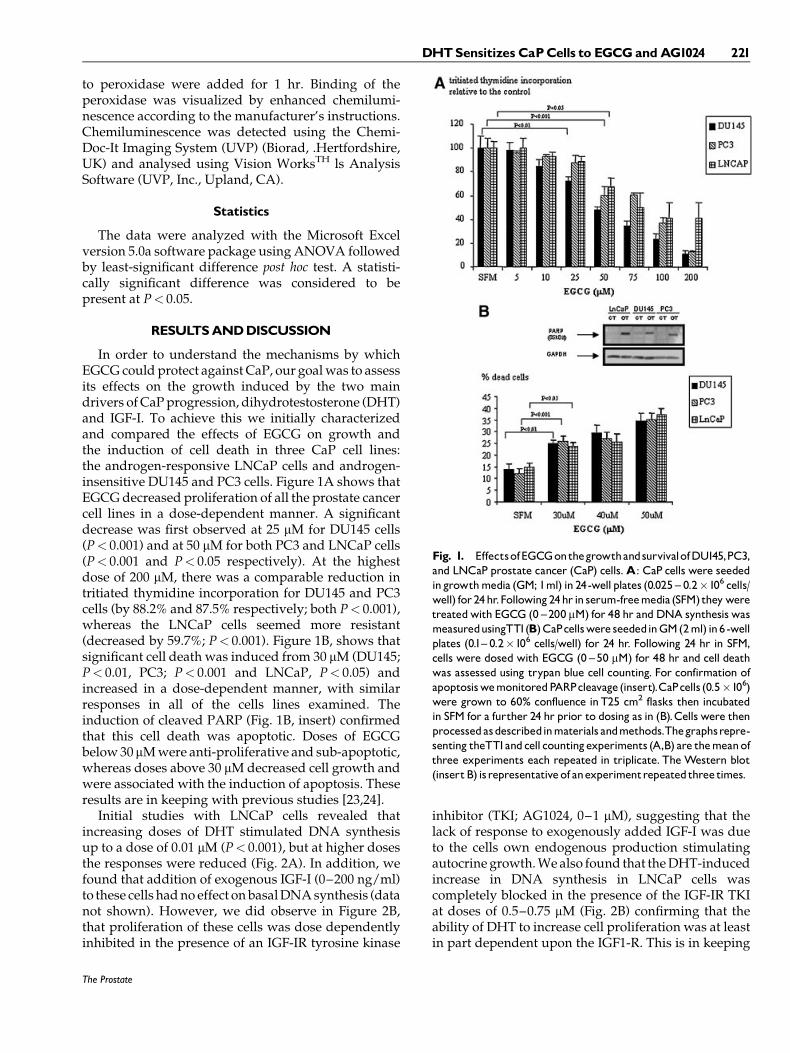
We then wished to assess the effects of EGCG on theactions of both IGF-I and DHT on prostate cancer cells.Using DU145 cells, which unlike LNCaP cells respondto addition of exogenous IGF-I, we show in Figure 3,that IGF-I induced a significant increase in tritiatedthymidine incorporation over 48 hr (P< 0.001), whichwas dose-dependently abrogated in the presence ofEGCG (10–30 mM). This effect of EGCG was associatedwith a reduction on the phosphorylation of the IGF-IRand p-MAPK (insert Fig. 3). We also found that EGCGwas able to decrease the basal phosphorylation of theIGF-IR in LNCaP cells (insert Fig. 3). Such an effect ofEGCG on the phosphorylation of the IGF-IR has beenshown previously in colon cancer cells [25] and inmouse 3T3 fibroblasts, in which the IGF-IR was foundto be a novel binding partner of EGCG [26].
However, previous data in the prostate havedemonstrated that oral infusion of green tea polyphe-nols over 24 weeks to the mouse model (TRAMP;transgenic adenocarcinoma of the mouse prostate)
The Prostate
Fig. 2. Effects of an IGF-IR inhibitor, AG1024 on DHT-inducedgrowth of LNCaP cells LNCaP cells were seeded in growth media(GM; 1ml) in 24-well plates (0.2�106 cells/well) for 24 hr.Following24 hr in serum-free media (SFM) they were treated with (A) DHT(0 ^100mM)either alone orþ/� (B)AG1024 (0 ^1mM).TTIwasper-formedasdescribedinFigure1B.C:CellswereseededasinFigure1Band following 24 hr in SFM were dosed with DHT (10 nM), AG1024(1mM)or thecombinationfor48hr andthencelldeathwas assessedasinFigure1B.Thegraphseachshowthemeanof threeexperimentseachrepeatedin triplicate.
Fig. 3. Effects of EGCG on IGF-induced growth of DU145 cells.DU145 cells were seeded in growth media (GM; 1 ml) in 24-wellplates (0.025�106 cells/well) for24hr.Following24hr in serum-freemedia (SFM) they were treated with IGF-I (25 ng/ml) þ/� EGCG(10 ^30mM) for 48hr.DNA synthesiswasmeasured as describedinFigure 1A. To assess activation of the IGF-I receptor (DU145 andLNCaP) and p-MAPK (DU145) cells were prepared as in Figure1B,dosed with IGF-I (25 ng/ml) (DU145) þ/� EGCG (20 mM) (DU145andLNCaP) for10minandthenlysedandrunonagelasinFigure1B.TheTTI graph shown in (A) is themean of three experiments eachrepeatedin triplicate.TheWesternblots (insert) arerepresentativeofexperimentsrepeatedthree times.
222 Thomas et al.

caused a reduction in the levels of IGF-I and increasedconcentrations of IGFBP-3 in the dorso-lateral prostateand that this was associated with a decrease in p-Aktand p-MAPK [27]. In Du145 and LNCaP cells,EGCG decreased levels of p-Akt but increased levelsof p-MAPK over a period of 12–24 hr [28].
As we had found that the IGF-IR played a role inthe ability of DHT to increase DNA synthesis and thatthe growth response to IGF-I was blocked by EGCG, wenext investigated the effect of EGCG on DHT-inducedproliferation. It has been shown that treatment withEGCG reduces circulating testosterone in murinemodels of CaP [29,30]. In addition, with in vivo modelsof CaP, inhibitors of 5 a-reductase slowed the growthof established tumors [31] and EGCG is capable ofblocking 5 a-reductase activity [32]. In vitro, EGCGhas also been shown to down-regulate expressionof androgen receptor mRNA in LNCaP cells [33].Figure 4A shows that in the presence of sub-apoptoticdoses of EGCG (10–20 mM), the growth responseto DHT was in fact significantly reversed (10 mM,P< 0.001 and 20 mM, P< 0.01) and blocked at the higherdose of 30 mM. In Figure 4B, we determined that in thepresence of androgen the cells became sensitized toEGCG such that a sub-apoptotic dose then induceddeath. In the presence of EGCG or DHT alone there wasno effect, but together there was a significant inductionof cell death (P< 0.01). We show photomicrographsin Figure 4C to illustrate that coincident with theinduction of apoptosis, there were fewer cells attachedto the plate with the combination of DHT and EGCGin relation to either on their own. We also establishedthat in contrast to DHT, exogenous IGF-I was unable tosensitize these cells to EGCG (data not shown), despitethe growth response to DHT being dependent onactivation of the IGF-IR.
Our data suggests that both green tea and the IGF-IRTKI are both effective in inhibiting cell growth andinducing death in CaP cells and they both become moreeffective in the presence of androgen. Our data furthersuggests that these agents might therefore be compro-mised if used in combination with anti-androgentherapy. This study highlights that potential combina-tion therapy has to be considered carefully in order todevelop effective chemoprevention and therapy ofprostate cancer.
ACKNOWLEDGMENTS
We thank The Ralph Shackman Trust for supportingthis work.
REFERENCES
1. Mohanty NK, Saxena S, Singh UP, Goyal NK, Arora RP.Lycopene as a chemopreventive agent in the treatment of high-grade prostate intraepithelial neoplasia. Urol Oncol 2005;23(6):383–385.
2. Bettuzzi S, Brausi M, Rizzi F, Castagnetti G, Peracchia G, Corti A.Chemoprevention of human prostate cancer by oral adminis-tration of green tea catechins in volunteers with high-gradeprostate intraepithelial neoplasia: A preliminary report from a
The Prostate
Fig. 4. Effects of EGCGonDHT-inducedgrowth of LNCaPcells.LNCaPcellswere seededingrowthmedia (GM; 1ml) (A) in 24-wellplates (0.2�106 cells/well) for 24 hr. Following 24 hr in serum-freemedia (SFM) they were treatedwith (A)DHT (0.01mM)þ/�EGCG(0 ^30 mM).TTI was performed as described in 1B and the graphshows the mean of three experiments each repeated in triplicate.Subpart B in 6-well plates (0.3�106 cells/well) for 24 hr.Following24 hr in serum-free media (SFM) they were treated with DHT(0.01mM)þ/�EGCG(10mM).Celldeathwas assessedasinFigure1Band thegraph shows themeanof three experiments eachrepeatedin triplicate.C: LNCaP cells were treated as in (B) and are repre-sentedpictorially(D).CellswereviewedusingaLeicaDMIRBmicro-scopeandphotographsweretakenusingaNikonE450digitalcamera(magnification100�).
DHTSensitizes CaPCells to EGCGandAG1024 223

one-year proof-of-principle study. Cancer Res 2006;66(2):1234–1240.
3. Nelson WG, De Marzo AM, Isaacs WB. Prostate cancer. N Engl JMed 2003;349(4):366–381.
4. Jian L, Xie LP, Lee AH, Binns CW. Protective effect of green teaagainst prostate cancer: A case-control study in southeast China.Int J Cancer 2004;108(1):130–135.
5. Bostwick DG, Qian J. High-grade prostatic intraepithelial neo-plasia. Mod Pathol 2004;17(3):360–379.
6. Bishara T, Ramnani DM, Epstein JI. High-grade prostaticintraepithelial neoplasia on needle biopsy: Risk of cancer onrepeat biopsy related to number of involved cores andmorphologic pattern. Am J Surg Pathol 2004;28(5):629–633.
7. Kronz JD, Allan CH, Shaikh AA, Epstein JI. Predicting cancerfollowing a diagnosis of high-grade prostatic intraepithelialneoplasia on needle biopsy: Data on men with more than onefollow-up biopsy. Am J Surg Pathol 2001;25(8):1079–1085.
8. Suzuki H, Ueda T, Ichikawa T, Ito H. Androgen receptorinvolvement in the progression of prostate cancer. Endocr RelatCancer 2003;10(2):209–216.
9. Debruyne F. Hormonal therapy of prostate cancer. Semin UrolOncol 2002;20(3 Suppl 1):4–9.
10. Gurumurthy S, Vasudevan KM, Rangnekar VM. Regulationof apoptosis in prostate cancer. Cancer Metastasis Rev 2001;20(3–4):225–243.
11. Chan JM, Stampfer MJ, Ma J, Gann P, Gaziano JM, Pollak M,Giovannucci E. Insulin-like growth factor-I (IGF-I) and IGFbinding protein-3 as predictors of advanced-stage prostatecancer. J Natl Cancer Inst 2002;94(14):1099–1106.
12. Woodson K, Tangrea JA, Pollak M, Copeland TD, Taylor PR,Virtamo J, Albanes D. Serum insulin-like growth factor I: Tumormarker or etiologic factor? A prospective study of prostatecancer among Finnish men. Cancer Res 2003;63(14):3991–3994.
13. Stattin P, Bylund A, Rinaldi S, Biessy C, Dechaud H, StenmanUH, Egevad L, Riboli E, Hallmans G, Kaaks R. Plasma insulin-like growth factor-I, insulin-like growth factor-binding proteins,and prostate cancer risk: A prospective study. J Natl Cancer Inst2000;92(23):1910–1917.
14. Furstenberger G, Senn HJ. Insulin-like growth factors andcancer. Lancet Oncol 2002;3(5):298–302.
15. Meinbach DS, Lokeshwar BL. Insulin-like growth factors andtheir binding proteins in prostate cancer: Cause or consequence?Urol Oncol 2006;24(4):294–306.
16. Cardillo MR, Monti S, Di Silverio F, Gentile V, Sciarra F, ToscanoV. Insulin-like growth factor (IGF)-I, IGF-II and IGF type Ireceptor (IGFR-I) expression in prostatic cancer. Anticancer Res2003;23(5A):3825–3835.
17. Wu JD, Haugk K, Coleman I, Woodke L, Vessella R, Nelson P,Montgomery RB, Ludwig DL, Plymate SR. Combined in vivoeffect of A12, a type 1 insulin-like growth factor receptorantibody, and docetaxel against prostate cancer tumors. ClinCancer Res 2006;12(20 Pt 1):6153–6160.
18. Le H, Arnold JT, McFann KK, Blackman MR. DHT andtestosterone, but not DHEA or E2, differentially modulate IGF-I, IGFBP-2, and IGFBP-3 in human prostatic stromal cells. Am JPhysiol Endocrinol Metab 2006;290(5):E952–E960.
19. Pandini G, Mineo R, Frasca F, Roberts CT Jr, Marcelli M, VigneriR, Belfiore A. Androgens up-regulate the insulin-like growth
factor-I receptor in prostate cancer cells. Cancer Res 2005;65(5):1849–1857.
20. Martin JL, Pattison SL. Insulin-like growth factor bindingprotein-3 is regulated by dihydrotestosterone and stimulatesdeoxyribonucleic acid synthesis and cell proliferation in LNCaPprostate carcinoma cells. Endocrinology 2000;141(7):2401–2409.
21. Burrows C, Holly JM, Laurence NJ, Vernon EG, Carter JV, ClarkMA, McIntosh J, McCaig C, Winters ZE, Perks CM. Insulin-likegrowth factor binding protein 3 has opposing actions onmalignant and nonmalignant breast epithelial cells that are eachreversible and dependent upon cholesterol-stabilized integrinreceptor complexes. Endocrinology 2006;147(7):3484–3500.
22. Griner RD, Bollag WB. Inhibition of [(3)H]thymidine transport isa nonspecific effect of PDMP in primary cultures of mouseepidermal keratinocytes. J Pharmacol Exp Ther 2000;294(3):1219–1224.
23. Ahmad N, Feyes DK, Nieminen AL, Agarwal R, Mukhtar H.Green tea constituent epigallocatechin-3-gallate and inductionof apoptosis and cell cycle arrest in human carcinoma cells. J NatlCancer Inst 1997;89(24):1881–1886.
24. Paschka AG, Butler R, Young CY. Induction of apoptosisin prostate cancer cell lines by the green tea component,(-)-epigallocatechin-3-gallate. Cancer Lett 1998;130(1–2):1–7.
25. Shimizu M, Deguchi A, Hara Y, Moriwaki H, Weinstein IB.EGCG inhibits activation of the insulin-like growth factor-1receptor in human colon cancer cells. Biochem Biophys ResCommun 2005;334(3):947–953.
26. Li M, He Z, Ermakova S, Zheng D, Tang F, Cho YY, Zhu F, MaWY, Sham Y, Rogozin EA, Bode AM, Cao Y, Dong Z. Directinhibition of insulin-like growth factor-I receptor kinase activityby (-)-epigallocatechin-3-gallate regulates cell transformation.Cancer Epidemiol Biomarkers Prev 2007;16(3):598–605.
27. Adhami VM, Siddiqui IA, Ahmad N, Gupta S, Mukhtar H. Oralconsumption of green tea polyphenols inhibits insulin-likegrowth factor-I-induced signaling in an autochthonous mousemodel of prostate cancer. Cancer Res 2004;64(23):8715–8722.
28. Siddiqui IA, Adhami VM, Afaq F, Ahmad N, Mukhtar H.Modulation of phosphatidylinositol-3-kinase/protein kinaseB- and mitogen-activated protein kinase-pathways by teapolyphenols in human prostate cancer cells. J Cell Biochem2004;91(2):232–242.
29. Kao YH, Hiipakka RA, Liao S. Modulation of endocrine systemsand food intake by green tea epigallocatechin gallate. Endo-crinology 2000;141(3):980–987.
30. Campbell JK, Stroud CK, Nakamura MT, Lila MA, Erdman JWJr. Serum testosterone is reduced following short-term phyto-fluene, lycopene, or tomato powder consumption in F344 rats.J Nutr 2006;136(11):2813–2819.
31. Tsukamoto S, Akaza H, Imada S, Koiso K, Shirai T, Ideyama Y,Kudo M. Chemoprevention of rat prostate carcinogenesis by useof finasteride or casodex. J Natl Cancer Inst 1995;87(11):842–843.
32. Liao S, Hiipakka RA. Selective inhibition of steroid 5 alpha-reductase isozymes by tea epicatechin-3-gallate and epigalloca-techin-3-gallate. Biochem Biophys Res Commun 1995;214(3):833–838.
33. Ren F, Zhang S, Mitchell SH, Butler R, Young CY. Teapolyphenols down-regulate the expression of the androgenreceptor in LNCaP prostate cancer cells. Oncogene 2000;19(15):1924–1932.
The Prostate
224 Thomas et al.