digestive system 1 ehs unit 6
TRANSCRIPT

Anatomy and Physiology of the Digestive System
ATS Unit 5

Goals
• Identify major organs and there functions in the digestive tract.
• Discuss micro and macro anatomy and physiology of the digestive tract.
• Discuss how the digestive tract impacts and interacts with other systems.

Overview of the Digestive System
• Functions: – Digestion of food substances– Absorption of nutrients– Excretion of waste
• The digestive system includes the entire alimentary canal (oral cavity to anus, approx. 30 feet) plus all of the accessory organs which produce and store enzymes or otherwise assist in digestion.

The Alimentary Canal
• Include (in order) the mouth, pharynx, esophagus, stomach, small intestine and large intestine.
• The passageway has four layers:– Innermost is called the mucosa, made of epithelium. – Next is the submucosa, contains blood vessels, nerve
fibers and connective tissue. – Third is the muscularis, which contains either skeletal
(voluntary) or smooth (involuntary) muscle. – The outermost layer is is called the serosa, also called
the visceral peritoneum.


Physiology of the Alimentary Canal
• Primary function is PERISTALSIS– Coordinated rhythmic muscle movements that
move food substance along in a bolus. It also helps in mixing and digestive food.
– The upper GI tract has skeletal muscle that allow for voluntary initiation of peristalsis. The lower GI tracts acts automatically.
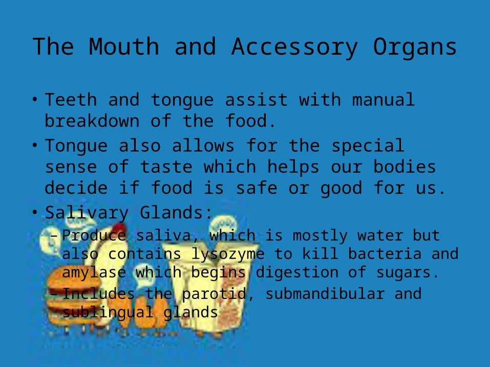
The Mouth and Accessory Organs
• Teeth and tongue assist with manual breakdown of the food.
• Tongue also allows for the special sense of taste which helps our bodies decide if food is safe or good for us.
• Salivary Glands: – Produce saliva, which is mostly water but also contains
lysozyme to kill bacteria and amylase which begins digestion of sugars.
– Includes the parotid, submandibular and sublingual glands

The Esophagus
• This is a 10 inch tube that connects the lower portion of the pharynx to the stomach. It passes through the diaphragm.
• It ends in a ring like muscular structure called the cardiac sphincter that prevent stomach contents from regurgitating.

The Stomach• Located just below the diaphragm and has three
portions: – Fundus (upper part)– Body (greater and lesser curvature or middle portion)– Pylorus (lower or distal portion)
• Has the same layers as the rest of the alimentary canal, but he inner mucosa has lots of folds called rugae.
• The pylorus ends in another sphincter (like the esophagus) called the pyloric sphincter which regulates food passing into the duodenum (small intestine).

Small Intestine
• Continues the process of digestion and does most of the absorption of nutrients. It has 3 portions: – Duodenum (most proximal)– Jejunum (middle)– Ileum (most distal)
• Similar to the inner lining of the stomach, the mucosa of the small intestine has folds called plicae circularis (increase surface area). It is also covered with villi.

Absorption in the Small Intestine
• As the food (now called chyme) bolus moves toward the distal end of the small intestine, more and more nutrients are absorbed (after being broken down as much as possible).
• The plicae and villi drastically increase surface area which helps with this, and each villus has capillaries which absorb and transport he nutrients.

The Large Intestine• About 5 feet long, this final part of the alimentary canal
focuses mostly on reabsorbing water and a few nutrients such as B vitamins and vitamin K, and producing/eliminating waste.
• It has 6 portions (proximal to distal)– Cecum (pouch like area below the ileocecal valve, attached is
the appendix)– Ascending Colon– Transverse Colon– Descending Colon– Sigmoid Colon– Rectum (including anus)

Physiology of the Large Intestine
• Inner Mucosa produces lots of mucus to allow the solidifying bolus of waste to move easily.
• The colon is gathered by the teniae coli (muscle bands) and formed into pockets called haustra.
• Vitamin absorption and (possibly) immune functions are heavily impacted by the large amount of coliform bacteria that lives in the large intestine.

Elimination of waste
• The Waste product we eliminate is called either feces or stool and is comprised of unabsorbable food parts like dense fiber and seeds, bacteria, waste products from metabolism, mucus, and gases.
• Cellulose if what we call fiber and is important in keeping the bowels healthy and regular.
• Feces should be moderate to dark brown in color. Other colors can mean disease, illness or unhealthy diet.