differentiation between mass-forming type peripheral ... › synapse › data › pdfdata ›...
TRANSCRIPT

Several CT features of hepatic abscesses have been re-ported (1-4). Some hepatic abscesses occasionally mim-
ic other hepatic tumors on CT, and some malignant he-patic tumors such as metastases, cholangiocarcinoma(CC), and leiomyosarcoma, may also mimic hepatic ab-scesses (5-12). Since the treatment strategy differsbased on the possibility of malignancy, differentiation ofhypovascular liver tumors such as CC or metastasisfrom hepatic abscesses on CT is important. A variety ofradiologic procedures have been used to evaluate focal
J Korean Radiol Soc 2005;53:343-352
─ 343 ─
Differentiation Between Mass-forming Type PeripheralCholangiocarcinoma and Hepatic Abscesses: Application of Artificial Neural Networks to CT Images1
Nak Jong Seong, M.D., Jeong Min Lee, M.D., Se Hyung Kim, M.D., Joon Koo Han, M.D., Young Jun Kim, M.D., Ji Hoon Kim, M.D., Jae Young Lee, M.D.,
Seong Ho Park, M.D.2, Byung Ihn Choi, M.D.
1Department of Radiology and the Institute of Radiation Medicine, SeoulNational University Hospital and 3Asan Medical Center Received April 25, 2005 ; Accepted August 4, 2005Address reprint requests to : Jeong Min Lee, M.D., Department ofRadiology, Seoul National University College of Medicine28, Yongon-dong, Chongno-gu, Seoul 110-744, Korea.Tel. 82-2-2072-3154 Fax. 82-2-743-6385 E-mail: [email protected]
Purpose: To determine which CT findings are useful for differentiating cholangiocar-cinomas (CC) from hepatic abscesses and also to determine whether artificial neuralnetworks (ANNs) improve radiologists’ performance.Materials and Methods: CT findings of 51 patients with mass-forming type CC and 70patients with hepatic abscesses were analyzed with morphologic, enhancing and otherancillary findings by three radiologists with differing levels of expertise independently.ANNs were constructed using statistically significant CT findings derived from theanalyses. The performances of the ANNs and the radiologists were evaluated using re-ceiver operating characteristic analysis.Results: CT findings of rim-like enhancement, lymphadenopathy, capsular retraction,focal bile duct dilatation and a solid component were significant features of CC (p<0.05). Findings of a clustered sign, multilayered enhancement, sharp margin, roundshape, and air-biliary gram were significant features of hepatic abscesses. The ANNsshowed better performance (AZ=0.9673, 98.0%, 97.1%, and 97.5%, respectively) thanthe resident (AZ=0.898, 78.4%, 81.4%, 80.2%) (p<0.05) in differentiating between thetwo diseases: (AZ, sensitivities, specificities, and overall accuracies). However, therewere no significant differences in the diagnostic performance of the ANNs and the twoboard-certified radiologists.Conclusion: Several CT findings are useful in differentiating CC from hepatic abscess-es and ANNs may improve the performance of a radiologist with little experience.
Index words : Abdomen, CTBile duct, neoplasmLiver, abscessComputers, neural networkDiagnostic radiography, observer performance
liver lesions, with CT currently considered one of themost important noninvasive diagnostic techniques forcharacterizing focal liver lesions (7, 8). Although CTfindings of these two diseases have been published inseveral articles (1-4, 13-15), differentiating CC fromhepatic abscesses, even using sonography, enhanced CTor MRI, remains a difficult task for radiologists becausethere is an overlapping spectrum of radiographic ap-pearances and clinical parameters between the two dis-eases (10, 11, 16, 17). However, to our knowledge, thereare no reports investigating the usefulness of helical CTfor differentiating CC from hepatic abscesses in a largepatient population.
In recent years artificial neural networks (ANNs) havebeen studied intensively computer scientists and havebeen shown to be a powerful tool for a variety of data-classification and pattern-recognition tasks. However,the usefulness of ANNs in diagnostic radiology has fo-cused mainly on chest and breast lesions and in the diag-nosis of pulmonary nodules, interstitial lung disease, pe-diatric lung lesions, and breast nodules (18-23).
The purpose of this study is to determine which CTfindings are useful for differentiating CC from hepaticabscesses and to compare the performance of ANNs ap-plied to CT images with the performance of radiologistswith differing levels of expertise in differentiating be-tween the two diseases using receiver operating charac-teristic (ROC) analysis.
Materials and Methods
Patient Population
In order to find patients diagnosed with hepatic ab-scesses and mass-forming type CC, a computerizedsearch of the electronic medical records including radio-logic, surgical, and pathologic reports from January 1998to March 2003 at the Seoul National UniversityHospital, was performed. Due to the retrospective na-ture of the study and because the institution’s patientssign a general consent form to cover all diagnostic stud-ies, neither institutional review board approval nor in-formed consent was necessary.
The computerized search of the electronic medicalrecords revealed 380 patients diagnosed with hepaticabscesses. Two abdominal radiologists, who did not par-ticipate in the image interpretation, reviewed the avail-able CT scans for these patients and selected patients forsubsequent analysis. Of these patients, 310 were exclud-ed from analysis for the following reasons: (a) no con-
trast-enhanced helical CT scan was available (n=75); (b)the presence of associated malignancy (n=125) such ashepatocellular carcinoma (n=32), gastric cancer (n=20),previous klatskin tumor (n=17), colorectal cancer(n=13), pancreatic cancer (n=10), other malignancies(n=17), and unknown origins (n=16); and (c) no histo-logic confirmation of hepatic abscesses or insufficientimaging follow-up (n=110). The remaining 70 patients(48 male, 22 female; mean age 58.2 years; range 29-91years) with hepatic abscesses were included in thisstudy. Pyogenic abscesses were confirmed by positivecultures from percutaneous aspirates in 31 patients.Five of these 31 patients had monomicrobial abscessesand 26 patients had polymicrobial abscesses. In the re-maining 39 patients that showed no growth of the ab-scess contents, a diagnosis of hepatic abscess was basedon symptoms including fever and abdominal pain, labo-ratory results such as leukocytosis, and the CT imagingfindings. All of these 39 patients responded to percuta-neous drainage and/or antimicrobial therapy, and subse-quent CT scanning revealed improvement of their he-patic abscesses.
The computerized search of the electronic medicalrecords revealed 175 patients diagnosed with intrahep-atic CC. Of these patients, 124 were excluded fromanalysis for the following reasons: (a) no contrast-en-hanced helical CT scan was available (n=107); (b) othertypes of CC were present (n=12; periductal infiltrationin nine patients and intraductal-growing in three); and(c) hyperattenuating mass (n=5) on CT scans. The re-maining 51 patients (37 male, 14 female; mean age 55.4years; range 40-80 years) with the mass-forming typeCC were included in the study. CC was confirmed bypercutaneous ultrasound-guided needle biopsy in 40cases and by surgery in 11 patients.
CT Acquisition
The CT examinations were performed on four differ-ent scanners (Somatom Plus S and Somatom Plus 4;Siemens Medical Systems, Erlangen, Germany;HiSpeed Advantage CT scanner; General ElectricMedical Systems, Milwaukee, WI; MX 8000; andMarconi Medical Systems, Highland Heights, OH). Dueto the retrospective nature of the study several differentCT techniques were used. In general, examinationswere performed using a spiral technique with 5-10 mmcollimation and 5-10 mm reconstruction intervals. TheX-ray tube voltage was 120-140 kV, and the currentvaried between 150 and 200 mA. During this period
Nak Jong Seong, et al : Differentiation Between Mass-forming Type Peripheral Cholangiocarcinoma and Hepatic Abscesses
─ 344 ─

(January 1998 to March 2003), the standard protocol fordynamic CT consisted of a total volume of 120-150 mLof nonionic intravenous contrast material (300-370 mgof iodine per mL) administered by power injection at arate of 2-3 mL/sec, with a scanning delay of 30 secondsfor the hepatic arterial phase (HAP) and of 65 secondsfor the portal venous phase (PVP). Of the 121 patientsanalyzed, 101 (56 with hepatic abscesses and 45 withCC) underwent dual-phase (HAP and PVP) CT and theremaining 20 (14 with hepatic abscesses and six withCC) underwent single-phase (PVP) CT.
Radiologists CT Interpretation
Each of the 121 CT image sets were analyzed retro-spectively and independently by three radiologists (onefaculty-level abdominal radiologist with 20 years experi-ence, one abdominal imaging fellow with 8 years experi-ence, and one senior resident with 3 years experience)who were unaware of the final diagnosis and of anyclinical or laboratory findings. When a patient had mul-tiple lesions, only the largest lesion was analyzed.
Based on previous reports on hepatic abscesses andCC (1-4, 7, 8, 13-15), several imaging features sugges-tive of hepatic abscesses and CC were chosen by thestudy coordinators. The three radiologists were given ashort summary of what had been reported as “typicalfindings” of hepatic abscesses and CC, including mor-phologic features and enhancing characteristics of thelesions and other findings such as lymphadenopathyand metastases (Table 1). The three radiologists inde-pendently reviewed all 121 CT scans at a picture archiv-ing and communications system (PACS) workstation(Marotech, Seoul, Korea) without knowledge of the tis-sue diagnosis or clinical course. During the analysis ofthe CT images, cases of CC and hepatic abscesses werepresented randomly. The readers were asked to grade
their degree of confidence for the presence of variousCT findings believed to be indicative of the two diseasesand for each diagnosis. During the review, the radiolo-gists were allowed to adjust the window width andlength to interpret the CT images. In the CT interpreta-tion, the portal venous phase (PVP) was used as the coreimage set because the lesions are seen the best duringPVP. The degree of confidence for each diagnosis wasgraded as follows: 1, definitely benign (abscess); 2, prob-ably benign; 3, possibly malignant; 4, probably malig-nant; and 5, definitely malignant. The degree of confi-dence for the presence of each of the CT findings indi-cating hepatic abscesses or CC was graded as follows: 1,definitely absent; 2, probably absent; 3, possibly pre-sent; 4, probably present; and 5, definitely present.
The CT findings were analyzed according to two cate-gories; the lesion itself and the associated findings. Thelesion was evaluated for margin (sharp vs ill-defined),configuration (unilocular vs clustered), shape (lobulatedvs round), the presence of a solid component and inter-nal air-density, and the pattern of enhancement (rim-like vs multilayered). Associated findings includedstones in the IHD or CBD, the presence of capsular re-traction, lymphadenopathy (short-axis diameter oflymph nodes more than 1 cm or the presence of centralnecrosis), focal biliary dilatation distal to the lesion, air-biliary gram, transient hepatic attenuation difference(THAD), ascites, pleural effusion, atelectasis of the low-er lung, and extrahepatic metastasis.
Extraction of Significant Imaging Features
A biostatistician participated in the study design andreview of the data. Statistical differences of the CT find-ings between abscesses and CC were analyzed usingFisher’s exact test and the Chi-square test. The odds ra-tio (OR) for each CT feature used to characterize the le-sion into an abscess or CC, was also obtained. The mag-nitude of the relevance of statistically significant CT pre-dictors of abscesses or CC was ranked according to theirP values and ORs. We used binary collapse of the ratedresponses for statistical analysis. Specifically, lesionsthat were graded with scores of 1 and 2 were classifiedas an abscess, and scores of 3, 4, and 5 were classified asCC. Each CT finding that was graded with a score of 1or 2, was classified as absent, whereas, scores of 3, 4,and 5 were as classified as having that CT finding.
Artificial Neural Networks
A brief search of possible ANN designs provided in-
J Korean Radiol Soc 2005;53:343-352
─ 345 ─
Table 1. Useful CT Findings in Differentiating PeripheralCholangiocarcinoma from Hepatic Abscesses
Cholangiocarcinoma Hepatic Abscess
Broad marginal transition Sharp marginal transitionSpiculated outer margin Round outer marginSolid component Cystic componentRim-like enhancement Multilayered enhancementLymph node enlargement Internal air densityCapsular retraction Clustered signFocal bile duct dilatation Air-biliary gram
Transient hepatic attenuationdifference
CT findings were derived from references 1-4 and 13-15.

sight into the type of design that would work for this ap-plication. Several designs were attempted and rated fortheir ability to generalize, once trained. In the presentstudy, fully connected, three-layer ANNs with a backpropagation algorithm were used. The ANNs consistedof one input layer, one hidden layer and one output lay-er. Statistically significant CT features derived from theChi-square or Fisher’s exact test were used as input vari-ables for the input layer. The hidden layer consisted of15 neurons connected to the input layer. A nonlinear,sigmoid function was used as a transfer function for eachof the neurons in the hidden layer and the output layer ofthe networks. The nonlinear function is expressed by:
neuron i and xiwi is the net input to the neuron, whichwas derived by multiplying the weight (w) by the inputvalue (x) of the unit. During the training process, theconnection weights between the neurons were adjustedusing the back propagation updating algorithm (23). Thehidden layer was connected to the output layer of a sin-gle neuron which indicated whether the CT findingswere classified as benign (abscess) or malignant (CC).
The training and testing of the ANNs were performedby means of a leave-one-out method. With this method,all of the cases in the database except for one were usedfor training; the case that was not used was used in thetesting of the trained ANNs. We used a fourfold cross-validation procedure for the training procedure. Each ofthe four different networks was trained until the error inthe training set reached the stopping criterion. This pro-cedure was repeated until every case in the databasewas used once for testing. The test results of the four dif-ferent networks were combined to calculate ANNs’ per-formance in terms of determining the malignancy of thelesion. The output values for the test were between 0and 1, which is used to represent the likelihood of malig-nancy. A threshold value of 0.5 was used, above whichall values were regarded to be malignant.
ROC Analysis
The individual performances of the three radiologistsand of the ANNs were evaluated by ROC analysis; areaswere calculated using a nonparametric method (24, 25).Binormal ROC curves were estimated using theMedCalc software, version 7.1 (MedCalc Software,Mariakerke, Belgium) which was used to obtain maxi-mum-likelihood estimates of binormal ROC curvesfrom the continuous ordinal-scale rating data. The AZ
was calculated to summarize the performance of eachradiologist and the ANNs in the task of classifying he-patic abscesses and CC. The univariate z-score test wasused to assess the significance of differences in the area(AZ) under the two estimated binormal ROC curves. Inaddition, sensitivity and specificity were calculated us-ing only those patients deemed to have CC by each radi-ologist and those with ANNs output levels of 0.5 orgreater. Sensitivity and specificity were presented with95% confidence intervals (CI).
Statistical Analysis
Statistical differences of the CT findings between ab-scesses and CC were analyzed using Fisher’s exact testand the Chi-square test. Interobserver agreementsamong the three radiologists for each CT finding and thefinal radiologic diagnosis were evaluated using theCronbach coefficient α, where an αvalue of greater than0.8 was considered to represent excellent agreementand αvalues of 0.70-0.80 represented good agreement.Values of less than 0.70 were considered to representpoor agreement (25, 26). Statistical analyses were per-formed using SPSS software, version 11.0 for Windows(SPSS Inc., Chicago, Ill). A probability value of less than0.05 was considered to indicate statistical significance.To compare the performance of each radiologist and theANNs, the sensitivity and specificity of the ANNs andeach reviewer were compared using the McNemar test,which is a nonparametric test for two related dichoto-mous variables.
Results
Interobserver agreements among the three radiolo-gists in recognizing the CT findings are presented inTable 2. With respect to the presence of air density with-in the lesion, the three radiologists were in completeagreement (α= 1.00). In terms of most of the CT fea-tures analyzed, almost perfect (for clustered appearanceand multilayered enhancement of the lesion, capsularretraction, focal biliary dilatation distal to the lesion, air-biliary gram, lymphadenopathy, THAD, atelectasis ofthe lower lungs, and pleural effusion) or substantialagreement (rim-like enhancement of the lesion, solidcomponent within the lesion, sharp margin, and config-uration of the lesion) was achieved. For specific diagno-sis, interobserver agreement among the three radiolo-gists was almost perfect (α= 0.907). In addition, interob-server agreement of each combination of two radiolo-
Nak Jong Seong, et al : Differentiation Between Mass-forming Type Peripheral Cholangiocarcinoma and Hepatic Abscesses
─ 346 ─
11+e-∑xiwi
f(∑i
xiwi)= , where f(∑i
xiwi) is the output of a
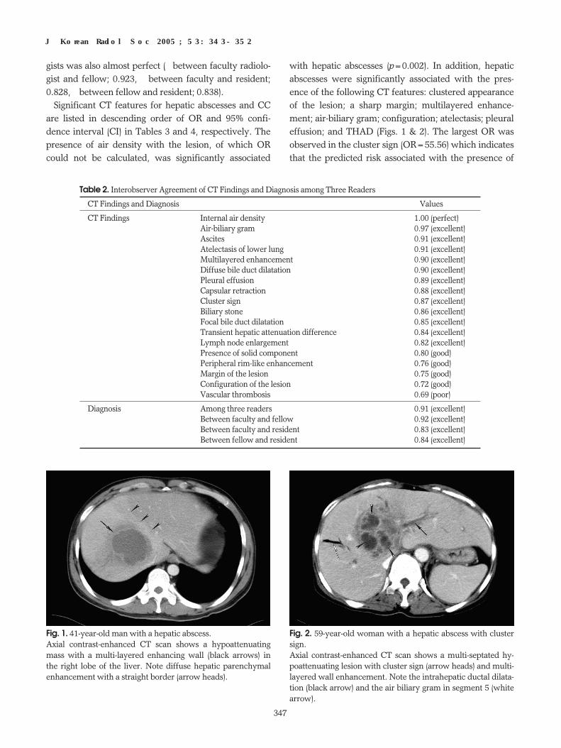
gists was also almost perfect (αbetween faculty radiolo-gist and fellow; 0.923, αbetween faculty and resident;0.828, αbetween fellow and resident; 0.838).
Significant CT features for hepatic abscesses and CCare listed in descending order of OR and 95% confi-dence interval (CI) in Tables 3 and 4, respectively. Thepresence of air density with the lesion, of which ORcould not be calculated, was significantly associated
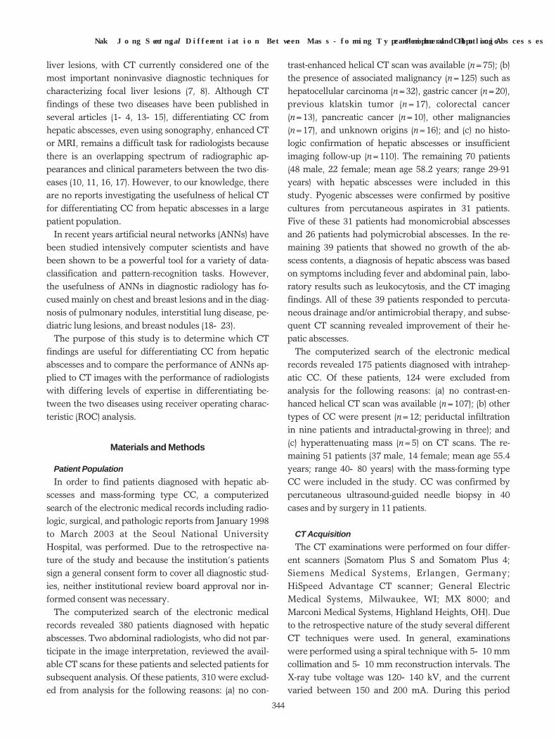
with hepatic abscesses (p=0.002). In addition, hepaticabscesses were significantly associated with the pres-ence of the following CT features: clustered appearanceof the lesion; a sharp margin; multilayered enhance-ment; air-biliary gram; configuration; atelectasis; pleuraleffusion; and THAD (Figs. 1 & 2). The largest OR wasobserved in the cluster sign (OR=55.56) which indicatesthat the predicted risk associated with the presence of
J Korean Radiol Soc 2005;53:343-352
─ 347 ─
Fig. 1. 41-year-old man with a hepatic abscess.Axial contrast-enhanced CT scan shows a hypoattenuatingmass with a multi-layered enhancing wall (black arrows) inthe right lobe of the liver. Note diffuse hepatic parenchymalenhancement with a straight border (arrow heads).
Fig. 2. 59-year-old woman with a hepatic abscess with clustersign.Axial contrast-enhanced CT scan shows a multi-septated hy-poattenuating lesion with cluster sign (arrow heads) and multi-layered wall enhancement. Note the intrahepatic ductal dilata-tion (black arrow) and the air biliary gram in segment 5 (whitearrow).
Table 2. Interobserver Agreement of CT Findings and Diagnosis among Three Readers
CT Findings and Diagnosis αValues
CT Findings Internal air density 1.00 (perfect)Air-biliary gram 0.97 (excellent)Ascites 0.91 (excellent)Atelectasis of lower lung 0.91 (excellent)Multilayered enhancement 0.90 (excellent)Diffuse bile duct dilatation 0.90 (excellent)Pleural effusion 0.89 (excellent)Capsular retraction 0.88 (excellent)Cluster sign 0.87 (excellent)Biliary stone 0.86 (excellent)Focal bile duct dilatation 0.85 (excellent)Transient hepatic attenuation difference 0.84 (excellent)Lymph node enlargement 0.82 (excellent)Presence of solid component 0.80 (good)Peripheral rim-like enhancement 0.76 (good)Margin of the lesion 0.75 (good)Configuration of the lesion 0.72 (good)Vascular thrombosis 0.69 (poor)
Diagnosis Among three readers 0.91 (excellent)Between faculty and fellow 0.92 (excellent)Between faculty and resident 0.83 (excellent)Between fellow and resident 0.84 (excellent)

the cluster sign on CT was 55.56 times higher for pa-tients with hepatic abscesses than those with CC.
The CT features of peripheral rim-like enhancement,lymphadenopathy, capsular retraction, focal biliary di-latation distal to the lesion, and a solid component with-in the lesion were significant for diagnosing CC (Figs. 3& 4). The largest OR was observed in the rim-like en-hancement of the lesion (OR = 49.78) (Fig. 3).
Tables 5 and 6 list the diagnostic performances of radi-ologists and the ANNs in terms of AZ, sensitivity andspecificity in diagnosing CC. The performance of theANNs (0.966, 95% CI: 0.916, 0.990), the faculty level ra-diologist (0.947, 95% CI: 0.891, 0.979), and the radiolog-ic fellow (0.946, 95% CI: 0.889, 0.978) were significantlybetter in terms of AZ than that of the resident (0.873,95% CI: 0.800, 0.926) based on the outcome of the z-score test (p<0.049). However, the performance be-tween the ANNs, faculty, and fellow were not signifi-cantly different (p>0.05). For diagnosing CC at a ratingof 3 or larger, the sensitivities of the faculty (48/51,94.1%; 95% CI: 0.835, 0.986), fellow (48/51, 94.1%; 95%CI: 0.835, 0.986), and the ANNs (49/51, 96.1%; 95% CI:0.865, 0.995) were significantly better than that of theresident (40/51, 78.4%; 95% CI: 0.652, 0.877) (p<0.05).However, the differences in sensitivity between the two
radiologists (faculty and fellow) and the ANNs were notstatistically significant according to the results of theMcNemar test (p>0.05). For diagnosing an abscess at arating of 1 or 2, the sensitivity was the same as the speci-ficity for diagnosing the CC of ANNs (68/70, 97.1%; 95%CI: 0.901, 0.997) and was statistically better than that ofthe resident (57/70, 81.4%; 95% CI: 0.706, 0.890) (p=0.005) (Table 6). However, the specificities from the
Nak Jong Seong, et al : Differentiation Between Mass-forming Type Peripheral Cholangiocarcinoma and Hepatic Abscesses
─ 348 ─
Fig. 3. 57-year-old man with cholangiocarcinoma. Axial contrast-enhanced CT scan shows an irregular masswith rim-like enhancement (black arrows) and capsular retrac-tion (arrow head).
Table 3. Significant CT Findings for Cholangiocarcinoma and their Odds Ratio
Odds Ratio
CT Findings Odds Ratio 95% Confidence Interval p value
Lower Upper
Peripheral rim-like enhancement 49.78 16.51 150.12 < 0.0001Lymph node enlargement 21.94 04.83 099.73 < 0.0001Capsular retraction 21.23 02.66 169.51 < 0.0001Focal bile duct dilatation 16.94 04.70 061.09 < 0.0001Presence of solid component 08.41 03.18 022.24 < 0.0001
Table 4. Significant CT Findings for Hepatic Abscesses and their Odds Ratio
Odds Ratio
CT Findings Odds Ratio 95% Confidence Interval p value
Lower Upper
Inner air density Specific finding - - 0.002Cluster sign 55.56 7.35 500.00 < 0.0001Multilayered enhancement 29.41 6.54 125 < 0.0001Sharp margin 21.28 8.00 55.56 < 0.0001Air-biliary gram 12.50 1.59 100 0.004Configuration 10.64 4.18 27.03 < 0.0001Atelectasis of lower lungs 09.43 3.72 23.81 < 0.0001Pleural effusion 06.90 2.44 19.61 < 0.0001THAD 02.75 1.30 5.81 0.01
THAD= transient hepatic attenuation difference.

three radiologists were not significantly different fromeach other (faculty; 63/70, 90.0%, 95% CI: 0.905, 0.954,fellow; 61/70, 87.1%, 95% CI: 0.771, 0.933, resident;57/70, 81.4%, 95% CI: 0.706, 0.890) (p> 0.05).
Discussion
Differentiating between hepatic abscesses and mass-forming type intrahepatic CC on CT remains a difficultproblem because a considerable overlap between their
J Korean Radiol Soc 2005;53:343-352
─ 349 ─
Fig. 4. 55-year-old man with cholangiocarcinoma presentingwith fever and vague right upper quadrant pain. Axial contrast-enhanced CT scan shows a large hypoattenuat-ing mass with focal bile duct dilatation (black arrow). Thepresence of a heterogeneously enhancing solid component ofthe mass (arrowheads) and the absence of a multi-layered en-hancing pattern or transient hepatic attenuation differencehelps to determine the diagnosis of cholangiocarcinoma.
Fig. 5. 56-year-old man with cholangiocarcinoma and accom-panying fever and leukocytosis.Axial contrast-enhanced CT scan shows a low attenuating le-
sion with multi-layered enhancement (arrow) in the right pos-terior segment of the liver, which mimics a hepatic abscess.The resident reviewer diagnosed this lesion as a hepatic ab-scess, but the other radiologists correctly diagnosed it ascholangiocarcinoma.
Table 5. Diagnostic Performances of Three Radiologists and ANNs for Diagnosing Cholangiocarcinoma using Receiver OperatingCharacteristic Analysis
AZ
Value 95% CI p value
Upper Lower For ANNs For Faculty For Fellow
Faculty 0.947 0.891 0.979 0.359 - 0.957Fellow 0.946 0.889 0.978 0.448 0.957 -Resident 0.873 0.800 0.926 0.012 0.049 0.041ANNs 0.966 0.916 0.990 - 0.359 0.448
Two-tailed p values were calculated between each reader and the ANNs from the univariate z-score test. ANNs = artificial neural net-works, CI = confidence interval
Table 6. Sensitivities and Specificities of Three Radiologists and ANNs for Diagnosing Cholangiocarcinoma
Sensitivity Specificity
Value95% CI p value
Value95% CI p value
Upper Lower For For For For For For ANNs Faculty Fellow Upper Lower ANNs Faculty Fellow
Faculty 94.1 (48/51) 0.835 0.986 1.000 - 1.000 90.0 (63/70) 0.805 0.954 0.165 - 0.791Fellow 94.1 (48/51) 0.835 0.986 1.000 1.000 - 87.1 (61/70) 0.771 0.933 0.055 0.791 -Resident 78.4 (40/51) 0.652 0.877 0.015 0.041 0.041 81.4 (57/70) 0.706 0.890 0.005 0.227 0.487ANNs 96.1 (49/51) 0.865 0.995 - 1.000 1.000 97.1 (68/70) 0.901 0.997 - 0.165 0.055
* Data in parentheses are the numbers from which the proportions were calculated. p values were calculated between each reader andthe ANNs from the McNemar chi-square test. ANNs = artificial neural networks, CI = confidence interval.

clinical presentations and radiologic appearance may ex-ist (9-11, 17, 27). Despite that, there have been manyreports on CT findings of hepatic abscesses (1-4) andCC (13-15), with CC often being misinterpreted as a he-patic abscess and mistreated with percutaneousdrainage which can cause tract seeding or peritonealspillage of the tumor (28). Many previous studies havedescribed simple observations of the CT findings foreach disease, but no study has attempted to provide dif-ferential points by dedicated statistical analysis or to de-velop diagnostic algorithms to differentiate them.Recently, several publications have focused on the ap-plication of ANNs for lesion classification in the field ofradiology (29-31). In this study, we attempted to con-struct ANNs to differentiate hepatic abscesses from CCon CT and to determine whether ANNs are able to im-prove radiologists’ performance in differentiating be-tween the two diseases.
The ANNs constructed for this study using significantCT findings derived from univariate analysis as multipleinput variables showed a high performance in differenti-ating between hepatic abscesses and CC. The AZ value,sensitivity and specificity of the ANNs were significant-ly greater than those of the resident and marginally bet-ter than those of the other radiologists. However, therewere no statistically significant differences between theANNs and the two radiologists. Considering the excel-lent agreement for each CT finding among the three ra-diologists, the low performance of the resident is per-haps explained by the fact that a resident may not beable to effectively organize all of the CT features system-atically. On the other hand, ANNs consistently andcomprehensively respond to all data that has been in-put. In addition, it was expected that the experienced ra-diologists were aware of the relative importance of thesignificant CT findings based on their experience indealing with various CT findings, which was used tohelp differentiate between the two diseases. Therefore,we are not surprised at the excellent performanceshown by the ANNs and the abdominal radiologists indifferentiating between the two diseases. We believethat the inclusion of a fellow and a resident as reviewersmakes our results and conclusions more applicable forindividuals with varying levels of experience.
In this study, hepatic abscesses were characterized bylesions having inner air-density, cluster sign, multilay-ered enhancement, sharp margin, and a lobulated con-figuration. In addition, hepatic abscesses were also morefrequently associated with air-biliary gram, atelectasis of
the lower lungs, pleural effusion, and transient hepaticattenuation difference (THAD). On the other hand, le-sions showing peripheral rim-like enhancement, capsu-lar retraction, focal bile duct dilatation distal to the le-sion, a solid component or which had lymph node en-largement, were more frequently associated with pa-tients with a mass-forming type CC. The results of thisstudy are consistent with those of previous reports (1-4,13-16).
In this study, ancillary findings as well as morphologicor enhancing features of the lesion were helpful in dif-ferentiating between the two diseases. The presence ofan air-biliary gram is one of the factors found to be a sig-nificant predictor for hepatic abscesses. Among the fourmain routes for hepatic abscesses, i.e. ascending, portal,hematogenous, and iatrogenic, ascending infectionswere the most common cause of hepatic abscesses.From this point of view, the result of the air-biliary gramwhich suggests free communication between the bileduct and the enteric system was a significant CT findingfor hepatic abscesses. We can also assume that reactivechanges such as atelectasis of the lower lungs, pleural ef-fusion or THAD associated with hyperemic changescould be observed more frequently in benign inflamma-tory lesions, and our results support this premise.
In conclusion, specific CT findings including multilay-ered, clustered or rim-like enhancement patterns can behelpful in diagnosing hepatic abscesses and mass-form-ing type CC. In addition, ANNs may potentially im-prove a radiologist’s performance and assist in differen-tiating CC from hepatic abscesses, especially in thetrainees with little experience in interpreting CT images.
References
1. Mathieu D, Vasile N, Fagniez PL, Segui S, Grably D, Larde D.Dynamic CT features of hepatic abscesses. Radiology 1985;154:749-752
2. Jeffrey RB Jr, Tolentino CS, Chang FC, Federle MP. CT of pyo-genic hepatic microabscesses: the cluster sign. AJR Am JRoentgenol 1988;151:487-489
3. Rubinson HA, Isikoff MB, Hill MC. Morphologic aspects of hepat-ic abscesses: a retrospective analysis. AJR Am J Roentgenol 1980;135:735-740
4. Halvorsen RA, Korobkin M, Foster WL, Silverman PM,Thompson WM. The variable CT appearance of hepatic abscesses.AJR Am J Roentgenol 1984;141:941-946
5. Subramanyam BR, Balthazar EJ, Raghavendra BN, Horii SC,Hilton S, Naidich DP. Ultrasound analysis of solid-appearing ab-scesses. Radiology 1983;146:487-491
6. Gabata T, Kadoya M, Matsui O, Kobayashi T, Kawamori Y,Sanada J, et al. Dynamic CT of hepatic abscesses: significance oftransient segmental enhancement. AJR Am J Roentgenol 2001;176:
Nak Jong Seong, et al : Differentiation Between Mass-forming Type Peripheral Cholangiocarcinoma and Hepatic Abscesses
─ 350 ─
675-6797. Mortele KJ, Ros PR. Cystic focal liver lesions in the adult: differen-
tial CT and MR imaging features. Radiographics 2001;21:895-9108. Murphy BJ, Casillas J, Ros PR, Morillo G, Albores-Saavedra J,
Rolfes DB. The CT appearance of cystic masses of the liver.Radiographics 1989;9:307-322
9. Ryan RS, Al-Hashimi H, Lee MJ. Hepatic abscesses in elderly pa-tients mimicking metastatic disease. Ir J Med Sci 2001;170:251-253
10. Jan YY, Yeh TS, Chen MF. Cholangiocarcinoma presenting aspyogenic abscess: is its outcome influenced by concomitant hepa-tolithiasis? Am J Gastroenterol 1998;93:253-255
11. Jin GY, Lee JM, Yu HC, Mun WS, Kim CS. Intraductal papillarycholangiocarcinoma with aneurismal dilation: a case of the mimic-king abscess. Hepatogastroenterology 2002;49:1523-1525
12. Gates LK Jr, Cameron AJ, Nagorney DM, Goellner JR, Farley DR.Primary leiomyosarcoma of the liver mimicking liver abscess. AmJ Gastroenterol 1994; 89: 1541-1543
13. Kim TK, Choi BI, Han JK, Jang HJ, Cho SG, Han MC. Peripheralcholangiocarcinoma of the liver: two-phase spiral CT findings.Radiology 1997;204:539-543
14. Choi BI, Han JK, Shin YM, Baek SY, Han MC. Peripheral cholan-giocarcinoma: comparison of MRI with CT. Abdom Imaging 1995;20:357-360
15. Choi BI, Lee JM, Han JK. Imaging of intrahepatic and hilar cholan-giocarcinoma. Abdom Imaging 2004;29:548-557
16. Maetani Y, Itoh K, Watanabe C, Shibata T, Ametani F, YamabeH,, et al. MR imaging of intrahepatic cholangiocarcinoma withpathologic correlation. AJR Am J Roentgenol 2001;176:1499-1507
17. Chan JH, Tsui EY, Luk SH, Fung AS, Yuen MK, Szeto ML, et al.Diffusion-weighted MR imaging of the liver: distinguishing hepaticabscess from cystic or necrotic tumor. Abdom Imaging 2001;26:161-165
18. Henschke CI, Yankelevitz DF, Mateescu I, Brettle DW, RaineyTG, Weingard FS. Neural networks for the analysis of small pul-monary nodules. Clin Imaging 1997;21:390-399
19. Asada N, Doi K, MacMahon H, Montner SM, Giger ML, Abe C, etal. Potential usefulness of an artificial neural network for differen-tial diagnosis of interstitial lung diseases: pilot study. Radiology1990;177:857-860
20. Ashizawa K, Ishida T, MacMahon H, Vyborny CJ, Katsuragawa S,
Doi K. Artificial neural networks in chest radiography: applicationto the differential diagnosis of interstitial lung disease. Acad Radiol1999;6:2-9
21. Ashizawa K, MacMahon H, Ishida T, Nakamura K, Vyborny CJ,Katsuragawa S, et al. Effect of an artificial neural network on radi-ologists’ performance in the differential diagnosis of interstitiallung disease using chest radiographs. AJR Am J Roentgenol 1999;172:1311-1315
22. Wu Y, Giger ML, Doi K, Vyborny CJ, Schmidt RA, Metz CE.Artificial neural networks in mammography: application to deci-sion making in the diagnosis of breast cancer. Radiology 1993;187:81-87
23. Cross SS, Harrison RF, Kennedy RL. Introduction to neural net-works. Lancet 1995;346:1075-1079
24. Metz CE. ROC methodology in radiologic imaging. Invest Radiol1986;21:720-733
25. Metz CE. Some practical issues of experimental design and dataanalysis in radiological ROC studies. Invest Radiol 1989;24:234-245
26. Blachar A, Federle MP, Ferris JV, Lacomis JM, Waltz JS, ArmfieldDR, et al. Radiologists’ performance in the diagnosis of liver tu-mors with central scars by using specific CT criteria. Radiology2002;223:532-539
27. Nino-Murcia M, Olcott EW, Jeffrey RB Jr, Lamm RL, Beaulieu CF,Jain KA. Focal liver lesions: pattern-based classification scheme forenhancement at arterial phase CT. Radiology 2000;215:746-751
28. Kaneko T, Nakao A, Oshima K, Iizuka A. Rapid progression of in-trahepatic cholangiocarcinoma after drainage of large cystic le-sions: report of a case. Surg Today 2000;30:1049-1052
29. Chen CM, Chou YH, Han KC, Hung GS, Tiu CM, Chiou HJ, et al.Breast lesions on sonograms: computer-aided diagnosis with near-ly setting-independent features and artificial neural networks.Radiology 2003;226:504-514
30. Nakamura K, Yoshida H, Engelmann R, MacMahon H,Katsuragawa S, Ishida T, et al. Computerized analysis of the likeli-hood of malignancy in solitary pulmonary nodules with use of arti-ficial neural networks. Radiology 2000;214:823-830
31. Abe H, Ashizawa K, Li F, Matsuyama N, Fukushima A, Shiraishi J,et al. Artificial neural networks for differential diagnosis of intersti-tial lung disease: results of a simulation test with actual clinicalcases. Acad Radiol 2004;11:29-37
J Korean Radiol Soc 2005;53:343-352
─ 351 ─

Nak Jong Seong, et al : Differentiation Between Mass-forming Type Peripheral Cholangiocarcinoma and Hepatic Abscesses
─ 352 ─
대한영상의학회지 2005;53:343-352
종괴형 간담도암과 간농양의 감별: CT소견의 인공신경망 적용1
1서울대학병원진단방사선과2울산의대아산병원진단방사선과
성낙종·이정민·김세형·한준구·김영준·김지훈·이재영·박성호2·최병인
목적: 간담도암과 간농양의 감별진단에 유용한 CT소견을 알아보고, 인공신경망이 진단방사선과 의사의 수행향상에 도
움이 되는지를 알고자 하였다.
대상과 방법: 51명의 종괴형 간담도암 환자와 70명의 간농양 환자의 CT소견을 3명의 방사선과의사가 병변의 형태와
조영소견에 따라 분석하였다. 인공신경망은 이 분석으로 얻어진 유의한 소견을 이용하여 훈련되었으며 인공신경망 및
방사선과의사의 수행능력은 Receiver Operating Characteristic Analysis로 평가하였다.
결과: 테두리조영증강, 림프절종대, 피막수축, 국소담관확대, 고형성분등은 간담도암의 중요 소견이었으며 군집성모양,
다층조영증강, 뚜렷한 경계, 둥근 모양과 담도내공기등은 간농양의 중요소견이었다(p<0.05). 인공신경망
(AZ=0.9673, 98.0%, 97.1%, and 97.5%)은 두 질환의 감별진단에 있어서 전공의(AZ=0.898, 78.4%, 81.4%,
80.2%)보다 좋은 수행능력을 보여주었다: (AZ, 민감도, 특이도, 정확성)(p<0.05) 그러나, 인공신경망과 전문의 사이
에는 수행능력에 유의한 차이는 없었다.
결론: 몇가지 CT소견은 간담도암과 간농양의 감별에 유용하며 인공신경망은 진단방사선과 전공의들의 수행능력 향상
에 도움을 준다.