differential transcription of fathead minnow immune...
TRANSCRIPT
Differential transcription of fathead minnow immune-related genesfollowing infection with frog virus 3, an emerging pathogen ofectothermic vertebrates
Kwang Cheng a, B. Lynn Escalon b, Jacques Robert c, V. Gregory Chinchar a,n,Natàlia Garcia-Reyero d
a Department of Microbiology, University of Mississippi Medical Center, Jackson, MS 39211, USAb US Army Corps of Engineers, Engineering Research and Development Center, Vicksburg, MS 39180, USAc Department of Microbiology and Immunology, University of Rochester Medical Center, Rochester, NY 14642, USAd Institute for Genomics, Biocomputing & Biotechnology, Mississippi State University, Starkville, MS 39759, USA
a r t i c l e i n f o
Article history:Received 30 January 2014Returned to author for revisions3 March 2014Accepted 13 March 2014Available online 1 April 2014
Keywords:RanavirusIridovirusAnti-viral immunityInterferonFHM microarrayFathead minnow cellsImmune-related genesFrog virus 318K knock out mutant
a b s t r a c t
Frog virus 3 (FV3) and other ranaviruses are responsible for die-offs involving wild, farmed, and captiveamphibians, fish, and reptiles. To ascertain which elements of the immune system respond to infection,we explored transcriptional responses following infection of fathead minnow cells with either wild type(wt) FV3 or a knock out (KO) mutant targeting the 18 kDa immediate early gene (18K). At 8 h postinfection we observed marked upregulation of multiple transcripts encoding proteins affecting innateand acquired immunity. Sequences expressed 4-fold or higher in wt-infected cells included transcriptsencoding interferon (IFN), IFN regulatory factors (IRFs), IFN stimulated genes (ISGs) such as Mx and MHCclass I, and interleukins IL-1β, IL-8, IL-17C and IL-12. Cells infected with the 18K KO mutant (Δ18K)showed qualitative differences and lower levels of induction. Collectively, these results indicate thatranavirus infection induced expression of multiple cellular genes affecting both innate and acquiredimmunity.
& 2014 Elsevier Inc. All rights reserved.
Introduction
During the past 30 years, ranaviruses have caused considerablemorbidity and mortality among ectothermic vertebrates, i.e.,amphibians, fish, and reptiles (Chinchar et al., 2009, 2011).Although infections with the chytrid fungus Batracochytriumdendrobatidis are responsible for the extinction of several amphi-bian species and have been viewed as the principal pathogenthreatening amphibians, ranavirus infections are common and arethe leading cause of localized die-offs among amphibians in NorthAmerica (Gray et al., 2009; Green et al., 2002). Moreover, ranavirusinfections affect multiple species throughout the world and it isthought that die-offs may push small populations with limitedgeographic ranges to extinction.
Frog Virus 3 is the best-characterized member and the typespecies of the genus Ranavirus (family Iridoviridae), a group oflarge, icosahedral, double-stranded DNA viruses (Chinchar, 2002;
Chinchar et al., 2009). Within the family Iridoviridae, two genera(Iridovirus and Chloriridovirus) infect invertebrates, whereas threegenera (Ranavirus, Lymphocystivirus, and Megalocytivirus) targetcold-blooded vertebrates (Jancovich et al., 2012). Among these fivegenera, ranaviruses and megalocytiviruses are currently viewed asemerging pathogens of fish and amphibians (Chinchar et al.,2009).
Ranaviruses are promiscuous pathogens and infect a widerange of species belonging to one or more taxonomic classes(Jancovich et al., 2012). For example, although originally isolatedfrom leopard frogs, FV3 and FV3-like viruses have been detected inother frog species, as well as from salamanders, fish, and turtles(Chinchar and Waltzek, 2014). Previous studies (Gantress et al.,2003; Tweedell and Granoff, 1968) demonstrated that althoughimmunocompetent adults confine ranavirus infection to the kid-ney and successfully recover, tadpoles fail to clear infection,develop systemic disease, and succumb to infection. The sensitiv-ity of tadpoles to infection likely reflects the fact that tadpoles lackfull expression of MHC class I molecules and are thought to bedeficient in the development of T cell responses (Robert and Ohta,2009). Moreover, metamorphosis imposes considerable metabolic
Contents lists available at ScienceDirect
journal homepage: www.elsevier.com/locate/yviro
Virology
http://dx.doi.org/10.1016/j.virol.2014.03.0140042-6822/& 2014 Elsevier Inc. All rights reserved.
n Corresponding author. Tel.: þ1 601 984 1743.E-mail address: [email protected] (V.G. Chinchar).
Virology 456-457 (2014) 77–86
costs on amphibians and heightens their susceptibility to severeranavirus infections (Rollins-Smith, 1998). In contrast, adult frogsdevelop protective innate and acquired responses, the latterinvolving the induction of anti-viral antibodies and cytotoxic Tcells (Maniero et al., 2006; Morales and Robert, 2007; Robert et al.,2005). While cellular genes play critical roles in protection andrecovery from ranavirus infection, viral genes are thought to playimportant roles as possible immune antagonists. For examplepoxviruses encode a dozen or more genes whose function is tocircumvent various aspects of the host immune response orenhance replication (Finlay and McFadden, 2006; Johnston et al.,2005; Johnston and McFadden, 2003; Seet et al., 2003; Wang et al.,2009). Ranaviruses likely encode functionally similar genes and,consistent with that view, a FV3 knock out mutant lacking the18 kDa immediate early gene (Δ18K-FV3) displayed reduced leth-ality following infection of Xenopus tadpoles (Chen et al., 2011).Likewise, an Ambystoma tigrinum virus (ATV) mutant lacking theviral homolog of eukaryotic initiation factor 2 (vIF-2α), a putativevirulence gene, was unable to prevent the phosphorylation andsubsequent inactivation of eIF-2α and displayed slightly lessvirulence in vivo (Jancovich and Jacobs, 2011)
In this study we explored whether FV3 infection inducedexpression of immune-related genes in fathead minnow (Pime-phales promelas, FHM) cells, a permissive epithelial line commonlyused to study the replication of FV3 and other ranaviruses. FHMcells were infected with either wt FV3 or Δ18K-FV3 and assayed todetermine whether immune-related genes were differentiallyexpressed. Expression was monitored using a 60 K-feature FHMmicroarray and results of several key genes were validated byquantitative real time RT-PCR (qPCR). As discussed below, weobserved induction of numerous immune-related genes in cellsinfected with wt- or Δ18K-FV3 virus suggesting that host imm-une responses likely play critical roles in combating ranavirusinfection.
Results
Induction of host immune-related transcripts
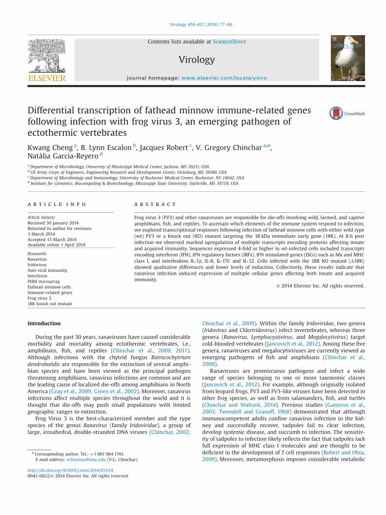
Ranavirus infections result in the progressive inhibition ofcellular RNA and protein synthesis (Raghow and Granoff, 1979;Tannenbaum et al., 1978, 1979). As a consequence, virus-inducedhost gene expression is likely a transient event that takes placewithin a window defined by virus-induced onset and virus-mediated inhibition of host protein and RNA synthesis. To deter-mine the time at which cellular immune-related transcripts werepresent, we infected FHM cells at a multiplicity of infectionsufficient to infect all cells (5 PFU/cell) and monitored expressionof the viral major capsid protein (MCP) gene and cellular tran-scripts encoding Mx and β actin by RT-PCR. Expression of the MCP,a late viral gene product, serves as a marker of infection, Mx, acellular anti-viral protein whose expression is induced by inter-feron (IFN), is a marker for virus-induced immune-related tran-scripts, and β actin is a constitutively expressed housekeepinggene that serves as an indicator for RNA integrity. As shown inFig. 1, MCP transcripts were not detected in uninfected cells,marginally expressed at 4 h p.i., abundant at 8 h, and maximallypresent at 16 h. Reflective of a low level of constitutive synthesis,Mx transcripts were present at reduced levels in mock-infectedcells, but were upregulated by 4 h p.i. and remained at high levelsthereafter. As expected, β-actin levels were abundant and constantthroughout infection. Based on these results, cellular gene expres-sion was examined at 8 h p.i. as this time point provides anexcellent opportunity for detecting cellular genes that weredifferentially regulated by virus infection.
Ranavirus-induced transcriptional changes
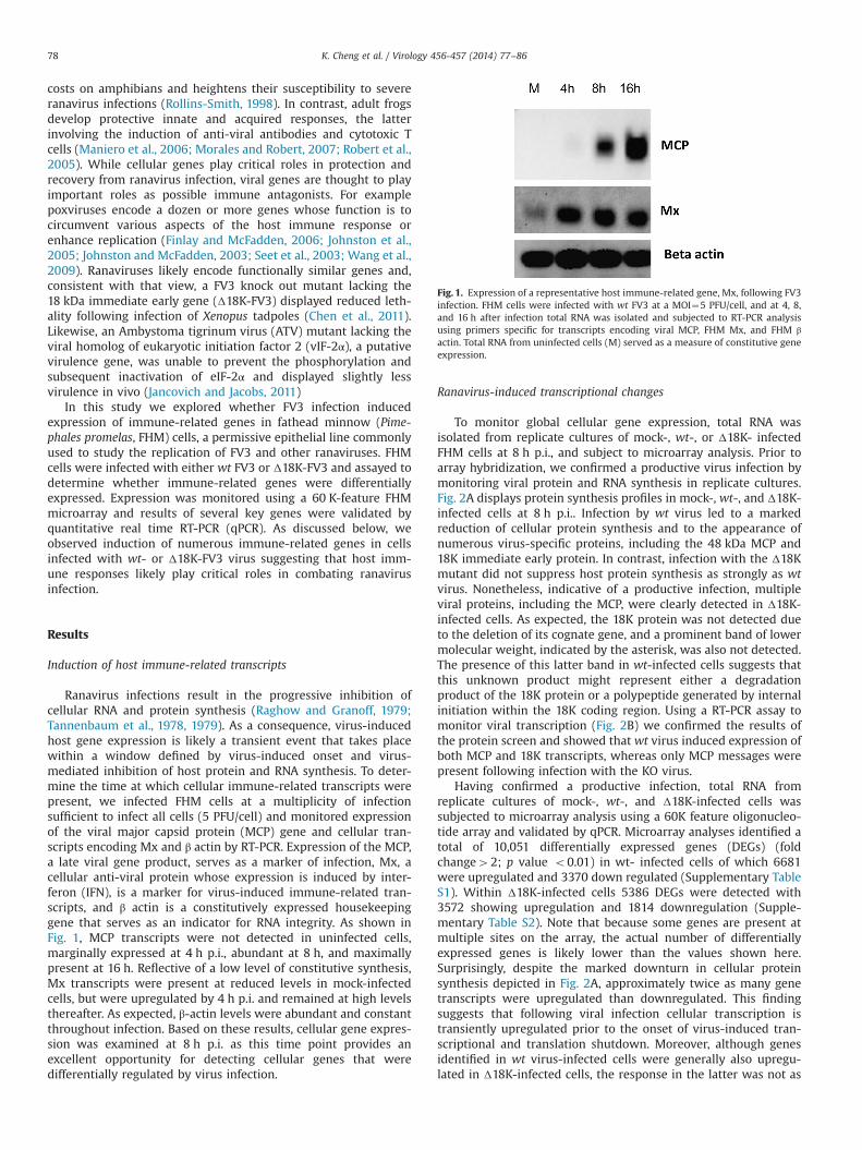
To monitor global cellular gene expression, total RNA wasisolated from replicate cultures of mock-, wt-, or Δ18K- infectedFHM cells at 8 h p.i., and subject to microarray analysis. Prior toarray hybridization, we confirmed a productive virus infection bymonitoring viral protein and RNA synthesis in replicate cultures.Fig. 2A displays protein synthesis profiles in mock-, wt-, and Δ18K-infected cells at 8 h p.i.. Infection by wt virus led to a markedreduction of cellular protein synthesis and to the appearance ofnumerous virus-specific proteins, including the 48 kDa MCP and18K immediate early protein. In contrast, infection with the Δ18Kmutant did not suppress host protein synthesis as strongly as wtvirus. Nonetheless, indicative of a productive infection, multipleviral proteins, including the MCP, were clearly detected in Δ18K-infected cells. As expected, the 18K protein was not detected dueto the deletion of its cognate gene, and a prominent band of lowermolecular weight, indicated by the asterisk, was also not detected.The presence of this latter band in wt-infected cells suggests thatthis unknown product might represent either a degradationproduct of the 18K protein or a polypeptide generated by internalinitiation within the 18K coding region. Using a RT-PCR assay tomonitor viral transcription (Fig. 2B) we confirmed the results ofthe protein screen and showed that wt virus induced expression ofboth MCP and 18K transcripts, whereas only MCP messages werepresent following infection with the KO virus.
Having confirmed a productive infection, total RNA fromreplicate cultures of mock-, wt-, and Δ18K-infected cells wassubjected to microarray analysis using a 60K feature oligonucleo-tide array and validated by qPCR. Microarray analyses identified atotal of 10,051 differentially expressed genes (DEGs) (foldchange42; p value o0.01) in wt- infected cells of which 6681were upregulated and 3370 down regulated (Supplementary TableS1). Within Δ18K-infected cells 5386 DEGs were detected with3572 showing upregulation and 1814 downregulation (Supple-mentary Table S2). Note that because some genes are present atmultiple sites on the array, the actual number of differentiallyexpressed genes is likely lower than the values shown here.Surprisingly, despite the marked downturn in cellular proteinsynthesis depicted in Fig. 2A, approximately twice as many genetranscripts were upregulated than downregulated. This findingsuggests that following viral infection cellular transcription istransiently upregulated prior to the onset of virus-induced tran-scriptional and translation shutdown. Moreover, although genesidentified in wt virus-infected cells were generally also upregu-lated in Δ18K-infected cells, the response in the latter was not as
Fig. 1. Expression of a representative host immune-related gene, Mx, following FV3infection. FHM cells were infected with wt FV3 at a MOI¼5 PFU/cell, and at 4, 8,and 16 h after infection total RNA was isolated and subjected to RT-PCR analysisusing primers specific for transcripts encoding viral MCP, FHM Mx, and FHM βactin. Total RNA from uninfected cells (M) served as a measure of constitutive geneexpression.
K. Cheng et al. / Virology 456-457 (2014) 77–8678
strong as following infection with wt virus (Table 2). With fewexceptions, putative immune related genes were generally upre-gulated after viral infection. However, some immune-related genes(NKEF, NK cell enhancing factor; ATG7, autophagy related gene 7;EIF2AK1, an eIF-2α kinase; C9, complement component 9, TLR5A,toll-like receptor 5A, and TRIM25, tripartite motif containingprotein 25) were modestly downregulated 2- to 4-fold afterinfection. While a number of genes involved in DNA (DNApolymerase A, POLA2; DNA polymerase B, POLB; DNA polymeraseE, POLE) and RNA synthesis (the transcription factor SP1) weredown-regulated after infection, other genes linked to RNA (e.g., thetranscription factors JUND and JUNB) and protein synthesis(e.g., EIF2S2, the β subunit of eukaryotic initiation factor 2) wereupregulated (Tables 2 and 3). It should be noted that the FHMcultures used in these experiments were confluent and thereforenot actively engaged in the synthesis of cellular macromolecules.We speculate that genes that underwent at least transient induc-tion were likely those that represent an immune response toinfection or play critical roles in the synthesis of viral geneproducts and/or viral replication and assembly. A selection ofdifferentially expressed immune-related and metabolic genes islisted in Tables 2 and 3 and their putative roles in viral replicationare described more fully in the Discussion.
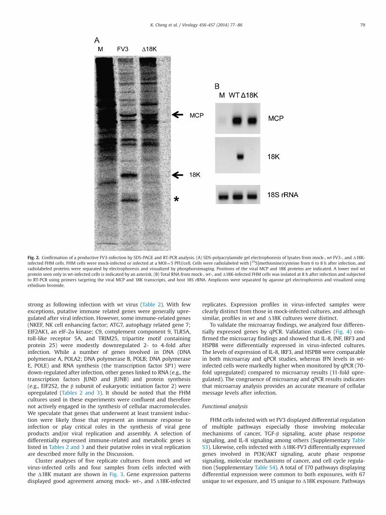
Cluster analyses of five replicate cultures from mock and wtvirus-infected cells and four samples from cells infected withthe Δ18K mutant are shown in Fig. 3. Gene expression patternsdisplayed good agreement among mock- wt-, and Δ18K-infected
replicates. Expression profiles in virus-infected samples wereclearly distinct from those in mock-infected cultures, and althoughsimilar, profiles in wt and Δ18K cultures were distinct.
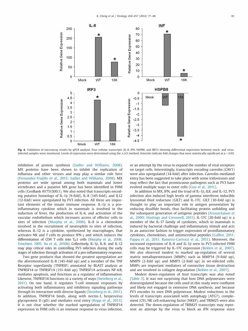
To validate the microarray findings, we analyzed four differen-tially expressed genes by qPCR. Validation studies (Fig. 4) con-firmed the microarray findings and showed that IL-8, INF, IRF3 andHSPB8 were differentially expressed in virus-infected cultures.The levels of expression of IL-8, IRF3, and HSPB8 were comparablein both microarray and qPCR studies, whereas IFN levels in wt-infected cells were markedly higher when monitored by qPCR (70-fold upregulated) compared to microarray results (11-fold upre-gulated). The congruence of microarray and qPCR results indicatesthat microarray analysis provides an accurate measure of cellularmessage levels after infection.
Functional analysis
FHM cells infected with wt FV3 displayed differential regulationof multiple pathways especially those involving molecularmechanisms of cancer, TGF-β signaling, acute phase responsesignaling, and IL-8 signaling among others (Supplementary TableS3). Likewise, cells infected with Δ18K-FV3 differentially expressedgenes involved in PI3K/AKT signaling, acute phase responsesignaling, molecular mechanisms of cancer, and cell cycle regula-tion (Supplementary Table S4). A total of 170 pathways displayingdifferential expression were common to both exposures, with 67unique to wt exposure, and 15 unique to Δ18K exposure. Pathways
Fig. 2. Confirmation of a productive FV3-infection by SDS-PAGE and RT-PCR analysis. (A) SDS-polyacrylamide gel electrophoresis of lysates from mock-, wt FV3-, and Δ18K-infected FHM cells. FHM cells were mock-infected or infected at a MOI¼5 PFU/cell. Cells were radiolabeled with [35S]methionine/cysteine from 6 to 8 h after infection, andradiolabeled proteins were separated by electrophoresis and visualized by phosphoroimaging. Positions of the viral MCP and 18K proteins are indicated. A lower mol wtprotein seen only in wt-infected cells is indicated by an asterisk. (B) Total RNA from mock-, wt-, and Δ18K-infected FHM cells was isolated at 8 h after infection and subjectedto RT-PCR using primers targeting the viral MCP and 18K transcripts, and host 18S rRNA. Amplicons were separated by agarose gel electrophoresis and visualized usingethidium bromide.
K. Cheng et al. / Virology 456-457 (2014) 77–86 79
unique to wt FV3 infected cells involved signaling controlled bythrombin, p38 MAPK, IL-2, or death receptors. Unique pathwaysdisplaying differential regulation after Δ18K-FV3 infection werethose for glycogen degradation and PXR/RXR activation. Collec-tively, pathway analysis suggests that FV3 infection induces, atleast transiently, the expression of multiple genes that regulatevarious aspects of cellular and viral metabolism.
Discussion
Differential gene expression in wild-type and Δ18K infected cells
FV3 infection resulted in the marked upregulation of multiplecellular transcripts including those that encode immune-relatedproteins such as IFN and the products of various IFN-stimulatedgenes (ISGs). These results are consistent with those seen follow-ing infection with influenza virus, smallpox virus, and a number ofranaviruses including FV3 where infection of Xenopus laevis wasshown to trigger the expression of TNFα, IL-1β, and IFNγ (Chenet al., 2011; Holopainen et al., 2012; Huang et al., 2011; Kash et al.,2004; Randall and Goodbourn, 2008; Rubins et al., 2004; Wu et al.,2012; Xu et al., 2010a). The marked upregulation of immune-related transcripts in FV3-infected FHM cells is in marked contrastto the marked progressive inhibition of cellular protein synthesisthat accompanies infection. The presence of immune-relatedtranscripts at a time of diminished protein synthesis likely reflects
their earlier induction and continued stability. We speculate thatthese transcripts are translated prior to the profound inhibition ofhost protein synthesis seen at later times after infection and serveto either reduce viral replication in infected cells or, if releasedfrom cells as is the case for IFN, trigger induction of an anti-viralstate in surrounding uninfected cells.
Despite the apparently large number of upregulated putativeimmune-related transcripts, FV3 infected FHM cells ultimatelyundergo cytopathic effect and die within 24 h. It is not knownwhether cultures infected at a lower MOI, where the vast majorityof cells are initially uninfected, would respond to the IFN inducedin, and released by, infected cells and display reduced levels ofviral replication due to the induction of anti-viral ISGs such as PKR(protein kinase R), OAS (2050 oligoadenylate synthase), RNase L(RNase L), ISG15, and other antiviral molecules (Randall andGoodbourn, 2008; Sadler and Williams, 2008). We speculate thatdespite the rapid replication of FV3 in vitro, the small size of FV3plaques in FHM monolayers after 5–7 days of culture may reflectthe inhibitory effects of IFN on surrounding uninfected cells.Moreover, while such protective effects might not be seen afterhigh multiplicity infection in vitro, they may play importantprotective roles in vivo where even a modest inhibition of virusreplication may allow host innate and adaptive immune systemssufficient time to successfully contain and resolve an ongoinginfection.
We observed that the level of induction of immune-relatedgenes in Δ18K KO-infected FHM cells was lower than that seenafter wt infection (Table 2) and that, with the exception of the 18Kprotein, there was little to no difference in the expression of virus-specific polypeptides in wt and Δ18K KO-infected cultures(Fig. 2A). Consistent with these observations, we previouslyshowed that knock down of 18K expression using anti-sensemorpholino oligonucleotides had little to no effect on viral geneexpression and virus production (Sample et al., 2007). Based onthose results, we concluded that 18K was not essential forreplication in FHM cells and hypothesized that 18K only playedan essential replicative role in certain cell types or that 18Kmodulated anti-viral immunity and virulence in vivo. The obser-vation that lower levels of immune-related transcripts wereinduced following infection with Δ18K may explain its reducedvirulence in vivo. Perhaps the viral 18K protein, by a mechanismyet to be elucidated, triggers a robust induction of cellularimmune-related transcripts (i.e., the amphibian version of a“cytokine storm”) that is responsible for the marked morbidityand mortality seen in vivo after wt infection. Conversely, mutantslacking the 18K gene trigger a modest induction of immune-related genes which protects the animal and contributes to viralclearance.
Immune-related genes upregulated after infection
Transcripts induced by FV3 infection include a number asso-ciated with innate immune responses to viral and bacterialinfections. In mammalian cells, engagement of TLR3 by viraldouble-stranded RNA (dsRNA) triggers signaling through IFNregulatory factor 3 (IRF3) and results in the induction of IFNβ,which acts in an autocrine fashion to induce IFNα (Murphy et al.,2008). IFNα subsequently binds the IFNα/β receptor and triggerssignaling through the JAK/STAT pathway leading to the synthesisof 200 or more IFN-stimulated genes (ISGs) that inhibit viralreplication (Sadler and Williams, 2008). In this study, we sawevidence for induction of IFNα (11-fold upregulated) in wt-infectedcells and several ISGs including MXD (9-fold up) and MHC class I(5-fold up), but did not detect upregulation of a piscine homolog ofPKR, a cellular product that in most mammalian systems lead tophosphorylation of eukaryotic initiation factor 2α and the global
Fig. 3. Heat map analysis of mock-, wt-, and Δ18K KO-infected FHM cells. Levelsof gene expression are indicated by color: blue lowest, yellow intermediate,red highest. Brackets across the top of the map indicate relatedness (clustering)among samples.
K. Cheng et al. / Virology 456-457 (2014) 77–8680
inhibition of protein synthesis (Sadler and Williams, 2008).MX proteins have been shown to inhibit the replication ofinfluenza and other viruses and may play a similar role here(Fernandez-Trujillo et al., 2013; Sadler and Williams, 2008). MXproteins are wide spread among both mammals and lowervertebrates and a putative MX gene has been identified in FHMcells (GenBank AY751300.1). We also noted that transcripts encod-ing putative homologs of IL-1β (9-fold), IL-8 (145-fold), and IL12(12-fold) were upregulated by FV3 infection. All three are impor-tant elements of the innate immune response. IL-1β is a pro-inflammatory cytokine which in mammals is involved in theinduction of fever, the production of IL-6, and activation of thevascular endothelium which increases access of effector cells tosites of infection (Murphy et al., 2008). IL-8 is a chemokineinvolved in the recruitment of neutrophils to sites of infection,whereas IL-12 is a cytokine, synthesized by macrophages, thatactivates NK and T cells to produce IFN-γ and which induces thedifferentiation of CD4 T cells into TH1 cells (Murphy et al., 2008;Trinchieri, 1995; Xu et al., 2010b). Collectively, IL-1β, IL-8, and IL-12may play critical roles in controlling FV3 infection during the earlystages of infection through activation of a pro-inflammatory response.
Two gene products that showed the greatest upregulation arethe aforementioned IL-8 (145-fold up) and a member of the TNFReceptor superfamily (TNFRSF) annotated by BLAST analysis asTNFRSF14 or TNFRSF1A (151-fold up). TNFRSF1A activates NF-ΚB,mediates apoptosis, and functions as a regulator of inflammation.Likewise, TNFRSF14 functions in a variety of ways (Steinberg et al.,2011). On one hand, it regulates T-cell immune responses byactivating both inflammatory and inhibitory signaling pathwaysthrough its interaction with diverse ligands (Steinberg et al., 2011).In addition, TNFRSF14 binds, along with nectin-1, herpesvirusglycoprotein D (gD) and mediates viral entry (Kopp et al., 2013).It is not clear whether the marked upregulation of TNFRSF14expression in FHM cells is an immune response to virus infection,
or an attempt by the virus to expand the number of viral receptorson target cells. Interestingly, transcripts encoding caveolin (CAV1)were also upregulated (14-fold) after infection. Caveolin-mediatedentry has been suggested to take place with some iridoviruses andmay reflect the fact that promiscuous pathogens such as FV3 haveevolved multiple ways to enter cells (Guo et al., 2011).
In addition to MX, IFN, and the triad of IL-1β, IL8, and IL-12, FV3infection also induced high levels of gamma interferon induciblelysosomal thiol reductase (GILT) and IL-17C. GILT (30-fold up) isthought to play an important role in antigen presentation byreducing disulfide bonds, thus facilitating protein unfolding andthe subsequent generation of antigenic peptides (Arunachalam etal., 2000; Hastings and Cresswell, 2011). IL-17C (20-fold up) is amember of the IL-17 family of cytokines, which in mammals isinduced by bacterial challenge and inflammatory stimuli and actsin an autocrine fashion to trigger expression of proinflammatorycytokines, chemokines, and antimicrobial peptides (Gaffen, 2011;Pappu et al., 2011; Ramirez-Carrozzi et al., 2011). Moreover, theincreased expression of IL-8 and IL-1β seen in FV3-infected FHMcells may be triggered by IL-17C expression (Beklen et al., 2007).We also observed modest to marked up-regulation of severalmatrix metalloproteinases (MMPs) such as MMP14 (9-fold up),MMP9 (2-fold up) and MMP3 (2-fold up) in wt-infected cells.MMPs are important mediators of connective tissue destructionand are involved in collagen degradation (Beklen et al., 2007).
Modest down-regulation of host transcripts was also noted(Table 3). It was not surprising that host DNA polymerases weredownregulated because the cells used in this study were confluentand likely not engaged in extensive DNA synthesis, and becauseFV3 encodes its own DNA polymerase. Modest reductions in thelevels of transcripts associated with autophagy (ATG7), comple-ment (C9), NK cell enhancing factor (NKEF), and TRIM25 were alsodetected. The down-regulation of TRIM25 transcripts may repre-sent an attempt by the virus to block an IFN response since
Fig. 4. Validation of microarray results by qPCR analysis. Four cellular transcripts (IL-8, IFN, HSPB8, and IRF3) showing differential expression between mock- and virus-infected samples were monitored. Levels of expression were determined using the ΔΔCt method. Asterisks indicate fold changes that were statistically significant at po0.05.
K. Cheng et al. / Virology 456-457 (2014) 77–86 81
TRIM25, an E3/ISG15 ligase, ubiquitinates the N-terminal CARDdomain of RIG-I (DDX58) and triggers IFN induction (Gack et al.,2007). The importance of this mechanism is underscored by theobservation that influenza A virus targets TRIM25 in an attempt toevade recognition by RIG-I (Gack et al., 2009). In FV3, downregulation of TRIM25 may allow the virus to escape detection.Likewise, down-regulation of the autophagy-related protein ATG7may be critical for successful replication because cytoplasmic viralassembly sites may be viewed as danger signals and triggerautophagosome formation. In contrast to the down regulationnoted above, we detected marked upregulation of several tran-scription factors (JUNB and JUND) as well as one of the subunitsof eIF-2. Upregulation may reflect the involvement of host RNApolymerase II and associated transcription factors in early viraltranscription, and the subsequent need to ramp up viral proteinsynthesis in contact-inhibited cells as they transition from host toviral protein synthesis.
Modulation of signaling pathways following FV3 infection
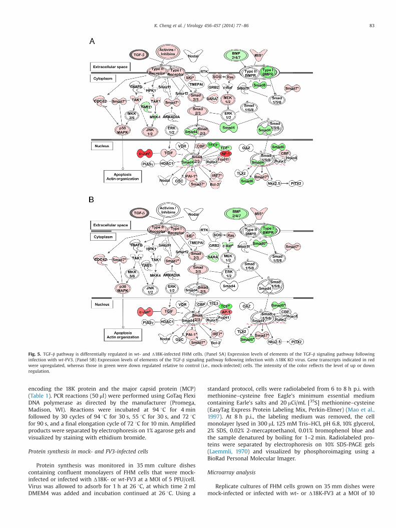
To identify host elements that modulate viral infection, weexamined specific pathways within wt- and the Δ18K-infectedcells that play key roles in cellular and viral metabolism. Since wt-and Δ18K-FV3 differed in virulence following infection of Xenopuslaevis tadpoles (Chen et al., 2011), we hypothesized that differ-ences in their ability to modulate the host immune response couldexplain this dichotomy. Several pathways common to both infec-tions were related to immune responsiveness, cancer, or apoptoticdeath. Some of these pathways involved signaling by acute phaseproteins, mitogen-activated protein kinase/extracellular signal-regulated kinase (ERK/MAPK), PI3K/AKT, TGF-β, and IL-8. Althoughelements of these pathways were differentially expressed follow-ing both wt- and Δ18K-infection, wt-infected cells displayed alarger number of differentially expressed genes (compare Supple-mentary Tables S1 and S2). A number of viruses have been foundto affect key signaling pathways such as those involving MAPK/ERK and PI3K/AKT. The MAPK/ERK pathway is involved in cellgrowth and proliferation and has been suggested to modulatereplication of several viruses including human immunodeficiencyvirus (HIV), bovine herpesvirus, and hepatitis B virus (Jacque et al.,1998; Zheng et al., 2003; Zhu et al., 2011). In addition, dysregula-tion of this pathway has been linked to viral pathogenicity (Zhaoet al., 2005). Interestingly, integrins, used as receptors by severalviruses (Schmidt et al., 2013) and involved in MAPK/ERK signaling(Yee and Hamerman, 2013), were down-regulated in both expo-sures. Likewise, the PI3K/AKT pathway is required for the efficientreplication of several viruses including influenza virus (Shin et al.,2007a, b) and human cytomegalovirus (Johnson et al., 2001). Asindicate therein, inhibition of the MAPK/ERK and PI3K/AKT path-ways led to reductions in virus yield, suggesting that elements ofthese pathways might be necessary for virus multiplication.
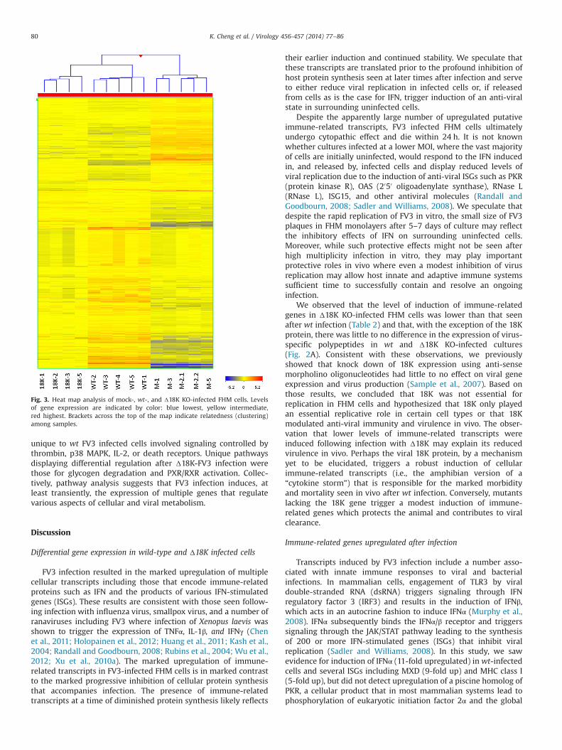
An example of a pathway that was differentially regulatedfollowing wt- and Δ18K-infection was that involving TGF-β signal-ing (Fig. 5). Notably activins/inhibins, members of the TGF-βsuperfamily, were upregulated after wt infection, but unaffectedfollowing infection with Δ18K-FV3. Activins are cytokines withcritical roles in infection and inflammation, and their inductionfollowing wt infection suggests a virus-induced immune response(de Kretser et al., 2012; Ebert et al., 2010; Robson et al., 2009).Another part of this signaling pathway, the Raf/MEK/ERK cascade,has an important role in cell growth, differentiation and survival.Pleschka et al. (Pleschka et al., 2001) showed that infection of cellswith influenza A virus led to activation of the Raf/MEK/ERKcascade, and inhibition of Raf signaling resulted in inhibitionof virus replication. While the cascade is mostly upregulated in
wt-infected cells, Raf is downregulated after Δ18K infection, whichcould explain its decreased virulence in vivo (Chen et al., 2011).
Lastly, the death receptor signaling pathway was significantlyenriched only after wt infection. Death receptor induced apoptosishas been identified as an important pathway in West Nile viruspathogenesis (Clarke et al., 2014). Moreover, caspase 8, an initiatorof the caspase cascade and a protein whose cognate transcript wasupregulated after FV3 infection, promotes apoptotic signaling afterinfection. Collectively, the data suggest that these pathways maybe involved in the pathogenesis of FV3 virus.
The ability of FV3 to markedly upregulate multiple immune-related functions while modestly downregulating others mayreflect viral adaptations to the realities of life as an intracellularparasite. Controlling viral replication by balancing replicativeneeds on one hand and host survival and immunity on the otheris thought to permit viral spread within a single organism andpopulation. Although microarray studies do not provide a com-plete picture of metabolic events within virus-infected cells andrequire proof that cognate immune-related proteins are differen-tially expressed, they clearly indicate that host cells vigorouslyrespond to ranavirus challenge. Moreover, since in vitro studiesutilizing a single cell type may not accurately reflect events in vivowhere multiple tissues and cell types interact and contribute todisease and resistance to disease, a better understanding ofcellular responses to ranavirus infection can only be gained byinfecting whole animals or specific subsets of immune cells, e.g.,macrophages and T cells. Nevertheless, functional analysis oftranscriptional data identified potential mechanisms involved inthe differential pathogenesis of FV3 and FV3-induced mortality.
Material and methods
Cells and virus
For biochemical analyses, wt FV3 (ATCC VR-569) was grown infathead minnow cells (ATCC No. CCL-42) in Dulbecco's modifiedEagle's medium (DMEM) containing 4% fetal bovine serum (D4) at26 1C in a humidified incubator in an environment of 95%air/5% CO2 (Granoff et al., 1966). To prepare viral stocks, FV3 waspropagated on monolayers of FHM cells grown in 150 cm2
flasksand incubated in Eagle's minimum essential medium with Hank'ssalts (HMEM) supplemented with 4% fetal bovine serum (H4).Virus stocks were generated by infecting FHM cells at a multi-plicity of infection (MOI) of 0.01 PFU/cell and harvesting whencytopathic effect was marked, i.e.,�5 days post infection (p.i.).Virions, released by three freeze-thaw cycles, were clarified by lowspeed centrifugation, and titers determined by plaque assay onFHM monolayers under an overlay of 0.75% methylcellulose. Aknock out mutant (Δ18K) lacking the entire 18 kDa immediateearly gene (Chen et al., 2011) was propagated as indicated above.
RT-PCR analysis of viral and cellular gene expression
To confirm a productive infection and determine the kinetics ofinduction of immune-related genes, RNA obtained from mock- andvirus-infected cultures was subjected to RT-PCR analysis. Conflu-ent monolayers of FHM cells grown on 35 mm dishes were mock-infected or infected with wt- or Δ18K-FV3 at a MOI¼5 PFU/cell.At the indicated times p.i. the cells were lysed with 1 ml Trizol(Invitrogen Life Technologies, Carlsbad, CA) and RNA isolated asdirected by the manufacturer. cDNA was synthesized from 1 mg oftotal RNA using random primers and SuperScripts III ReverseTranscriptase as directed by the supplier (Invitrogen Life Technol-ogies). Amplification of cDNA was performed using primersspecific for FHM 18S rRNA, Mx, and β-actin and FV3 transcripts
K. Cheng et al. / Virology 456-457 (2014) 77–8682
encoding the 18K protein and the major capsid protein (MCP)(Table 1). PCR reactions (50 μl) were performed using GoTaq FlexiDNA polymerase as directed by the manufacturer (Promega,Madison, WI). Reactions were incubated at 94 1C for 4 minfollowed by 30 cycles of 94 1C for 30 s, 55 1C for 30 s, and 72 1Cfor 90 s, and a final elongation cycle of 72 1C for 10 min. Amplifiedproducts were separated by electrophoresis on 1% agarose gels andvisualized by staining with ethidium bromide.
Protein synthesis in mock- and FV3-infected cells
Protein synthesis was monitored in 35 mm culture dishescontaining confluent monolayers of FHM cells that were mock-infected or infected with Δ18K- or wt-FV3 at a MOI of 5 PFU/cell.Virus was allowed to adsorb for 1 h at 26 1C, at which time 2 mlDMEM4 was added and incubation continued at 26 1C. Using a
standard protocol, cells were radiolabeled from 6 to 8 h p.i. withmethionine–cysteine free Eagle's minimum essential mediumcontaining Earle's salts and 20 mCi/mL [35S] methionine–cysteine(EasyTag Express Protein Labeling Mix, Perkin-Elmer) (Mao et al.,1997). At 8 h p.i., the labeling medium was removed, the cellmonolayer lysed in 300 mL 125 mM Tris–HCl, pH 6.8, 10% glycerol,2% SDS, 0.02% 2-mercaptoethanol, 0.01% bromophenol blue andthe sample denatured by boiling for 1–2 min. Radiolabeled pro-teins were separated by electrophoresis on 10% SDS-PAGE gels(Laemmli, 1970) and visualized by phosphoroimaging using aBioRad Personal Molecular Imager.
Microarray analysis
Replicate cultures of FHM cells grown on 35 mm dishes weremock-infected or infected with wt- or Δ18K-FV3 at a MOI of 10
Fig. 5. TGF-β pathway is differentially regulated in wt- and Δ18K-infected FHM cells. (Panel 5A) Expression levels of elements of the TGF-β signaling pathway followinginfection with wt-FV3. (Panel 5B) Expression levels of elements of the TGF-β signaling pathway following infection with Δ18K KO virus. Gene transcripts indicated in redwere upregulated, whereas those in green were down regulated relative to control (i.e., mock-infected) cells. The intensity of the color reflects the level of up or downregulation.
K. Cheng et al. / Virology 456-457 (2014) 77–86 83
PFU/cell. At 8 h p.i., total RNA was isolated from one set ofreplicates for RT-PCR and qPCR analysis using Trizol (InvitrogenLife Technologies, Carlsbad, CA). RNA was isolated from theremaining replicates using RNeasy (Qiagen, Valencia, CA, USA)and subjected to microarray analysis. RNA was DNase-treated,quantified using a Nanodrop ND-1000 spectrophotometer(Wilmington, DE), and its quality assessed prior to microarray
analysis using an Agilent 2100 Bioanalyzer (Santa Clara, CA). RNAwas stored at �80 1C until analyzed.
RNA from four (Δ18K FV3-infected) or five (mock- and wt FV3-infected) biological replicates was analyzed using custom fatheadminnow 60,000 gene arrays (GPL17098) purchased from AgilentTechnologies. cRNA labeling was performed using an AgilentQuick-AMP one color kit and hybridization was performed follow-ing the manufacturer's recommendations (One-color microarrayhybridization protocol, v 6.5; Agilent). An Agilent high-resolutionscanner (Model No. G2565CA) was used to acquire microarrayimages and data was processed using Agilent's Feature Extractionsoftware v10.7 (Agilent). Raw microarray data from this study havebeen deposited at the Gene Expression Omnibus website (http://www.ncbi.nlm.nih.gov/geo/; GSE53739).
Bioinformatics
Raw microarray data were imported into GeneSpring versionGX11 (Agilent), and normalized using quantile normalizationfollowed by median scaling across all samples. To identify differ-entially expressed genes (DEGs), one-way Analysis of Variance(ANOVA) was performed followed by pair-wise comparison and
Table 1Primers used for RT-PCR and qPCR.
Gene Forward (F) and reverse (R) primersa Use
FHM 18S rRNA F GTAGACGTCAACAAGATGTGGATCC RT-PCRR CAGGAAGGTAATGTTCTTTGTGGCCG
FHM β actin F CCGTGCTGTCTGGAGGTA RT-PCRR AAGGAGCAAGGGAGGTGATTTC
FHM Mx1 F TGGCATGGGAGAATCAGTTACAAG RT-PCRR TGCCCCAGCCATCTCATCC
FV3 MCP F TTATGGTGCAGAACGTCACA RT-PCRR AGCCTTGTGGTGTTTTCGTA
FV3 18K F GCCCAAGAATGTCTTTTCCA RT-PCRR CGGTCAGTCTCCAGGTTTTC
FHM interleukin-8 (IL-8) F CCCTCCTAGCCCTCACTGTAAA qPCRR GGATCTTCTCAATGACCTTCTT
FHM heat shock protein 8B (HSPB8) F CGAGCAGTACGCGTGGGAGTC qPCRR AGCGTGATGGGGTAGCCGATGAAC
FHM interferon (INF) F CAACAACATCATGACCCGCTACCT qPCRR GTTCTCTGCCTCCGTTCTGTCCTT
FHM interferon regulatory factor 3 (IRF3) F AGCATGCTTTGAGACAGGAC qPCRR CACGAAGAGCGCTACGGAAGTT
a Primer sequences were designed using PrimerSelect (DNAStar, Madison, WI).
Table 2Differential expression of cellular genes following infection with wt FV3 or theΔ18K knock out mutant: upregulated transcripts.
Category Gene Fold change
wt Δ18K
IFN, IFN-related, IFN-induced GILT 30 9IFNα 11 4MXD 9 NDa
IRF2 8 4IRF3 5 2IRF1 3 2ADAR 4 4
Cytokines & chemokines IL8 145 70IL17C 20 18IL-12a 12 12SOCS3 12 7IL11 10 7IL1β 9 4CXCR7 4 2RIG-I 3 3IL-12b 2 ND
Pathogen recognition and immune signaling TNFRSF14 151 100NFKBIA 44 23TRAF3 20 12TLR18 7 5CASP9 6 3TLR5B 5 ND
Antigen presentation ICLP2 63 11MHC class I 5 4
Metabolic EIF2S2 96 24JUND 77 40CAV1 14 8JUNB 6 4
a ND, not differentially expressed.
Table 3Differential expression of cellular genes following infection with wt FV3 or theΔ18K knock out mutant: down-regulated transcripts.
Category Gene Fold change
wt Δ18K
Immune-related ATG7 �5 �3NKEF �4 �4C9 �3 �4TRIM 25 �3 �3TLR5A �3 �2IL-6R �3 �2EIF2AK �3 NDa
Metabolic SP1 �4 �3POLA2 �3 �2HSPB8 �4 �5POLB �2 NDPOLE �3 �2
a ND, not differentially expressed.
K. Cheng et al. / Virology 456-457 (2014) 77–8684
Benjamini-Hochberg multiple testing correction (po0.05). Hier-archical clustering was performed with GeneSpring (Agilent Tech-nologies). Functional analysis and identification of upstreamregulators of pathways were performed using the human orthologgenes of fathead minnow DEGs and Ingenuity Pathway Analysissoftware (IPA, Redwood City, CA).
Quantitative RT-PCR (qPCR)
To validate microarray results, qPCR was performed usingprimers (Table 1) targeting FHM interleukin-8 (IL-8), INF, heatshock protein 8B (HSP8B), IRF3, and 18S rRNA (control). Tocompare directly microarray and qPCR results, replicate RNAsamples were prepared from mock- and virus-infected cells at8 h p.i. as indicated above. Relative expression levels of putativeimmune-related genes were determined based on Ct (thresholdcrossing) values using the 2�ΔΔCt method (Livak and Schmittgen,2001).
Quadruplicate reactions were performed using cDNA templatesprepared from RNA isolated from mock- or virus-infected FHMcells. Reactions were carried out in 96-well plates in a 25 μl finalreaction volume consisting of 12.5 mL of RT2 SYBR Green qPCRMaster Mix (SABiosciences, Frederick, USA), 0.5 mL each of forwardand reverse primers (10 mM each), 1 mL cDNA, and sterile nuclease-free water to 25 μl. Amplification conditions were 1 cycle at 95 1Cfor 1 min followed by 30 cycles of 95 1C for 20 s and 55 1C for1 min. To verify that a single product was synthesized, melt curveswere generated for each product and only primer pairs and cDNAsthat generated a single peak were used in the final analyses.The PCR efficiencies for each primer set were Z90%. The samethreshold was used for each sample on a given plate; CT valuesr31 were considered positive.
Acknowledgments
This work was partially funded by the US Army EnvironmentalQuality Research Program (including BAA 11-4838), the NationalScience Foundation (IOS 07-42711), the University of MississippiMedical Center, and the National Institutes of Health (R24-AI-059830). Permission for publishing has been granted by the Chiefof Engineers, US Army Corps of Engineers.
Appendix A. Supporting information
Supplementary data associated with this article can be found inthe online version at http://dx.doi.org/10.1016/j.virol.2014.03.014.
References
Arunachalam, B., Phan, U.T., Geuze, H.J., Cresswell, P., 2000. Enzymatic reduction ofdisulfide bonds in lysosomes: characterization of a gamma-interferon-inducible lysosomal thiol reductase (GILT). Proc. Natl. Acad. Sci. U. S. A. 97,745–750.
Beklen, A., Ainola, M., Hukkanen, M., Gurgan, C., Sorsa, T., Konttinen, Y.T., 2007.MMPs, IL-1, and TNF are regulated by IL-17 in periodontitis. J. Dent. Res. 86,347–351.
Chen, G., Ward, B.M., Yu, K.H., Chinchar, V.G., Robert, J., 2011. Improved knockoutmethodology reveals that frog virus 3 mutants lacking either the 18Kimmediate-early gene or the truncated vIF-2α gene are defective for replicationand growth in vivo. J. Virol. 85, 11131–11138.
Chinchar, V.G., 2002. Ranaviruses (family Iridoviridae): emerging cold-bloodedkillers. Arch. Virol. 147, 447–470.
Chinchar, V.G., Hyatt, A., Miyazaki, T., Williams, T., 2009. Family iridoviridae: poorviral relations no longer. Curr. Topics Microbiol. Immunol. 328, 123–170.
Chinchar, V.G., Robert, J., Storfer, A.T., 2011. Ecology of viruses infecting ectothermicvertebrates – the impact of ranavirus infections on amphibians. In: Hurst, C.J.(Ed.), Studies in Viral Ecology. Wiley-Blackwell, Hoboken, NJ, pp. 231–259
Chinchar, V.G., Waltzek, T.B., 2014. Ranaviruses: not just for frogs. PLoS. Pathog. 10,e1003850.
Clarke, P., Leser, J.S., Quick, E.D., Dionne, K.R., Beckham, J.D., Tyler, K.L., 2014. Deathreceptor-mediated apoptotic signaling is activated in the brain followingInfection with West Nile virus in the absence of a peripheral immune response.J. Virol. 88, 1080–1089.
de Kretser, D.M., O'Hehir, R.E., Hardy, C.L., Hedger, M.P., 2012. The roles of activin Aand its binding protein, follistatin, in inflammation and tissue repair. Mol. Cell.Endocrinol. 359, 101–106.
Ebert, S., Nau, R., Michel, U., 2010. Role of activin in bacterial infections: a potentialtarget for immunointervention? Immunotherapy 2, 673–684.
Fernandez-Trujillo, M.A., Garcia-Rosado, E., Alonso, M.C., Castro, D., Alvarez, M.C.,Bejar, J., 2013. Mx1, Mx2 and Mx3 proteins from the gilthead seabream (Sparusaurata) show in vitro antiviral activity against RNA and DNA viruses. Mol.Immunol. 56, 630–636.
Finlay, B.B., McFadden, G., 2006. Anti-immunology: evasion of the host immunesystem by bacterial and viral pathogens. Cell 124, 767–782.
Gack, M.U., Albrecht, R.A., Urano, T., Inn, K.S., Huang, I.C., Carnero, E., Farzan, M.,Inoue, S., Jung, J.U., Garcia-Sastre, A., 2009. Influenza A virus NS1 targets theubiquitin ligase TRIM25 to evade recognition by the host viral RNA sensor RIG-I. Cell Host Microbe 5, 439–449.
Gack, M.U., Shin, Y.C., Joo, C.H., Urano, T., Liang, C., Sun, L., Takeuchi, O., Akira, S.,Chen, Z., Inoue, S., Jung, J.U., 2007. TRIM25 RING-finger E3 ubiquitin ligase isessential for RIG-I-mediated antiviral activity. Nature 446, 916–920.
Gaffen, S.L., 2011. Recent advances in the IL-17 cytokine family. Curr. Opin.Immunol. 23, 613–619.
Gantress, J., Maniero, G.D., Cohen, N., Robert, J., 2003. Development and character-ization of a model system to study amphibian immune responses to irido-viruses. Virology 311, 254–262.
Granoff, A., Came, P.E., Breeze, D.C., 1966. Viruses and renal carcinoma of Ranapipiens. I. The isolation and properties of virus from normal and tumor tissue.Virology 29, 133–148.
Gray, M.J., Miller, D.L., Hoverman, J.T., 2009. Ecology and pathology of amphibianranaviruses. Dis. Aquat. Organ. 87, 243–266.
Green, D.E., Converse, K.A., Schrader, A.K., 2002. Epizootiology of sixty-fouramphibian morbidity and mortality events in the USA 1996 – 2001. Ann. N.Y. Acad. Sci. 969, 323–339.
Guo, C.J., Liu, D., Wu, Y.Y., Yang, X.B., Yang, L.S., Mi, S., Huang, Y.X., Luo, Y.W., Jia, K.T.,Liu, Z.Y., Chen, W.J., Weng, S.P., Yu, X.Q., He, J.G., 2011. Entry of Tiger frog virus(an Iridovirus) into HepG2 cells via a pH-dependent, atypical, caveola-mediatedendocytosis pathway. J. Virol. 85, 6416–6426.
Hastings, K.T., Cresswell, P., 2011. Disulfide reduction in the endocytic pathway:immunological functions of gamma-interferon-inducible lysosomal thiolreductase. Antioxid. Redox Signal. 15, 657–668.
Holopainen, R., Tapiovaara, H., Honkanen, J., 2012. Expression analysis of immuneresponse genes in fish epithelial cells following ranavirus infection. FishShellfish Immunol. 32, 1095–1105.
Huang, Y., Huang, X., Yan, Y., Cai, J., Ouyang, Z., Cui, H., Wang, P., Qin, Q., 2011.Transcriptome analysis of orange-spotted grouper (Epinephelus coioides)spleen in response to Singapore grouper iridovirus. BMC Genomics 12, 556.
Jacque, J.M., Mann, A., Enslen, H., Sharova, N., Brichacek, B., Davis, R.J., Stevenson,M., 1998. Modulation of HIV-1 infectivity by MAPK, a virion-associated kinase.EMBO J. 17, 2607–2618.
Jancovich, J.K., Chinchar, V.G., Hyatt, A., Miyazaki, T., Williams, T., Zhang, Q.Y., 2012.Family Iridoviridae. In: King, A.M.Q., Adams, M.J., Carstens, E.B., Lefkowitz, E.J.(Eds.), Virus Taxonomy: Classification and Nomenclature of Viruses. NinthReport of the International Committee on Taxonomy of Viruses. Elsevier,Amsterdam, pp. 193–210
Jancovich, J.K., Jacobs, B.L., 2011. Innate immune evasion mediated by the Ambys-toma tigrinum virus eukaryotic translation initiation factor 2 alpha homologue.J. Virol. 85, 5061–5069.
Johnson, R.A., Wang, X., Ma, X.L., Huong, S.M., Huang, E.S., 2001. Human cytome-galovirus up-regulates the phosphatidylinositol 3-kinase (PI3-K) pathway:inhibition of PI3-K activity inhibits viral replication and virus-induced signal-ing. J. Virol. 75, 6022–6032.
Johnston, J.B., Barrett, J.W., Nazarian, S.H., Goodwin, M., Ricciuto, D., Wang, G.,McFadden, G., 2005. A poxvirus-encoded pyrin domain protein interacts withASC-1 to inhibit host inflammatory and apoptotic responses to infection.Immunity 23, 587–598.
Johnston, J.B., McFadden, G., 2003. Poxvirus immunomodulatory strategies: currentperspectives. J. Virol. 77, 6093–6100.
Kash, J.C., Basler, C.F., Garcia-Sastre, A., Carter, V., Billharz, R., Swayne, D.E.,Przygodzki, R.M., Taubenberger, J.K., Katze, M.G., Tumpey, T.M., 2004. Globalhost immune response: pathogenesis and transcriptional profiling of type Ainfluenza viruses expressing the hemagglutinin and neuraminidase genes fromthe 1918 pandemic virus. J. Virol. 78, 9499–9511.
Kopp, S.J., Karaba, A.H., Cohen, L.K., Banisadr, G., Miller, R.J., Muller, W.J., 2013.Pathogenesis of neonatal herpes simplex 2 disease in a mouse model is dependenton entry receptor expression and route of inoculation. J. Virol. 87, 474–481.
Laemmli, U.K., 1970. Cleavage of structural proteins during the assembly of thehead of bacteriophage T4. Nature 227, 680–685.
Livak, K.J., Schmittgen, T.D., 2001. Analysis of relative gene expression data usingreal-time quantitative PCR and the 2�ΔΔCT method. Methods 25, 402–408.
Maniero, G.D., Morales, H., Gantress, J., Robert, J., 2006. Generation of a long-lasting,protective, and neutralizing antibody response to the ranavirus FV3 by the frogXenopus. Dev. Comp. Immunol. 30, 649–657.
K. Cheng et al. / Virology 456-457 (2014) 77–86 85
Mao, J., Hedrick, R.P., Chinchar, V.G., 1997. Molecular characterization, sequenceanalysis, and taxonomic position of newly isolated fish iridoviruses. Virology229, 212–220.
Morales, H.D., Robert, J., 2007. Characterization of primary and memory CD8 T-cellresponses against ranavirus (FV3) in Xenopus laevis. J. Virol. 81, 2240–2248.
Murphy, K., Travers, P., Walport, M., 2008. Janeway's Immunobiology, 7th ed.Garland Science, New York and London
Pappu, R., Ramirez-Carrozzi, V., Sambandam, A., 2011. The interleukin-17 cytokinefamily: critical players in host defence and inflammatory diseases. Immunology134, 8–16.
Pleschka, S., Wolff, T., Ehrhardt, C., Hobom, G., Planz, O., Rapp, U.R., Ludwig, S., 2001.Influenza virus propagation is impaired by inhibition of the Raf/MEK/ERKsignalling cascade. Nat. Cell Biol. 3, 301–305.
Raghow, R., Granoff, A., 1979. Macromolecular synthesis in cells infected by frogvirus 3. X. Inhibition of cellular protein synthesis by heat-inactivated virus.Virology 98, 319–327.
Ramirez-Carrozzi, V., Sambandam, A., Luis, E., Lin, Z., Jeet, S., Lesch, J., Hackney, J.,Kim, J., Zhou, M., Lai, J., Modrusan, Z., Sai, T., Lee, W., Xu, M., Caplazi, P., Diehl, L.,de Voss, J., Balazs, M., Gonzalez Jr., L., Singh, H., Ouyang, W., Pappu, R., 2011. IL-17C regulates the innate immune function of epithelial cells in an autocrinemanner. Nat. Immunol. 12, 1159–1166.
Randall, R.E., Goodbourn, S., 2008. Interferons and viruses: an interplay betweeninduction, signalling, antiviral responses and virus countermeasures. J. Gen.Virol. 89, 1–47.
Robert, J., Morales, H., Buck, W., Cohen, N., Marr, S., Gantress, J., 2005. Adaptiveimmunity and histopathology in frog virus 3-infected Xenopus. Virology 332,667–675.
Robert, J., Ohta, Y., 2009. Comparative and developmental study of the immunesystem in Xenopus. Developmental dynamics: an official publication of theAmerican Association of Anatomists 238, 1249–1270.
Robson, N.C., Wei, H., McAlpine, T., Kirkpatrick, N., Cebon, J., Maraskovsky, E., 2009.Activin-A attenuates several human natural killer cell functions. Blood 113,3218–3225.
Rollins-Smith, L.A., 1998. Metamorphosis and the amphibian immune system.Immunol. Rev. 166, 221–230.
Rubins, K.H., Hensley, L.E., Jahrling, P.B., Whitney, A.R., Geisbert, T.W., Huggins, J.W.,Owen, A., Leduc, J.W., Brown, P.O., Relman, D.A., 2004. The host response tosmallpox: analysis of the gene expression program in peripheral blood cells in anonhuman primate model. Proc. Natl. Acad. Sci. U. S. A. 101, 15190–15195.
Sadler, A.J., Williams, B.R., 2008. Interferon-inducible antiviral effectors. Nat. Rev.Immunol. 8, 559–568.
Sample, R., Bryan, L., Long, S., Majji, S., Hoskins, G., Sinning, A., Olivier, J., Chinchar,V.G., 2007. Inhibition of iridovirus protein synthesis and virus replication byantisense morpholino oligonucleotides targeted to the major capsid protein,the 18 kDa immediate-early protein, and a viral homolog of RNA polymerase II.Virology 358, 311–320.
Schmidt, K., Keller, M., Bader, B.L., Korytar, T., Finke, S., Ziegler, U., Groschup, M.H.,2013. Integrins modulate the infection efficiency of West Nile virus into cells. J.Gen. Virol. 94, 1723–1733.
Seet, B.T., Johnston, J.B., Brunetti, C.R., Barrett, J.W., Everett, H., Cameron, C., Sypula,J., Nazarian, S.H., Lucas, A., McFadden, G., 2003. Poxviruses and immuneevasion. Annu. Rev. Immunol. 21, 377–423.
Shin, Y.K., Liu, Q., Tikoo, S.K., Babiuk, L.A., Zhou, Y., 2007a. Effect of the phospha-tidylinositol 3-kinase/Akt pathway on influenza A virus propagation. J. Gen.Virol. 88, 942–950.
Shin, Y.K., Liu, Q., Tikoo, S.K., Babiuk, L.A., Zhou, Y., 2007b. Influenza A virus NS1protein activates the phosphatidylinositol 3-kinase (PI3K)/Akt pathway bydirect interaction with the p85 subunit of PI3K. J. Gen. Virol. 88, 13–18.
Steinberg, M.W., Cheung, T.C., Ware, C.F., 2011. The signaling networks of theherpesvirus entry mediator (TNFRSF14) in immune regulation. Immunol. Rev.244, 169–187.
Tannenbaum, J., Goorha, R., Granoff, A., 1978. Inhibition of vesicular stomatitis virusreplication by frog virus 3. Selective action on secondary transcription. Virology89, 560–569.
Tannenbaum, J., Goorha, R., Granoff, A., 1979. The inhibition of vesicular stomatitisvirus protein synthesis by frog virus 3. Virology 95, 227–231.
Trinchieri, G., 1995. Interleukin-12: a proinflammatory cytokine with immunor-egulatory functions that bridge innate resistance and antigen-specific adaptiveimmunity. Annu. Rev. Immunol. 13, 251–276.
Tweedell, K., Granoff, A., 1968. Viruses and renal carcinoma of Rana pipiens. V.Effect of frog virus 3 on developing frog embryos and larvae. J. Natl. Cancer. Inst.40, 407–410.
Wang, F., Barrett, J.W., Shao, Q., Gao, X., Dekaban, G.A., McFadden, G., 2009.Myxoma virus selectively disrupts type I interferon signaling in primary humanfibroblasts by blocking the activation of the Janus kinase Tyk2. Virology 387,136–146.
Wu, M.S., Chen, C.W., Liu, Y.C., Huang, H.H., Lin, C.H., Tzeng, C.S., Chang, C.Y., 2012.Transcriptional analysis of orange-spotted grouper reacting to experimentalgrouper iridovirus infection. Dev. Comp. Immunol. 37, 233–242.
Xu, D., Wei, J., Cui, H., Gong, J., Yan, Y., Lai, R., Qin, Q., 2010a. Differential profiles ofgene expression in grouper Epinephelus coioides, infected with Singaporegrouper iridovirus, revealed by suppression subtractive hybridization and DNAmicroarray. J. Fish Biol.77, 341–360.
Xu, M., Mizoguchi, I., Morishima, N., Chiba, Y., Mizuguchi, J., Yoshimoto, T., 2010b.Regulation of antitumor immune responses by the IL-12 family cytokines, IL-12,IL-23, and IL-27. Clin. Dev. Immunol. 2010.
Yee, N.K., Hamerman, J.A., 2013. beta(2) integrins inhibit TLR responses byregulating NF-kappaB pathway and p38 MAPK activation. Eur. J. Immunol. 43,779–792.
Zhao, L.J., Wang, L., Ren, H., Cao, J., Li, L., Ke, J.S., Qi, Z.T., 2005. Hepatitis C virus E2protein promotes human hepatoma cell proliferation through the MAPK/ERKsignaling pathway via cellular receptors. Exp. Cell. Res. 305, 23–32.
Zheng, Y., Li, J., Johnson, D.L., Ou, J.H., 2003. Regulation of hepatitis B virusreplication by the ras-mitogen-activated protein kinase signaling pathway.J. Virol. 77, 7707–7712.
Zhu, L., Ding, X., Zhu, X., Meng, S., Wang, J., Zhou, H., Duan, Q., Tao, J., Schifferli, D.M.,Zhu, G., 2011. Biphasic activation of PI3K/Akt and MAPK/Erk1/2 signalingpathways in bovine herpesvirus type 1 infection of MDBK cells. Vet. Res. 42, 57.
K. Cheng et al. / Virology 456-457 (2014) 77–8686