differential techniques used for detection … · differential techniques used for detection of...
TRANSCRIPT

Page 1
Journal of Parasitic Diseases: Diagnosis and Therapy, Volume 1, Number 1, 2016
DIFFERENTIAL TECHNIQUES USED FOR DETECTION OF CRYPTOSPORIDIUM OOCYSTS IN STOOL SPECIMENS
Sumaira Shams, Department of Zoology Abdul Wali Khan University, UCS Shankar Campus, Mardan 23200, KPK Pakistan
Saima Khan, Department of Zoology Abdul Wali Khan University, UCS Shankar Campus, Mardan 23200, KPK Pakistan
Asar Khan, Department of Zoology Abdul Wali Khan University, UCS Shankar Campus, Mardan 23200, KPK Pakistan
Iftikhar Khan, Department of Zoology Abdul Wali Khan University, UCS Shankar Campus, Mardan 23200, KPK Pakistan
Muhammad Ijaz, Department of Zoology Abdul Wali Khan University, UCS Shankar Campus, Mardan 23200, KPK Pakistan
Atta Ullah, Department of Zoology Abdul Wali Khan University, UCS Shankar Campus, Mardan 23200, KPK Pakistan
*Corresponding author: Saima Khan, Department of Zoology Abdul Wali Khan University, UCS Shankar Campus, Mardan 23200, KPK Pakistan, E-mail: [email protected]
Received: August 15, 2016; Accepted: September 07, 2016; Published: October 20, 2016
Review Article
ABSTRACT
Cryptosporidium is a coccidian protozoon belongs to phylum Apicomplexa, and an important etiologic agent of diarrheal disease in both immune-competent and immune-compromised hosts. There are many diagnostic techniques available for detecting and diagnosing of cryptosporidium spp. The microscopic methods are used in many studies but they are not reliable as compared to the molecular and serological methods. Most of these methods are time consuming and need expert skills to be carry out for clinical investigations. The detection and diagnosis of cryptosporidiosis in Pakistan is narrowed to major research laboratories and research centers therefore, it does not feature in protocols for routine investigations in many clinical laboratories. This review deals with the differential currently available diagnostic methods and their utilization as a standard for implementation of routine clinical diagnosis against cryptosporidium infection.
Keywords: Cryptosporidiosis; diagnosis methods; microscopic methods; PCR; serological tests
INTRODUCTION
Cryptosporidium is a coccidian protozoon belongs to phylum Apicomplexa, and an important etiologic agent of diarrheal disease in both immune-competent and immune-compromised hosts. To date, at least 10 species of Cryptosporidium have been considered to be valid (Fayer et al., 2000). While, different studies have identified five species of Cryptosporidium (C. parvum, C. hominis, C. meleagridis, C. felis, and C. canis) with different reservoir hosts (Xiao et al., 2004). It is estimated that about 30% adult population in advanced thrifts and 60% in developing countries have serologic evidence of infection with this organism (Cheng et al., 2005). In Asia, the organism has been detected in 2-20% of all the cases of acute diarrhea (Aye et al., 1994). Still, only a minority of people is detected with clinical disease, accordingly Cryptosporidium testing is imperious in diarrheal patients, as this will provide a stronger representation of the occurrence of the infection in the numerous environmental areas of the Biosphere (Beauty et al., 2014).
http://www.alliedacademies.org/parasitic-diseases-diagnosis-therapy/ISSN: 2591-7846
Page 2
Journal of Parasitic Diseases: Diagnosis and Therapy, Volume 1, Number 1, 2016
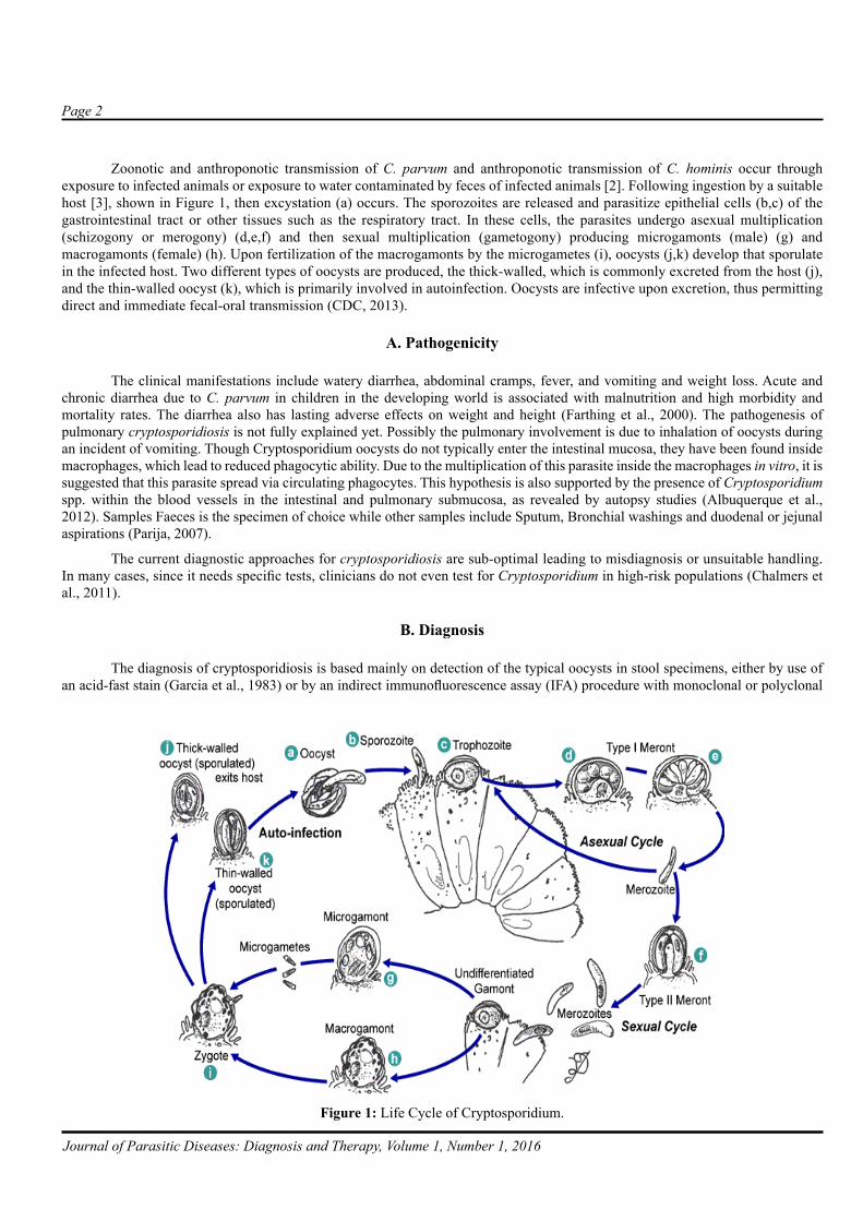
Zoonotic and anthroponotic transmission of C. parvum and anthroponotic transmission of C. hominis occur through exposure to infected animals or exposure to water contaminated by feces of infected animals [2]. Following ingestion by a suitable host [3], shown in Figure 1, then excystation (a) occurs. The sporozoites are released and parasitize epithelial cells (b,c) of the gastrointestinal tract or other tissues such as the respiratory tract. In these cells, the parasites undergo asexual multiplication (schizogony or merogony) (d,e,f) and then sexual multiplication (gametogony) producing microgamonts (male) (g) and macrogamonts (female) (h). Upon fertilization of the macrogamonts by the microgametes (i), oocysts (j,k) develop that sporulate in the infected host. Two different types of oocysts are produced, the thick-walled, which is commonly excreted from the host (j), and the thin-walled oocyst (k), which is primarily involved in autoinfection. Oocysts are infective upon excretion, thus permitting direct and immediate fecal-oral transmission (CDC, 2013).
A. Pathogenicity
The clinical manifestations include watery diarrhea, abdominal cramps, fever, and vomiting and weight loss. Acute and chronic diarrhea due to C. parvum in children in the developing world is associated with malnutrition and high morbidity and mortality rates. The diarrhea also has lasting adverse effects on weight and height (Farthing et al., 2000). The pathogenesis of pulmonary cryptosporidiosis is not fully explained yet. Possibly the pulmonary involvement is due to inhalation of oocysts during an incident of vomiting. Though Cryptosporidium oocysts do not typically enter the intestinal mucosa, they have been found inside macrophages, which lead to reduced phagocytic ability. Due to the multiplication of this parasite inside the macrophages in vitro, it is suggested that this parasite spread via circulating phagocytes. This hypothesis is also supported by the presence of Cryptosporidium spp. within the blood vessels in the intestinal and pulmonary submucosa, as revealed by autopsy studies (Albuquerque et al., 2012). Samples Faeces is the specimen of choice while other samples include Sputum, Bronchial washings and duodenal or jejunal aspirations (Parija, 2007).
The current diagnostic approaches for cryptosporidiosis are sub-optimal leading to misdiagnosis or unsuitable handling. In many cases, since it needs specific tests, clinicians do not even test for Cryptosporidium in high-risk populations (Chalmers et al., 2011).
B. Diagnosis
The diagnosis of cryptosporidiosis is based mainly on detection of the typical oocysts in stool specimens, either by use of an acid-fast stain (Garcia et al., 1983) or by an indirect immunofluorescence assay (IFA) procedure with monoclonal or polyclonal
Figure 1: Life Cycle of Cryptosporidium.

Page 3
Journal of Parasitic Diseases: Diagnosis and Therapy, Volume 1, Number 1, 2016
antibodies (Sterling et al., 1986). The definitive diagnosis requires microscopical detection of the parasite in tissues or body fluids. However, clinical, endoscopic, immunologic, and molecular techniques all have a place in the diagnosis and clinical assessment of cryptosporidiosis. Newer tools, such as immune-fluorescent assays and antigen-capture enzyme-linked immune-sorbent assays (ELISA), which are now commonly used in diagnostic laboratories, improve the sensitivity and specificity of the test. Polymerase-chain-reaction based techniques are available as research tests (Xian et al., 2002).
C. Microscopic Methods
Some studies showed that there is no difference among these methods; however, others have found that formalin-ether and sodium chloride flotations are the most sensitive (Zierdt, 1984., Casemore et al., 1985) (Table 1). The Sheather's sugar solution gives results equal or better than those obtained with formalin-ether or formalin-ethyl acetate (Delfín et al., 1988). As the Formalin-ethyl acetate sedimentation concentration technique is already performed routinely in most clinical microbiology laboratories, thus preparation of the acid-fast smear for detecting Crvptosporidium sp. in the same sediment may reduce cost and technical time. The acid-fast smear provides a permanent record of the results, but the preparation and reading of the smear require some technical expertise (McNabb et al., 1985). The Iodine-saline wet mount is Recommended as a routine screening procedure for Crvptosporidium detection but its record is not permanent (Arora et al., 2009).
Staining Methods
The use of Romanowsky stain to show endogenous stages in gut mucosa smears (Figure 2). The method is relatively insensitive but retains a useful role as an additional technique in cases of doubtful morphology (Casemore et al., 1985). The best common technique of detecting Cryptosporidiosis is acid-fast staining approaches, with or without a stool sample. Acid-fast staining, or Ziehl-Neelsen stain, is used for the detection of acid-fast mycobacteria. As Cryptosporidium is acid-fast, it retains a red/pink color (Stanford, 2006). The modified Zeihl-Nelson stained smears shows 70-78% sensitivity and specificity respectively (Johnston et al., 2003).
The malachite green method is a practical, safe, and sensitive method of detecting Cryptosporidium oocysts in stool specimens than other others. When the light green stain is used, the yeasts did not always take up the stain, greatly confusing diagnoses. Similarly, negative staining with nigrosin also results in yeasts remaining unstained (Pohjola, 1984). The negative staining technique of Heine can be the first choice for screening of slides for Cryptosporidium Spp. Uncertain or confusingly positive samples can be confirmed, using the modified Ziehl-Neelsen staining or other, more classy techniques (Potters et al., 2010).
The dimethyl sulfoxide modification of an acid-fast technique was applied to direct fecal smears to monitor cryptosporidiosis in non-human primates. Brilliantly stained pink oocysts against a pale green background demonstrated well-preserved internal morphology and facilitated rapid, simple, noninvasive diagnosis without fluorescent or phase-contrast microscopy (Bronsdon,
Table 1Various microscopic techniques for detection of Cryptosporidium Oocysts
(Vohra et al., 2012)Microscopic/Staining methods
Concentration techniques Floatation methods
• Sheater`s Sugar solution• Zinc sulfate,• Saturated salt solution
Sedimentation methods • Formalin ether• Formalin ethyl-acetate)
Iodine-saline wet mount• H & E (Hematoxylin & eosin) stain• Romanowsky stains• Modified acid fast stain• Safranine-methylene blue staining• Negative staining• DMSO modified acid fast staining• Geimsa stains• Leishman’s stain

Page 4
Journal of Parasitic Diseases: Diagnosis and Therapy, Volume 1, Number 1, 2016
1984). Some studies find that Giemsa staining technique is problematic because of the prolonged staining period, the perilous decolorization stage, and poor colour disparity, which necessitates oil-immersion microscopy (Henriksen et al., 1980). Recent studies suggested that the use of Leishman’s stain could be effective in making a convincing diagnosis of cryptosporidiosis with a little experience and effort but it is not available and used for flexible examination in most of the laboratories at grass root level. (Brar et al., 2016).
Serological Methods
The sero-diagnostic practices are used to screen the revelation of Cryptosporidium sp. has been limited now to some laboratories. Antibodies specific to Cryptosporidium sp. have been detected by an IFA practice in sera obtained from persons who improved from long-established infections (Campbell, 1983; Current, 1983) (Table 2).
Antibody Detection Methods
Specific anti-Cryptosporidium IgG, IgM, or both were also detected, by an enzyme-linked immunosorbent assay (ELISA), in the sera of 95% of patients with cryptosporidiosis at the time of medical presentation and in 100% within 2 weeks of presentation (Ungar et al., 1986). Rendering to one serologic survey, >50% of persons with no known infection may have anti-Cryptosporidium IgG, suggesting topical experience to the parasite (Ungar et al., 1988).
D. Antigen Detection Tests
Immunochromatographic Dipstick Tests (ICT)/Rapid Strip Tests
Rapid antigen detection assays have the advantage of being more precise, rapid, simple and cost-effective modality, offering a relevant alternative method to the routine examination method and provide the added sensitivity required to confirm infections in patients with low parasite numbers the sensitivity and specificity of these test reported to be 98.8% and 100% respectively against cryptosporidium spps. (Garcia et al., 2003) (Tables 3).
E. Enzyme Immunoassay (EIA)
The EIA does not depend on microscopy skills, it is highly sensitive and specific, and thus useful for screening large numbers of specimens. There are four EIA tests been presented for the detection of cryptosporidial antigens in faecal samples? These kits are supposedly greater to conformist microscopic examination and therefore show good correlation with monoclonal antibody-based immunofluorescence assays. Kit sensitivities and specificities ranged from 66.3-100% and 93-100%, respectively (CDC). Enzyme-linked immunosorbent assays (ELISAs) have been reported to be up to 10 times more sensitive than acid-fast
Figure 2: (A) Cryptosporidiosis Stool Sample Stained with Safranin-Methylene Blue 1500x (Baxby et al., 1984), (B) Stained by a Hot Ziehl-Neelson Method and Decolorized by Acid-Alcohol 1500x (Baxby et al., 1984), (C) Histological Observation of Crypto Using Hematoxylin and Eosin (H & E) Staining Procedure (Ogata et al., 2009), (D) Giemsa Staining Methods for Cryptosporidium Investigation in Stool Specimen (Ogata et al., 2009), (E) Faecal Smear Stained with Leishman Stain Showing Hollow, Round But Unstained Bodies Resembling Cryptospridia 1000x (Brar et al., 2016), (F) Morphology of Fluorescent Mycobacteria in a Tissue Section Using Auramine-O Stain 1000x (Kommareddi et al., 1984).

Page 5
Journal of Parasitic Diseases: Diagnosis and Therapy, Volume 1, Number 1, 2016
staining (Katanik et al., 2001), making the ELISA method currently the “gold standard” for antigen detection in infected stool samples (Chappell et al., 2002).
F. Direct Fluorescent Antibody (DFA) assay
These assays employ fluorescein-labeled antibodies, which are kept in contradiction of Cryptosporidium oocysts cell wall antigens and permit the conception of the intact parasites, providing a definitive analysis. There is 99.8% to 100% sensitivity and specificity for Cryptosporidium reported by using viable DFA test and MERIFLUOR DFA test (Zimmerman, 1995; Needham, 1995).
G. Reverse Passive Haemagglutination (RPH) Technique
Through sensitization of sheep erythrocytes with a Cryptosporidium anti-oocyte monoclonal antibody, we have standardized a simplified laboratory technique to diagnose human infections with Cryptosporidium species (Farrington et al., 1994).
H. Flow Cytometry Method
The flow cytometry method for measuring oocysts in mouse fecal specimens suggests significantly enhanced sensitivity and reproducibility of parasite load calculation in mice infected with C. parvum. The claim of this method should be predominantly beneficial in the in-vivo evaluation of possible anti-cryptosporidial agents, where sensitive detection of treatment-persuaded parasite load variation is required. Cryptosporidium oocysts, as isolated particles, can be detected by flow cytometry (FC) if labeled with a suitable fluorescent label Analyses of stool samples from persistently infected mice confirmed that the flow cytometry method was almost 10 times more sensitive than conventional immunofluorescent assay (Arrowood et al., 1995).
Table 3Commercially available tests for Cryptosporidium antigen detection in stool samples (CDC, 2012)
Kit/Assay Name Manufacturer Test TypeCrypto CELISA Cellabs EIA
PARA-TECT™Cryptosporidium Antigen 96 Medical Chemical Corporation EIAProSpecT Rapid Remel EIACryptosporidium TechLab EIACryptosporidium Wampole EIA
Crypto CEL Cellabs IFAXPect Crypto Remel Rapid
Merifluor Meridian DFAColorPAC* Becton Dickinson Rapid
ImmunoCard STAT!* Meridian RapidXPect Remel Rapid
Table 2Showing different serological and molecular methods used for detection of
Cryptosporidium spp. in stool specimenSerological methods
• Antibody detection methods• Immunochromatographic dipstick tests (ICT)• Enzyme immunoassays (EIAs)• Direct fluorescent antibody (DFA) assay• Reverse passive haemagglutination (RPH) method
Flow cytometryMolecular methods
• Real time PCR• Multiplex real-time PCR• FISH• LAMP• PRAC

Page 6
Journal of Parasitic Diseases: Diagnosis and Therapy, Volume 1, Number 1, 2016
I. Molecular Method (PCR)
Due to its extreme sensitivity and specificity, the PCR-based detection of microorganisms in clinical samples is an attractive option (Fricker et al., 1998). Some researchers have reviewed various methods for PCR-based detection of Cryptosporidium in clinical samples and drinking water. While some investigators have found high sensitivity for PCR-based assays (one oocyst) and suggest that these assays are more sensitive than microscopic analysis of acid-fast smears; unfortunately, no large relative study has been performed to govern the ideal primers, PCR conditions, or stool extraction methods to use with clinical samples (Morgan et al., 1998). The Polymerase Chain Reaction assay were used for the detect Cryptosporidium in clinical samples, the overall prevalence in the stool samples of children with diarrhea was 20% in South Africa (Higgins et al., 2001). The PCR assay was found to be 97-100% Sensitive and specified for diagnosis of Cryptosporidium (Bialek et al., 2002).
J. Real Time PCR
The detection of Cryptosporidium spp. and genotypes in human clinical samples by Real-time PCRs have been described by many researchers. Yet, no one identify C. hominis and C. parvum and detect the other Cryptosporidium spp. by amplifying a region which can be directly sequenced to identify species/genotype (Amar et al., 2004, Limor et al., 2002, Tanriverdi et al., 2003). The development of real-time PCR assays is capable of detecting low numbers of oocysts and genotyping C. parvum (Tanriverdi et al., 2002).
K. Multiplex Real Time PCR
Some novel investigations demonstrate that there is no difference in the performance of the amplification for specific targets in the individual assays as compared with the multiplex PCR, therefore the multiplex PCR could be used with identical assurance as the individual assays. PCR inhibition by fecal elements is known to be a thoughtful problem. However, there was no evidence of inhibition of the amplification in any of these samples with this DNA isolation method. Detection of parasite-specific DNA appears to be more sensitive than microscopy, as has been shown for C. parvum infections (Verweij et al., 2004).
L. Fluorescence In Situ Hybridization (FISH)
Fluorescence In Situ Hybridization (FISH) using rRNA-targeted oligonucleotide probes this method relies on the hybridization of synthetic oligonucleotide probes to exact regions within the rRNA of the organism. This assay is based on species-specific probes, while FISH has been applied for the detection of Cryptosporidium oocysts in water samples. Therefore, it is a dependable substitute to PCR. It can discriminate between the two foremost species C. parvum and C. hominis in human infections. (Alagappan et al., 2009).
M. Loop Mediated Isothermal Amplification (LAMP)
Recently, parasitologists have started employing LAMP technology for detection of Cryptosporidium spp. (Bakheit et al., 2008). LAMP is a unique amplification technique with tremendously high specificity and sensitivity able to distinguish between a single nucleotide differences (Parida et al., 2008). It is categorized by the use of six different primers precisely designed to identify eight distinct regions on a mark gene, with amplification only occurring if all primers bind and form a product (Mori et al., 2001).
N. RPAC (Recombinase Polymerase Amplification based Cryptosporidium) assay
The recently RPA-based Cryptosporidium assay (RPAC assay) was established and enhanced by means of DNA from human stool samples spiked with pathogen. The overall performance of the unified assay is analogous to or better than polymerase chain reaction (PCR), without demanding the use of thermal cycling apparatus. This platform can easily be improved to detect DNA from multiple pathogens (Crannel et al., 2014).
O. Extra Intestinal Cryptosporidiosis diagnosis
The extra intestinal cryptosporidiosis (Biliary/Pulmonary) is currently diagnosed by the following methods based on the condition of infection and level of parasite occurrence.

Page 7
Journal of Parasitic Diseases: Diagnosis and Therapy, Volume 1, Number 1, 2016
Biliary Cryptosporidiosis
Ultrasonography-
Computerized tomography-
Endoscopic retrograde cholangiopancreatography (ERCP)-
Percutaneous liver biopsy-
Serum alkaline phosphatase-
Serum Aminotransferases- (Wilcox et al., 1998)
Pulmonary Cryptosporidiosis
Cryptosporidium Oocysts have been recognized in sputum samples, tracheal aspirates, broncho-alveolar lavage fluid, brush biopsy specimens, and alveolar exudate obtained from lung biopsy (Current et al., 1991). Owing to occurrence of acid-fast oocysts in sputum sample and absence of any other pathogenic organism by microscopy or culture examination, Cryptosporidium was measured as an etiological agent for the pulmonary pathology (Shrikhande et al., 2009).
Prophylactic Measures
From a perspective of disease control, the preventive hygiene procedures are the most substantial tools in the fight against cryptosporidiosis in farm animals, in order to destroy external forms of the parasite and to prevent their spread among animals and from the environment to the host (Angus, 1990) (Figures 3).
Anti-parasitic Therapy
The ideal therapy for cryptosporidiosis comprises care to fluids and electrolytes, anti-motility agents, anti-parasitic drugs, nutritious support and setback of immunosuppression (Lima et al., 2011). However, the Nitazoxanide suggestively curtails the extent of diarrhea and can decline the risk of mortality in underfed children (Amadi et al., 2002). While its efficacy has also been confirmed in adults (Rossignol et al., 2006). Trials of anti-parasitic drugs in patients with AIDS and cryptosporidiosis have been unsatisfactory. Nitazoxanide, Paromomycin, and azithromycin are incompletely vigorous. The grouping of anti-retroviral therapy that contains an HIV protease inhibitor associated with theatrical precision in many cases (White 7th Ed, 2009). The enhancement is probable to result from immune reconstruction but may, in part, replicate the anti-parasitic action of protease inhibitors. Use of somewhat active anti-parasitic drugs i.e nitazoxanide or paromomycin combined with azithromycin should be painstaking along with initiating anti-retroviral treatment (Cabada et al., 2010, Pantenburg et al., 2010).
CONCLUSION
The current developments guise promising in diagnosis of Cryptosporidium infections. A big concern in diagnostic tests involves expertise and skills for better understanding of organism biology. More studies are needed to develop such techniques which are less expensive, need no extra skills or expertise, can be perform easily, gives rapid results and can be easily available for clinical diagnosis.
70
98.8
98.8
66.3
97
78
100
100
93
100
M O D I F I E D A C I D -F A S T S T A I N
D I R E C T F L U O R E S C E N T
A N T I B O D Y A S S A Y
R A P I D T E S T E N Z Y M E I M M U N O A S S A Y
M O L E C U L A R M E T H O D S ( P C R )
Sensitivity (%) Specificity (%)
Figure 3: Comparison of Different Techniques for Detection of Cryptosporidium.

Page 8
Journal of Parasitic Diseases: Diagnosis and Therapy, Volume 1, Number 1, 2016
FUTURE PERSPECTIVE
Animal models are needed for understanding the mechanisms by which the parasite damages the host mucosa, and to study the host immune response to extra-intestinal infections of Cryptosporidium. Drug designing and testing against cryptosporidiosis can also be study using suitable animal model.
REFERENCES
Alagappan A., Bergquist P.L., Ferrari B.C. (2009). Development of a Two-Color Fluorescence In Situ Hybridization technique for species level identification of Human-Infectious Cryptosporidium spp. Appl Environ Microbiol, 75, 5996-5998.
Albuquerque Y.M.M.D., Silva M.C.F., Lima A.L.M.D.A., Magalhães V. (2012). Pulmonary cryptosporidiosis in AIDS patients, an underdiagnosed disease. Jornal Brasileiro de Pneumologia, 38, 530-532.
Amadi B., Mwiya M., Musuku J.,Watuka A., Sianongo S., Ayoub A., Kelly P. (2002). Effect of nitazoxanide on morbidity and mortality in Zambian children with cryptosporidiosis: a randomised controlled trial. Lancet, Nov 2. 360:1375-1380.
Amar, C.F., Dear P.H., McLauchlin J. (2004). Detection and identification by real time PCR/RFLP analyses of Cryptosporidium species from human faeces. Lett Appl Microbiol, 38, 217-222.
Angus K.W. (1990). Cryptosporidiosis in ruminants. In: Dubey JP, Speer CA, Fayer R, editors. Cryptosporidiosis of man and animals. Boca Raton: CRC Press, 83-103.
Arora D.R., Arora B. (2009). AIDS-associated parasitic diarrhoea. Indian J Med Microbiol, 27, 185.
Arrowood M.J., Hurd M.R., Mead J.R. (1995). A new method for evaluating experimental cryptosporidial parasite loads using immunofluorescent flow cytometry. J Parasitol, 404-409.
Aye T., Moe K.Y.A.W., Nyein M.M., Swe T.H.A.N. (1994). Cryptosporidiosis in Myanmar infants with acute diarrhea. The Southeast Asian Journal of Tropical Medicine and Public Health, 25, 654-656.
Bakheit M.A., Torra D., Palomino L.A., Thekisoe O.M., Mbati P.A., Ongerth J., Karanis P. (2008) Sensitive and specific detection of Cryptosporidium species in PCR-negative samples by loop-mediated isothermal DNA amplification and confirmation of generated LAMP products by sequencing, Vet Parasitol,158, 11-22.
Baxby D., Blundell N., Hart C.A. (1984). The development and performance of a simple, sensitive method for the detection of Cryptosporidium oocysts in faeces. Journal of Hygiene, 93, 317-323.
Beauty E.O., Uchechukwu U.N., Udem C.S., Okonkwo F.O. (2014). “Comparative Diagnostic Techniques for Cryptosporidium Infection” Molecules, 19, 2674-2683.
Bialek R., Binder N., Dietz K., Joachim A., Knobloch J., Zelck U.E. (2002). Comparison of fluorescence, antigen and PCR assays to detect Cryptosporidium parvum in faecal specimens. Diagn. Microbiol Infect Dis, 43, 283-288.
Brar A.P.S., Sood N.K., Singla L.D., Kaur P., Gupta K., Sandhu B.S. (2016). Validation of Romanowsky staining as a novel screening test for the detection of faecal cryptosporidial oocysts. Journal of Parasitic Diseases, 1-3.
Bronsdon M.A. (1984). Rapid dimethyl sulfoxide-modified acid-fast stain of Cryptosporidium oocysts in stool specimens. J Clin Microbiol, 19, 952-953.
Cabada M.M., White A.C. Jr. (2010). Treatment of cryptosporidiosis: do we know what we think we know? Curr Opin Infect Dis, 23: 494-499.
Campbell P.N., Current W.L. (1983). Demonstration of serum antibodies to Cryptosporidium sp. in normal and immunodeficient humans with confirmed infections, J Clin Microbiol,18, 165-169.
Casemore D.P., Armstrong M., Sands R.L.,(1985). Laboratory diagnosis of cryptosporidiosis. J Clin Pathol, 38, 1337-1341.
Page 9
Journal of Parasitic Diseases: Diagnosis and Therapy, Volume 1, Number 1, 2016
CDC (2016) DPDx. Diagnostic Procedures for Stool Specimens; Detection of Parasite antigens.
CDC (2013) Cryptosporidiosis. Diagnostic Procedures for Stool Specimens; Detection of Parasite antigens.
Chalmers R.M., Campbell B.M., Crouch N., Charlett A., Davies A.P.J. (2011). J Med Microbiol, 60, 1598-1604.
Chappell C.L., Okhuysen P.C. (2002). Cryptosporidiosis. Curr Opin Infec Dis, 15, 523-527.
Cheng, A.C., McDonald J.R., Nathan M.D. (2005). Infectious diarrhoea in developed and developing Countries. J Clin Gastroenterol, 39, 757-773.
Crannell, Z.A., Castellanos-Gonzalez A., Irani A., Rohrman B., White A.C., Richards-Kortum R. (2014). Nucleic acid test to diagnose cryptosporidiosis: lab assessment in animal and patient specimens. Analytical Chemistry, 86, 2565-2571.
Current W.L., Garcia L.S. ( 1991). Cryptosporidiosis. Clin Microbiol Rev,4, 325-358.
Delfín, M., E. Sanjurjo, C.M. Findlay & L.M. Gordeeva (1988). Cryptosporidium sp. in children with diarrhea in Cuba. Meditsinskaia parazitologiia i parazitarnye bolezni, (4), 36-39.
Farrington M., Winters S., Walker C., Miller R., Rubenstein D. (1994). Cryptosporidium antigen detection in human feces by reverse passive hemagglutination assay. J Clin Microbiol, 32, 2755-2759.
Farthing MJ. (2006). Clinical aspects of human cryptosporidiosis. Contrib Microbiol, 6, 50-74.
Fayer R., Morgan U., Upton SJ. (2000). Epidemiology of Cryptosporidium: transmission, detection and identification. Int J Parasitol, 30, 1305-1322.
Fricker C.R., Crabb J.H. (1998). Water-borne cryptosporidiosis: detection methods and treatment options. Adv Parasitol, 40, 241-278.
Garcia LS., Shimizu RY., Novak S., Carroll M.,Chan F. (2003). Commercial assay for detection of Giardia lamblia and Cryptosporidium parvum antigens in human fecal specimens by rapid solid-phase qualitative immunochromatography. J Clin Microbiol 41, 209-212.
Garcia L.S., Bruckner D.A., Brewer T.C., Shimizu R.Y. (1983). Techniques for the recovery and identification of Cryptosporidium oocysts from stool specimens. J Clin Microbiol, 18, 185-190.
Griffiths J.K. (1998) Human cryptosporidiosis: epidemiology, transmission, clinical disease, treatment, and diagnosis. Adv Parasitol, 40, 37-83.
Henriksen S.A., Pohlenz J.F. (1980). Staining of cryptosporidia by a modified Ziehl-Neelsen technique. Acta Veterinaria Scandinavica, 22, 594-596.
Higgins J.A., Fayer R., Trout J.M., Xiao L., Lal A.A., Kerby S., Jenkins M.C. (2001). Real-time PCR for the detection of Cryptosporidium parvum. J. Microbiol Meth, 47, 323-337.
http://www.cdc.gov/dpdx/cryptosporidiosis/dx.html.
http://www.mcdinternational.org/trainings/malaria/english/DPDx5/HTML/Frames/AF/Cryptosporidiosis/body_Cryptosporidiosis_page1.htm#Life Cycle
https://web.stanford.edu/class/humbio103/ParaSites2006/Cryptosporidiosis/treatment.html
Johnston S.P., Melissa M.B., Michael J.B., Causer L., Patricia P.W. (2003). Evaluation of Three Commercial Assays for Detection of Giardia and Cryptosporidium Organisms in Fecal Specimens Evaluation of Three Commercial Assays for Detection of Giardia and Cryptosporidium Organisms in Fecal Specimens”. J Clin Microbiol, 41, 623-626.
Katani M.T., Schneider S.K., Rosenblatt J.E., Hall G.S., Procop G.W. (2001). Evaluation of Color PAC Giardia/Cryptosporidium rapid assay and ProSpecT Giardia/Cryptosporidium microplate assay for detection of Giardia and Cryptosporidium in fecal specimens. J Clin Microbiol, 39, 4523-4525

Page 10
Journal of Parasitic Diseases: Diagnosis and Therapy, Volume 1, Number 1, 2016
Katsumata T., Hosea D., Ranuh I.G., Uga S., Yanagi T., Kohno S. (2000). Short report: possible Cryptosporidium muris infection in humans. American Journal of Tropical Medicine and Hygiene, 62, 70-72.
Kommareddi S., Abramowsky C.R., Swinehart G.L., Hrabak L. (1984). Nontuberculous mycobacterial infections: comparison of the fluorescent auramine-O and Ziehl-Neelsen techniques in tissue diagnosis. Human Pathol, 15, 1085-1089.
Lima AAM., Samie A., Guerrant R.L. (2011). Cryptosporidiosis. In: Guerrant RL, Walker DH, Weller PF, eds. Tropical Infectious Diseases. Philadelphia, Pa: Elsevier-Churchill Livingstone, 640-663.
Limor J.R., Lal A.A., Xiao L. (2002) Detection and differentiation of Cryptosporidium parasites that are pathogenic for humans by real-time PCR, J Clin Microbiol,40, 2335-2338.
McNabb S.J., Hensel D.M., Welch D.F., Heijbel H., McKee G.L., Istre G.R.,(1985).Comparison of sedimentation and flotation techniques for identification of Cryptosporidium sp. oocysts in a large outbreak of human diarrhea. J Clin Microbiol, 22, 587-589.
Morgan U.M., Thompson R.C.A. (1998). PCR detection of Cryptosporidium: the way forward. Parasitology Today, 14,241-245.
Mori Y., Nagamine K.,Tomita N., Notomi T., (2001) Detection of loop-mediated isothermal amplification reaction by turbidity derived from magnesium pyrophosphate formation. Biochem Biophys Res Commun, 289,150-154.
Ogata, S., Suganuma, T., Okadac, C., Inoue, K., Kinoshita, A., Sato, K. (2009). A case of sporadic intestinal cryptosporidiosis diagnosed by endoscopic biopsy. Acta Medica Okayama, 63, 287-291.
Pantenburg B., White A.C. Jr. (2010). Nitazoxanide. In: Grayson ML, ed. Kucer’s The Use of Antibiotics. 6th ed. London, United Kingdom: Hodder Arnold, 2132-2139.
Parida M., Sannarangaiah S., Dash P.K., Rao P.V., Morita K. (2008) Loop mediated isothermal amplification (LAMP): a new generation of innovative gene amplification technique; perspectives in clinical diagnosis of infectious diseases, Rev Med Virol, 18, 407-421.
Parija S.C. ( 2007) Textbook of Medical Parasitology: Protozoology & Helminthology Text and Colour atlas (New Delhi, Chennai,India: All India Publishers & Distributers).
Pohjola, S. (1984). Negative staining method with nigrosin for the detection of cryptosporidial oocysts: a comparative study. Res Vet Sci, 36, 217-219.
Potters I., Van Esbroeck M., (2010). Negative staining technique of Heine for the detection of Cryptosporidium spp.: a fast and simple screening technique. The Open Parasitology Journal, 4.
Rossignol JF., Kabil SM., el-Gohary Y., Younis A.M. (2006). Effect of nitazoxanide in diarrhea and enteritis caused by Cryptosporidium species. Clin Gastroenterol Hepatol, 4, 320-324.
Shrikhande S.N., Chande C.A., Shegokar V.R., Powar R.M., (2009) Pulmonary cryptosporidiosis in HIV negative, immunocompromised host, Indian J. Pathol. Microbiol,52, 267-268.
Sterling C.R., Arrowood M. (1986). Detection of Cryptosporidium sp. infections using a direct immuno-fluorescent assay. Pediatr Infect Dis, 5: S139-S142.
Tanriverdi S., Arslan M.O., Akiyoshi D.E., Tzipori S., Widmer G. (2003) Identification of genotypically mixed Cryptosporidium parvum populations in humans and calves. Mol Biochem Parasitol,130, 13-22.
Ungar B.L., Soave R., Fayer R., Nash T.E. (1986) Enzyme immunoassay detection of immunoglobulin M and G antibodies to Cryptosporidium in immunocompetent and immunocompromised persons. J Infect Dis,153, 570-578.
Ungar, B.L., Gilman R.H., Lanata C.F., Perez-Schael, I. (1988) Seroepidemiology of Cryptosporidium infection in two Latin American populations, J Infect Dis, 157, 551-556.
Verweij J.J., Blange R.A., Templeton K., Schinkel J., Brienen E.A.T., van Rooyen M.A.A., van Leishout L. (2004) Polerman AM,

Page 11
Journal of Parasitic Diseases: Diagnosis and Therapy, Volume 1, Number 1, 2016
Simulatnous detection ofEntamoeba histolytica, Giardia lamblia, and Cryptosporidium parvum in fecal samples using multiplex real-time PCR. J Clin Microbiol, 42, 1220-1223.
Vohra P., Sharma M., Chaudhary U. (2012). A comprehensive review of diagnostic techniques for detection of cryptosporidium parvum in stool samples. IOSR J Pharm 2,15-26.
White A.C. Jr. (2009). Cryptosporidiosis species. In: Mandell GL, Bennett JE, Dolin R, ed. Principles and Practice of Infectious Diseases. 7th ed. Philadelphia, Pa: Elsevier Churchill Livingstone, 3547-3560.
Wilcox C.M., Mönkemüller K.E. (1998) Hepatobiliary diseases in patients with AIDS: focus on AIDS cholangiopathy and gallbladder disease. Dig Dis., 16, 205-213.
Chen X.M., Janet S.K., Carlos E., Paya V., Larusso N.F. (2002). “Cryptosporidiosis”. N Engl J Med, 346, 22
Xiao L., Lal A.A., Jiang J. (2004). Detection and differentiation of Cryptosporidium oocysts in water by PCR-RFLP. Methods Mol Biol, 268, 163-176.
Zierdt W.S. (1984). Concentration and identification of Cryptosporidium sp. by use of a parasite concentrator. J Clin Microbiol, 20, 860-861.
Zimmerman S.K., Needham C.A. (1995). Comparison of conventional stool concentration and preserved-smear methods with Merifluor Cryptosporidium/Giardia direct immune-fluorescence assay and ProSpecT Giardia EZ microplate assay for detection of Giardia lamblia. J Clin Microbiol, 33, 1942-1943.