differential regulation of the serotonin transporter by ... · pdf filedelivery rate of sert...
TRANSCRIPT
Differential Regulation of the Serotonin Transporter byVesicle-Associated Membrane Protein 2 in Cells ofNeuronal versus Non-Neuronal OriginHeidi Kaastrup Muller1*, Marie Kragballe1, Anja Winther Fjorback2, Ove Wiborg1
1 Translational Neuropsychiatry Unit, Department of Clinical Medicine, Aarhus University Hospital, Risskov, Denmark, 2 Stereology and Electron Microscopy Laboratory,
Centre for Stochastic Geometry and Advanced Bioimaging, Department of Clinical Medicine, University of Aarhus, Aarhus, Denmark
Abstract
The serotonin transporter (SERT) is a key regulator of serotonergic signalling as it mediates the re-uptake of synapticserotonin into nerve terminals, thereby terminating or modulating its signal. It is well-known that SERT regulation is adynamic process orchestrated by a wide array of proteins and mechanisms. However, molecular details on possiblecoordinated regulation of SERT activity and 5-HT release are incomplete. Here, we report that vesicle-associated membraneprotein 2 (VAMP2), a SNARE protein that mediates vesicle fusion with the plasma membrane, interacts with SERT. This wasdocumented in vitro, through GST pull-down assays, by co-immunoprecipitation experiments on heterologous cells and rathippocampal synaptosomes, and with FRET analysis in live transfected HEK-293 MSR cells. The related isoforms VAMP1 andVAMP3 also physically interact with SERT. However, comparison of the three VAMP isoforms shows that only VAMP2possesses a functionally distinct role in relation to SERT. VAMP2 influences 5-HT uptake, cell surface expression and thedelivery rate of SERT to the plasma membrane differentially in HEK-293 MSR and PC12 cells. Moreover, siRNA-mediatedknock-down of endogenous VAMP2 reduces 5-HT uptake in CAD cells stably expressing low levels of heterologous SERT.Deletion and mutant analysis suggest a role for the isoform specific C-terminal domain of VAMP2 in regulating SERTfunction. Our data identify a novel interaction between SERT and a synaptic vesicle protein and support a link between 5-HTrelease and re-uptake.
Citation: Muller HK, Kragballe M, Fjorback AW, Wiborg O (2014) Differential Regulation of the Serotonin Transporter by Vesicle-Associated Membrane Protein 2 inCells of Neuronal versus Non-Neuronal Origin. PLoS ONE 9(5): e97540. doi:10.1371/journal.pone.0097540
Editor: Michael A. Fox, Virginia Tech Carilion Research Institute, United States of America
Received January 29, 2014; Accepted April 21, 2014; Published May 30, 2014
Copyright: � 2014 Muller et al. This is an open-access article distributed under the terms of the Creative Commons Attribution License, which permitsunrestricted use, distribution, and reproduction in any medium, provided the original author and source are credited.
Funding: This study was supported by Grants from the Lundbeck Foundation, Augustinus Foundation, Aase and Ejnar Danielsens Foundation, Else and MogensWedell-Wedellsborgs Foundation, Direktør Jacob Madsen and Hustru Olga Madsens Foundation, and Kong Christian den Tiendes Foundation. The funders had norole in study design, data collection and analysis, decision to publish, or preparation of the manuscript.
Competing Interests: The authors have declared that no competing interests exist.
* E-mail: [email protected]
Introduction
The serotonin transporter (SERT) is well-known for its
involvement in brain serotonin (5-HT) homeostasis as it
determines the magnitude and duration of serotonergic signalling
[1,2]. SERT is the primary target for a number of widely
prescribed antidepressants and is also a site of action for
psychostimulants [3,4]. Both transcriptional and posttranslational
modifications of SERT have been associated with susceptibility to
affective disorders and treatment response to antidepressants. The
control of SERT transport capacity via intrinsic and trafficking
mediated events is subject to regulation by various mechanisms
including modulation by transporter substrates and antagonists,
signaling molecules and to a large extent by direct protein-protein
interactions [5]. This is emphasized by the diverse nature of SERT
interacting proteins identified to date, which includes proteins
involved in neurotransmitter release, cellular signaling, trafficking
through the secretory pathway and presynaptic targeting. The idea
that presynaptic release of serotonin is coupled to its subsequent
re-uptake by SERT in a regulatory manner was first put forward
along with the identification of syntaxin 1A as a direct interacting
partner of SERT [6,7]. The membrane-associated syntaxin 1A is a
key component of the synaptic vesicle release machinery. It
combines with the vesicle-associated membrane protein 2
(VAMP2) and the synaptosome-associated protein of 25 kDa
(SNAP25) to form the soluble N-ethylmaleimide-sensitive factor
attachment protein receptor (SNARE) complex essential for fusion
of synaptic vesicles to the plasma membrane [8]. Syntaxin 1A
binds specifically to the N-terminal of SERT and regulates both
transporter trafficking and 5-HT transport activity through
mechanisms that involve subcellular redistribution and changes
in the transport stoichiometry of SERT [6,9,10].
Syntaxin 1A also interacts with the N-terminal of the closely
related transporters for GABA, glycine, norepinephrine and
dopamine, suggesting a common regulatory mechanism and a
possible general link between exocytosis of neurotransmitters and
the re-uptake by the cognate transporters (reviewed in [11]). Other
SNARE proteins are possibly involved in this process, and we
therefore tested for possible interactions between SERT and the
two proteins, VAMP2 and SNAP25. We found that VAMP2, but
not SNAP25, physically interacts with SERT and differentially
modulates SERT uptake and cell surface expression in cell lines of
different origin. A functional interaction between the synaptic
vesicle protein VAMP2 and SERT may suggest a physiologically
relevant mechanism linking SERT cell surface abundance to
synaptic activity.
PLOS ONE | www.plosone.org 1 May 2014 | Volume 9 | Issue 5 | e97540
Materials and Methods
Animals and Ethics StatementMale Sprague-Dawley rats weighing 330–400 g (Taconic MB,
Denmark) were pair-housed and maintained in standard condi-
tions with a 12-h light/dark cycle and free access to food and
water. Rats were killed by decapitation and the cerebellum and
hippocampus were dissected on an ice-cold tile, frozen with dry ice
powder and stored at -80uC. All animal procedures and protocols
were approved by the Danish Committee for the Welfare and Use
of Laboratory Animals (2007/561-1378) and conducted in
accordance with the European Communities Council Directive
of 22 September 2010 (2010/63/EU).
ReagentsThe polyclonal goat C-20 antibody (Santa Cruz Biotechnology),
recognizing the C-terminal domain of SERT, was used for the
detection of SERT by immunoblotting as well as for co-
immunoprecipitation from transfected HEK-293 MSR cells. For
co-immunoprecipitation from rat hippocampal crude synapto-
somes, a rabbit polyclonal antibody raised against the N-terminal
of SERT was used (NSERT) [12]. This antibody was kindly
provided by Dr. Christopher Tate, Medical Research Council
Laboratory of Molecular Biology (Cambridge, UK). Additional
antibodies used were rabbit anti-VAMP2 (Synaptic System), rabbit
anti-HA (Santa Cruz Biotechnology) and mouse anti-b-actin
(Sigma-Aldrich). Horseradish peroxidase-conjugated secondary
antibodies were obtained from Thermo Scientific. All other
reagents were purchased from Sigma-Aldrich unless otherwise
stated.
ConstructsTo obtain full-length cDNAs encoding VAMP1, VAMP2,
VAMP3, SNAP25 and syntaxin 1A, first strand cDNA was
synthesized from total RNA isolated from rat cerebellum as
previously described [13] and subjected to PCR using appropriate
primer pairs containing restriction sites for subcloning into the
pGEX-KG bacterial expression vector (Amersham Biosciences)
and/or the mammalian expression vector pcDNA3 (Invitrogen).
Alanine substitution was introduced into pcDNA3-VAMP2 at
methionine 46 (M46A) using the QuickChange site-directed
mutagenesis system (Stratagene). Rat SERT in pcDNA3 was
kindly provided by Dr. Jana Haase, Conway Institute of
Biomolecular and Biomedical Research, Dublin, Ireland.
Cell culture and transfectionHEK-293 MSR cells (Invitrogen) stably expressing the macro-
phage scavenger receptor for strong adherence to culture plates
were cultured in DMEM (BioWhitaker) supplemented with 10%
fetal calf serum (Gibco Life Technologies), 0.1 mM non-essential
amino acids (Gibco Life Technologies), 100 U/ml penicillin and
100 mg/ml streptomycin (BioWhitaker), and 600 mg/ml Geneticin
(Invitrogen). Exponentially growing cells were transiently trans-
fected in suspension using Lipofectamine 2000 (Invitrogen)
according to the manufacturer’s instructions. PC12 cells were
maintained in DMEM supplemented with 2 mM glutamine,
0.1 mM non-essential amino acids, 5% fetal calf serum and 10%
horse serum. Adherent PC12 cells were transfected using
Lipofectamine 2000 according to standard protocol in serum-free
medium. For all transfection experiments the pcDNA3 vector was
used to equalize total DNA input. CAD cells stably expressing low
levels of hSERT were kindly provided by Steffen Sinning, TNU,
Aarhus University Hospital, Denmark. CAD cells which are
derived from the Cath. a line established from mouse catechol-
aminergic CNS neurons [14] possess morphological characteristics
similar to neurons when differentiated by removal of serum from
the culture medium [15]. The stably transfected CAD cells were
maintained in a 1:1 ratio of DMEM/Ham’s F-12 medium with
8% fetal bovine serum 100 U/ml penicillin, 100 mg/ml strepto-
mycin and 2 mg/ml blasticidin (Invivogen). For differentiation
CAD cells were incubated in serum-free growth medium. All cell
lines were grown in a humidified environment containing 5% CO2
and at a constant temperature at 37uC.
siRNA transfectionThree different stealth siRNA oligonucleotides [ID#278658
(siRNA-1), ID#278659 (siRNA-2), and ID#278660 (siRNA-3)]
were purchased as predesigned from Ambion and tested for their
effectiveness at silencing VAMP2 expression. siRNA-3 was chosen
for further experiments. Stealth RNAi negative control duplex
containing medium GC content was purchased from Invitrogen.
CAD cells were plated in growth medium without antibiotics and
allowed to differentiate in serum-free medium for 36 h. Differen-
tiated CAD cells were transfected with different concentrations of
siRNAs using the Lipofectamine 2000 (Invitrogen) stealth RNAi
protocol with dilutions prepared in Opti-MEM I Reduced Serum
Medium and a ratio of 1 ml of Lipofectamine 2000 for every
20 pmol of siRNA. At 48 h post transfection, 5-HT uptake assays
were performed as described below.
ImmunoblottingSamples were mixed with SDS sample buffer (125 mM Tris-
HCl, pH 6.8, 20% glycerol, 4% SDS, 0.02% bromphenol blue,
and 125 mM dithiothreitol (DTT)), incubated at 50uC for 15 min
and resolved by SDS-polyacrylamide gel electrophoresis using
10% precast NuPAGE gels (Invitrogen) with a MOPS buffer
system. Proteins were transferred onto nitrocellulose membranes
using the iBlot dry blotting system (Invitrogen). The membranes
were blocked with 5% dry milk in TBS-T (50 mM Tris-HCl,
pH 8.0, 150 mM NaCl, and 0.5% Tween 20) for 1 h at RT and
probed with the primary antibodies: goat-anti-SERT (C-20)
(1:1,000), rabbit anti-VAMP2 (1:1,000 or 1:200 for endogenous
levels), rabbit anti-HA (1:1,000) and mouse anti-b-actin (1:1,000)
overnight at 4uC followed by incubation with the appropriate
HRP-conjugated secondary antibody for 2 h at RT: rabbit anti-
goat antibody (1:10,000), goat anti-rabbit antibody (1:10,000), goat
anti-mouse antibody (1:2,000). Immunoreactive bands were
visualized using ECL Advance Western Blotting Detection
Reagent (GE Healthcare Life Sciences). The chemiluminescent
signals were captured on a KODAK Image Station 440, and
relative intensities were quantified by the KODAK 1D3.6 Image
Analysis Software.
Preparation of rat hippocampal crude synaptosomesHippocampi from Sprague Dawley rats were homogenized in
10% (w/v) ice-cold buffer containing 0.32 M sucrose, 20 mM
HEPES pH 7.4, and protease inhibitors (Roche Applied Science).
After centrifugation of the homogenate at 8006g for 10 min, the
supernatant was centrifuged at 12,0006g for 10 min. The
resultant pellet, designated the crude synaptosomal fraction, was
resuspended in lysis buffer containing 0.2% digitonin and protease
inhibitors in PBS.
GST pull-downGST (glutathione S-transferase) fusion constructs of VAMP2,
SNAP25 and syntaxin 1A were expressed in E.coli and induced
with 1 mM isopropyl-1-thio-b-D-galactopyranoside at 30uC for
Functional Interaction between SERT and VAMP2
PLOS ONE | www.plosone.org 2 May 2014 | Volume 9 | Issue 5 | e97540
4 h. The cells were harvested by centrifugation at 45006g for
10 min and the pellets were resuspended in 150 OD/ml
Spermidin mix (20 mM Spermidin, 200 mM NaCl and 2 mM
EDTA). 26 volume of sucrose was added (10% sucrose, 0.1 mM
DTT, protease inhibitors in PBS) followed by addition of Brij-58
to a final concentration of 0.25%. Lysozyme was added to a final
concentration of 0.5 mg/ml, and after 1 h of incubation, the
lysates were homogenized using a Dounce Homogenizer (10
strokes). The GST construct was recovered from the supernatant
after centrifugation at 12,0006g for 45 min (GST fraction). The
pellets from the remaining three lysates were homogenized in 5 ml
cold STE buffer (10 mM Tris-HCl pH 8.0, 1 mM EDTA,
150 mM NaCl). Sarkosyl and DTT were added to final
concentrations of 1.25% and 10 mM, respectively. The lysates
were incubated on ice for 15 min, homogenized, and centrifuged
at 12,0006g for 45 min. The supernatants with the GST fusion
proteins of VAMP2, SNAP25 and syntaxin 1A were recovered
and diluted with Triton X-100 in STE buffer to a final
concentration of 2%. The supernatants including the GST
fraction were incubated with glutathione-sepharose 4B beads
(GE Healthcare Life Sciences) for 2 h at RT, washed 3 times with
PBS, and maintained at 4uC. 20 mg of GST or GST fusion
protein, immobilized to glutathione beads, were incubated with
500 mg of total protein from transfected HEK-293 MSR cells
(prepared in 5 mM CHAPS, 50 mM Tris-HCl pH 7.4, 150 mM
NaCl, protease inhibitors). The beads were washed three times in
5 mM CHAPS lysis buffer, and proteins were eluted in SDS
sample buffer and analyzed by immunoblotting.
ImmunoprecipitationTransfected HEK-293 MSR cells were washed with ice-cold
PBS and lysed in PBS buffer containing 0.5% digitonin (or
alternative detergent as specified in the result section) and protease
inhibitors at 4uC for 1 h. Samples were centrifuged for 10 min at
12,0006g to remove cellular debris. Supernatants (500 mg of total
protein) were incubated with anti-SERT (C-20), anti-NSERT, or
control goat IgG. Co-immunoprecipitation on rat brain was
performed by incubating aliquots of hippocampal crude synapto-
somes with rabbit anti-NSERT or control rabbit non-immune
serum for 2 h at 4uC. Immunocomplexes were captured by
incubating with protein G-agarose beads (Santa Cruz Biotechnol-
ogy) at 4uC overnight. Beads were washed three times in lysis
buffer, and proteins were eluted in SDS-sample buffer.
5-[3H]HT uptake assayCells grown in monolayer on 24-well plates (poly-L-lysine-
coated for PC12 cells), were washed and incubated in TB buffer
(10 mM HEPES, pH 7.5, 150 mM NaCl, 2 mM KCl, 1 mM
CaCl2, 1 mM MgCl2) for 15 min at 37uC. The uptake assay was
performed at RT and started by the addition of 5-[3H]HT and
terminated 6 min later by three washes with ice-cold TB buffer
containing 1 mM paroxetine. Aspirated cells were lysed with
Microscint 20 (Packard,) and the accumulated radioactive
neurotransmitter was quantified on a Packard Topcounter.
Specific 5-[3H]HT uptake was determined by subtracting the
amount of 5-[3H]HT accumulated in the presence of 10 mM
paroxetine. The kinetic data were analyzed by nonlinear
regression of the Michaelis-Menten equation (SigmaPlot 12.0).
Cell surface biotinylationTransfected cells grown in 6-well plates were washed three times
with ice-cold PBS2+ (PBS containing 1 mM MgCl and 0.1 mM
CaCl2) and incubated in 1.0 mg/ml sulfo-NHS-biotin (Pierce) in
PBS2+ on ice for 30 min with gentle agitation. The cells were
washed three times with ice-cold quench buffer (100 mM glycine
in PBS2+) and incubated for an additional 30 min in quench
buffer on ice. Cells were washed with PBS2+, lysed in 1% Triton
X-100 prepared in PBS2+, and incubated with NeutrAvidin beads
(Pierce) for 1 h at RT. Beads were washed three times in lysis
buffer, and bound (biotinylated) proteins were eluted in SDS
sample buffer. To label SERT that was continuously inserted into
the plasma membrane, cells were washed three times in PBS2+
pre-warmed to 37uC and then incubated in PBS2+ containing
1.5 mg/ml biotin at 37uC for different periods of time. After
incubation, the cells were rinsed quickly with ice-cold quench
buffer to stop protein trafficking and to quench unbound biotin.
Control cells were incubated with ice-cold biotinylation solution to
quantify the amount of biotinylated SERT at time zero. The
remaining steps of the procedure were as described above.
Confocal and FRET microscopy on live HEK-293 MSR cellsHEK-293 MSR cells were transiently transfected as described
above and grown on circular cover glasses for 48 hours. The
growth medium was replaced with prewarmed PBS and the cells
were maintained at 37uC throughout image acquisition. The
samples were imaged on a Zeiss LSM-confocal microscope using a
40x/1.2NA C-Apochromat objective. The following filtersets were
used to discriminate between CFP- and YFP-fluorescence: CFP,
excitation at 458 nm and emission at 469–501 nm; YFP,
excitation at 514 nm and emission at 533–576 nm. Forster
Resonance Energy Transfer (FRET) measurements were carried
out as previously described [16] with an additional FRET filter set
with excitation of the donor CFP at 458 nm and emission
detection of the acceptor YFP at 533–576 nm. 8–10 donor-only,
acceptor-only and FRET images were captured for all samples
with all filters under identical settings. Images were analyzed, and
the apparent FRET value (EApp) was calculated using the ImageJ-
based PFRET software of Wallrabe and Periasamy [17]. In this
module, a possible dependence of donor- and acceptor-spectral
bleed-throughs (DSBT and ASBT) on donor- and acceptor-signal
levels is taken into account. Furthermore, with the software it is
possible to obtain not only bleed-through corrected FRET channel
images (PFRET images) but also Eapp images as well as Eapp
values from ROIs’ which can be used to plot Eapp as a function of,
e.g., acceptor-signal level. For our calculations we assumed that G
= 1. All calculations/corrections were performed on background
subtracted images. Bleed-through correction was done on a pixel
by-pixel basis while FRET efficiency values (Eapp) were calculated
on a pixel-by-pixel basis (for Eapp images) or for automatically
defined regions of interests (ROI’s) of size 5 pixels (for Eapp
histograms). Lower bounds for signal levels used in bleed-through
and direct acceptor excitation corrections were set to 25. In the
acquisition of images from the donor and acceptor-only samples
particular care was taken to avoid saturated pixels. For all donor-
and acceptor-only samples 5–6 images were acquired for bleed
through correction.
Results
Interaction between SERT and VAMP2To explore the possibility that SERT interacts with additional
members of the SNARE complex we performed in vitro binding
assays using immobilized GST fusion proteins. Lysates from HEK-
293 MSR cells transfected with SERT were incubated with fusion
proteins of GST and the full-length VAMP2, SNAP25 and
syntaxin 1A. The results revealed a direct interaction between
VAMP2 and SERT and confirmed the previously reported
interaction between syntaxin 1A and SERT. SNAP25 and GST
Functional Interaction between SERT and VAMP2
PLOS ONE | www.plosone.org 3 May 2014 | Volume 9 | Issue 5 | e97540
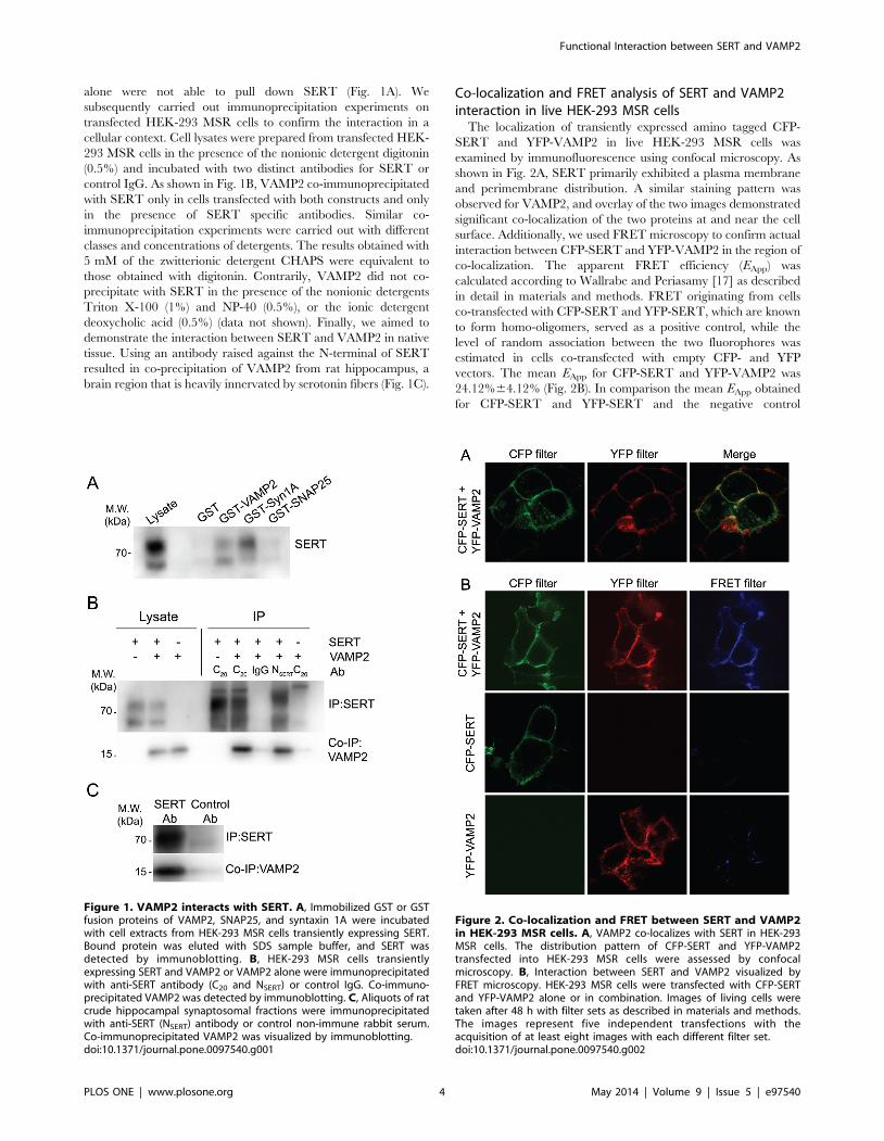
alone were not able to pull down SERT (Fig. 1A). We
subsequently carried out immunoprecipitation experiments on
transfected HEK-293 MSR cells to confirm the interaction in a
cellular context. Cell lysates were prepared from transfected HEK-
293 MSR cells in the presence of the nonionic detergent digitonin
(0.5%) and incubated with two distinct antibodies for SERT or
control IgG. As shown in Fig. 1B, VAMP2 co-immunoprecipitated
with SERT only in cells transfected with both constructs and only
in the presence of SERT specific antibodies. Similar co-
immunoprecipitation experiments were carried out with different
classes and concentrations of detergents. The results obtained with
5 mM of the zwitterionic detergent CHAPS were equivalent to
those obtained with digitonin. Contrarily, VAMP2 did not co-
precipitate with SERT in the presence of the nonionic detergents
Triton X-100 (1%) and NP-40 (0.5%), or the ionic detergent
deoxycholic acid (0.5%) (data not shown). Finally, we aimed to
demonstrate the interaction between SERT and VAMP2 in native
tissue. Using an antibody raised against the N-terminal of SERT
resulted in co-precipitation of VAMP2 from rat hippocampus, a
brain region that is heavily innervated by serotonin fibers (Fig. 1C).
Co-localization and FRET analysis of SERT and VAMP2interaction in live HEK-293 MSR cells
The localization of transiently expressed amino tagged CFP-
SERT and YFP-VAMP2 in live HEK-293 MSR cells was
examined by immunofluorescence using confocal microscopy. As
shown in Fig. 2A, SERT primarily exhibited a plasma membrane
and perimembrane distribution. A similar staining pattern was
observed for VAMP2, and overlay of the two images demonstrated
significant co-localization of the two proteins at and near the cell
surface. Additionally, we used FRET microscopy to confirm actual
interaction between CFP-SERT and YFP-VAMP2 in the region of
co-localization. The apparent FRET efficiency (EApp) was
calculated according to Wallrabe and Periasamy [17] as described
in detail in materials and methods. FRET originating from cells
co-transfected with CFP-SERT and YFP-SERT, which are known
to form homo-oligomers, served as a positive control, while the
level of random association between the two fluorophores was
estimated in cells co-transfected with empty CFP- and YFP
vectors. The mean EApp for CFP-SERT and YFP-VAMP2 was
24.12%64.12% (Fig. 2B). In comparison the mean EApp obtained
for CFP-SERT and YFP-SERT and the negative control
Figure 1. VAMP2 interacts with SERT. A, Immobilized GST or GSTfusion proteins of VAMP2, SNAP25, and syntaxin 1A were incubatedwith cell extracts from HEK-293 MSR cells transiently expressing SERT.Bound protein was eluted with SDS sample buffer, and SERT wasdetected by immunoblotting. B, HEK-293 MSR cells transientlyexpressing SERT and VAMP2 or VAMP2 alone were immunoprecipitatedwith anti-SERT antibody (C20 and NSERT) or control IgG. Co-immuno-precipitated VAMP2 was detected by immunoblotting. C, Aliquots of ratcrude hippocampal synaptosomal fractions were immunoprecipitatedwith anti-SERT (NSERT) antibody or control non-immune rabbit serum.Co-immunoprecipitated VAMP2 was visualized by immunoblotting.doi:10.1371/journal.pone.0097540.g001
Figure 2. Co-localization and FRET between SERT and VAMP2in HEK-293 MSR cells. A, VAMP2 co-localizes with SERT in HEK-293MSR cells. The distribution pattern of CFP-SERT and YFP-VAMP2transfected into HEK-293 MSR cells were assessed by confocalmicroscopy. B, Interaction between SERT and VAMP2 visualized byFRET microscopy. HEK-293 MSR cells were transfected with CFP-SERTand YFP-VAMP2 alone or in combination. Images of living cells weretaken after 48 h with filter sets as described in materials and methods.The images represent five independent transfections with theacquisition of at least eight images with each different filter set.doi:10.1371/journal.pone.0097540.g002
Functional Interaction between SERT and VAMP2
PLOS ONE | www.plosone.org 4 May 2014 | Volume 9 | Issue 5 | e97540
consisting of cytosolic CFP and YFP was 14.29%63.73% and
4.18%61.19%, respectively.
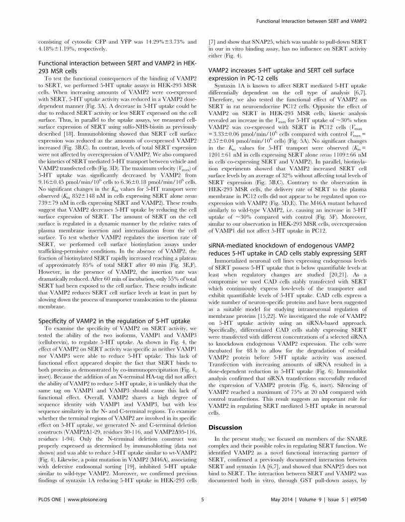
Functional interaction between SERT and VAMP2 in HEK-293 MSR cells
To test the functional consequences of the binding of VAMP2
to SERT, we performed 5-HT uptake assays in HEK-293 MSR
cells. When increasing amounts of VAMP2 were co-expressed
with SERT, 5-HT uptake activity was reduced in a VAMP2 dose-
dependent manner (Fig. 3A). A decrease in 5-HT uptake could be
due to reduced SERT activity or less SERT expressed on the cell
surface. Thus, in parallel to the uptake assays, we measured cell-
surface expression of SERT using sulfo-NHS-biotin as previously
described [18]. Immunoblotting showed that SERT cell surface
expression was reduced as the amounts of co-expressed VAMP2
increased (Fig. 3B,C). In contrast, levels of total SERT expression
were not affected by overexpression of VAMP2. We also compared
the kinetics of SERT mediated 5-HT transport between vehicle and
VAMP2 transfected cells (Fig. 3D). The maximum velocity (Vmax) of
5-HT uptake was significantly decreased by VAMP2 from
9.1660.45 pmol/min/106 cells to 6.3660.18 pmol/min/106 cells.
No significant changes in the Km values for 5-HT transport were
observed (Km 8526148 nM in cells expressing SERT alone versus
739679 nM in cells expressing SERT and VAMP2). These results
suggest that VAMP2 decreases 5-HT uptake by reducing the cell
surface expression of SERT. The amount of SERT on the cell
surface is regulated in a dynamic manner by the relative rates of
plasma membrane insertion and internalization from the cell
surface. To test whether VAMP2 regulates the insertion rate of
SERT, we performed cell surface biotinylation assays under
trafficking-permissive conditions. In the absence of VAMP2, the
fraction of biotinylated SERT rapidly increased reaching a plateau
of approximately 85% of total SERT after 40 min (Fig. 3E,F).
However, in the presence of VAMP2, the insertion rate was
dramatically reduced. After 60 min of incubation, only 55% of total
SERT had been exposed to the cell surface. These results indicate
that VAMP2 reduces SERT cell surface levels at least in part by
slowing down the process of transporter translocation to the plasma
membrane.
Specificity of VAMP2 in the regulation of 5-HT uptakeTo examine the specificity of VAMP2 on SERT activity, we
tested the ability of the two isoforms, VAMP1 and VAMP3
(cellubrevin), to regulate 5-HT uptake. As shown in Fig. 4, the
effect of VAMP2 on SERT activity was specific as neither VAMP1
nor VAMP3 were able to reduce 5-HT uptake. This lack of
functional effect appeared despite the fact that SERT binds to
both proteins as demonstrated by co-immunoprecipitation (Fig. 4,
inset). Because the addition of an N-terminal HA-tag did not affect
the ability of VAMP2 to reduce 5-HT uptake, it is unlikely that the
same tag on VAMP1 and VAMP3 should cause this lack of
functional effect. Overall, VAMP2 shares a high degree of
sequence identity with VAMP1 and VAMP3, but with less
sequence similarity in the N- and C-terminal regions. To examine
whether the terminal regions of VAMP2 are involved in its specific
effect on 5-HT uptake, we generated N- and C-terminal deletion
constructs (VAMP2D1-29, residues 30-116, and VAMP2D95-116,
residues 1-94). Only the N-terminal deletion construct was
properly expressed as determined by immunoblotting (data not
shown) and was able to reduce 5-HT uptake similar to wt-VAMP2
(Fig. 4). Likewise, a point mutation in VAMP2 (M46A), associating
with defective endosomal sorting [19], inhibited 5-HT uptake
similar to wild-type VAMP2. Moreover, we confirmed previous
findings of syntaxin 1A reducing 5-HT uptake in HEK-293 cells
[7] and show that SNAP25, which was unable to pull-down SERT
in our in vitro binding assay, has no influence on SERT activity
either (Fig. 4).
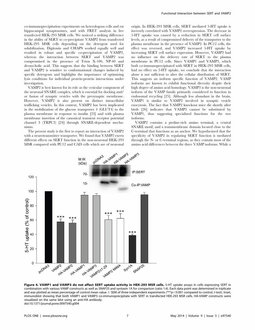
VAMP2 increases 5-HT uptake and SERT cell surfaceexpression in PC-12 cells
Syntaxin 1A is known to affect SERT mediated 5-HT uptake
differentially dependent on the cell type of analysis [6,7].
Therefore, we also tested the functional effect of VAMP2 on
SERT in rat neuroendocrine PC12 cells. Opposite the effect of
VAMP2 on SERT in HEK-293 MSR cells, kinetic analysis
revealed an increase in the Vmax for 5-HT uptake of ,30% when
VAMP2 was co-expressed with SERT in PC12 cells (Vmax
= 3.3360.06 pmol/min/106 cells compared with control Vmax =
2.5760.04 pmol/min/106 cells) (Fig. 5A). No significant changes
in the Km values for 5-HT transport were observed (Km =
1201661 nM in cells expressing SERT alone versus 1109666 nM
in cells co-expressing SERT and VAMP2). In parallel, biotinyla-
tion experiments showed that VAMP2 increased SERT cell
surface levels by an average of 32% without affecting total levels of
SERT expression (Fig. 5B,C). Contrary to the observation in
HEK-293 MSR cells, the delivery rate of SERT to the plasma
membrane in PC12 cells did not appear to be regulated upon co-
expression with VAMP2 (Fig. 5D,E). The M46A mutant behaved
similarly to wild-type VAMP2, i.e. causing an increase in 5-HT
uptake of ,30% compared with control (Fig. 5F). Moreover,
similar to our observation in HEK-293 MSR cells, overexpression
of VAMP1 did not affect 5-HT uptake in PC12.
siRNA-mediated knockdown of endogenous VAMP2reduces 5-HT uptake in CAD cells stably expressing SERT
Immortalized neuronal cell lines expressing endogenous levels
of SERT possess 5-HT uptake that is below quantifiable levels at
least when regulatory changes are studied [20,21]. As a
compromise we used CAD cells stably transfected with SERT
which continuously express low-levels of the transporter and
exhibit quantifiable levels of 5-HT uptake. CAD cells express a
wide number of neuron-specific proteins and have been suggested
as a suitable model for studying intraneuronal regulation of
membrane proteins [15,22]. We investigated the role of VAMP2
on 5-HT uptake activity using an siRNA-based approach.
Specifically, differentiated CAD cells stably expressing SERT
were transfected with different concentrations of a selected siRNA
to knockdown endogenous VAMP2 expression. The cells were
incubated for 48 h to allow for the degradation of residual
VAMP2 protein before 5-HT uptake activity was assessed.
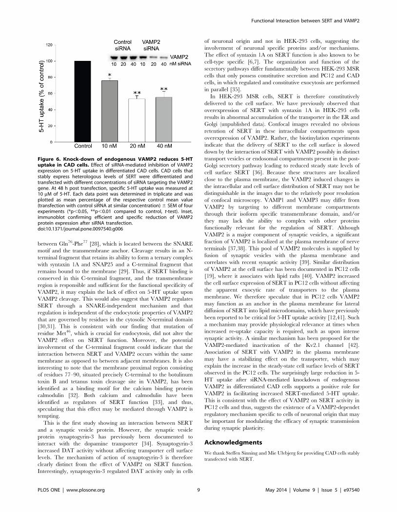
Transfection with increasing amounts of siRNA resulted in a
dose-dependent reduction in 5-HT uptake (Fig. 6). Immunoblot
analysis confirmed that siRNA transfections successfully reduced
the expression of VAMP2 protein (Fig. 6, inset). Silencing of
VAMP2 reached a maximum of 75% at 20 nM compared with
control transfections. This result suggests an important role for
VAMP2 in regulating SERT mediated 5-HT uptake in neuronal
cells.
Discussion
In the present study, we focused on members of the SNARE
complex and their possible roles in regulating SERT function. We
identified VAMP2 as a novel functional interacting partner of
SERT, confirmed a previously documented interaction between
SERT and syntaxin 1A [6,7], and showed that SNAP25 does not
bind to SERT. The interaction between SERT and VAMP2 was
documented both in vitro, through GST pull-down assays, by
Functional Interaction between SERT and VAMP2
PLOS ONE | www.plosone.org 5 May 2014 | Volume 9 | Issue 5 | e97540
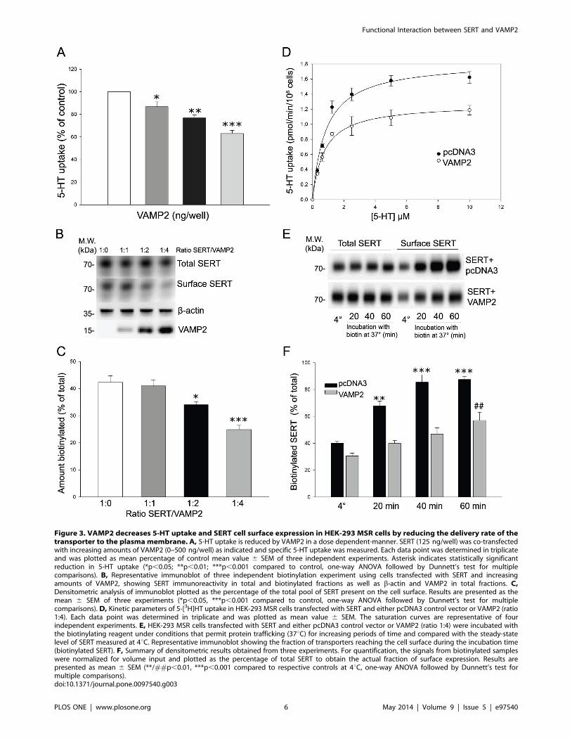
Figure 3. VAMP2 decreases 5-HT uptake and SERT cell surface expression in HEK-293 MSR cells by reducing the delivery rate of thetransporter to the plasma membrane. A, 5-HT uptake is reduced by VAMP2 in a dose dependent-manner. SERT (125 ng/well) was co-transfectedwith increasing amounts of VAMP2 (0–500 ng/well) as indicated and specific 5-HT uptake was measured. Each data point was determined in triplicateand was plotted as mean percentage of control mean value 6 SEM of three independent experiments. Asterisk indicates statistically significantreduction in 5-HT uptake (*p,0.05; **p,0.01; ***p,0.001 compared to control, one-way ANOVA followed by Dunnett’s test for multiplecomparisons). B, Representative immunoblot of three independent biotinylation experiment using cells transfected with SERT and increasingamounts of VAMP2, showing SERT immunoreactivity in total and biotinylated fractions as well as b-actin and VAMP2 in total fractions. C,Densitometric analysis of immunoblot plotted as the percentage of the total pool of SERT present on the cell surface. Results are presented as themean 6 SEM of three experiments (*p,0.05, ***p,0.001 compared to control, one-way ANOVA followed by Dunnett’s test for multiplecomparisons). D, Kinetic parameters of 5-[3H]HT uptake in HEK-293 MSR cells transfected with SERT and either pcDNA3 control vector or VAMP2 (ratio1:4). Each data point was determined in triplicate and was plotted as mean value 6 SEM. The saturation curves are representative of fourindependent experiments. E, HEK-293 MSR cells transfected with SERT and either pcDNA3 control vector or VAMP2 (ratio 1:4) were incubated withthe biotinylating reagent under conditions that permit protein trafficking (37uC) for increasing periods of time and compared with the steady-statelevel of SERT measured at 4uC. Representative immunoblot showing the fraction of transporters reaching the cell surface during the incubation time(biotinylated SERT). F, Summary of densitometric results obtained from three experiments. For quantification, the signals from biotinylated sampleswere normalized for volume input and plotted as the percentage of total SERT to obtain the actual fraction of surface expression. Results arepresented as mean 6 SEM (**/##p,0.01, ***p,0.001 compared to respective controls at 4uC, one-way ANOVA followed by Dunnett’s test formultiple comparisons).doi:10.1371/journal.pone.0097540.g003
Functional Interaction between SERT and VAMP2
PLOS ONE | www.plosone.org 6 May 2014 | Volume 9 | Issue 5 | e97540
co-immunoprecipitation experiments on heterologous cells and rat
hippocampal synaptosomes, and with FRET analysis in live
transfected HEK-293 MSR cells. We noticed a striking difference
in the ability of SERT to co-precipitate VAMP2 from transfected
HEK-293 MSR cells depending on the detergent used for
solubilization. Digitonin and CHAPS worked equally well and
resulted in robust and specific co-precipitation of VAMP2,
whereas the interaction between SERT and VAMP2 was
compromised in the presence of Trion X-100, NP-40 and
deoxycholic acid. This suggests that the binding between SERT
and VAMP2 is sensitive to conformational changes induced by
specific detergents and highlights the importance of optimizing
lysis conditions for individual protein-protein interactions under
investigation.
VAMP2 is best known for its role as the vesicular component of
the neuronal SNARE complex, which is essential for docking and/
or fusion of synaptic vesicles with the presynaptic membrane.
However, VAMP2 is also present on distinct intracellular
trafficking vesicles. In this context, VAMP2 has been implicated
in the mobilization of the glucose transporter 4 (GLUT4) to the
plasma membrane in response to insulin [23] and with plasma
membrane insertion of the canonical transient receptor potential
channel 3 (TRPC3) [24] through SNARE-dependent mecha-
nisms.
The present study is the first to report an interaction of VAMP2
with a neurotransmitter transporter. We found that VAMP2 exerts
different effects on SERT function in the non-neuronal HEK-293
MSR compared with PC12 and CAD cells which are of neuronal
origin. In HEK-293 MSR cells, SERT mediated 5-HT uptake is
inversely correlated with VAMP2 overexpression. The decrease in
5-HT uptake was caused by a reduction in SERT cell surface
levels, as a result of compromised delivery of the transporter to the
plasma membrane in the presence of VAMP2. In PC12 cells, the
effect was reversed, and VAMP2 increased 5-HT uptake by
increasing SERT cell surface expression. However, VAMP2 had
no influence on the delivery rate of SERT to the plasma
membrane in PC12 cells. Since VAMP1 and VAMP3, which
both co-immunoprecipitated with SERT in HEK-293 MSR cells,
had no effect on 5-HT uptake, we conclude that the interaction
alone is not sufficient to alter the cellular distribution of SERT.
This suggests an isoform specific function of VAMP2. VAMP
isoforms are known to exhibit functional diversity despite their
high degree of amino acid homology. VAMP3 is the non-neuronal
isoform of the VAMP family primarily considered to function in
endosomal recycling [25]. Although less abundant in the brain,
VAMP1 is similar to VAMP2 involved in synaptic vesicle
exocytosis. The fact that VAMP2 knockout mice die shortly after
birth [26] indicates that VAMP2 cannot be substituted by
VAMP1, thus suggesting specialized functions for the two
isoforms.
VAMP2 contains a proline-rich amino terminal, a central
SNARE motif, and a transmembrane domain located close to the
C-terminal that functions as an anchor. We hypothesized that the
specificity of VAMP2 in regulating SERT function is mediated
through the N- or C-terminal regions, as they contain most of the
amino acid differences between the three VAMP isoforms. While a
Figure 4. VAMP1 and VAMP3 do not affect SERT uptake activity in HEK-293 MSR cells. 5-HT uptake assays in cells expressing SERT incombination with various VAMP constructs as well as SNAP25 and syntaxin 1A for comparison (ratio 1:4). Each data point was determined in triplicateand was plotted as mean percentage of control mean value 6 SEM of three independent experiments (***p,0.001 compared to control, t-test). Inset,immunoblot showing that both VAMP1 and VAMP3 co-immunoprecipitate with SERT in transfected HEK-293 MSR cells. HA-VAMP constructs werevisualized on the same blot using an anti-HA antibody.doi:10.1371/journal.pone.0097540.g004
Functional Interaction between SERT and VAMP2
PLOS ONE | www.plosone.org 7 May 2014 | Volume 9 | Issue 5 | e97540
C-terminal deletion construct (VAMP2D95-116) failed to express
properly, the N-terminal deletion construct VAMP2D1-29 was
able to reduce 5-HT uptake to a level similar to wt-VAMP2. This
indirectly points toward a functional role for the isoform specific
C-terminal transmembrane region of VAMP2. Previous studies
have shown that proteolytic cleavage of VAMP2 by botulinum
toxin B [6] and tetanus toxin [27] has no effect on 5-HT uptake in
cultures of rat thalamocortical neurons and rat synaptosomes,
respectively. This is in contrast with proteolytic cleavage of
syntaxin 1A with botulinum toxin C, which decreases SERT
activity and plasma membrane levels in thalamocortical neurons
[6]. Botulinum toxin B and tetanus toxin both cleave VAMP2
Figure 5. VAMP2 increases SERT uptake and cell surface expression in PC12 cells. A, Kinetic parameters of 5-[3H]HT uptake in PC12 cellstransfected with SERT and either pcDNA3 control vector or VAMP2 (ratio 1:4). Each data point was determined in triplicate and was plotted as meanvalue 6 SEM. The saturation curves are representative of three independent experiments. B, Representative immunoblot of biotinylation experimenton PC12 cells transfected with SERT in combination with pcDNA3 or VAMP2 (ratio 1:4), showing SERT immunoreactivity in total and biotinylatedfractions. C, Densitometric analysis of immunoblots plotted as the percentage of the total pool of SERT present on the cell surface. Results arepresented as the mean 6 SEM of three experiments (*p,0.05, t-test). D, PC12 cells transfected with SERT and either pcDNA3 control vector or VAMP2(ratio 1:4) were incubated with the biotinylating reagent under conditions that permit protein trafficking (37uC) for increasing periods of time andcompared with the steady-state level of SERT measured at 4uC. Representative immunoblot showing the fraction of transporters reaching the cellsurface during the incubation time (biotinylated SERT). E, Summary of densitometric results obtained from three experiments. For quantification, thesignals from biotinylated samples were normalized for volume input and plotted as the percentage of total SERT to obtain the actual fraction ofsurface expression. Results are presented as mean 6 SEM (#p,0.05, **/##p,0.01, ***p,0.001 compared to respective controls at 4uC, one-wayANOVA followed by Dunnett’s test for multiple comparisons). F, 5-HT uptake assays in PC12 cells expressing SERT in combination with pcDNA3control vector, VAMP2, VAMP1 or the VAMP2 mutant M46A (ratio 1:4). Data are expressed as the mean percentage of control mean 6 SEM of valuesobtained in four experiments performed in triplicates (***p,0.001 compared to control, t-test).doi:10.1371/journal.pone.0097540.g005
Functional Interaction between SERT and VAMP2
PLOS ONE | www.plosone.org 8 May 2014 | Volume 9 | Issue 5 | e97540
between Gln76-Phe77 [28], which is located between the SNARE
motif and the transmembrane anchor. Cleavage results in an N-
terminal fragment that retains its ability to form a ternary complex
with syntaxin 1A and SNAP25 and a C-terminal fragment that
remains bound to the membrane [29]. Thus, if SERT binding is
conserved in this C-terminal fragment, and the transmembrane
region is responsible and sufficient for the functional specificity of
VAMP2, it may explain the lack of effect on 5-HT uptake upon
VAMP2 cleavage. This would also suggest that VAMP2 regulates
SERT through a SNARE-independent mechanism and that
regulation is independent of the endocytotic properties of VAMP2
that are governed by residues in the cytosolic N-terminal domain
[30,31]. This is consistent with our finding that mutation of
residue Met46, which is crucial for endocytosis, did not alter the
VAMP2 effect on SERT function. Moreover, the potential
involvement of the C-terminal fragment could indicate that the
interaction between SERT and VAMP2 occurs within the same
membrane as opposed to between adjacent membranes. It is also
interesting to note that the membrane proximal region consisting
of residues 77–90, situated precisely C-terminal to the botulinum
toxin B and tetanus toxin cleavage site in VAMP2, has been
identified as a binding motif for the calcium binding protein
calmodulin [32]. Both calcium and calmodulin have been
identified as regulators of SERT function [33], and thus,
speculating that this effect may be mediated through VAMP2 is
tempting.
This is the first study showing an interaction between SERT
and a synaptic vesicle protein. However, the synaptic vesicle
protein synaptogyrin-3 has previously been documented to
interact with the dopamine transporter [34]. Synaptogyrin-3
increased DAT activity without affecting transporter cell surface
levels. The mechanism of action of synaptogyrin-3 is therefore
clearly distinct from the effect of VAMP2 on SERT function.
Interestingly, synaptogyrin-3 regulated DAT activity only in cells
of neuronal origin and not in HEK-293 cells, suggesting the
involvement of neuronal specific proteins and/or mechanisms.
The effect of syntaxin 1A on SERT function is also known to be
cell-type specific [6,7]. The organization and function of the
secretory pathways differ fundamentally between HEK-293 MSR
cells that only possess constitutive secretion and PC12 and CAD
cells, in which regulated and constitutive exocytosis are performed
in parallel [35].
In HEK-293 MSR cells, SERT is therefore constitutively
delivered to the cell surface. We have previously observed that
overexpression of SERT with syntaxin 1A in HEK-293 cells
results in abnormal accumulation of the transporter in the ER and
Golgi (unpublished data). Confocal images revealed no obvious
retention of SERT in these intracellular compartments upon
overexpression of VAMP2. Rather, the biotinylation experiments
indicate that the delivery of SERT to the cell surface is slowed
down by the interaction of SERT with VAMP2 possibly in distinct
transport vesicles or endosomal compartments present in the post-
Golgi secretory pathway leading to reduced steady state levels of
cell surface SERT [36]. Because these structures are localized
close to the plasma membrane, the VAMP2 induced changes in
the intracellular and cell surface distribution of SERT may not be
distinguishable in the images due to the relatively poor resolution
of confocal microscopy. VAMP1 and VAMP3 may differ from
VAMP2 by targeting to different membrane compartments
through their isoform specific transmembrane domain, and/or
they may lack the ability to complex with other proteins
functionally relevant for the regulation of SERT. Although
VAMP2 is a major component of synaptic vesicles, a significant
fraction of VAMP2 is localized at the plasma membrane of nerve
terminals [37,38]. This pool of VAMP2 molecules is supplied by
fusion of synaptic vesicles with the plasma membrane and
correlates with recent synaptic activity [39]. Similar distribution
of VAMP2 at the cell surface has been documented in PC12 cells
[19], where it associates with lipid rafts [40]. VAMP2 increased
the cell surface expression of SERT in PC12 cells without affecting
the apparent exocytic rate of transporters to the plasma
membrane. We therefore speculate that in PC12 cells VAMP2
may function as an anchor in the plasma membrane for lateral
diffusion of SERT into lipid microdomains, which have previously
been reported to be critical for 5-HT uptake activity [12,41]. Such
a mechanism may provide physiological relevance at times when
increased re-uptake capacity is required, such as upon intense
synaptic activity. A similar mechanism has been proposed for the
VAMP2-mediated inactivation of the Kv2.1 channel [42].
Association of SERT with VAMP2 in the plasma membrane
may have a stabilizing effect on the transporter, which may
explain the increase in the steady-state cell surface levels of SERT
observed in the PC12 cells. The surprisingly large reduction in 5-
HT uptake after siRNA-mediated knockdown of endogenous
VAMP2 in differentiated CAD cells supports a positive role for
VAMP2 in facilitating increased SERT-mediated 5-HT uptake.
This is consistent with the effect of VAMP2 on SERT activity in
PC12 cells and thus, suggests the existence of a VAMP2-dependet
regulatory mechanism specific to cells of neuronal origin that may
be important for modulating the efficacy of synaptic transmission
during synaptic plasticity.
Acknowledgments
We thank Steffen Sinning and Mie Ulvbjerg for providing CAD cells stably
transfected with SERT.
Figure 6. Knock-down of endogenous VAMP2 reduces 5-HTuptake in CAD cells. Effect of siRNA-mediated inhibition of VAMP2expression on 5-HT uptake in differentiated CAD cells. CAD cells thatstably express heterologous levels of SERT were differentiated andtransfected with different concentrations of siRNA targeting the VAMP2gene. At 48 h post transfection, specific 5-HT uptake was measured at10 mM of 5-HT. Each data point was determined in triplicate and wasplotted as mean percentage of the respective control mean value(transfection with control siRNA at similar concentration) 6 SEM of fourexperiments (*p,0.05, **p,0.01 compared to control, t-test). Inset,immunoblot confirming efficient and specific reduction of VAMP2protein expression after siRNA transfection.doi:10.1371/journal.pone.0097540.g006
Functional Interaction between SERT and VAMP2
PLOS ONE | www.plosone.org 9 May 2014 | Volume 9 | Issue 5 | e97540
Author Contributions
Conceived and designed the experiments: HKM MK AWF OW.
Performed the experiments: HKM MK AWF. Analyzed the data: HKM
MK AWF OW. Contributed reagents/materials/analysis tools: HKM MK
AWF OW. Wrote the paper: HKM OW.
References
1. Zahniser NR, Doolen S (2001) Chronic and acute regulation of Na+/Cl- -
dependent neurotransmitter transporters: drugs, substrates, presynaptic recep-tors, and signaling systems. Pharmacol Ther 92: 21–55. S0163-7258(01)00158-9
[pii].2. Zhou FC, Tao-Cheng JH, Segu L, Patel T, Wang Y (1998) Serotonin
transporters are located on the axons beyond the synaptic junctions: anatomical
and functional evidence. Brain Res 805: 241–254. S0006-8993(98)00691-X [pii].3. Andersen J, Kristensen AS, Bang-Andersen B, Strømgaard K (2009) Recent
advances in the understanding of the interaction of antidepressant drugs withserotonin and norepinephrine transporters. Chem Commun (Camb) 3677–3692.
10.1039/b903035m [doi].
4. Han DD, Gu HH (2006) Comparison of the monoamine transporters fromhuman and mouse in their sensitivities to psychostimulant drugs. BMC
Pharmacol 6: 6. 1471-2210-6-6 [pii];10.1186/1471-2210-6-6 [doi].5. Steiner JA, Carneiro AM, Blakely RD (2008) Going with the flow: trafficking-
dependent and -independent regulation of serotonin transport. Traffic 9: 1393–1402. TRA757 [pii];10.1111/j.1600-0854.2008.00757.x [doi].
6. Quick MW (2002) Role of syntaxin 1A on serotonin transporter expression in
developing thalamocortical neurons. Int J Dev Neurosci 20: 219–224.S0736574802000217 [pii].
7. Haase J, Killian AM, Magnani F, Williams C (2001) Regulation of the serotonintransporter by interacting proteins. Biochem Soc Trans 29: 722–728.
8. Sudhof TC (2004) The synaptic vesicle cycle. Annu Rev Neurosci 27: 509–547.
10.1146/annurev.neuro.26.041002.131412 [doi].9. Ciccone MA, Timmons M, Phillips A, Quick MW (2008) Calcium/calmodulin-
dependent kinase II regulates the interaction between the serotonin transporterand syntaxin 1A. Neuropharmacology 55: 763–770. S0028-3908(08)00204-9
[pii];10.1016/j.neuropharm.2008.06.018 [doi].
10. Quick MW (2003) Regulating the conducting states of a mammalian serotonintransporter. Neuron 40: 537–549. S0896627303006056 [pii].
11. Kristensen AS, Andersen J, Jørgensen TN, Sørensen L, Eriksen J, et al. (2011)SLC6 neurotransmitter transporters: structure, function, and regulation.
Pharmacol Rev 63: 585–640. pr.108.000869 [pii];10.1124/pr.108.000869 [doi].12. Magnani F, Tate CG, Wynne S, Williams C, Haase J (2004) Partitioning of the
serotonin transporter into lipid microdomains modulates transport of serotonin.
J Biol Chem 279: 38770–38778. 10.1074/jbc.M400831200 [doi];M400831200[pii].
13. Muller HK, Wegener G, Popoli M, Elfving B (2011) Differential expression ofsynaptic proteins after chronic restraint stress in rat prefrontal cortex and
hippocampus. Brain Res 1385: 26–37. S0006-8993(11)00377-5 [pii];10.1016/
j.brainres.2011.02.048 [doi].14. Suri C, Fung BP, Tischler AS, Chikaraishi DM (1993) Catecholaminergic cell
lines from the brain and adrenal glands of tyrosine hydroxylase-SV40 T antigentransgenic mice. J Neurosci 13: 1280–1291.
15. Qi Y, Wang JK, McMillian M, Chikaraishi DM (1997) Characterization of aCNS cell line, CAD, in which morphological differentiation is initiated by serum
deprivation. J Neurosci 17: 1217–1225.
16. Fjorback AW, Pla P, Muller HK, Wiborg O, Saudou F, et al. (2009) Serotonintransporter oligomerization documented in RN46A cells and neurons by
sensitized acceptor emission FRET and fluorescence lifetime imaging micros-copy. Biochem Biophys Res Commun 380: 724–728. S0006-291X(09)00108-9
[pii];10.1016/j.bbrc.2009.01.128 [doi].
17. Wallrabe H, Periasamy A (2005) Imaging protein molecules using FRET andFLIM microscopy. Curr Opin Biotechnol 16: 19–27. S0958-1669(04)00167-3
[pii];10.1016/j.copbio.2004.12.002 [doi].18. Muller HK, Wiborg O, Haase J (2006) Subcellular redistribution of the
serotonin transporter by secretory carrier membrane protein 2. J Biol Chem 281:28901–28909. M602848200 [pii];10.1074/jbc.M602848200 [doi].
19. Grote E, Kelly RB (1996) Endocytosis of VAMP is facilitated by a synaptic
vesicle targeting signal. J Cell Biol 132: 537–547.20. Yammamoto H, Tanaka S, Tanaka A, Hide I, Seki T, et al. (2013) Long-term
exposure of RN46A cells expressing serotonin transporter (SERT) to a cAMPanalog up-regulates SERT activity and is accompanied by neural differentiation
of the cells. J Pharmacol Sci 121: 25–38. DN/JST.JSTAGE/jphs/12229FP
[pii].21. Yoffe JR, Borchardt RT (1982) Characterization of serotonin uptake in cultured
pheochromocytoma cells. Comparison with norepinephrine uptake. MolPharmacol 21: 368–373.
22. Li Y, Hou LX, Aktiv A, Dahlstrom A (2007) Studies of the central nervoussystem-derived CAD cell line, a suitable model for intraneuronal transport
studies? J Neurosci Res 85: 2601–2609. 10.1002/jnr.21216 [doi].
23. Bryant NJ, Govers R, James DE (2002) Regulated transport of the glucose
transporter GLUT4. Nat Rev Mol Cell Biol 3: 267–277. 10.1038/nrm782
[doi];nrm782 [pii].
24. Singh BB, Lockwich TP, Bandyopadhyay BC, Liu X, Bollimuntha S, et al.
(2004) VAMP2-dependent exocytosis regulates plasma membrane insertion of
TRPC3 channels and contributes to agonist-stimulated Ca2+ influx. Mol Cell
15: 635–646. 10.1016/j.molcel.2004.07.010 [doi];S1097276504004162 [pii].
25. McMahon HT, Ushkaryov YA, Edelmann L, Link E, Binz T, et al. (1993)
Cellubrevin is a ubiquitous tetanus-toxin substrate homologous to a putative
synaptic vesicle fusion protein. Nature 364: 346–349. 10.1038/364346a0 [doi].
26. Schoch S, Deak F, Konigstorfer A, Mozhayeva M, Sara Y, et al. (2001) SNARE
function analyzed in synaptobrevin/VAMP knockout mice. Science 294: 1117–
1122. 10.1126/science.1064335 [doi];294/5544/1117 [pii].
27. Inserte J, Najib A, Pelliccioni P, Gil C, Aguilera J (1999) Inhibition by tetanus
toxin of sodium-dependent, high-affinity [3H]5-hydroxytryptamine uptake in rat
synaptosomes. Biochem Pharmacol 57: 111–120. S0006-2952(98)00281-0 [pii].
28. Schiavo G, Benfenati F, Poulain B, Rossetto O, Polverino de LP, et al. (1992)
Tetanus and botulinum-B neurotoxins block neurotransmitter release by
proteolytic cleavage of synaptobrevin. Nature 359: 832–835. 10.1038/
359832a0 [doi].
29. Hayashi T, McMahon H, Yamasaki S, Binz T, Hata Y, et al. (1994) Synaptic
vesicle membrane fusion complex: action of clostridial neurotoxins on assembly.
EMBO J 13: 5051–5061.
30. Hosoi N, Holt M, Sakaba T (2009) Calcium dependence of exo- and endocytotic
coupling at a glutamatergic synapse. Neuron 63: 216–229. S0896-
6273(09)00463-2 [pii];10.1016/j.neuron.2009.06.010 [doi].
31. Koo SJ, Puchkov D, Haucke V (2011) AP180 and CALM: Dedicated endocytic
adaptors for the retrieval of synaptobrevin 2 at synapses. Cell Logist 1: 168–172.
10.4161/cl.1.4.18897 [doi];2159-2780-1-4-10 [pii].
32. de Haro L, Quetglas S, Iborra C, Leveque C, Seagar M (2003) Calmodulin-
dependent regulation of a lipid binding domain in the v-SNARE synaptobrevin
and its role in vesicular fusion. Biol Cell 95: 459–464. S0248490003000765 [pii].
33. Jayanthi LD, Ramamoorthy S, Mahesh VB, Leibach FH, Ganapathy V (1994)
Calmodulin-dependent regulation of the catalytic function of the human
serotonin transporter in placental choriocarcinoma cells. J Biol Chem 269:
14424–14429.
34. Egana LA, Cuevas RA, Baust TB, Parra LA, Leak RK, et al. (2009) Physical and
functional interaction between the dopamine transporter and the synaptic vesicle
protein synaptogyrin-3. J Neurosci 29: 4592–4604. 29/14/4592 [pii];10.1523/
JNEUROSCI.4559-08.2009 [doi].
35. Khvotchev MV, Ren M, Takamori S, Jahn R, Sudhof TC (2003) Divergent
functions of neuronal Rab11b in Ca2+-regulated versus constitutive exocytosis.
J Neurosci 23: 10531–10539. 23/33/10531 [pii].
36. Melikian HE (2004) Neurotransmitter transporter trafficking: endocytosis,
recycling, and regulation. Pharmacol Ther 104: 17–27. S0163-7258(04)00104-
4 [pii];10.1016/j.pharmthera.2004.07.006 [doi].
37. Sankaranarayanan S, Ryan TA (2000) Real-time measurements of vesicle-
SNARE recycling in synapses of the central nervous system. Nat Cell Biol 2:
197–204. 10.1038/35008615 [doi].
38. Taubenblatt P, Dedieu JC, Gulik-Krzywicki T, Morel N (1999) VAMP
(synaptobrevin) is present in the plasma membrane of nerve terminals. J Cell
Sci 112 (Pt 20): 3559–3567.
39. Dittman JS, Kaplan JM (2006) Factors regulating the abundance and
localization of synaptobrevin in the plasma membrane. Proc Natl Acad
Sci U S A 103: 11399–11404. 0600784103 [pii];10.1073/pnas.0600784103
[doi].
40. Chamberlain LH, Burgoyne RD, Gould GW (2001) SNARE proteins are highly
enriched in lipid rafts in PC12 cells: implications for the spatial control of
exocytosis. Proc Natl Acad Sci U S A 98: 5619–5624. 10.1073/pnas.091502398
[doi];091502398 [pii].
41. Samuvel DJ, Jayanthi LD, Bhat NR, Ramamoorthy S (2005) A role for p38
mitogen-activated protein kinase in the regulation of the serotonin transporter:
evidence for distinct cellular mechanisms involved in transporter surface
expression. J Neurosci 25: 29–41. 25/1/29 [pii];10.1523/JNEUROSCI.3754-
04.2005 [doi].
42. Lvov A, Chikvashvili D, Michaelevski I, Lotan I (2008) VAMP2 interacts
directly with the N terminus of Kv2.1 to enhance channel inactivation. Pflugers
Arch 456: 1121–1136. 10.1007/s00424-008-0468-7 [doi].
Functional Interaction between SERT and VAMP2
PLOS ONE | www.plosone.org 10 May 2014 | Volume 9 | Issue 5 | e97540