differential effects of startle on reaction time for finger and arm
TRANSCRIPT

1
Running head: Differential effects of startle on RT
Differential effects of startle on reaction time for finger and arm movements
Anthony N. Carlsen, Romeo Chua, J. Timothy Inglis, David J. Sanderson, and Ian M. Franks
School of Human Kinetics, University of British Columbia, Vancouver, Canada.
Correspondence address:
Anthony N. Carlsen
210 – 6081 University Boulevard
University of British Columbia
Vancouver, BC V6T 1Z1
Canada
Ph. (604) 822-3400
Fax. (604) 822-6842
Email: [email protected]
Page 1 of 39 Articles in PresS. J Neurophysiol (November 12, 2008). doi:10.1152/jn.00878.2007
Copyright © 2008 by the American Physiological Society.
2
Abstract
Recent studies employing a reaction time (RT) task have reported that a preprogrammed
response could be triggered directly by a startling acoustic stimulus (115-124 dB) presented
along with the usual “go” signal. It has been suggested that details of the upcoming response
could be stored subcortically and are accessible by the startle volley, directly eliciting the correct
movement. However, certain muscles (e.g. intrinsic hand) are heavily dependent on cortico-
motoneuronal connections, and thus would not be directly subject to the subcortical startle volley
in a similar way to muscles whose innervations include extensive reticular connections. In the
present study fourteen participants performed 75 trials in each of two tasks within a RT
paradigm: an arm extension task, and an index finger abduction task. In 12 trials within each
task, the regular “go” stimulus (82 dB) was replaced with a 115 dB startling stimulus. Results
showed that in the arm task, the presence of a startle reaction led to significantly shorter latency
arm movements compared to the effect of the increased stimulus intensity alone. In contrast, for
the finger task, no additional decrease in RT due to startle was observed. Taken together these
results suggest that only movements that involve muscles more strongly innervated by
subcortical pathways are susceptible to response advancement by startle.
Keywords: Startle, corticospinal, motor program, reaction time
Page 2 of 39
3
Introduction
Recent studies employing a reaction time (RT) paradigm, have reported that premotor RT
(time from stimulus presentation to EMG onset) in voluntary aiming tasks was substantially
shortened when an unexpected loud acoustic stimulus (130 dB) was presented along with the
usual “go” signal (Valls-Solé et al. 1995, 1999). Based on these findings, it was suggested that a
loud startling stimulus could be used to directly elicit a pre-programmed response without the
usual voluntary command. Specifically, Valls-Solé et al. stated that “the whole motor
programme [could] be triggered [by the startle] without the typical command from the cerebral
cortex” (1999 p.937). This statement was mainly supported by the drastic nature of the RT
decrease observed. That is, premotor RT (PMT) was very short compared to a control (no startle)
condition. In the fastest reactions, PMT was 65 ms (Valls-Solé et al. 1999). In contrast, RTs of
180 ms are normally observed in response to visual stimuli, while RTs of 140 ms or more are
commonly observed in response to acoustic stimuli (Brebner and Welford 1980). Valls-Solé et
al. (1999) argued that because of the fixed amounts of time needed both to convert the acoustic
stimulus to neural signals and for neural transmission, it was unlikely that cortical loops were
involved in the initiation of movements in which PMT was less than 65 ms. Thus they suggested
that sufficient details of a prepared movement may have been stored subcortically, possibly in
the brainstem and spinal centres that were accessible to the startle volley, so that in some cases it
could be triggered early. Several later experiments replicated and extended these findings (e.g.
Carlsen et al. 2003b, 2004b, 2007; Castellote et al. 2007; Cressman et al. 2006; MacKinnon et al.
2007; Siegmund et al. 2001). For example, it was found that response kinematics and EMG
patterns are unchanged between control and startle elicited movements (Carlsen et al. 2004b),
Page 3 of 39

4
and that the paradigm can be extended to different effectors and movement types such as
saccades (Castellote et al. 2007) and anticipatory postural adjustments (MacKinnon et al. 2007).
The defining requirement for a response to be elicited by a startle appears to be pre-
programming. That is, in order for a startle to elicit a movement at short latencies it must have
been prepared in advance. When a startle was presented in a choice RT paradigm, it was found
that PMT was unaffected by the startle. This indicated that the RT shortening effect of startle
was not simply due to increased neural excitability allowing for faster response transmission,
since a similar speeding effect would be observed whether or not a response could be pre-
programmed. It was only when the response was certain beforehand (i.e. a simple RT task) that
the response was speeded by a startle (Carlsen et al. 2004a). Although this result supports the
suggestion that a stored program is triggered early by a startle, it does not necessitate subcortical
program storage (e.g. Carlsen et al. 2004b; Valls-Solé et al. 1999) since it cannot rule out an
undescribed, fast transcortical route.
While it has been traditionally thought that corticospinal connections are extremely
important in the control of individual finger movements, recent preliminary data indicate that
some reticulospinal connections with distal finger muscles exist, and modulate their activity with
movements of the finger (Baker and Riddle 2007; Soteropoulos et al. 2007). However, these
reticulospinal connections are seen less frequently than corticospinal connections (Baker and
Riddle 2007), and thus may be less functionally effective. This is evidenced by earlier studies
showing that following permanent lesions of the corticospinal tract, monkeys were unable to
produce individual finger movements for tasks such as eating and grooming, although they
recovered the ability to use more proximal muscles for climbing and walking (Lawrence and
Kuypers 1968). Similar, although more variable, observations have been made in humans
Page 4 of 39
5
following stroke (e.g. Carroll 1965; Wade et al. 1983). More recently, similar deficits in fine
finger control have been shown in monkeys through reversible chemical inactivation of primary
motor cortex (Brochier et al. 1999). The aim of the current experiment was to determine if a
startle acts to shorten RT in a finger movement (abduction of the index finger) that is thought to
be strongly mediated by corticospinal connections. It was hypothesised that if this finger
movement were speeded by a startle, it would suggest that the startle effect likely includes a
transcortical component. If, however, the movement was not speeded by startle, it would support
the suggestion that for other types of movements (e.g. arm extension, see Carlsen et al. 2004b),
motor programs can be stored subcortically, and that the startle can act to trigger pre-
programmed movements without the involvement of cortex.
Method
Participants
Fourteen participants (9M, 5F; age 24 +/- 5 years) with no obvious upper body
abnormalities, or sensory or motor dysfunctions volunteered to participate in the study. All
participants gave written informed consent, and the study was conducted in accordance with the
ethical guidelines set by the University of British Columbia.
Apparatus and task
Participants performed two tasks on separate days. These will be referred to as the finger
task and the arm task.
For the finger movement task, participants sat in a height-adjustable chair with their right
arm secured to a table pointing forward and in a semi-pronated position. The arm was positioned
so that the shoulder was both flexed and abducted approximately 30 deg with the elbow flexed at
Page 5 of 39

6
30 deg. The hand was also secured to the table using a Velcro strip attached to a clip and passing
around fingers 3-5 which were bent 90 deg at the proximal interphalangeal joint leaving the
index finger (digitus II) free to move. A simple contact switch requiring 0.04 N to close (i.e.
simply resting the finger in the switch was sufficient to close it) was placed under the end of the
outstreched index finger on the medial surface so that upwards movement (abduction) of the
index finger opened the switch (see Fig. 1). The finger movement task was a rapid finger
abduction which was just sufficient to open the switch using only the finger muscles, following
an acoustic stimulus.
(INSERT FIGURE 1 ABOUT HERE)
For the arm task, participants sat in a height-adjustable chair outfitted with an automobile
racing harness (Racer Components Inc.) in order to constrain any movement to the right elbow
joint. The right arm was secured, in a pronated position with the palm down, to a custom-made
aluminium manipulandum that moved in the transverse plane with an axis of rotation at the
elbow. The starting position (90 degrees of flexion at the elbow with the shoulder flexed 30
degrees) was indicated by a mechanical stop. Participants were instructed to perform a 20 deg
arm extension movement to a fixed target as quickly and as accurately as possible following an
acoustic stimulus. For both tasks participants were offered a monetary bonus for fast reactions.
Instrumentation and stimuli
Trials started with a warning consisting of a short acoustic tone (100 ms, 300 Hz, 80 dB)
generated by the computer using a 16 bit sound card (Creative SoundBlaster 16®) and standard
computer speakers (Juster® sp-691n). A variable foreperiod of 2 - 3 sec. spanned the time
between the end of the warning tone and the imperative stimulus. A computer program then
generated the imperative stimulus consisting of a narrow band noise pulse (1 kHz, 40ms
Page 6 of 39

7
duration). The signal was amplified and presented via a loudspeaker (<1 ms rise time) placed
directly behind the head of the participant. The intensity of the acoustic “go” signal (imperative
stimulus) was either 82 dB (control trials) or 115 dB (startle trials), and was measured using a
sound level meter (Cirrus Research, model CR: 252B) at a distance of 30 cm from the
loudspeaker (approximately the distance to the ears of the participant).
It was previously shown that 124 dB was the most effective intensity for eliciting a startle
response (i.e. EMG activity in the sternocleidomastiod muscle) and startle RT facilitation in the
greatest proportion of trials. In contrast, lower intensities resulted in a higher proportion of trials
in which a startle response was not observed. Although RT was decreased in non startle trials, it
was shown that when a startle response was observed, there was an associated further dramatic
decrease in premotor RT to 80 ms irrespective of stimulus intensity (Carlsen et al. 2007). Here
we used 115 dB so that several startled (SCM activity present) and non-startled (no SCM
activity) loud (115 dB stimulus) trials would be achieved from each participant. This was done in
order to control for stimulus intensity facilitation (e.g. Woodworth 1938, p.318) where increases
in intensity are associated with decreases in RT.
Each participant performed 75 trials in each of the 2 tasks, comprised of 57 control, 12
startle, and 6 catch (no acoustic imperative stimulus) trials. Catch trials were included to
discourage false starts. Startle trials and catch trials occurred randomly amongst the control trials
with the stipulation that no 2 consecutive trials were startle trials, and no startles occurred within
the first 5 trials. The approximate trial to trial interval was 10s, although this varied due to the
random foreperiod (see above).
For the arm movement task, surface Electromyographic (EMG) data were collected from
the right elbow prime movers: the long head of the biceps brachii (BIC), and the lateral head of
Page 7 of 39
8
triceps brachii (TRI), as well as from the startle response indicator sternocleidomastoid (SCM).
For the finger task, EMG data were collected from the first dorsal interosseous (FDI, primary
index finger abductor), as well as from SCM. The recording sites were prepared and cleansed in
order to decrease electrical impedance and then bipolar preamplified Ag/AgCl surface EMG
electrodes were attached in the middle of the muscle bellies parallel to the line of force of the
muscles. These electrodes were connected via shielded cabling to an external amplifier system
(Therapeutics Unlimited Inc., model 544). A grounding electrode was placed on the participant’s
left radial styloid process. Arm angular displacement data in the arm task was collected using a
potentiometer attached to the pivot point of the manipulandum. Time of initiation of finger
displacement in the finger task was monitored using the contact switch described above. All raw
data were digitally sampled for 2 sec at 1 kHz (National Instruments® PCI-6023E) using a
personal computer running a customized program written with LabVIEW® software (National
Instruments Inc.). Data collection was automatically initiated 500 ms prior to the imperative
stimulus.
Target and feedback
The target for the arm task was a fixed point in space located at 20 degrees of angular
displacement into extension with respect to the right arm’s starting position. A computer screen
placed directly in front of the participant provided real time position feedback. The position of
the manipulandum was represented with a yellow marker line (1 cm tall) whose movement in the
horizontal plane corresponded directly to movement of the manipulandum. The starting position
of the marker was approximately in the centre of the computer screen. The target was
represented by a stationary blue target line (1 cm tall), located 10 cm from the right edge of the
screen. After each trial, feedback information including target error (deg), and RT (ms) was
Page 8 of 39
9
displayed on the same computer monitor display. For the finger task, RT (ms) was displayed
following each trial.
Training
Participants were allowed to practice the task prior to testing to familiarize themselves
with the task and equipment. The participants were instructed that they would first hear a
warning tone, followed by a variable foreperiod, and finally a “go” tone (imperative stimulus).
Instructions emphasised fast reaction times and fast movement times. Participants were also
instructed that the loudness of the stimulus would be variable. For each task, participants
received a single block of 10 practice trials in which no startle tone occurred.
Data reduction and analysis
For the arm task, movement onset (Displacement RT) was defined as the first point of a
change of more than 0.2 deg of angular displacement from the starting position following the
stimulus. For the finger task, movement onset was the moment that the finger switch registered a
positive voltage greater than 1V. Additional variables were calculated for the arm task. The final
position of the movement was defined as the first point at which angular velocity remained
below 8 deg/sec for at least 150 ms. Movement time was defined as the time (in ms) between
movement onset and final position.
For both tasks, surface EMG burst onsets were defined as the point at which the EMG
first began a sustained rise above baseline levels. The location of this point was determined by
first displaying the EMG pattern on a computer monitor with a superimposed line indicating the
point at which rectified, filtered EMG activity increased to more than 2 standard deviations
above baseline (mean of 100 ms of EMG activity preceding onset). Onset was then verified by
Page 9 of 39
10
visually locating and manually adjusting the onset mark to the point at which the activity first
increased on the raw EMG trace. This method allows for correction of errors due to the strictness
of the algorithm. Premotor RT (PMT) was defined as EMG onset in the TRI muscle for the arm
task and EMG onset in the FDI for the finger task. PMT was the main measure of RT for the
current study as it represents an estimate of the total central processing time. EMG offsets were
marked in a similar fashion, using a mean of EMG activity following the end of movement as a
baseline level to account for any residual activity time between bursts. These were also verified
and manually adjusted, with the activity between EMG onset and EMG offset being defined as a
distinct burst. PMTs greater than 1000 ms and less than 40 ms were discarded if observed in the
control (82 dB) condition
Startle trials in which SCM activity was not present prior to 120 ms following the
stimulus were treated separately from startle trials in which SCM activity was observed as this
distinction can be used to control for the effect of stimulus intensity vs. the effect of startle
(Carlsen et al. 2007). This led to the designation of a third stimulus category for analysis (see
Results section).
Statistical analyses
Dependent measures were analyzed using 2 factor (task), or 3 factor (stimulus) repeated
measures analysis of variance (ANOVA), where appropriate, to determine if differences existed
between tasks and/or conditions. EMG and kinematic measures were not analyzed between tasks
as the mechanical features of the tasks differed greatly. Several dependent measures were
available for the arm task only, (e.g. final position) and were analyzed using a 3 factor (stimulus)
repeated measures ANOVA. Proportion variables were subjected to an arcine square root
transform prior to analysis. Greenhouse-Geisser corrected degrees of freedom were used to
Page 10 of 39
11
correct for violations of the assumption of sphericity. Differences with a probability of less than
.05 were considered to be significant. Tukey’s Honestly Significant Difference (HSD) post-hoc
tests were administered to determine the locus of the differences.
Results
Raw data from a single participant is shown in Figure 2 exemplifying the differences
observed between the conditions in both the arm task (left panels) and the finger task (right
panels). The presence of startle-related EMG activity in sternocleidomastoid (SCM) was used to
categorize startle trials into trials in which SCM activity was present (SCM+) vs. absent (SCM-).
The dashed line allows comparison of PMT observed in SCM+ trials with PMT observed in the
control and SCM- conditions. Although the 115 dB stimulus led to shorter PMT in both
movement tasks, only in the arm task did the presence of a startle response (SCM+) lead to a
further reduction in PMT.
(INSERT FIGURE 2 ABOUT HERE)
Startle response
In both the finger task and arm task EMG activity in SCM was observed in startle trials
for all participants. There was no significant difference in SCM onset latency between the arm
task (72.6 +/- 16.2 ms) and the finger task (76.2 +/- 15.7 ms), F(1,13) = 1.213, p = .291.
Calculations showed that effect size was low, partial eta squared (ηp2) = .085, indicating that only
8.5% of any difference observed was attributable to the difference between tasks. Although SCM
activity was not observed in all 115 dB (startle) trials, the proportion of trials in which SCM
activity was present (SCM+) vs. absent (SCM-) did not differ between tasks, F(1,13) = .119, p =
.736, ηp2 = .009, with a SCM response being observed in 59.5 +/- 25.3% of startle trials in the
Page 11 of 39

12
arm task and in 59.5 +/- 22.1% of startle trials in the finger task. Thus dependent measures were
analyzed between three stimulus conditions: Control trials, SCM+ trials, and SCM- trials. This
made it possible to investigate the effect of stimulus intensity separately from the effect of a
startle response as both SCM+ and SCM- trials involved the same stimulus (115 dB).
Premotor reaction time
Since participants were not always startled by the 115 dB stimulus, as defined by EMG
activity in SCM (Carlsen et al. 2007), premotor RT (i.e. time from stimulus to prime mover
EMG onset) was analyzed between control trials, SCM+ trials, and SCM- trials for each task.
Results are presented in Figures 3 & 4. For the arm task, a significant main effect for stimulus
was found, F(2,26) = 48.894, p < .001, ηp2 = .790. Post-hoc tests revealed that PMT was shortest
for SCM+ trials (p < .05), with PMT for SCM- trials being both significantly longer than for
SCM+ trials and shorter than control trials (p <.05, see Fig. 3A). This difference is further
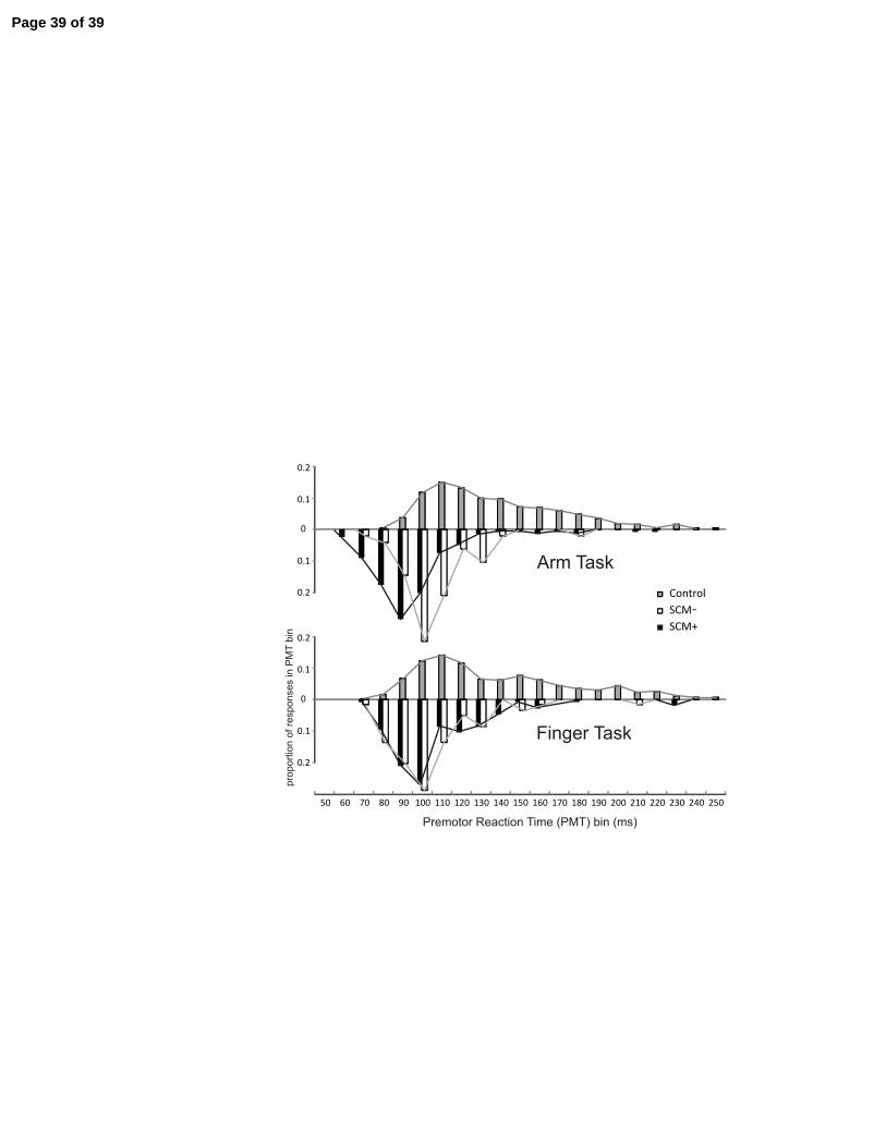
illustrated in Figure 4, where it can be seen that the SCM+ and SCM- PMT distributions are
separated. For the finger task, a significant main effect for stimulus was also found, F(2,26) =
18.580, p < .001, ηp2 = .588. However, while post-hoc tests revealed that control trial PMT was
significantly longer than both 115 dB conditions (p < .05), post-hoc tests showed no difference
(p > .05) in PMT between SCM+ and SCM- trials (see Fig. 3B). In fact, Tukey’s post-hoc
calculations showed that a PMT difference of 15.5 ms between conditions was required to reach
significance at α = .05, while only a negligible mean PMT difference of 0.05 ms was observed
between these conditions. Secondary analysis also showed that there was no significant
difference, F(1,13) < .001, p = .995, ηp2
< .001, between the two 115 dB stimulus conditions for
the finger task. This can also be seen in Figure 4, where there is considerable overlap between
the SCM+ and SCM- PMT distributions for the finger task. Using the Arm task mean PMT
Page 12 of 39
13
difference between the SCM+ and SCM- conditions as an estimate, it was calculated that the
effect size for the task was >1.36. Given the sample size (14) it was calculated that the power to
detect any difference that existed between these two conditions was .996. No catch trial false
starts were observed and all were discarded from analysis.
(INSERT FIGURES 3 & 4 ABOUT HERE)
Kinematic and EMG measures
For the arm task, displacement RT, movement final position and movement time (time
from displacement onset to final position) were analyzed between stimulus conditions.
Displacement RT was also calculated and analyzed for the finger task. Results are presented in
Table 1. None of the kinematic measures were significantly different between stimulus
conditions except displacement RT for both the arm task, F(2,26) = 63.366, p <.001, ηp2
= .830,
and for the finger task, F(2,26) = 15.932, p <.001, ηp2
= .551. Post hoc tests showed the same
pattern as observed for premotor RT: that is displacement RT was significantly different between
SCM+ and SCM- in the arm task (p <.05), but not different in the finger task.
(INSERT TABLE 1 ABOUT HERE)
In addition to premotor RT (see above) EMG measures were analyzed and are presented
in Table 1. Initial agonist durations (TRI for the arm task; FDI for the finger task) were analyzed
for differences between stimulus conditions, however no significant differences were observed
for either task, ηp2
= .017, .166 for the finger and arm tasks respectively. Secondly, burst onset
timing was analyzed for differences between stimulus conditions for the arm task. While a
characteristic triphasic pattern of activity (agonist-antagonist-agonist) was observed for the arm
task, no significant differences in burst timing were observed between the stimulus conditions.
Page 13 of 39

14
Discussion
Previous investigations have demonstrated that during a RT task, pre-programmed
movements can be elicited early if the “go” signal is accompanied by a startling acoustic
stimulus (Carlsen et al. 2004a, b; Castellote et al. 2007; Cressman et al. 2006; Siegmund et al.
2001; Valls-Solé et al. 1999). Although it has been suggested that the startle effect acts by
releasing a motor program that was stored subcortically (Carlsen et al. 2004b; Valls-Solé et al.
1999), there have been no direct tests of this hypothesis. In the present study, a 115 dB startling
stimulus replaced the usual RT “go” stimulus on several trials of two movement tasks. Here we
show that for a finger abduction movement, which appears to be more strongly mediated by
corticospinal connections, the loud stimulus led to a reduction in PMT, yet no difference was
observed whether or not a startle response was observed. For an arm extension movement
however, when a startle reaction was detected in response to the loud stimulus, a larger decrease
in premotor RT was observed than when no startle response was elicited. These data indicate that
in order for early response triggering by startle to occur, the movement must involve more
extensive subcortical brainstem connections.
In order to infer any effect of a startle on RT, it is important to measure the presence of a
startle response in the participants. Without a startle response, the acoustic stimulus is simply
“loud.” It may be suggested that the startle effect is merely an extreme case of stimulus intensity
facilitation (e.g. Woodworth 1938, p.318) where increases in intensity are associated with
decreases in RT, however, it has been recently shown that when a startle reaction was detected,
the RT facilitation was different and larger than that brought on by increases in stimulus intensity
alone (Carlsen et al. 2007): A startle response can be detected using EMG activity in SCM, as it
has been shown to be the electrophysiological indicator of startle in muscle EMG that is the
Page 14 of 39

15
among most reliable and one of the last to habituate to repeated stimuli (Brown et al. 1991). This
SCM activity was associated with substantially shorter RTs at all stimulus intensities (93 – 123
dB). Thus, irrespective of the intensity of the stimulus, if SCM activity was observed, mean PMT
was shortened to approximately 80 ms (Carlsen et al. 2007). However, lower intensities were
also associated with a lower probability of observing a startle (i.e. SCM) response (see Carlsen et
al. 2007). In the present study, an acoustic stimulus intensity (115 dB) was chosen that would
elicit a startle response in about half of the “loud” trials. Although varying numbers of trials in
which a startle response was (SCM+) or was not (SCM-) detected between participants (yet all
exhibited some SCM+ and some SCM- trials), the mean proportion of SCM+ 115dB trials was
not different between the two tasks (59.5% for both the arm and finger tasks). Thus by
comparing dependent measures between Control trials, 115 dB SCM+ trials, and 115dB SCM-
trials, it was possible to examine the effect of the stimulus intensity on RT separately from the
effect of an overt startle response.
Premotor RT (PMT) results for the arm task were similar to those reported in previous
studies (Carlsen et al. 2003a, 2004a, b, 2007; Cressman et al. 2006; Siegmund et al. 2001; Valls-
Solé et al. 1995, 1999). Specifically, when a startle reaction (i.e. EMG activity in SCM) was
detected in trials where a startling stimulus (115dB, SCM+) replaced the usual “go” stimulus
(82dB, control), mean PMT was dramatically shortened from 135 ms to 85 ms (Fig 3A).
However, many individual trials with PMT much shorter than this were observed (see Fig. 4).
Previously, it was hypothesised that under certain circumstances the details of the motor program
are stored subcortically and can be triggered directly by the startle (Valls-Solé et al. 1999).
This subcortical storage and triggering hypothesis (Valls-Solé et al. 1999) is based on
interconnections between the neural pathways involved in both voluntary reactions and startle.
Page 15 of 39

16
The startle reflex pathway involves connections between the cochlear nucleus and the caudal
reticular formation, with the giant neurons of the nucleus reticularis pontis caudalis (NRPc)
acting as control neurons for the startle reflex (Yeomans and Frankland 1996; Koch 1999). In
addition, voluntary movement preparation-related activity has also been recorded from the NRPc
in animal models (Buford and Davidson. 2004; Schepens and Drew. 2004). Thus it was
suggested that the startle reflex may interact with the voluntary response at the level of the
reticular formation (Carlsen et al. 2004b; Rothwell 2006; Rothwell et al. 2002) leading to early
release of the intended movement. This was suggested because of the drastic nature of the RT
decrease observed, and because estimates of stimulus transduction and nerve conduction delays
precluded a transcortical pathway for responses with PMTs of less than 65 ms (Valls-Solé et al.
1999). This value was calculated by summing the time between an acoustic stimulus and the first
volley of activity arriving at the auditory cortex (35 ms, Erwin and Buchwald 1986), with the
time required for neural conduction between the primary motor cortex and the muscles (20-
30ms, Jones et al. 1996; Rothwell 1997). This left almost no time for cortico-cortical
transmission, let alone any cortical processing for the shortest RTs observed. However, these
previous results could not rule out an unknown fast transcortical route. In the present study, the
fastest PMT observed in SCM+ trials (arm task) was 52 ms, with many more less than 80 ms,
replicating the observed response speeding effect due to startle. Importantly, these fast reactions
all belong to the same RT distribution which is significantly different from the control trial
distribution (Fig. 4).
In the arm task, PMT for the 115 dB trials where no startle response was detected (SCM-)
was 111 ms; significantly longer than for SCM+ trials (85 ms), which also agrees well with
previous studies that have shown that PMT for startle facilitated responses are different than
Page 16 of 39

17
stimulus intensity facilitated responses (Carlsen et al. 2003a, 2007). It was argued that only when
SCM activity was observed, there was sufficient activation to directly trigger a response that was
stored subcortically (Carlsen et al. 2003a, 2007). Otherwise, PMT was sufficiently long (based
on the above calculation) to allow the involvement of a normal cortical route for response
initiation. Thus in the current study, it appears that SCM- trials were only affected by the
increased intensity of stimulus (Kohfeld 1969; Luce 1986; Woodworth 1938, p.318) and not by a
triggering effect due to startle.
Previously it was suggested that the speeding effect due to startle may simply be due to a
later voluntary response adding on to an earlier startle reflex, resulting in an apparent decrease in
voluntary RT where none truly existed (Siegmund et al. 2001). However, unless somehow
seamlessly blended together, this would result in differences in EMG timing characteristics. For
example the duration of the initial agonist burst as well as the time from EMG onset until
antagonist onset would be lengthened. This was exemplified in an experiment in which
participants produced a required arm extension movement to a target located at 20, 40 or 60 deg
from the starting position. Although the burst durations were different between the movement
distances, when startled, no differences were observed in either the kinematic or EMG
characteristics (Carlsen et al. 2004b). This provided evidence that the startle triggered the
intended movement and was not simply a movement superimposed on an early startle. In the
current experiment, as in previous studies, the movement produced when participants were
startled (or simply had a loud stimulus) was indistinguishable from that produced in response to
the control stimulus (except for RT differences). That is, no differences in either EMG timing
patterns or response kinematics were observed between the conditions for either the finger task
or the arm task. Additionally, the small non-significant difference shown in the initial agonist
Page 17 of 39
18
burst duration data (Table 1) was opposite to the hypothesised outcome if a startle added onto a
later voluntary response. If that were the case, the burst durations observed in response to startle
should be longer than control, not shorter as observed here.
In the finger task, however, a somewhat different RT result was observed compared to
the arm task. That is, like for the arm task, PMT was significantly shorter for the 115 dB
stimulus compared to control when the primary movement task was index finger abduction.
However, while some responses were observed at what would be considered “startle like”
latencies in the finger task following the 115 dB stimulus, the PMT distributions were not
different whether or not a startle (SCM) response was observed (Figs. 3B & 4). Given that the
power to detect a difference that existed between these two conditions was .996, this implies
there was a very low probability of a Type II error (< .005), and a high probability that no PMT
difference existed between these conditions.
In addition, mean PMT in both SCM+ and SCM- trials was sufficiently long (> 106 ms,
Table 1) to allow for traditional transcortical pathways to be used to initiate the response, and
was also similar to mean PMT for SCM- trials in the arm task (Fig. 3, Table 1). Thus, the results
of the present experiment indicate that unlike the arm task, the finger task was not directly
triggered by startle. Indeed, it appears that the response pathway may be different for the finger
task compared to the arm task due to the differential effect of a startle reaction on RT between
the two tasks. Therefore, it is suggested that the RT advancement observed in the finger task may
have been due to stimulus intensity facilitation, which is thought to have a perceptual basis, and
is the result of faster cortical perceptual processing (Levick 1973). This is in contrast to the
hypothesised subcortical origin for the RT speeding effect of startle.
Page 18 of 39
19
This result is important in determining the mechanism of the startle effect. As previously
stated, there is considerable evidence that the intrinsic muscles of the hand (such as FDI) are
strongly mediated by cortico-motoneuronal connections (Brochier et al. 1999; Carroll 1965;
Krakauer and Ghez 2000; Lawrence and Kuypers 1968; Wade et al. 1983). However, while
preliminary reports indicate that some subcortical connections exist with the distal finger
muscles in the monkey (Baker and Riddle 2007, Soteropoulos et al. 2007), these are weak
compared to corticospinal effects. Additionally, stimulation of the reticulospinal tracts in cats
and monkeys has not been reported to evoke paw or hand movements (Drew and Rossignol
1990; Davidson and Buford 2006). If the mechanism of startle advancement is to release a motor
program that is stored in subcortical structures, as has been proposed (Carlsen et al. 2004b;
Valls-Solé et al. 1999), a movement involving intrinsic hand muscles, such as the one used in the
current experiment, should not be triggered by startle. This is precisely what was observed in the
current investigation. On the other hand, reticulospinal pathways and parallel subcortical (e.g.
reticulospinal / rubrospinal) connections in the voluntary activation pathway can have strong
effects on more proximal limb muscles such as the elbow prime movers (Buford and Davidson
2004; Davidson and Buford 2006; Drew and Rossignol 1990; Schepens and Drew 2004).
Furthermore, there appears to be strong projections to the reticular formation from cortical
preparatory areas (e.g. Keizer and Kuypers 1989), as well as from cerebellar nuclei with
preparatory activity (Allen et al. 1997). Thus, since PMT in the finger task was not facilitated by
startle (over and above the effect of stimulus intensity), while PMT in the arm task was
facilitated by startle, it appears that the involvement of subcortical (particularly reticulospinal)
pathways for voluntary activation are a requirement to elicit a prepared movement via startle.
Page 19 of 39
20
As noted earlier, the cortical preparatory areas and cerebellar nuclei could create a
preparatory state in the reticulospinal neurons such that when the startle occurred, those prepared
brainstem neurons were the ones most likely to respond produce an output. There is also
evidence that spinal cord interneurons can display preparatory motor activity for wrist
movements in monkeys (Prut et al. 2001; Prut and Fetz 1999). Buford and Davidson (2004)
noted this and argued that in addition to preparatory activity in the reticulospinal system,
corticospinal projections from the premotor areas to the spinal cord could set a state in the spinal
cord that would be favourable for transmission of brainstem outputs to the appropriate muscles.
Drew has suggested this type of cortical gating of brainstem output as an explanation for the
observation that the effects of reticulospinal outputs to upper limb muscles can vary with the
phase of locomotion or if the limb is standing or reaching (Drew et al. 2004; Schepens and Drew
2006). Hence, there is ample evidence for preparation in the brainstem and the spinal cord that
could serve to direct the output of startle-evoked responses from the brainstem to appropriate
proximal upper limb muscles.
The most likely alternative explanation for our findings is that startle evoked responses
can be observed in intrinsic hand muscles through a direct subcortical route, but our intensity
here was simply not high enough to evoke these effects because thresholds are higher in this
pathway (Brown et al. 1991; Rothwell 2006). We chose an intensity high enough to elicit a
startle in about 60% of the trials. It is possible that if we had used a higher intensity (e.g. 124 dB,
see Carlsen et al. 2007) such that a much higher percentage of trials included a startle, we could
have overcome this threshold and observed a startle-associated advancement of response
latencies in finger muscles. However, if a startle was elicited in all trials there would be no way
to reliably differentiate between stimulus intensity facilitated and startle elicited responses.
Page 20 of 39

21
Nevertheless, this remains a possibility requiring further study. It should be noted that acoustic
stimulus intensities of 130 dB or greater should not be used due to the possibility of damaging
the auditory apparatus (see NIOSH 1998).
It has been suggested that at least part of the RT advancement observed in startle trials
may be due to increased neural activation. In some cases, startle activity may lower the
thresholds of spinal circuits, resulting in sub-threshold activation of alpha motor neurons. Thus
when the voluntary response is triggered, less time is required for the central command to reach
the muscles. Although one experiment involving a choice RT task found no evidence of
decreased RTs due to startle (Carlsen et al. 2004a), others have described some RT advancement
in a choice RT task (Kumru et al. 2006; Oude Nijhuis et al. 2007; Reynolds and Day 2007). One
explanation for this result was that the increased neural excitability may have been responsible
for at least some of the response shortening observed. Indeed, an alternative explanation for the
differential effect of startle between the two tasks in the current experiment involves the startle
response threshold of the hand muscles. Specifically, hand muscles such as FDI may have much
higher thresholds required to elicit a startle response, as outlined above, indicating that a
different pathway is used for activation due to startle. This pathway possibly involves caudo-
rostral pattern of activation beginning at the NRPc resulting in late corticospinal startle activation
of the hand muscles. Therefore even though a startle response was detected in SCM on some
trials, there was not necessarily sufficient activation in the FDI pathway to lead to a further
decrease in PMT. The current PMT results could be explained if the only effect of startle was to
increase neural excitability, however, it seems unlikely that simply a decrease in neural
transmission time could account for the entirety of the 45 ms PMT decrease (larger for the fastest
reactions) from 133 to 88 ms observed in the arm task. A similar argument has been made
Page 21 of 39

22
previously (Valls-Solé et al. 1999). Although decreased conduction time may explain some of
the observed PMT decrease, it is our contention that a modified response pathway stemming
from the elicitation of a startle response is also at least partially responsible for the observed
PMT decrease.
Conclusion
The present experiment investigated the effect of a startling acoustic stimulus on the
performance of a finger abduction movement and an arm extension movement within the context
of a RT task. Consistent with previous literature, the presence of a startle response (activity in
SCM) during the arm movement task resulted in the early release of the intended response. In
contrast, no increased difference was observed in PMT for the finger abduction task when a
startle response was observed. Since voluntary activation of the intrinsic muscles of the hand
depends largely on corticospinal connections, we feel that the most likely explananation for these
results is that only movements involving muscles with strong subcortical circuit connections as
part of the voluntary response pathway are susceptible to full response speeding by startle.
Page 22 of 39
23
Grants
This research was supported by a grant from the Natural Sciences and Engineering Research
Council of Canada (NSERC) awarded to IM Franks.
Page 23 of 39
24
References
Allen G, Buxton RB, Wong EC, Courchesne E. Attentional Activation of the Cerebellum
Independent of Motor Involvement. Science 275: 1940-1943, 1997.
Baker SN, Riddle CN. The macaque reticulospinal tract forms monosynaptic connections with
motoneurons in the cervical spinal cord controlling distal arm and hand muscle. Program No.
191.3. 2007 Abstract Viewer and Itinerary Planner. Washington, DC: Society for Neuroscience,
2007.
Brochier T, Boudreau M-J, Pare M, Smith AM. The effects of muscimol inactivation of small
regions of motor and somatosensory cortex on independent finger movements and force control
in the precision grip. Exp Brain Res 128: 31–40, 1999.
Brebner JMT, Welford AT. Introduction: An historical background sketch. In: Reaction
Times, edited by AT Welford. London: Academic Press, 1980.
Brown P, Rothwell JC, Thompson PD, Britton TC, Day BL, Marsden CD. New observations
on the normal auditory startle reflex in man. Brain 114: 1891-1902, 1991.
Buford JA, Davidson AG. Movement-related and preparatory activity in the reticulospinal
system of the monkey. Exp Brain Res 159: 284-300, 2004.
Page 24 of 39

25
Carlsen AN, Chua R, Inglis JT, Sanderson DJ, Franks IM. Startle response is dishabituated
during a reaction time task. Exp Brain Res 152: 510-518, 2003a.
Carlsen AN, Chua R, Inglis JT, Sanderson DJ, Franks IM. Can prepared responses be stored
subcortically? Exp Brain Res 159: 301-309, 2004a.
Carlsen AN, Chua R, Inglis JT, Sanderson DJ, Franks IM. Prepared movements are elicited
early by startle. J Mot Behav 36: 253-264, 2004b.
Carlsen AN, Dakin CJ, Chua R. Franks IM. Startle produces early response latencies that are
distinct from stimulus intensity effects. Exp Brain Res 176: 199-205, 2007.
Carlsen AN, Hunt MA, Inglis JT, Sanderson DJ, Chua R. Altered triggering of a prepared
movement by a startling stimulus. J Neurophysiol 89: 1857-1863, 2003b.
Carroll D. Hand function in hemiplegia. J Chronic Dis 18: 493–500, 1965.
Castellote, JM, Kumru H, Queralt A, Valls-Solé J. A startle speeds up the execution of
externally guided saccades. Exp Brain Res 177: 129-136, 2007.
Cressman EK, Carlsen AN, Chua R, Franks IM. Temporal uncertainty does not affect
response latencies of movements produced during startle reactions. Exp Brain Res 171: 278-282,
2006.
Page 25 of 39

26
Davidson AG, Buford JA. Bilateral actions of the reticulospinal tract on arm and shoulder
muscles in the monkey: stimulus triggered averaging. Exp Brain Res 173: 25-39, 2006.
Drew T, Prentice S, Schepens B. Cortical and brainstem control of locomotion. Prog Brain Res
143: 251-261, 2004.
Drew T, Rossignol S. Functional organization within the medullary reticular formation of intact
unanesthetized cat. I. Movements evoked by microstimulation. J Neurophysiol 64: 767–781,
1990.
Erwin RJ, Bushwald JS. Midlatency auditory evoked responses: differential recovery cycle
characteristics. Electroenceph Clin Neurophysiol 64: 417-423, 1986.
Ghez C, Krakauer J. The organization of movement. In: Principles of Neural Science (4th
ed),
edited by Kandel ER, Schwartz JH, Jessel TM. New York, NY: McGraw-Hill, 2000.
Jones KE, Calancie B, Hall A, Bawa P. Comparison of peripheral Ia and corticomotoneuronal
composite EPSPs in human motoneurons. Electroenceph Clin Neurophysiol 101: 431-437, 1996.
Keizer K, Kuypers HJGM. Distribution of corticospinal neurons with collaterals to the lower
brain stem reticular formation in monkey (Macaca fascicularis). Exp Brain Res 74: 311-318,
1989.
Page 26 of 39

27
Koch M. The neurobiology of startle. Prog Neurobiol 59: 107-128, 1999.
Kohfeld DL. Effects of the intensity of auditory and visual ready signals on simple reaction
time. J Exp Psychol 82: 88-95, 1969.
Krakauer J, Ghez C. Voluntary movement. In: Principles of Neural Science (4th
ed), edited by
Kandel ER, Schwartz JH, Jessel TM. New York, NY: McGraw-Hill, 2000.
Kumru H, Urra X, Compta Y, Castellote JM, Turbau J, Valls-Solé J. Excitability of
subcortical motor circuits in Go/noGo and forced choice reaction time tasks. Neurosci Lett 406:
66-70, 2006.
Lawrence DG, Kuypers HGJM. The functional organization of the motor system in the
monkey. I. The effects of bilateral pyramidal lesions. Brain 91: 1–14, 1968.
Levick WR. Variation in the response latency of cat retinal ganglion cells. Vision Res 13: 837-
853, 1973.
Luce RD. Response Times: Their Role in Inferring Elementary Mental Organization. New York,
NY: Oxford University Press, 1986.
Page 27 of 39
28
MacKinnon CD, Bissig D, Chiusano J, Miller E, Rudnick L, Jager C, Zhang Y, Mille M-L,
Rogers MW. Preparation of anticipatory postural adjustments prior to stepping. J Neurophysiol
97: 4368-4379, 2007.
McDowell JE, Brown GG, Lazard N, Camchonga J, Sharp R, Krebs-Thomson K, Eylerc
LT, Braff DL, Geyer MA. The neural correlates of habituation of response to startling tactile
stimuli presented in a functional magnetic resonance imaging environment. Psychiat Res
Neuroim 148: 1-10, 2006.
Nambu A, Yoshida S, Jinnai K. Projection on the motor cortex of thalamic neurons with
pallidal input in the monkey. Exp Brain Res 71: 658-662, 1988.
National Institute for Occupational Safety and Health (NIOSH). Publication No. 98-126:
Criteria for a Recommended Standard. Cincinnati: CDC, 1998.
Oude Nijhuis LB, Janssen L, Bloem BR, vanDijk JG, Gielen SC, Borm GF, Overeem S.
Choice reaction times for human head rotations are shortened by startling acoustic stimuli,
irrespective of stimulus direction. J Physiol 514: 97-109, 2007.
Parma M, Zanchetti A. Ascending reticular influences upon thalamically evoked
pyramidal discharges. Am J Physiol 185: 614-616, 1956.
Page 28 of 39
29
Prut Y, Fetz EE. Primate spinal interneurons show pre-movement instructed delay activity.
Nature 401: 590-594, 1999.
Prut Y, Perlmutter SI, Fetz EE. Distributed processing in the motor system: spinal cord
perspective. Prog Brain Res 130: 267-278, 2001.
Reynolds RF, Day BL. Fast visuomotor processing made faster by sound. J Physiol 583: 1107-
1115, 2007.
Rothwell JC. Techniques and mechanisms of action of transcranial stimulation of the human
motor cortex. J Neurosci Meth 74: 113-122, 1997.
Rothwell JC. The startle reflex, voluntary movement, and the reticulospinal tract. In: Brainstem
Function and Dysfunction, edited by Cruccu G, Hallett M. Amsterdam: Elsevier, 2006.
Rothwell JC, MacKinnon CD, Valls-Solé J. Role of brainstem-spinal projections in voluntary
movement. Movement Disord 17: S27-S29, 2002.
Salami M, Itami C, Tsumoto T, Kimura F. Change of conduction velocity by regional
myelination yields constant latency irrespective of distance between thalamus and cortex. Proc
Nat Acad Sci 100: 6174-6179, 2003.
Schepens B, Drew T. Independent and convergent signals from the pontomedullary reticular
Page 29 of 39

30
formation contribute to the control of posture and movement during reaching in the cat. J
Neurophysiol 92: 2217-2238, 2004.
Schepens B, Drew T. Descending signals from the pontomedullary reticular formation are
bilateral, asymmetric, and gated during reaching movements in the cat. J Neurophysiol 96: 2229-
2252, 2006.
Siegmund GP, Inglis JT, Sanderson DJ. Startle response of human neck muscles sculpted by
readiness to perform ballistic head movements. J Physiol 535: 289–300, 2001.
Skinner RD, Homma Y, Garcia-Rill E. Arousal mechanisms related to posture and
locomotion: 2. Ascending modulation. Prog Brain Res 143: 291-298, 2004.
Soteropoulos DS, Williams ER, Baker SN. Primate reticular neurones modulate activity with a
slow finger movement task. Program No. 191.4. 2007 Abstract Viewer and Itinerary Planner.
Washington, DC: Society for Neuroscience, 2007.
Stelmack RM, Knott V, Beauchamp CM. Intelligence and neural transmission time: a brain
stem auditory evoked potential analysis. Personality Indiv Diff 34: 97-107, 2003.
Stockard JJ, Stockard JE, Sharbrough FW. Detection and localization of occult lesions with
brain stem auditory responses. Mayo Clin Proc 52: 761, 1977.
Page 30 of 39

31
Takakusaki K, Saitoh K, Harada H, Kashiwayanagi M. Role of basal ganglia–brainstem
pathways in the control of motor behaviors. Neurosci Res 50: 137-151, 2004.
Valls-Solé J, Rothwell JC, Goulart F, Cossu G, Muñoz E. Patterned ballistic movements
triggered by a startle in healthy humans. J Physiol 516.3: 931-938, 1999.
Valls-Solé J, Solé A, Valldeoriola F, Muñoz E, Gonzalez LE, Tolosa ES. Reaction time and
acoustic startle in normal human subjects. Neurosci Lett 195: 97-100, 1995.
Wade DT, Langton-Hewer R, Wood VA, Skilbeck CE, Ismail HM. The hemiplegic arm after
stroke: measurement and recovery. J Neurol Neurosurg Psychiatry 46: 521–524, 1983.
Woodworth RS. Experimental Psychology. New York, NY: Henry Holt, 1938.
Yeomans JS, Frankland PW. The acoustic startle reflex: neurons and connections. Brain Res
Rev 21: 301-314, 1996.
Page 31 of 39

32
Table Legends
Table 1. Mean (+/- 1 SD) EMG and kinematic data values for each task and stimulus type.
Page 32 of 39
33
Figure Legends
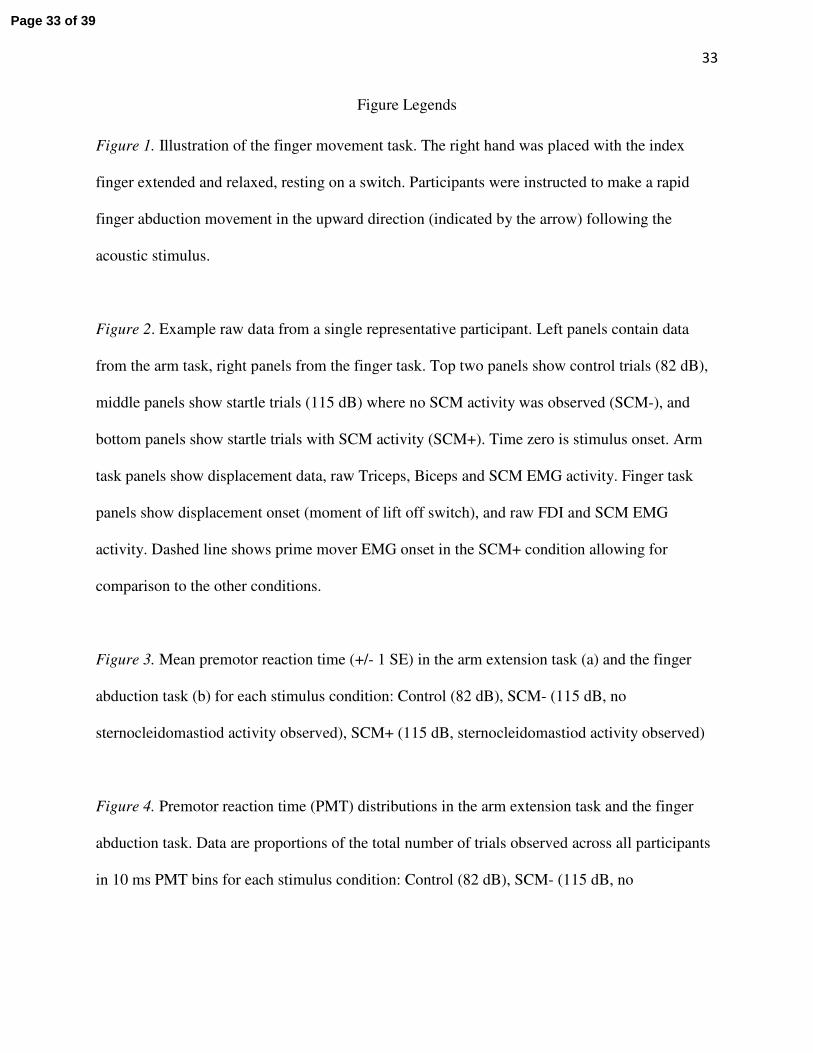
Figure 1. Illustration of the finger movement task. The right hand was placed with the index
finger extended and relaxed, resting on a switch. Participants were instructed to make a rapid
finger abduction movement in the upward direction (indicated by the arrow) following the
acoustic stimulus.
Figure 2. Example raw data from a single representative participant. Left panels contain data
from the arm task, right panels from the finger task. Top two panels show control trials (82 dB),
middle panels show startle trials (115 dB) where no SCM activity was observed (SCM-), and
bottom panels show startle trials with SCM activity (SCM+). Time zero is stimulus onset. Arm
task panels show displacement data, raw Triceps, Biceps and SCM EMG activity. Finger task
panels show displacement onset (moment of lift off switch), and raw FDI and SCM EMG
activity. Dashed line shows prime mover EMG onset in the SCM+ condition allowing for
comparison to the other conditions.
Figure 3. Mean premotor reaction time (+/- 1 SE) in the arm extension task (a) and the finger
abduction task (b) for each stimulus condition: Control (82 dB), SCM- (115 dB, no
sternocleidomastiod activity observed), SCM+ (115 dB, sternocleidomastiod activity observed)
Figure 4. Premotor reaction time (PMT) distributions in the arm extension task and the finger
abduction task. Data are proportions of the total number of trials observed across all participants
in 10 ms PMT bins for each stimulus condition: Control (82 dB), SCM- (115 dB, no
Page 33 of 39

34
sternocleidomastiod activity observed), SCM+ (115 dB, sternocleidomastiod activity observed).
PMT bin values are upper limits for that bin.
Page 34 of 39

Task Arm
Finger
Stimulus Control SCM+ SCM-
Control SCM+ SCM-
EMG Measures
Premotor RT (ms) 135.1 (12.5) 85.3 (9.5) 110.8 (18.3) *† 139.2 (17.3) 106.3 (18.7) 106.3 (17.8) *
Initial agonist burst duration (ms) 96.8 (15.3) 92.2 (14.9) 89.8 (18.1) 90.0 (20.8) 93.6 (28.1) 90.5 (26.0)
TR1 to BIC inter-onset time (ms) 77.2 (34.5) 73.5 (25.6) 68.5 (25.2) - - -
TR1 to TR2 inter-onset time (ms) 152.0 (40.3) 156.8 (39.5) 146.2 (31.6) - - -
Kinematic Measures
Displacement RT (ms) 199.8 (21.5) 149.6 (11.5) 165.6 (13.6) *† 178.3 (16.8) 143.6 (26.0) 139.5 (20.1) *
Final position (deg) 20.1 (1.4) 21.6 (3.3) 21.7 (3.6) - - -
Movement time (ms) 303.3 (29.1) 325.1 (50.5) 319.2 (42.1) - - -
Note. Standard deviations in parentheses, * signifies a significant main effect for auditory tone condition, † signifies a significant difference
between trials in which sternocleidomastiod sctivity was present (SCM+) and absent (SCM-). TR1 is initial triceps burst. BIC is biceps burst. TR2 is second
triceps burst. Premotor reaction time (RT) is time from stimulus to initial agonist EMG onset. Displacement RT is time from stimulus to onset of
displacement.
�
Page 35 of 39

Page 36 of 39

Page 37 of 39

Page 38 of 39
Page 39 of 39