differential effect of the damage to the … · differential effect of the damage to the lateral...
TRANSCRIPT

ACTA NEUROBIOL. EXP. 1989, 49: 153-169
DIFFERENTIAL EFFECT OF THE DAMAGE TO THE LATERAL HYPOTHALAMIC AREA ON HIPPOCAMPAL THETA RHYTHM
DURING WAKING AND PARADOXICAL SLEEP
E JURKOWLANIEC, W. TROJNIAR, T. OZOROWSKA and J. TOKARSKI
Department of Animal Physiology, University of Gda6sk 24 Kladki St., 80-822 Gdafisk, Poland
Key words: hippocampal theta rhythm, lateral hypothalamus, lesion, waking, para- doxical sleep
Abstract. Hippocampal theta rhythm was analyzed in rats subjected to bilateral, electrolytic lesions of the lateral hypothalamic (LH) region at different levels of its rostro-caudal axis. It was found that damage to the LH disturbed the hippocampal theta activity both during waking and paradoxical sleep. The main effect consisted in the lowering of the theta frequency. Typically, a decrease of frequency was accompanied by an increase of amplitude during waking, and an amplitude fall during paradoxical sleep. Extensive lesions increased the amount of rhythmic slow activity during waking and induced long trains of immobility- related theta. The general picture of impairments of the hippocampal theta rhythm in particular subjects depended on the size of the lesion and, to some extent, also on its localization within the LH. It is concluded that LH region contains systems of fibers which transmit impulses from the brain stem reticular formation to the prosencephalic structures gen- erating the rhythmicity of theta.
INTRODUCTION
It is well established that the rhythmic slow activity (theta rhythm) of the dorsal hippocampus is generated in the system involving the medial septum and diagonal band of Broca, enthorinal cortex and the

hippocampus itself (for review see 3). At least two independent gener- ators of theta activity were found in different cellular components of the dorsal hippocampus and the dentate gyrus (2, 9, 30). It is also well known that the hippocampal rhythmic activity depends on the inputs from the reticular nuclei of the lower brain stem (1, 15, 22, 27-29). The exact nature of these influences is still to be cleared up, however, as the theta activity survives well pretrigeminal (7, 17, 35) and cerveau isole (8, 16, 24, 35) transections, although afterwards it shows a number of ab- normalities.
On the basis of the electrophysiological and lesion experiments (1, 15, 22, 27-29) it was found that ascending fibers essential for the hippo- campal theta follow the dorsal and medial longitudinal fasciculi, central tegmental tract and the medial forebrain bundle. The latter pathway contains also fibers involved in low voltage fast activity.
The damage to the medial forebrain bundle at the level of the lateral hypothalamus evokes disturbances in the hippocampal theta rhythm (4, 11, 12, 23). In rats they consist in the decrease in the ampli- tude and frequency of motor-related theta and the appearance of long trains of theta rhythm during immobility, the phenomenon which is very rarely observed in normal rats.
In our previous investigations (23) in which we studied quantitative sleep-waking relations and cortical EEG activity in lateral hypothalamic rats, we observed that LH animals suffered also from disturbances in the hippocampal rhythmic slow activity. In the present paper we analyzed these disturbances.
In rats theta rhythm in the dorsal hippocampus accompanies volunt- ary behaviors (25, 26, 30, 32, 33) and paradoxical phase of sleep (20, 21, 25, 34). As we did not find data concerning the influence of LH damage on paradoxical sleep theta, we have analyzed it in the present paper separately from the changes of theta activity dGring waking. Moreover, we have tried to relate the observed abnormalities in waking and sleep theta to the localization of the damage within the LH.
MATERIAL AND METHOD
The experiment was carried out on 18 male albino rats of the Wistar strain, weighing 250-300 g on the day of surgery. The animals were kept in individual home cages with food and water ad lib., in an artificially maintained 12 :12 h lightldark cycle.
The animals were implanted, under Nembutal anesthesia, with elec- trodes for lesion bilaterally in the region of the lateral hypothalamus, and with EEG recording electrodes. A bipolar, concentric electrode was

placed in the CAI pyramidal cell layer of the left dorsal hippocampus; a cortical screw electrode was positioned over the right occipital cortex and a reference electrode - on the os frontale. A silver wire electrode was sutured into the neck muscles for recording the EMG activity.
The localization of lesion electrodes varied in different animals along the rostro-caudal axis of LH and stereotaxic coordinates were as follows: 1.2-2.8 mm posterior to the bregma, 1.5-1.8 mm lateral to the midline and 7.7-9.0 below the surface of the skull. The hippocampal recording electrode was implanted 2.5-2.8 mm posterior to the bregma, 2.5 rnrn lateral to the midline, and 2.5-3.5 mm below the skull surface. The neocortical recording electrode was screwed 5 mm posterior to the bregma, 3 mm lateral to the midline at a depth of 1 rnrn below the skull surface. The construction and implantation of electrodes was des- cribed in detail elsewhere (23).
EEG recording began after 10 days of recovery period, during which the rats were adapted to the experimental conditions. The recording was carried out in glass cages measuring 260 X 260 X 400 mm, placed in an illuminated, sound attenuating chamber. The experiment lasted 1 hour daily and was performed always at the same time (11:OO-12:OO a.m.). As we found earlier, this part of the daily cycle allows to record all types of the rat's EEG activity (waking, slow wave sleep and paradoxical sleep patterns) as they transit spontaneously from one to another. The hippocampal theta rhythm was recorded bipolarly from the concentric electrode between the inside and the outside pole, and for comparison also monopolarly between each pole of the concentric electrode and the reference electrode, using 16 channels Medicor polygraph (passband 0.53- 50.0 Hz). The animals were continuously observed by the experimenter through a camera connected to a monitoring system and their behavior (waking, rearing, probable sleep etc.) was noted concomitant with EEG records.
Normal EEG pattern was recorded for 3 days. Then the rats were subjected, under light ether anesthesia, to electrolytic lesions made by passing 1.2-2.0 mA anodal current for 15-20 s through previously im- planted electrodes.
Starting from the day following the brain damage, the EEG pattern was recorded for 7-8 consecutive days and then on the l l th, 14th, 18th, and in some animals periodically up to the 36th postlesion day. Regard- less whether the EEG recording was performed or not on the particular day, up to the end of the experiment the animals were put in the re- cording chamber every day at the time of the experimental session.
All rats were observed for behavioral disturbances (somnolence, in- gestive impairments, body weight loss) resulting from the brain damage.

The rats which were aphagic and adipsic or extremely hypophagic and hypodipsic for more than 2 days were artificially fed and watered by means of a gastric tube. In the rats with anterior LH lesions the rectal temperature was measured at the end of each recording session.
After the completion of the experiment the rats were treated with an overdose of ether anesthesia, the brains were removed from the skull and placed in 10°/o Formalin. After fixation, brain sections 30 pm thick were cut using a frozen tissue technique. The sections were stain- ed with cresyl violet for cell bodies.
All records were visually inspected for the presence of hippocampal theta rhythm during waking and paradoxical sleep, and their amounts in experimental hour were assessed. Three 10 s samples of waking and sleep theta were taken randomly from each prelesion and postlesion daily record. Peak-to-peak frequency of theta rhythm was determined by counting the number of theta waves in 10 s samples. Amplitude was measured with the use of a transparent plastic ruler (millimeter scale). Amplitudes and the frequency of theta waves in each postlesion day were compared with a preoperative baseline. Depending on distribution, the data were statistically analyzed with the use of the Student's t-test or nonparametric Mann-Whitney U test.
RESULTS
Prelesion hippocampal theta activity. Before the lesion, the rhythmic slow activity (theta rhythm) in the hippocampal records appeared during waking mainly in association with exploratory behaviors (waking around the cage, sniffing, rearing) and with head movements. In normal rats we have not observed theta activity during immobile waking. Our observ- ations concerning the relation of theta rhythm to behavior were com- patible with those described previously (25, 26, 30, 32, 33). The frequency of waking theta varied from 6.0 to 7.6 Hz. This may reflect differences in the intensity of particular movements (20, 25, 33). The amplitude of theta waves showed a marked interindividual variability (average values: 140-440 pV), which was probably due to slight variations in the localiza- tion of electrodes' tips within the dorsal hippacampus (18). However, the amplitude was relatively stable intraindividually.
Similarly to the previous reports (19, 21, 25, 34), the theta rhythm was also recorded in the paradoxical phase of sleep. Its amplitude varied from 200 to 780 pV in different subjects and its frequency was 7.0- 8.0 Hz.
Theta rhythm in the hippocampus was accompanied in the neocortex either by desynchronization or by rhythmic synchronous activity re-

sembling the hippocampal theta, especially during paradoxical sleep. As was suggested, such cortical activity is probably volume-conducted from the hippocampus (6) .
Before the lesion, the mean duration of waking theta in the hippo- campus was l l O / o of the total recording time. Paradoxical sleep theta took 13O/o.
Abnormalities in theta rhythm after LH damage. Damage to the lateral hypothalamus resulted in a number of abnormalities in the hippo- campal rhythmic slow activity. The intensity and duration of these dist- urbances corresponded with the size of the lesion.
The main change concerned the frequency of theta waves. It was depressed during waking in 8 rats and during paradoxical sleep in 12 subjects. Typically, a decrease of frequency was accompanied by an in- crease of amplitude (n = 6 ) in waking and by an amplitude fall (n = 6) in paradoxical sleep. Unfortunately, in 3 rats an assessment of amplitude of paradoxical sleep theta was impossible because of either extreme shortening (e.g. only one episode in the entire recording session) or even complete cessation of this phase of sleep in the early postlesion period, or because of unsatisfactory quality of EEG records.
Disturbances in theta rhythm during waking and paradoxical sleep in particular rats subjected to brain damage. Explanations: 'It" increase in comparison with the baseline; "4" decrease in com-
parison with the baseline; "=" lack of change; "-" not analyzed
Waking theta Paradoxical sleep theta Rat - -- - .- . - - - - -
frequency amplitude amount frequency amplitude amount - - - - - -- . -- -- - - - -- 10 J J -1 -- -- - - - 9 J t - .. -1- 3. =
32 4 t ? 4 4 -
.t 44 -1 t t J 45 -
-1 -1 . .- -- f f 4 4
28 4 f t - -. - = --
29 -1 t - - = - - -- 1- -- --
27 - - - - - - -1 J J

Out of 18 investigated rats, simultaneous abnormalities in the waking and sleep theta of a typical course (with only minor deviations) occurred in 5 subjects, mainly those with the most extensive lesions of the LH region. In two animals there were impairments in the waking theta only, and further 4 were disturbed only in paradoxical sleep. In 4 other rats the changes of theta rhythm were atypical, for instance a decrease of amplitude of the waking theta without change of frequency or a de- crease of frequency of the paradoxical sleep theta accompanied by an increase of its amplitude. Three rats did not show any changes of hip- pocampal activity after LH damage. Table I summarizes the results obtained in particular subjects.
Figures 1-3 demonstrate the examples of the main three types of disturb'ances in the hippocampal theta activity. Each figure consists of diagrams of the amplitude and frequency of the theta rhythm during waking and paradoxical sleep and of segments of polygraph records taken before the damage and in the early postlesion period. In particular figures some postlesion days are omitted, usually due to unsatisfactory quality of EEG records. Figures 4-6 illustrate anatomical verification of the lesion placements in rats shown in Figs. 1-3.
Figure 1 illustrates an example (rat no. 32) of severe disturbances in both waking and paradoxical sleep theta, lasting up to the end of the experiment. Regardless of the state of vigilance, LH lesions affected mainly the frequency of theta rhythm. Its depression (by 0.5-1.7 Hz during waking and by 0.6-1.8 Hz during paradoxical sleep) was highly significant and persisted at least up to the 18th postlesion day. Changes in amplitude were not as consistent, particularly during sleep. In this animal the increase of amplitude of the waking theta ranged from 40 to 150 pV in particular postlesion days, and the amplitude fall was 110- 120 pV during paradoxical sleep. This constituted 13-45O/o and 21-24"o of the prelesion baseline respectively. In other animals from this group the changes were similar, although the relative intensity and duration of disturbances might differ in particular subjects.
Figure 2 shows the example of a rat (no. 28) in which a relatively large lesion of the relevant area caused disturbances exclusively in the waking theta. The hippocampal activity during paradoxical sleep re- mained virtually unchanged. In the presented animal the frequency was decreased by 0.6-1.5 Hz. The increase of amplitude of the waking theta was relatively small (about 40 pV, i.e. 10°/a of the baseline). The changes in theta activity were observed in the first week after the lesion. The other animal from this group (no. 29) showed a 3 days' marked increase of amplitude (100-140 pV, i.e. 24-34"/0 of the baseline) and a comparable (0.9 Hz) fall of frequency.

WAKING
uu nr
FRLOUENCY
bW - b
*"*
POSTOPERATIVE DAYS
* *\\$!f#ib\fl q!j/iy&/y4/hv//b \/~((/fl;f~fi/(!(\~~l \YA!H~~/;~AY&: 8 f j , .dj\i/\&;~i!~t,,h I
OEFORE AFTER BEFORE L E S I O N
AFTER - L E S I O N
Fig. 1. The example of disturbances in theta rhythm both in waking and para- doxical sleep (rat no. 32). Top: diagrams of frequency and amplitude of theta waves; each point denotes mean (fSD) value counted from 9 samples (3 in each day) before LH lesion (marked as day O), and from 3 samples in each postlesion day. The level of significant difference from the baseline: * P < 0.05, ** P < 0.01, *I* P < 0.001. Bottom: A, bipolar; B, monopolar records of hippocampal theta
rhythm. Calibrations: 400 pV; 1 S.
U4 HI W A K I N G
e'J% r 8
POSTOPERATIVE DAYS
FREQUENCY
BtFORt A F I f R BEFORE AFTER - L E S I O N L E S I O N
Fig. 2. The example of disturbances in theta rhythm exclusiveIy in waking (rat no. 28). Explanations as in Fig. 1. The experiment was concluded on the 7th
postlesion day.
6110-
5m-
6 - 6 &,i\$/ ' 6
6/"' "'6 I..
- 5 *'I

Figure 3 shows an example (rat no. 27) of exclusive disturbances in paradoxical sleep theta of a typical course. The decrease of frequency in this animal ranged from 0.7 to 0.8 Hz, and the amplitude fall was 50-80 pV (21-33O/o of the baseline). These changes, although not regular, lasted up to the end of the experiment (usually 18 days). In one rat from this group they were particularly long-lasting and persisted up to the 36th postlesion day (the last day of the experiment).
POSTOPfRATIVE OATS
urn H I
BfFOPf I F l E R LICFORE 1 F T f R -
LESION i t S lON
t m
, 600-
s o 0
100.
300-
2rn.
Fig. 3. The example of disturbances in theta rhythm exclusively in paradoxical sleep (rat no 27). Explanations as in Fig. 1.
The other characteristic abnormality in hippocampal EEG observed after LH damage is the appearance in some animals, mainly those with extensive damage to LH, of long trains of theta rhythm during imrnobil- ity. Occasionally there were also episodes of extremely slow (4.4-4.5 Hz) waking theta, never observed before the lesion.
Six rats showed a marked elevation of the amount of theta activity during waking (Table I). Without exception they belonged to a group with the largest lesions of the relevant area. Accordingly they suffered from the most severe behavioral depression (hypokinesia, or even som- nolence) and thus the theta activity occurred mainly during states of immobility.
As we have described elsewhere (23) damage to the LH region dis- turbs quantitative sleep-waking relations towards the shortening of sleep,
D '*' '&<?.-~-& - P C - . . . . . . , . r 5 - . . . , . . . - O d l 1 3 5 6 1 8 11 1 i l a O 2 1 I 3 5 6 ? 8 11 U 18
P W . P W4NlNG PARAOOXICAL SLEEP
.s d 6
, J b+..o-a-~~6-6-b-b 6-ik \h,~u-a .. *;is FREQUENCV
6
5
b
4MPLITUDE A 3 1 A h & A - . P
4 ,-a-• I-A-L u -2 ,
/
mainly its slow wave phase, but to some extent also paradoxical sleep. Of the animals analyzed in the present paper - 5 showed a marked shortening of paradoxical sleep (Table I), in 3 of them there was a com- plete or almost complete cessation of this stage in the early postlesion period.
We have not found disturbances in body temperature in anterior LH lesioned animals.
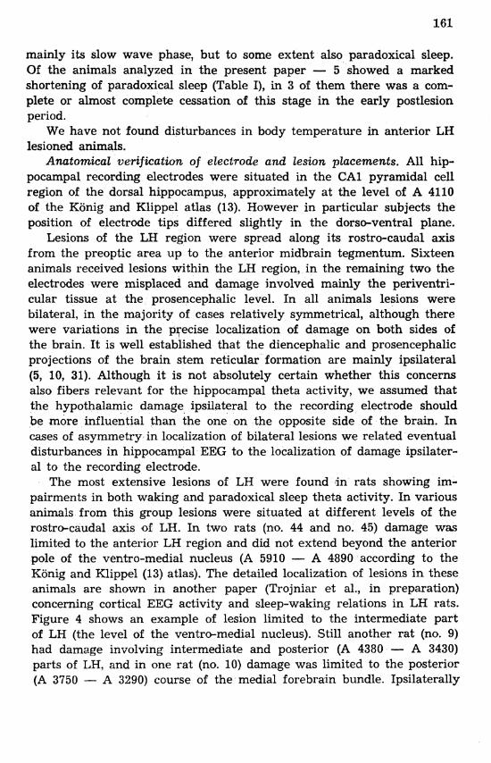
Anatomical verification of electrode and lesion placements. All hip- pocampal recording electrodes were situated in the CAI pyramidal cell region of the dorsal hippocampus, approximately at the level of A 4110 of the Konig and Klippel atlas (13). However in particular subjects the position of electrode tips differed slightly in the dorso-ventral plane.
Lesions of the LH region were spread along its rostro-caudal axis from the preoptic area up to the anterior midbrain tegmentum. Sixteen animals received lesions within the LH region, in the remaining two the electrodes were misplaced and damage involved mainly the periventri- cular tissue at the prosencephalic level. In all animals lesions were bilateral, in the majority of cases relatively symmetrical, although there were variations in the precise localization of damage on both sides of the brain. It is well established that the diencephalic and prosencephalic projections of the brain stem reticular formation are mainly ipsilateral (5, 10, 31). Although it is not absolutely certain whether this concerns also fibers relevant for the hippocampal theta activity, we assumed that the hypothalamic damage ipsilateral to the recording electrode should be more influential than the one on the opposite side of the brain. In cases of asymmetry in localization of bilateral lesions we related eventual disturbances in hippocampal EEG to the localization of damage ipsilater- a1 to the recording electrode.
The most extensive lesions of LH were found in rats showing im- pairments in both waking and paradoxical sleep theta activity. In various animals from this group lesions were situated at different levels of the rostro-caudal axis of LH. In two rats (no. 44 and no. 45) damage was limited to the anterior LH region and did not extend beyond the anterior pole of the ventro-medial nucleus (A 5910 - A 4890 according to the Kijnig and Klippel (13) atlas). The detailed localization of lesions in these animals are shown in another paper (Trojniar et al., in preparation) concerning cortical EEG activity and sleep-waking relations in LH rats. Figure 4 shows an example of lesion limited to the intermediate part of LH (the level of the ventro-medial nucleus). Still another rat (no. 9) had damage involving intermediate and posterior (A 4380 - A 3430) parts of LH, and in one rat (no. 10) damage was limited to the posterior (A 3750 - A 3290) course of the medial forebrain bundle. Ipsilaterally

Fig. 4. Anatomical localization of lesions (shaded areas) in the rat shown in Fig. 1 4no. 32). Reconstruction of the damage was superimposed on plates taken from the atlas by KiSnig and Klippel (13). Unilateral hippocampal electrode was implant-
ed on the left side of the brain
to the hippocampal recording electrode the lesions dissected totally or almost totally the medial forebrain bundle, with the exception of the rat with the most posterior localization of the lesion (no. lo), in which the medial forebrain bundle was dissected in about 50°/0. All lesions were located in the close proximity of the capsula interna and/or cerebral peduncle involving pericapsular tissue. Lesions to the capsula interna were small or altogether absent.
On the contralateral side the lesions were similarly located as far as the rostro-caudal level is concerned. There were however some asym- metries (Fig. 4).
In two rats showing exclusive disturbances in the waking theta, damage to the lateral hypothalamus proper, ipsilaterally to the hippo- campal electrode was small (less than 114 of its coronal section, Fig. 5) or absent (rat no. 29). Instead, large lesions involved the capsula interna

Fig.
A 3990
in Fig. 2 (no.

and the optic tract together with the adjacent tissue. Contralaterally, in both rats lesions involved the medial border of the capsula interna and damaged totally (rat no. 29) or partially (Fig. 5) the lateral hypo- thalamic-medial forebrain bundle area.
Generally, rats showing impairments of hippocampal activity ex- clusively in the paradoxical sleep have small to medium-sized lesions within the LH region, which destroyed the area of the medial forebrain bundle only partially (Fig. 6). In some cases the medial border of the capsula interna was also invaded. Contralaterally the damage also in- volved the LH region, at relatively similar rostro-caudal level, but some- times there were some asymmetries. The same concerns the animals with an atypical course of theta changes after the brain damage.
A 4230
A CllO
Fig. 6. Anatomical localization of lesions in the rat shown in Fig. 3 (no. 27). Explanations as in Fig. 4.
No disturbances in the hippocampal theta were found in rats with either small lesions of the posterior LH region, or an extensive damage, but located outside the LH area.
The increase of the amount of theta activity during waking after brain damage was found in rats with the most extensive lesions of the LH region. They belonged to the first two groups described above.

DISCUSSION
The major findings of the present experiment are as follows: (i) damage to the lateral hypothalamic region disturbs hippocampal theta rhythm both during waking and paradoxical sleep; (ii) the main effect consists in the lowering of theta waves frequency, which typically is accompanied by an increase of amplitude during waking and amplitude fall during paradoxical sleep; (iii) extensive lesions increase the amount of slow rhythmic activity during waking and induce long trains of immobility-related theta; (iv) general picture of impairments of hip- pocampal activity in particular subjects depends on the size of the lesion and its localization within the LH region.
The effect of lateral (4, 11, 12) and posterior (20) hypothalamic damage on the theta rhythm during waking has already been described earlier. Our results, in general, are compatible with those reported previously. We also found that LH lesions greatly depress the frequency of theta rhythm, and release immobility-related theta activity, rarely observed in normal rats.
In rats the frequency of waking theta is correlated with the intensity of movement it accompanies. Extensive lateral hypothalamic lesions are known to produce akinesia or even somnolence (14). We observed it also in our animals (23). The first impression is that the slowing down of theta activity may. be secondary to the depression of locomotor activity. It is not so, however. Similarly to other authors (4, 12) we recorded this slower theta rhythm even during episodes of quite normal movements.
A decrease of body temperature (32) does not seem to contribute to the fall of theta frequency either, because we did not observe such a change in our LH rats. Similar observations were also made by other authors (4, 12). It seems therefore that damage to the LH region impairs some more basic mechanism involved in the regulation of frequency of the theta rhythm. As was found in the present experiment, this impair- ments concern also hippocarnpal activity during paradoxical sleep.
The effects of hypothalam5c lesions on the amplitude of theta are more vague. Kolb and Whishaw (12) found a depression of both am- plitude and frequency of the waking theta in the early postlesion period. De Ryck and Teiteilbaum (4) and Robinson and Whishaw (20) did not observe great changes in their lateral and posterior hypothalamic rats. In our experiment there was a different effect of LH damage on the amplitude of theta rhythm during waking and paradoxical sleep. Contr- ary to previous reports, during waking we observed rather an increase of amplitude accompanying a depression of frequency. During paradoxic-
3 - Acta Neurobiol. Exp. 4189

a1 sleep, usually both amplitude and frequency were decreased. How- ever, it should be pointed that the change in amplitude was not as im- pressive as the change of frequency. Its magnitude varied in different animals, in the majority of cases it was not particularly long-lasting, and sometimes appeared after a period of unchanged activity in the early postlesion days.
In particular rats investigated in this work, the lesions differed in size, precise localization within the LH region and sometimes in sym- metry. Accordingly, there were variations in disturbances of the hip- pocampal function. Having such a material, we tried to draw a con- clusion as to the neuroanatomical basis of the observed changes in theta activity. The main finding is that the severity of impairments depends on the size of the lesion. Only large lesions involving the lateral hypo- thalamic-medial forebrain area (total or almost total dissection) ipsil- aterally to the recording electrode produced impairments of the theta activity both during waking and paradoxical sleep. It appeared that the rostro-caudal level of the lesion was irrelevant, and the same results were obtained with damage at the anterior, intermediate and posterior LH level.
One may speculate that to cause exclusive disturbances of the waking theta, a destruction of the lateral hypothalamic-medial forebrain bundle area is not absolutely necessary. In two animals we found pronounced impairments when large lesions, ipsilateral to the recording electrode, were placed within the capsula interna and optic tract, omitting the main body of LH. Of course such a conclusion is possible only with an assumption that the lesion ipsilateral to the hippocampal electrode is decisive for theta disturbances. We feel that this hypothesis is worth checking in further experiments.
Incomplete lesions of LH caused either exclusive impairments of hip- pocampal activity during paradoxical sleep or inconsistent changes in both waking and paradoxical sleep theta. On the basis of this material we feel that the theta activity in paradoxical sleep is more sensitive to LH damage than that in waking.
Extensive lesions of LH region caused that the waking EEG activity was dominated by the theta rhythm in hippocampal records. Similar observations were reported after pretrigeminal (7, 17, 35) and cerveau is016 (8, 16, 24, 35) transections in rats and cats and after medial pontine lesions in rats (12). However, LH animals were not so deeply affected and the rhythmic, slow activity of the hippocampus was at times inter- mitted by other patterns.
All these data suggest that the lateral hypothalamus and neighbouring structures (e.g. capsula interna) contain systems of fibers which trans-

mit impulses from the brain stem reticular formation to the prosenc- ephalic structures generating theta rhythmicity. These fibers seems to determine the amount, frequency and possibly also amplitude of the theta rhythm both during waking and paradoxical sleep.
This investigztion was supported by Project CPBP 04.01.6.18.
REFERENCES
1. ANCHEL, H. and LINDSLEY, D. B. 1972. Differentiation of two reticulo-hypo- thalamic systems regulating hippocampal activity. EEG Clin. Neurophysiol. 32: 209-226.
2. BLAND, B. H. and WHISHAW, I. Q. 1976. Generators and topography of hippo- campal theta (RSA) in the anaesthetized and freely moving rat. Brain Res. 118: 259-280.
3. BUZSAKI, G., LEUNG, L. W. S. and VANDERWOLF, C. H. 1983. Cellular bases of hippocampal EEG in the behaving rat. Brain Res. Rev. 6: 139-171.
4. DE RYCK, M. and TEITELBAUM, P. 1978. Neocortical and hippocampal EEG in normal and lateral hypothalamic-damaged rats. Physiol. Behav. 20: 403- 409.
5. EDWARDS, S. B. and DE OLMOS, J. S. 1976. Autoradiographic studies of the projections of the midbrain reticular formation: ascending projections of nucleus cuneiformis. J. Comp. Neurol. 165: 417-432.
6. GERBRANDT, L. K., LAWRENCE, J. C., ECKHARDT, J. J. and LLOYD, R. L. 1978. Origin of neocortically monitored theta rhythm in the curarized rat. EEG Clin. Neurophysiol. 45: 454-467.
7. GOTTESMANN, C., USER, P. and ZERNICKI, B. 1980. The acute pretrigeminal rat. Acta Neurobiol. Exp. 40: 993-998.
8. GOTTESMANN, C., ZERNICKI, B. and GANDOLFO, G. 1981. Hippocampal theta activity in the acute cerveau is016 cat. Acta Neurobiol. Exp. 41: 251- 255.
9. GREEN, K. F. and RAWLINS, J. N. P. 1979. Hippocampal theta in rats under urethane: generators and phase relations. EEG Clin. Neurophysiol. 47: 420- 429.
10. JONES, B. E. and YANG T. 2. 1985. The efferent projections from the reticular formation and the locus coeruleus studied by anterograde and retrograde axonal transport in the rat. J. Comp. Neurol. 242: 56-92.
11. KOLB, B., DODIC, R. and WHISHAW, I. Q. 1979. Effects of serial lateral de- struction on feeding behavior, body weight and neocortical and hippocampal EEG activity. Exp. Neurol. 66: 263-276.
12. KOLB, B. and WHISHAW, I. Q. 1977. Effects of brain lesions and atropine on hippocampal and neocortical electroencephalograms in the rat. Exp. Neurol. 56: 1-22.
13. KONIG, J. F. R. and KLIPPEL, R. A. 1963. The rat brain: a stereotaxic atlas of the forebrain and lower parts of the brain stem. Williams and Wilkins, Baltimore.
14. LEVITT, D. R. and TEITELBAUM, P. 1975. Somnolence, akinesia and sensory activation of motivated behavior in the lateral hypothalamic syndrome. Proc. Natl. Acad. Sci. 72: 2819-2823.

15. MACADAR, A. W., CHALUPA, L. M. and LINDSLEY, D. B. 1974. Differentiation of brain stem loci which affect hippocampal and neocortical electrical activity. Exp. Neurol. 43: 499-514.
16. OLMSTEAD, C. E. and VILLABLANCA, J. R. 1977. Hippocampal theta rhythm persists in the permanent isolated forebrain in the cat. Brain Res. Bull. 2: 93-100.
17. RADIL-WEISS, T., ZERNICKI, B. and MICHALSKI, A. 1976. Hippocampal theta activity in the acute pretrigeminal cat. Acta Neurobiol. Exp. 36: 517- 534.
18. ROBINSON, T. E. 1980. Hippocampal rhythmic slow activity (RSA; theta): a critical analysis of selected studies and discussion of possible species- differences. Brain Res. Rev. 2: 69-101.
19. ROBINSON, T. E., KRAMIS, R. C. and VANDERWOLF, C. H. 1977. Two types of cerebral activation during active sleep: relations to behavior. Brain Res. 124: 544-549.
20. ROBINSON, T. E. and WHISHAW, I. Q. 1974. Effects of posterior hypothalamic lesions on voluntary behavior and hippocampal electroencephalograms in the rat. J. Comp. Physiol. Psychol. 86: 768-786.
21. SANO, K., IWAHARA, S., SEMBA, S., SANO, A. and YAMAZAKI, S. 1973. Eye movements and hippocampal theta activity in rats. EEG Clin. Neuro- physiol. 35: 621-625.
22. TORII, S. 1961. Two types of pattern of hippocampal electrical activity induced by stimulation of hypothalamus and surrounding parts of rabbit's brain. Jap. J. Physiol. 11: 147-157.
23. TROJNIAR, W., JURKOWLANIEC, E., ORZEL-GRYGLEWSKA, J. and TOKAR- SKI, J. 1987. The effect of lateral hypothalamic lesions on spontaneous EEG pattern in rats. Acta Neurobiol. Exp. 47: 27-43.
24. USER, P., GIOANNI, H. and GOTTESMANN, C. 1980. Intermediate stage of sleep and acute cerveau isole preparation in the rat. Acta Neurobiol. Exp. 40: 521-525.
25. VANDERWOLF, C. H. 1969. Hippocampal electrical activity and voluntary mo- vement in the rat. EEG Clin. Neurophysiol. 26: 407-418.
26. VANDERWOLF, C. H. 1975. Neocortical and hippocampal activation in relation to behavior: effects of atropine, eserine, phenothiazines and amphetamine. J. Comp. Physiol. Psychol. 88: 300-323.
27. VERTES, R. P. 1981. An analysis of ascending brain stem systems involved in hippocampal synchronization and desynchronization. J. Neurophysiol. 46: 1140-1159.
28. VERTES, R. P. 1982. Brain stem generation of the hippocampal EEG. Progr. Neurobiol. 19: 159-186.
29. VERTES, R. P. 1984. Brainstem control of the events of REM sleep. Progr. Neurobiol. 22: 241-288.
30. WALLENSTEIN, M. C. 1976. Anatomical and behavioral mapping of rhythmic slow wave activity in rat hippocampus. Physiol. Behav. 17: 515-522.
31. WATSON, R. T., HEILMAN, K. M., MILLER, B. D. and KING, F. A. 1974. Neglect after mesencephalic reticular formation leoions. Neurology 24: 294- 298.
32. WHISHAW, I. Q. and VANDERWOLF, C. H. 1971. Hippocampal EEG and behavior: effects of variation in body temperature and relation of EEG to vibrissae movement, swimming and shivering. Physiol. Behav. 6: 391-397.

33. WHISHAW, I. Q. and VANDERWOLF, C. H. 1973. Hippocampal EEG and behavior. Changes in amplitude and frequency of RSA (theta rhythm) associated with spontaneous and learned movement patterns in rats and cats. J. Behav. Biol. 8: 461-484.
34. WINSON, J. 1972. Interspecies differences in the occurrence of theta. Behav. Biol. 7: 479-487.
35. ZERNICKI, B., GANDOLFO, G., GLIN, L. and GOTTESMANN, C. 1984. Cerveau isole and pretrigeminal rats. Acta Neurobiol. Exp. 44: 159-177.
Accepted 20 January 1989