differences in coronary flow and myocardial metabolism at ... · myocardial oxygen consumption and...
TRANSCRIPT
JACC VoL 10, NO.1July 1987:53-62
53
Differences in Coronary Flow and Myocardial Metabolism at Rest andDuring Pacing Between Patients With Obstructive and Patients WithNonobstructive Hypertrophic Cardiomyopathy
RICHARD O. CANNON Ill, MD, FACC, WILLIAM H. SCHENKE, BA,
BARRY J. MARON, MD, FACC, CYNTHIA M, TRACY, MD, FACC, MARTIN B. LEON, MD, FACC,
JOHN E. BRUSH, JR., MD, DOUGLAS R, ROSING, MD, FACC, STEPHEN E. EPSTEIN, MD, FACC
Bethesda, Maryland
Fifty patients with hypertrophic cardiomyopathy underwent invasive study of coronary and myocardial hemodynamics in the basal state and during the stress ofpacing. The 23 patients with basal obstruction (averageleft ventricular outflow gradient, 77 ± 33 mm Hg; leftventricular systolic pressure, 196 ± 33 mm Hg, mean± 1 SD) had significantly lower coronary resistance (0.85± 0.18 versus 1.32 ± 0.44 mm Hg-min/ml, p < 0.001)and higher basal coronary flow (l06 ± 20 versus 80 ±25 ml/min, p < 0.001) in the anterior left ventricle,associated with higher regional myocardial oxygen consumption (12.4 ± 3.6 versus 8.9 ± 3.3 ml oxygen/min,p < 0.001) compared with the 27 patients without obstruction (mean left ventricular systolic pressure 134 ±18 mm Hg, p < 0.001).
Myocardial oxygen consumption and coronary bloodflow were also significantly higher at paced heart ratesof 100 and 130 beats/min (the anginal threshold for 41of the 50 patients) in patients with obstruction comparedwith those without. In patients with obstruction, transmural coronary flow reserve was exhausted at a heartrate of 130beats/min; higher heart rates resulted in more
The most characteristic feature of hypertrophic cardiomyopathy is hypertrophy of the left ventricle without obvious physiologic explanation (I). In many patients withthis disease, an intraventricular pressure gradient can bedemonstrated, localized to the site of apposition of the mitralvalve with the septum (2-5). However, controversy exists
From the Cardiovascular Diagnosis Section, Cardiology Branch, National Heart, Lung, and Blood Institute, National Institutes of Health.Bethesda, Maryland.
Manuscript received October 27, 1986; revised manuscript receivedJanuary 14, 1987, accepted January 26, 1987.
Address for reprints: Richard O. Cannon III, MD. Building 10. Room7B-15, National Institutes of Health, Bethesda, Maryland 20892.
© 1987 by the American College of Cardiology
severe metabolic evidence of ischemia with all patientsexperiencing chest pain, associated with an actual increase in coronary resistance. Patients without obstruction also demonstrated evidence of ischemia at heartrates of 130 and 150 beats/min, with 25 of 27 patientsexperiencing chest pain. In this group, myocardial ischemia occurred at significantly lower coronary flow, highercoronary resistance and lower myocardial oxygen consumption, suggesting more severely impaired flow delivery in this group compared with those with obstruction. Abnormalities in myocardial oxygen extraction andmarked elevation in filling pressures during stress werenoted in both groups.
Thus, obstruction to left ventricular outflow is associated with high left ventricular systolic pressure andoxygen consumption and therefore has important pathogenetic importance to the precipitation of ischemia inpatients with hypertrophic cardiomyopathy. Patientswithout obstruction may have greater impairment incoronary flow delivery during stress.
(J Am Call Cardiol 1987;10:53-62)
as to whether this phenomenon represents true mechanicalobstruction to left ventricular systolic ejection (5-10), Insymptomatic patients with fixed mechanical obstruction toleft ventricular outflow, such as aortic stenosis, a high leftventricular systolic pressure results in elevated absolute basalcoronary flow and myocardial oxygen consumption (11-13),and limited coronary flow reserve has been demonstratedintraoperatively (14). The present study was performed toexamine whether patients with and patients without basalobstruction to left ventricular outflow differ with regard tocoronary and myocardial hemodynamics and therefore verify whether the subaortic gradient and elevated intraventricular pressures in hypertrophic cardiomyopathy are ofpathophysiologic consequence,
0735-1097/87/$3.50

54 CANNON ET AL.CORONARY FLOW IN HYPERTROPHIC CARDIOMYOPATHY
JACC Vol. 10. No. IJuly 1987:53-62
Methods
Patient selection. We studied 50 patients with an echocardiographic diagnosis of hypertrophic cardiomyopathy.All patients had a hypertrophied, nondilated left ventriclein the absence of another cardiac or systemic disease capableof causing myocardial hypertrophy (I) and had angiographically normal epicardial coronary arteries. Thirty-six patients were men and 14 were women, ranging in age from22 to 60 years (mean 43). No patient had previously undergone cardiac surgery. All patients were severely symptomatic (New York Heart Association functional class III toIV) and 48 of the 50 described chest pain as a major symptom. This study represents a consecutive series of patientswith hypertrophic cardiomyopathy who underwent the catheterization study protocol. Patients were considered to havebasal obstruction to left ventricular outflow if their basalpressure gradient was ;::::30 mm Hg. Data from 18 of thesepatients have been reported previously (15). Informed consent for this study was obtained from all patients.
Echocardiographic studies. A combined M-mode andtwo-dimensional echocardiographic examination was performed in each of the 50 study patients. M-mode echocardiograms were performed with an Irex System II ultrasoundunit with either a 2.25 or 3.5 MHz transducer. Two-dimensional echocardiograms were performed with either aVarian V-3400 or ATL (Advanced Technology Laboratory,Inc.) MK-500 ultrasound system with 2.25 or 3.5 MHztransducers, respectively. In 42 of the 50 study patients inwhom two-dimensional echocardiograms were of adequatequality, assessment of the magnitude of left ventricular hypertrophy was made primarily from the parasternal shortaxis cross-sectional plane, as previously described (16). Theventricle was divided into four regions that identified theanterior and posterior ventricular septum and the lateral andposterior left ventricular free wall. Wall thickness of eachregion was measured directly from the television monitorwith calipers, using a calibration scale produced by theinstrument. A wall thickness index was calculated by addingthe measurements of maximal wall thickness obtained ineach of the four segments into which the ventricle had beendivided. The calculated score was considered a quantitativeexpression of the overall magnitude of left ventricular hypertrophy. All measurements were performed by one investigator without knowledge of the catheterization studyresults.
Cardiac catheterization: coronary flow and myocardial metabolism. All medications, including f3-adrenergic blockers and calcium channel blockers, were terminated at least 48 hours or 5 drug half-lives before cardiaccatheterization. After sedation with diazepam, 10mg orally,patients were taken to the catheterization laboratory, usuallyat 8:00 AM in the fasting state and without any other premedication. A Baim thermodilution flow catheter (17) (Ele-
cath Corp.) was introduced into the right atrium through theright internal jugular vein. The great cardiac vein, which isthe recipient of blood from the left anterior descending arterysystem, was cannulated through the coronary sinus. Handinjections of contrast dye were recorded on cine film, demonstrating the catheter tip at the junction of the anteriorinterventricular vein and great cardiac vein without adjacentmarginal veins in all cases. The catheter position was keptconstant throughout the study by frequent inspection of therelation of the electrodes on the catheter to bony landmarksby fluoroscopy. The thermodilution technique for determining great cardiac vein flow has been described previously(18,19). All temperature determinations were fed directlyinto an IBM PClXT computer, averaging 20 seconds ofinfusate-blood mixture for each flow determination, withon-line calculation of great cardiac vein flow using a standard formula (17).
This method of coronary flow measurement was chosenfor two reasons: First, in our experience, the catheter advanced into the great cardiac vein is stable in position withminimal or no movement even during pacing, and reproducible flow measurments may be achieved at rest and during pacing. Second, measurements of venous flow from theanterior left ventricular wall may allow estimation of coronary flow and myocardial metabolism in that portion of theventricle most abnormal in the majority of patients withhypertrophic cardiomyopathy, that is, the anterior septumand free wall (16). Coronary sinus flow measurements werenot recorded because of concern for right atrial reflux duringpacing (20). Coronary resistance was calculated as the meanblood pressure divided by the great cardiac vein flow.
A 20 gauge catheter was placed in the left brachial arteryfor arterial pressure measurements. An 8F end-hole pigtailcatheter (in 31 patients, a transducer-tipped catheter, MillarInstruments) was advanced into the left ventricle. Cardiacoutput and pulmonary artery wedge pressures were measured with a thermodilution catheter in the pulmonary artery.Cardiac index was calculated as cardiac output divided bythe body surface area. Systemic vascular resistance indexwas calculated as mean systemic blood pressure divided bythe cardiac index. Arterial and left ventricular pressures andelectrocardiographic (ECG) monitor leads I, aVF and V3
or V5 were recorded with each flow measurement in thegreat cardiac vein. Lactate samples were obtained from thegreat cardiac vein and brachial artery and immediately transferred to tubes containing sodium fluoride and potassiumoxalate for inhibition of glycolysis, with immediate centrifugation at 4°C at 5,000 rpm for 5 minutes. The supernatant was then processed for lactate content on a DuPontautomatic clinical analyzer by a modification of the technique of Marbach and Weil (21). Lactate consumption wascalculated as the difference between the arterial and greatcardiac vein lactate concentrations multiplied by the greatcardiac vein flow. Oxygen content was determined with a
JACC Vol. 10, No. IJuly 1987:53-62
CANNONET AL.CORONARY FLOW IN HYPERTROPHIC CARDIOMYOPATHY
55
Lex-Oj-Con oxygen analyzer (Lexington Instruments) atrest and during pacing. Myocardial oxygen consumption inthe anterior circulation was calculated as the difference between the arterial and great cardiac vein oxygen contentmultiplied by the great cardiac vein flow.
Diagnostic catheterization: ventricular outflow obstruction. Before the pacing coronary flow study, all patients underwent diagnostic right and left heart catheterization. Obstruction to left ventricular outflow was defined asthe presence of an intraventricular gradient of ::::30 mm Hglocalized to the left ventricular outflow tract during pullbackof an end-hole catheter. Care was taken to exclude artifactualdistortion of the left ventricular pressure by catheter entrapment as described by Wigle et al. (22). Although maneuvers to provoke an outflow gradient, including the Valsalva maneuver, amyl nitrite inhalation and isoproterenolinfusion, were used to assess the presence and severity ofa left ventricular outflow gradient in many patients, onlythe basal left ventricular outflow gradient was used to distinguish patients with from those without obstruction to leftventricular outflow in this study. In 47 patients, coronaryarteriography, using multiple angulated views, was performed after completion of hemodynamic studies. In theremaining three patients, coronary arteriography was performed before referral to the Cardiology Branch, NationalHeart, Lung, and Blood Institute. Left ventriculography wasperformed in 45 study patients. Left ventricular ejectionfraction was estimated from the left ventriculogram in the45° right anterior oblique projection using the area-lengthmethod of Sandler and Dodge (23).
Pacing coronary flow study. The study protocol wasinitiated at least 20 minutes after the use of angiographiccontrast material to eliminate any effects of the dye oncoronary flow and myocardial metabolism (24). Great cardiac vein flow, lactate and oxygen content were determinedat rest as were measurements of cardiac output, mean pulmonary artery wedge pressure and left ventricular peak systolic and end-diastolic pressures. Pacing through the coronary sinus thermodilution catheter was initiated at a heartrate of 100 beats/min in the 45 patients whose basal heartrates were < 100 beats/min, and increased by increments of10 beats/min at I to 2 minute intervals up to a heart rate of
150 beats/min in 48 patients. Atropine, 0.5 to 1.0 mg, wasgiven intravenously to seven patients to facilitate rapid atrialpacing. In two patients, pacing was terminated at 130beats/min because of the severity of chest pain. Arterial andleft ventricular pressures and great cardiac vein flow weremeasured at each heart rate. Left ventricular end-diastolicpressure was measured at heart rates of 100, 130 and 150beats/min. At heart rates of 100 and 130 beats/min, the leftventricular end-diastolic pressure was measured after suddentermination of pacing in 31 patients and during pacing inthe rest, with an average of pressures over a 10 secondinterval, excluding the first four postpacing beats. Afterpacing for 2 minutes at a heart rate of 150 beats/min, theleft ventricular end-diastolic pressure was measured immediately after termination of pacing in 48 patients usingthe same criteria. Cardiac output determinations were madein triplicate at heart rates of 130 and 150 beats/min andaveraged. Blood specimens from the great cardiac vein andbrachial artery were obtained at heart rates of 130 and 150beats/min for oxygen and lactate content. ECG analysis wasperformed only in the immediate postpacing ECG, whichwas compared with the record obtained before pacing.
Statistical analyses. Data were analyzed by the twotailed Student's t test for paired and unpaired data whenappropriate, with the probability (p) value <0.05 consideredstatistically significant. Regression analyses were performedwhen indicated. All group data are reported as mean ± 1SD.
ResultsEchocardiographic evaluation (Table 1). In each of
the 50 study patients, hypertrophy of the anterior ventricularseptum (thickness > IS mm) was identified by both M-modeand two-dimensional echocardiography. In 42 patients (21with obstruction, 21 without obstruction), two-dimensionalechocardiographic studies were of satisfactory quality topermit estimation of the magnitude and extent of left ventricular hypertrophy. The maximal thickness of the anteriorseptum (the region of the left ventricle drained in part bythe great cardiac vein) was 20 ± 3 mm in patients withobstruction and 20 ± 6 mm in those without obstruction.
Table 1. Quantitation of Left Ventricular Hypertrophy
HCM-N HCM-O p Value
No. of patientsMale/femaleMaximal thickness of LV (mm)Thickness of anterior septum (mm)Index of overall LV hypertrophy (mm)
2117/4
22 ± 520 ± 661 ± 15
2114/7
19 ± 520 ± 360 ± 14
NSNSNSNSNS
HCM-N and HCM-O = nonobstructive and obstructive hypertrophic cardiomyopathy, respectively; LV =left ventricle.

56 CANNON ET AL.CORONARY FLOW IN HYPERTROPHIC CARDIOMYOPATHY
JACC Vol. 10. No. IJuly 1987:53-62
There was no significant difference in the distribution orseverity of hypertrophy between patients with and withoutbasal left ventricular outflow obstruction.
Angiographic evaluation. By study inclusion criteria,all patients had entirely normal epicardial coronary arteries.Basal coronary flow in the left coronary artery appeared inmost patients to be higher than normal as evidenced by rapidtransit of contrast medium injected into the coronary ostia .There was often a complete cessation of dye movement insystole, with rapid transit in diastole . Systolic compressionof septal perforating vessels was noted in 19 (86%) of 22patients with obstruction and 16 (64%) of2S patients withoutobstruction whose coronary arteriograms were available forreview. Systolic compression of epicardial vessels was notedin one patient with obstruction and two patients withoutobstruction. Left ventriculography generally demonstratedan elongated, hyperdynamic ventricle with massive hypertrophy of papillary muscles , giving a "ballerina slipper"appearance. Evidence of mitral regurgitation was noted in19 (86%) of 22 patients with obstruction and 8 (35%) of23 patients without obstruction whose ventriculograms wereavailable for review. The angiographic ejection fraction wasincreased in both groups: 81 ± 8% in the nonobstructivegroup, 81 ± 10% in the obstructive group.
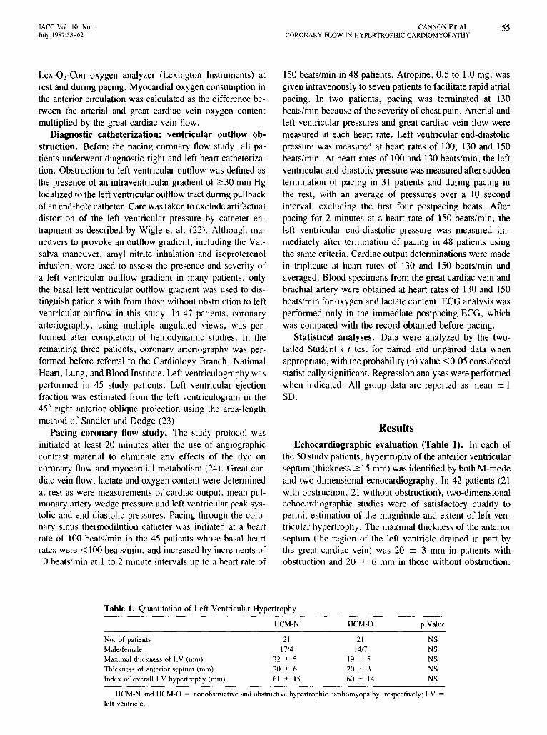
Basal coronary and myocardial hemodynamics (Table2, Fig. 1 and 2). In the basal state, 23 patients had anintraventricular gradient of 2:30 mm Hg (77 ± 33) localizedto the left ventricular outflow tract; 27 patients had either aminimal or no basal gradient (3 ± 7 mm Hg). There wasa significant relation between the basal left ventricular systolic pressure and great cardiac vein flow (r = 0.51, P <0.001) (Fig . IA) and coronary resistance (r = 0.41 , P <0.01) (Fig. 28), although wide variation was noted. Whereasthe left ventricular systolic pressure was higher in patientswith obstruction, the mean systemic pressure was significantly lower in patients with than in those without obstruction (88 ± 12 versus 96 ± 13 mm Hg, p < 0.05) . Additionally, the basal heart rate was higher in patients withobstruction (83 ± 13 versus 75 ± 11 beats/min, p < 0.005).The basal great cardiac vein flow was significantly higher(106 ± 20 versus 80 ± 25 mllmin, p < 0.(01) and coronaryresistance lower (0.85 ± 0.18 versus 1.32 ± 0.44 mmHg-min/rnl, p < 0.001) in the patients with obstruction (Fig.2) and was associated with greater myocardial oxygen consumption in the anterior left ventricular septum and freewall than that in patients without obstruction (12.4 ± 3.6versus 8.9 ± 3.3 ml oxygen/min, p < 0.01). Myocardialoxygen consumption, even when normalized for heart rate,was still significantly higher in the group with obstruction(0.15 ± 0.05 versus 0.12 ± 0.04 ml oxygen/min, p <0.025). Left ventricular filling pressures, as estimated bythe left ventricular end-diastolic and mean pulmonary arterywedge pressures, were similar for the two groups.
Coronary and myocardial hemodynamics during pacing (Table 2, Fig. 2 to 4). In the 45 patients whose basalheart rate was < 100 beats/min, atrial pacing was initiatedat 100 beats/min. Two patients (one with and one withoutobstruction ) experienced chest pain at this heart rate. Theleft ventricular systolic pressure was significantly higher(181 ± 30 versus 136 ± 21 mm Hg, p < 0.001) and meansystemic blood pressure significantly lower (91 ± 16 versus100 ± 14 mm Hg, p < 0.05) in the patients with than inthose without obstruction to left ventricular outflow . Coronary resistance decreased in both groups in response to thestress of pacing, but was significantly lower (0.76 ± 0.27versus 1.09 ± 0.44 mm Hg-min/rnl, p < 0.005) and thegreat cardiac vein flow higher (132 ± 46 versus 104 ± 38milmin, p < 0.05) in patients with obstruction. The leftventricular end-diastolic pressure was minimally increasedfrom basal measurements at this heart rate .
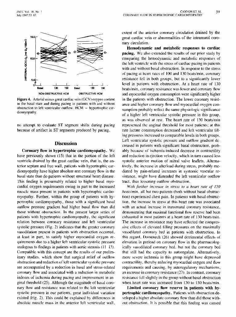
Pacing at a heart rate of 130 beats/min (performed ineach of the 50 patients) resulted in anginal chest pain in allbut 9 patients, regardless of the presence or absence ofoutflow obstruction. 111 response to this stress , mean systemic pressure rose in both groups, and was no longer significantly different. Additionally, the left ventricular systolicpressure fell in the group with basal outflow obstruction,resulting in a reduction of outflow gradient to 49 ± 30 mmHg. Still, the left ventricular systolic pressure was significantly higher in the group with obstruction (173 ± 28 versus132 ± 25 mm Hg, p < 0.001), as was coronary flow (155± 40 versus 125 ± 42 ml/rnin, p < 0.025) and myocardialoxygen consumption (18.1 ± 6.8 versus 13.0 ± 4.2 mloxygen/min, p < 0.01) . Coronary resistance was significantly lower in the group with obstruction (0.67 ± 0.17versus 0.95 ± 0.42 mm Hg-min/ml, p < 0.005). Metabolicevidence for ischemia was apparent in both groups at thisheart rille, along with a decline in lactate consumption,reflecting decreased lactate extraction compared with baseline measurements , and actual lactate production in fourpatients (Fig. 3). Further evidence for ischemia was thesignificant elevation in left ventricular end-diastolic pressurein both groups compared with the basal state . Despite theonset of ischemia in the majority of patients in both groups,there was no overall increase in oxygen extraction in eithergroup (Fig. 4).
At a heart rate of 150 beats/min in 48 patients, all but2 patients (both in the group without basal obstruction)experienced chest pain, often to a severe degree . The meansystemic pressure further increased in both groups. As thecardiac index fell in both groups compared with basal measurements, the increase in mean systemic pressure in bothgroups was related to an increase in systemic vascular resistance . Although the left ventricular systolic pressure andoutflow gradient fell further in the group with basal obstruction, the left ventricular systolic pressure was still signifi-
cl;-<()-()~ <...... 0Lh ~
""-100'0 .
''''' zo
Table 2. Hemody namic Data During Pacing Study in 50 Patients With Hypertrophic Cardiomyopathy
(A - V) O2 = arte ria l - great ca rd iac vein oxygen diffe rence ; GCV = grea t ca rdiac vein ; LV EDP = left ve ntr icu lar end-diasto lic pressure ; MV02 = myocard ialoxygen co nsump tion in the anterior c irculation I(A - V) O 2 X GCV flow]; O2 = oxygen; PCW = mean pu lmonary artery wedge pressure ; oth er abbrev iations as inTabl e 1.
Basal Pacin g: 100 beats/mi n Paci ng : 130 beats/min
HCM -N HCM -O P Value HCM-N HCM -O P Value HCM -N HCM -O P Value
75 :t: 11 83 :t: 13 < 0.005 100 100 130 13096 :t: 13 88 :t: 12 < 0.05 100 :t: 14 9 1 :t: 16 < 0 .05 103 :t: 16 98 :t: 13
134 ::!: 18 196 ::!: 33 < 0.00 1 136 ± 2 1 181 :t: 30 < 0 .00 1 132 :t: 25 173 :t: 28 < 0 .00 1
3 ::!: 7 77 :t: 33 < 0.00 1 3 ± 7 64 :t: 27 < 0 .00 1 1 :t: 4 49 :t: 30 < 0 .00 1
3. 1 :t: 0 .6 3.0 :t: 0.4 3 .0 :t: 0 .7 3 .0 ± 0.4
32 .7 ± 8. 1 30 .1 :t: 6 .7 35 ,9 :t: 9 .8 32 ,6 :t: 7.0
17.9 ± 2.0 18. 1 :t 3.0
6. 7 = 1.5 6.5 = 1.711.2 ± 1.6 11.6 = 1.9
8 .9 :t: 3.3 12.4 ± 3.6 < 0 .00 125.4 ± 20.3 30 .0 :t: 22.3
17 :t: 7 16 ± 6 18 :t: 7 18 ± 714 = 7 12 = 4oo f 27 oof 23 I of 26 I of 19
7.0 ± 1.1 6 .8 = 1.7 7.2 :t: 1.5 7.2 ± 1.610 .9 :t: 1.4 11.5 = 2.0 11.3 ± 2.0 11.4 :t 2.013 .0 =4 .2 18. 1 = 6 .8 < 0. 0 1 15. 2 :t: 5 . 1 16 .1 ± 5 .420 .8 :t: 29 .0 17 .0 = 23.4 3.0 ± 51.8 - 20 .8 = 35.3
22 :t: 10 2 1 ± 8 27 :t: 10 28 ± 718 ± 9 19 :t: 4 22 ± 10 2 1 :t: 722 o f 27 19 of 23 24 of 26 22 o f 22
8;;0oz>;;0
-<.."
5::E:z:t-<-em;;0-I
23-e:tn~;;o()
2>oz:::: z-<0Oz-e m>-1:i >-<t"""
18 .5 ± 3.2
140 ± 320 .76 :t: 0 . 15
Pacing : 150 beats/min
HCM -N HCM -O P Valu e
150 150107 = 2 1 103 ± 14
133 = 25 157 :t: 25 < 0 .005
::!: 4 32 :t: 25 < 0.00 1
2.7 ± 0 .8 2.7 :t: 0 .5
42.7 ± 13.8 39 .6 ± 10.1
18.4 ± 2.2
130 ± 4 10 .90 :t: 0 .31
< 0. 025< 0 .005
18 .1 ± 2.8
155 ± 400 .67 :t: 0. 17
17.8 ± 2.2
125 = 420 .95 ± 0.42
< 0 .05< 0 .005
132 :t: 460 .76 :t: 0 .27
104 ± 381.09 :t: 0.44
< 0.00 1< 0.00 1
106 :t: 200 .85 :t 0 .18
80 ± 251.32 :t 0.44
Heart rate (bea ts/min)Mean sys temic pressu re
(mm Hg)LV systo lic pressure
(mrn Hg)LV o utflow gradient
(mm Hg)Cardiac index
(liters/min per m2)
Systemic vascul arresistance index[(mm Hg-rnin -rrr ' j/liter]
GC V flow (rnl/rnin )
Coron ary resistance(mm Hg-rnin/ml j
Arte ria l O2
(ml O2/100 ml)GC V O2 (ml 0 21100 ml)(A - V) O2 (m l O2/ 100
ml)MV02 (ml 0 2/min)Lactate con sumption
(mM 'mllm in)LVEDP (m m Hg)PCW (rnrn Hg)Pat ient s with chest
pai n/No . of pat ient s
VI-..I

58 CANNON ET AL.CORONARY FLOW IN HYPERTROPHIC CARDIOMYOPATHY
JACC Vol. 10, No. IJuly 1987:53-62
150r - .51
0 p <.001A •:; • 0
~ •] 0 0
~100 0
90
0u.> o Obllruetivt HCMu • • Non..()bllructive HCMCl ••
w B •u 2.5Z •<- r - .41la] 2.0 p<,01~.~ •>;" 1.5a:J:<E 1.0Ze~-0 0.5u
"C..,t/100 150 200 250 300
LEFT VENTRICULAR SYSTOLIC PRESSUREImmHgl
Figure 2. Relation of (A) great cardiac vein (GCY) flow and (B)coronary resistance in the basal state to left ventricular systolicpressure in obstructive and nonobstructive hypertrophic cardiomyopathy (HeM).
wa: A ++:::>
LLLJV)V)
~- 200lI- ..uJ:- E-' E0_
r-r---r-1f-V)>V) 100>....
0z:-g B •••wECl';;, 20>0x_O!....<z-0c_a:f-<lI- 10u::EO::J>w::E Z
0u
c
~]~o....u.>UCl
was no overall change in oxygen extraction in either group:10of 22 patients with basal obstruction and IS of 26 patientswithout basal obstruction actually extracted less oxygen thanduring the basal state (Fig. 4).
Electrocardiographic changes during study. Despiteprecipitation of ischemia that was often severe symptomatically and metabolically, only seven patients (three with andfour without basal obstruction) demonstrated discernible STsegments shifts (depression in all seven cases) in one ormore of the three monitored leads (I, aYF or Y3 or Ys)recorded immediately after cessation of pacing. There was
Figure 3. Lactateconsumptionin the basal state and during pacingat rates of 130 and ISO beats/min in patients with hypertrophiccardiomyopathy (HCM) with and without obstruction to left ventricular outflow. Lactate consumption <0 indicates production oflactate by the myocardium.
150
+
100 130
PACING
++
I
Basal
o
0.5Oq'L-----l__-L__-L-__L-_
wU
~ _ 1.5tiE(jic::! 'f. 1.0> ..a: z< E~ Ea:-ou
Figure 1. A, Left ventricular (LY) systolic pressure; B, myocardial oxygen consumption in the anterior coronary circulation;C, great cardiac vein (GCY) flow; and D, coronary resistance inthe anterior circulation for patients with hypertrophic cardiomyopathy. With (open circles) and without (closed circles) obstruction to left ventricular outflow. Mean values with I SD areplotted in the basal state and during pacing. *p < 0.05, **p <0.025, ***p < 0.01, +p < 0.005, + +p < 0.001 versus patientswithout obstruction to left ventricular outflow.
cantly higher in that group compared with the group withoutbasal obstruction (157 ± 25 versus 133 ± 25 mm Hg, p< 0.005). Coronary flow and resistance changed little inthe group without obstruction, but coronary flow actuallyfell and resistance rose in the group with basal obstructionat a heart rate of 150 beats/min compared with a heart rateof 130 beats/min (0.67 ± 0.17 to 0.76 ± 0.15 mmHg-rnin/ml, p < 0.01). At a heart rate of 150 beats/min,left ventricular filling pressures increased markedly in bothgroups, and there was clear metabolic evidence of myocardial ischemia: 14 of 22 patients with basal obstructionand II of 26 patients without basal obstruction producedlactate (Fig. 3). Despite evidence of severe ischemia, there
:; 100e Z
~ ~~
~ 50
! iz~ 0III U 0:::I0CD
~ Z2 -50~~w
!;( ::J0
t- ou IE -100~
JACC Vol. 10, No.1July 1987:53-62
CANNON ET AL.CORONARY FLOW IN HYPERTROPHIC CARDIOMYOPATHY
59
extent of the anterior coronary circulation drained by thegreat cardiac vein or abnormalities of the intramural coronary circulation.
Hemodynamic and metabolic responses to cardiacpacing. We also extended the results of our prior study bycomparing the hemodynamic and metabolic responses ofthe left ventricle with the stress of cardiac pacing in patientswith and without basal obstruction. In response to the stressof pacing at heart rates of 100 and 130 beats/min, coronaryresistance fell in both groups, but to a significantly lowerlevel in patients with obstruction. At a heart rate of 130beats/min, coronary resistance was lower and coronary flowand myocardial oxygen consumption were significantly higherin the patients with obstruction. The lower coronary resistance and higher coronary flow and myocardial oxygen consumption probably reflect the same physiologic significanceof a higher left ventricular systolic pressure in this group,as was observed at rest. The heart rate of 130 beats/minrepresented the anginal threshold for most patients; at thisrate lactate consumption decreased and left ventricular filling pressures increased to comparable levels in both groups.Left ventricular systolic pressure and outflow gradient decreased in patients with significant basal obstruction, probably because of ischemia-induced decrease in contractilityand reduction in ejection velocity, which in turn caused lesssystolic anterior motion of mitral valve leaflets. Alternatively, the increase in afterload during stress, probably mediated by pain-related increases in systemic vascular resistance, might have distended the left ventricular outflowtract, thus lessening outflow obstruction.
With further increase in stress to a heart rate of J50beats/min, all but two patients (both without basal obstruction) experienced chest pain. In patients with basal obstruction, the increase in stress at this heart rate was associatedwith an actual increase in transmural coronary resistance,demonstrating that maximal functional flow reserve had beenexhausted in most patients at a heart rate of 130 beats/min.The increase in resistance may have reflected the compressive effects of elevated filling pressures on the maximallyvasodilated coronary bed in patients with obstruction. Inthis regard, Domenech (26) showed detrimental effects ofelevation in preload on coronary flow in the pharmacologically vasodilated coronary bed, but not the coronary bedthat still had the capacity to autoregulate. Alternatively,more severe ischemia in this group might have depressedcontractility. thereby reducing myocardial oxygen and flowrequirements and causing, by autoregulatory mechanisms,an increase in coronary resistance (27). In contrast, coronaryresistance fell slightly in the group without basal obstructionwhen heart rate was increased from 130 to 150 beats/min.
Limited coronary flow reserve in patients with hypertrophic cardiomyopathy. Patients with obstruction developed a higher absolute coronary flow than did those without obstruction. It is possible that this finding was caused
OBSTRUCTIVE HCM
15.0
~;0
-Er! -Er -Er
0 10.0su~..J
~II:W...II:s 5,0
Besal 1:'> 150 Basal I:.> 150
no attempt to evaluate ST segment shifts during pacingbecause of artifact in ST segments produced by pacing.
Figure 4. Arterial minusgreatcardiacvein(GCY)oxygencontentin the basal state and during pacing in patients with and withoutobstruction to left ventricular outflow. HCM = hypertrophic cardiomyopathy.
Discussion
Coronary flow in hypertrophic cardiomyopathy. Wehave previously shown (15) that in the portion of the leftventricle drained by the great cardiac vein, that is, the anterior septum and free wall, patients with hypertrophic cardiomyopathy have higher absolute rest coronary flow in thebasal state than do patients without structural heart disease.This finding is presumably related to higher basal myocardial oxygen requirements owing in part to the increasedmuscle mass present in patients with hypertrophic cardiomyopathy. Further, within the group of patients with hypertrophic cardiomyopathy, those with a significant basaloutflow pressure gradient had higher basal flow than didthose without obstruction. In the present larger series ofpatients with hypertrophic cardiomyopathy, the significantrelation between coronary resistance and left ventricularsystolic pressure (Fig. 2) indicates that the greater coronaryvasodilation present in patients with obstruction occurred,at least in part, to satisfy higher myocardial oxygen requirements due to a higher left ventricular systolic pressureanalogous to findings in patients with aortic stenosis (\ 1-13).Compatible with this concept are the results of our preliminary studies, which show that surgical relief of outflowobstruction and reduction of left ventricular systolic pressureare accompanied by a reduction in basal and stress-relatedcoronary flow and associated with a reduction in metabolicindexes of ischemia during pacing and improvement in anginal threshold (25). Although the magnitude of basal coronary flow and resistance was related to the left ventricularsystolic pressure in our patients, great individual variationexisted (Fig. 2). This could be explained by differences inabsolute muscle mass in the anterior left ventricular wall,
NON-OBSTRUCTIVE HCM

60 CANNON ET AL.CORONARY FLOW IN HYPERTROPHIC CARDIOMYOPATHY
JACC Vol. 10. No. IJuly 1987:53-62
Abnorm.102 - MYOCARDIAL - tCoron.ry.xtr.ctlon ISCHEMIA flow
I teol.ry/ ....l...nc.
tLV Filling Pre••ures
Abnorm.102 _ MYOCARDIAL ..... t eoroNry.xtrlCtlon ISCHEMIA flow
I teol.ry# ........nce
ttv Filling Pre..u....r
Reducedpeek flowcepec:1ty
H1llhba..1.nd .t_lnducedf1ow~ul~t.
OBSTRucnVE HYPERTROPHIC CARDIOMYOPATHYSTRESS
Hlllhba.., 1.nd.t......lnduced ...... _f1ow~ul~t.
LIMITED FLOW RESERVE
~
NON·OBSTRUCTIVE HYPERTROPHIC CARDIOMYOPATHYSTRESS
1--- Reduced
__ ..----- peekflowcepec:1ty
LIMITED FLOW RESERVE
l
cardial coronary artery limitation to coronary flow (42) andin patients with ischemia due to coronary artery disease (43).In fact, in many patients in both groups, less oxygen wasextracted during stress compared with the basal state. Wecould not demonstrate a relation between myocardial oxygenextraction and the presence and severity of obstruction, severity of ischemia by lactate production, absolute flow response to stress or elevation of left ventricular filling pressures. Thus, the mechanisms responsible for this findingcannot be determined from the present study, but one possibility might be arteriovenous shunting within the myo-
Figure 5. Proposed mechanisms of myocardial ischemia in hypertrophic cardiomyopathy (large arrows indicate mechanisms ofgreater importance than those indicated by small arrows). Toppanel, Patients with significant basal left ventricular (LV) outflowobstruction have high basal flow requirements primarily becauseof high left ventricular systolic pressure and wall stress. Flowrequirements increase further with stress, rapidly exhausting peakflow capacity, which in absolute terms may be relatively normal.Abnormal oxygen extraction capacity may also contribute to oraggravate ischemia. Elevated filling pressures resulting from ischemia have a deleterious compressive effect on the maximally vasodilated transmural coronary bed resulting in increased coronaryresistance and a decrease in flow. Lower panel, Patients with littleor no obstruction and lower left ventricular systolic pressure havelower basal flow requirements (although higher than that of patientswithout structural heart disease [15]). During stress, flow reservemay be compromised by greater impairment in the capacity foraugmenting coronary flow delivery, resulting in ischemia at lowercoronary flow and myocardial oxygen consumption than in patientswith obstruction. Abnormal oxygen extraction capacity may contribute to or aggravate ischemia. Elevation of filling pressuresrelated to ischemia appears to have a less deleterious effect oncoronary flow than that in patients with obstruction.
by the greater muscle mass present in patients with obstruction, although there were no group differences in the echocardiographically estimated extent or severity of hypertrophy (Table 1). Another explanation might be that patientswith obstruction have greater capillary density and thus greaterabsolute coronary flow capacity than do patients withoutobstruction, although higher flows during stress in this groupdid not prevent ischemia. Indeed, the peak flows achievedby many patients with obstruction were similar to thoseachieved after dipyridamole infusion in patients with a normal left ventricle using the same method of coronary flowmeasurement (28).
Thus, it seems that the greater peak coronary flow ofpatients with obstruction compared with that of patientswithout obstruction is due to a greater impairment in thecapacity for augmenting coronary flow delivery of the lattergroup rather than an augmented capacity of the former.However, although patients with obstruction had lowercoronary resistance and higher coronary flow at a heart rateof 130 beats/min, the variation was large. Thus, many patients with obstruction appear to have impairment in coronary flow delivery as well. Possible explanations for impaired coronary flow delivery in the patients with obstructionare I) greater dependence of myocardial perfusion on diastole because of systolic vascular compression with prolonged systolic ejection time (29-31), and 2) lower coronaryperfusion pressure secondary to the subcoronary obstruction(Table 2). Additional possible explanations for impairedcoronary flow for either the patients with or those withoutobstruction are I) abnormalities in diastolic relaxation, whichmight impair early filling of the coronary reservoir (32-36),2) high left ventricular filling pressures in response to ischemia with resulting compression of subendocardial coronarymicrocirculation (15),3) reduction in normal cross-sectionalarea of the coronary microcirculation due to replacement byabnormal myocellular architecture or fibrosis (37,38), 4)small vessel disease with diminished luminal area of intramural coronary arteries (39-41), and 5) systolic compression of septal perforating coronary arteries (which occurredslightly but not significantly more frequently in patients withbasal obstruction). Which of these considerations were mostimportant in limiting appropriate coronary flow deliveryduring stress cannot be determined from our study.
Oxygen extraction by ischemic myocardium. Therewas evidence of severe myocardial ischemia in both groupsin response to high paced heart rates. Although more patientswith basal obstruction produced lactate across the coronarybed, and the group response to pacing stress was lactateproduction (Fig. 3), the mean value of lactate consumptionof patients with and without obstruction did not differ significantly. As noted previously (15), there was no augmentation of oxygen extraction from the coronary circulation ineither group, despite metabolic evidence of severe ischemia.This is in contrast to the response seen in dogs with epi-

JACC Vol. 10, No.July 1987:53-62
CANNON ET ALCORONARY FLOW IN HYPERTROPHIC CARDIOMYOPATHY
6\
cardium in the presence of high subendocardial compressiveforces.
We have already outlined the major mechanisms we believe to be potentially responsible for precipitating ischemia. However, an additional contributing factor may bethe high ventricular pressure occurring in a ventricle withobstruction to left ventricular outflow, in which there isasymmetric distribution and magnitude of hypertrophy unlike the concentric, uniform hypertrophy present in valvularaortic stenosis. Thus, transmural wall stress may vary markedly within various regions of the same ventricle, that is,lowest in the most hypertrophied region and highest in regions of normal wall thickness. These latter regions mightbe particularly susceptible to the development of stressinduced ischemia.
Limitations of study. We have previously discussed(15) the advantages and disadvantages of estimating coronary flow by the thermodilution method. Ideally, measurement of coronary flow and calculation of resistance per unitmass would allow greater in sight as to mechanisms responsible for the limited flow reserve in hypertrophic cardiomyopathy. Unfortunately, there characteristicallyis markedheterogeneity in the distribution and amount of hypertrophyin a given patient and from patient to patient in this disease.This means that the mass of left ventricle supplied by acoronary artery (for contrast densitometric or Doppler determinations of flow velocity) or drained by the great cardiacvein (for thermodilution measurements) in a given patientcannot be defined, thereby making it impossible to normalize flow for left ventricular mass. Inert gas washouttechniques, which do measure flow per unit mass, requiretoo long a sampling period (up to 20 minutes) during stressto be practical and safe. Although no attempt was made tonormalize flow to mass in our study, we do not believe thedifferences we observed between the patients with and thosewithout obstruction could be explained by difference in mass.The reason for this is that there was no group difference inthe severity of hypertrophy between patients with and patients without obstruction to left ventricular outflow (Tablel). Further, it seems unlikely that the great cardiac veinanatomy would differ between the two groups, and thereforeunlikely that different drainage patterns exist. In furthersupport of the assumption that the higher great cardiac veinflow in patients with obstruction reflects a true increase inmyocardial oxygen demand, and not anatomic differencesin coronary venous drainage, is the observation (25) thatoperative relief of outflow obstruction reduces basal andstress-induced flow, lessens the severity of ischemia in response to stress and lowers anginal threshold.
Despite this limitation. we believe that the thermodilutionestimate of coronary flow in this study provides insight asto the mechanisms responsible for the limitation in flowreserve in patients with hypertrophic cardiomyopathy (Fig.5). For example, in most patients with obstruction, we were
able to demonstrate that limited flow reserve is related primarily to elevated basal flow and stress requirements, ratherthan to an impaired peak flow capacity, which is equal tovalues observed by us in patients with a normal left ventricleand coronary arteries after dipyridamole administration (28).In contrast, limited flow reserve in patients without obstruction does appear to be due to an impairment in peak flowcapacity in most patients.
Another limitation in this study is the calculation ofcoronary resistance. derived by mean arterial pressure dividedby great cardiac vein flow. This ignores the contribution ofelevated left ventricular filling pressures on the coronaryperfusion pressure, an effect that undoubtedly differs acrossthe ventricular wall, being greatest in the endocardium. Thepostpacing end-diastolic left ventricular and pulmonary artery occlusive pressures were similar in the two groups, andthus the difference in calculated coronary resistance duringpacing between patients with and patients without obstruction is unlikely to be related to omission of the effect of leftventricular filling pressure on coronary perfusion pressure.
Conclusions. The pathogenetic mechanisms causingmyocardial ischemia differ between patients with obstructive and nonobstructive hypertrophic cardiomyopathy.Coronary flow reserve in patients with obstructive hypertrophic cardiomyopathy is limited because of high basal andstress-induced coronary flow requirements related to a highleft ventricular systolic pressure, a situation analogous tothat in valvular aortic stenosis, and in some patients, becauseof limited maximal flow capacity. Patients with nonobstructive hypertrophic cardiomyopathy develop ischemia as aresult of severe impairment of maximal coronary flow capacity.
We thank Imogene Surrey for excellent secretarial assistance. Additionally,the technical assistance and support of Beverly Barber, Cecilia Bergamo,Donna Jo Fleagle, Terry Rumble, Carroll L. Hanson, Greg Johnson, RitaMincemoyer , Carl Newman, Annette Stine. Ronald White and Judith B.Winkler are gratefully acknowledged.
ReferencesI. Maron BJ, Epstein SE. Hypertrophic cardiomyopathy: a discussion
of nomenclature. Am J Cardiol 1979;43:1242-4.
2. Goodwin JF, Hollman A, Cleland WP, Teare D. Obstructive cardiomyopathy simulating aortic stenosis. Br Heart J 1960;22:403-14.
3. Wigle ED, Heimbecker RO, Gunton RW. Idiopathic ventricular septalhypertrophy causing muscular subaortic stenosis. Circulation 1962;26:325-40.
4. Braunwald E, Lambrew CT, Rockoff SO, Ross J, Morrow AG. Idiopathic hypertrophic subaortic stenosis. I. A description of the diseasebased upon an analysis of 64 patients. Circulation I964;30(suppl IV):IV-3-2l3.
5. Ross J, Braunwald E, Gault JH, Mason DT, Morrow AG. The mechanism of the interventricular pressure gradient in idiopathic hypertrophic subaortic stenosis. Circulation 1966;34:558-78.
6. Criley JM, Lewis KB, White RI, Ross RS. Pressure gradients withoutobstruction. A new concept of "hypertrophic subaortic stenosis."Circulation 1965;32:881-7.
62 CANNON ET AL.CORONARY FLOW [N HYPERTROPHIC CARDIOMYOPATHY
JACC Vol. [0, No. [July 1987:53-62
7. Murgo JP, Alter BR, Dorethy JF, Altobelli SA, McGranahan GM.Dynamics of left ventricular ejection in obstructive and non-obstructivehypertrophic cardiomyopathy. J Clin Invest 1980;66:1369-82.
8. Maron BJ, Gottdiener JS, Arce J, Rosing DR, Wesley YE, EpsteinSE. Dynamic subaortic obstruction in hypertrophic cardiomyopathy:analysis by pulsed Doppler echocardiography. J Am Coli Cardiol1985;6:1-15.
9. Bonow RO, Ostrow HG, Rosing DR, et al. Dynamic pressure-volumealterations during left ventricular ejection in hypertrophic cardiomyopathy: evidence for true obstruction to left ventricular outflow(abstr). Circulation 1984;70(suppl 11):11-17.
10. Criley JM, Siegel RJ. Has 'obstruction' hindered our understandingof hypertrophic cardiomyopathy? Circulation 1985;72:1148-54.
II. Fallen EL, Elliott WC, Gorlin R. Mechanisms of angina in aorticstenosis. Circulation 1967;36:480-8.
12. Trenouth RS, Phelps NC, Neill WA. Determinants of left ventricularhypertrophy and oxygen supply in chronic aortic valve disease. Circulation 1976;53:644-50.
13. Bertrand ME, LaBlanche JM, Tilmant PY, Thieuleux FP, DelforgeMR, Carre AG. Coronary sinus blood flow at rest and during isometricexercise in patients with aortic valve disease. Am J Cardiol 1981;47:199-205.
14. Marcus ML, Doty DB, Hiratzka LF, Wright CB, Eastham CL. Decreased coronary reserve: a mechanism for angina pectoris in patientswith aortic stenosis and normal coronary arteries. N Engl J Med1982;307:1362-6.
15. Cannon RO, Rosing DR, Maron BJ, et al. Myocardial ischemia inpatients with hypertrophic cardiomyopathy: contribution of inadequatevasodilator reserve and elevated left ventricular filling pressures. Circulation 1985;71:234-43.
16. Spirito P, Maron BJ, Chiarella F, et al. Diastolic abnormalities inpatients with hypertrophic cardiomyopathy: relation to magnitude ofleft ventricular hypertrophy. Circulation 1985;72:310-6.
17. Baim DS, Rothman MT, Harrison DC. Improved catheter for regionalcoronary sinus flow and metabolic studies. Am J Cardiol 1980;46:997-1000.
18. Ganz W, Tamura K, Marcus HS, Donoso R, Yoshida S, Swan HJe.Measurement of coronary sinus blood flow by continuous thermodilution in man. Circulation 1971;44:181-95.
[9. Pepine CJ, Mehta J, Webster WW, Nichols WW. In vivo validationof a thermodilution method to determine regional left ventricular bloodflow in patients with coronary disease. Circulation 1978;58:795-802.
20. Mathey DG, Chatterjee K, Tyberg JV, Lekven J, Brundage B, ParmleyWW. Coronary sinus reflux: a source of error in the measurement ofcoronary sinus blood flow. Circulation 1978;57:778-86.
21. Marbach EP, Weil MH. Rapid enzymatic measurement of blood lactateand pyruvate. Clin Chem 1964;13:314-25.
22. Wigle ED, Marquis Y, Auger P. Muscular subaortic stenosis: initialleft ventricular inflow tract pressure in the assessment of intraventricular pressure differences in man. Circulation 1967;35:1100-17.
23. Sandler H, Dodge HT. The use of single plane angiocardiograms forthe calculation of left ventricular volume in man. Am Heart J 1968;75:325-34.
24. Wisneski JA, Gertz EW, Neese R, et al. Myocardial metabolic alterations after contrast angiography. Am J Cardiol 1982;50:239-45.
25. Cannon RO, Rosing DR, McIntosh CL, Epstein SE. Hypertrophiccardiomyopathy: improved hemodynamics, metabolism, and anginal
threshold following surgical relief of outflow obstruction (abstr). Circulation 1985;72(suppl II1):II1-447.
26. Domenech RJ. Regional diastolic coronary blood flow during diastolicventricular hypertension. Cardiovasc Res 1978;12:639-45.
27. Henry PD, Eckberg D, Gault JH, Ross J. Depressed inotropic stateand reduced myocardial oxygen consumption in the human heart. AmJ Cardiol 1973;31:300-6.
28. Cannon RO, Schenke WH, Leon MB, Rosing DR, Urqhart J, EpsteinSE. Limited coronary flow reserve after dipyridamole in patients withergonovine-induced coronary vasoconstriction. Circulation (in press).
29. Pichard AD, Meller J, Teichholz LE, Lipnick S, Gorlin R, HermanMV. Septal perforator compression (narrowing) in idiopathic hypertrophic subaortic stenosis. Am J Cardiol 1977;40:310-4.
30. Ruddy T, Henderson A, Rakowski H, Wigle ED. Systolic constrictionof the coronary arteries in hypertrophic cardiomyopathy (abstr). Circulation 1981;64:IV-239.
31. Pasternac A, Noble J, Streulens Y, Elie R, Henschke C, BourassaMG. Pathophysiology of chest pain in patients with cardiomyopathiesand normal coronary arteries. Circulation 1982;65:778-89.
32. Brutsaert DL, Housmans PR, Goethals MA. Dual control of relaxation:its role in the ventricular function in the mammalian heart. Circ Res1980;47:637-52.
33. St John Sutton MG, Tajik AJ, Smith HC, Ritman EL. Angina inidiopathic hypertrophic subaortic stenosis. A clinical correlate of regional left ventricular dysfunction: A videometric and echocardiographic study. Circulation 1980;61:561-8.
34. Hanrath P, Mathey DG, Siegert R, Bleifield W. Left ventricular relaxation and filling pattern in different forms of left ventricular hypertrophy: an echocardiographic study. Am J Cardiol 1980;45: 15-23.
35. Bonow RO, Rosing DR, Bacharach SL, et al. Effects of verapamilon left ventricular systolic function and diastolic filling in patientswith hypertrophic cardiomyopathy. Circulation 1981;64:787-96.
36. Wigle ED. Hypertrophic cardiomyopathy: the importance of the siteand extent of hypertrophy. A review. Prog Cardiovasc Dis 1985;28:1-83.
37. Roberts WC, Ferrans VJ. Morphologic observations in the cardiomyopathies. In: Fowler NO, ed. Myocardial Disease. New York:Grune & Stratton, 1973:59-115.
38. Maron BJ, Roberts WC. Quantitative analysis of cardiac muscle celldisorganization in the ventricular septum of patients with hypertrophiccardiomyopathy. Circulation 1979;59:689-706.
39. Maron 8J, Epstein SE, Roberts We. Hypertrophic cardiomyopathyand transmural myocardial infarction without significant artherosclerosis of the extramural coronary arteries. Am J Cardiol 1979;43:1086-102.
40. James TH, Marshall TK. De subitaneis mortibus XII. Asymmetricalhypertrophy of the heart. Circulation 1975;51: 1149-66.
41. Maron BJ, Wolfson JK, Epstein SE, Roberts We. Intramural "smallvessel") coronary artery disease in hypertrophic cardiomyopathy. JAm Coli Cardiol 1986;8:545-57.
42. Weiss HR. Effect of coronary artery occlusion on regional arterial andvenous O2 saturation, O2 extraction, blood flow, and O2 consumptionin the dog heart. Circ Res 1980;47:400-7.
43. Messer JV, Wagman RJ, Levine HJ, Neill WA, Krasnow N, GorlinR. Patterns of human myocardial oxygen extraction during rest andexercise. J Clin Invest 1962;41:725-42.