dicephalus dipus dibrachius twins: report of an autopsy case
TRANSCRIPT

a
Autopsy and Case Reports. ISSN 2236-1960. Copyright © 2014. This is an Open Access article distributed of terms of the Creative Commons Attribution Non-Commercial License which permits unrestricted non-commercial use, distribution, and reproduction in any medium provided article is properly cited.
INTRODUCTION
Conjoined twins are a rare abnormality that has
been described since antiquity,1,2 and represent 1%
of all cases of monozygotic twins.3 Epidemiologically,
dicephalus dipus dibrachius twins are an even rarer
form of conjoined twins that is characterized by a
gestational conceptus possessing two arms, two legs,
a trunk, and two heads.1,2,4 The fetuses frequently die
before or shortly after birth, which is usually caused by
cardiorespiratory malformations.2 This article reports on
the diagnosis and subsequent autopsy of dicephalus
dipus dibrachius twins.
a Department of Pathology – Faculdade de Medicina de Botucatu – Universidade Estadual Paulista Júlio de Mesquita Filho – Botucatu/SP - Brazil. b Department of Genetics – Instituto de Biociências – Universidade Estadual Paulista Júlio de Mesquita Filho – Botucatu/SP - Brazil.
Article / Autopsy Case Report Artigo / Relato de Caso de Autópsia
Dicephalus dipus dibrachius twins: report of an autopsy case
Cristiano Claudino Oliveiraa, Claudia Aparecida Rainhob, Maria Aparecida Custódio Dominguesa
Oliveira CC, Rainho CA, Domingues MAC. Dicephalus dipus dibrachius twins: report of an autopsy case. Autopsy Case Rep [Internet]. 2014;4(2):21-6. http://dx.doi.org/10.4322/acr.2014.018
ABSTRACT
Dicephalus dipus dibrachius twins are a rare form of conjoined twins. An autopsy of conjoined dicephalus twins is reported. The diagnosis was performed during the pregnancy and the family received a judicial authorization for termination of pregnancy. The preterm newborn progressed to cardiac arrest and died following a court order to terminate the pregnancy. The conceptus presented two arms, two legs, a trunk, two heads, and a single umbilical cord. The two spinal columns presented vertebral fusion in the sacral region. The heart exhibited complex malformations. The external genitalia were female; and cytogenetic analysis confirmed female sex (46, XX). This analysis also corroborated the etiopathogenic hypotheses described for this abnormality, which proposes failures in embryonic formation rather than specific chromosomal alterations. Current identification of cases by ultrasound permits medical management and multidisciplinary action with the family, enabling the legal termination of pregnancy.
Keywords Autopsy; Twins, Conjoined; Cytogenetics; Embryology; Fetal Diseases.
CASE REPORT AND AUTOPSY FINDINGS
The case initially came to light during standard prenatal consultations. It was the first child of a non-consanguineous couple and the pregnancy had proceeded uneventful until the abnormality was discovered. The preterm infants were born vaginally at 21 weeks gestational age, following termination of the pregnancy accompanied by judicial authorization, due to malformation (conjoined twins), which was diagnosed by ultrasound.
The twins were born with an Apgar score of 1-1-1; measured 27.0 cm in length and weighed 560 g. The head circumferences were 19.5 cm (left twin)

Autopsy and Case Reports 2014; 4(2):21-26
Dicephalus dipus dibrachius twins: report of an autopsy case
22
and 18.5 cm (right twin). The chest girth measured 17.5 cm and the abdominal circumference measured 16.5 cm. The twins survived for three hours and during this period presented bradycardia, cardiac arrest, and death.
After family consent, the autopsy was performed according to the standard protocols of our department, namely: anthropometric measures, individual organ dissection and selection of fragments for histological analysis. During the procedure, fragments of heart, brain, skin, bone marrow, and lung were collected for cytogenetic karyotype examination. Intracardiac puncture was also performed to collect blood for the same exam.
Visual examination revealed that the conceptus was sustained by a single umbilical cord and had one trunk, two arms, two legs, and two heads, confirming the diagnosis of dicephalus dipus dibrachius twins.2 Dicephalus twins have only one pair of legs, but a variable degree of duplication of the upper body. In the subcategory of dicephalus dibrachius, there is one trunk, two arms, and two heads and necks situated next to each other2,5,6 (Figure 1A). The two spinal columns presented vertebral fusion in the sacral region (Figures 1B).
Evaluation of the internal organs revealed the presence of two brains (Figure 1C), two normal lungs in each fetus, one aorta, two trachea, two esophagi, two
Figure 1. Examination of A - dicephalus dipus dibrachius twins; B - Vertebral fusion of two spinal columns in the sacral region; C - Presence of two brains.
Autopsy and Case Reports 2014; 4(2):21-26
Oliveira CC, Rainho CA, Domingues MAC.
23
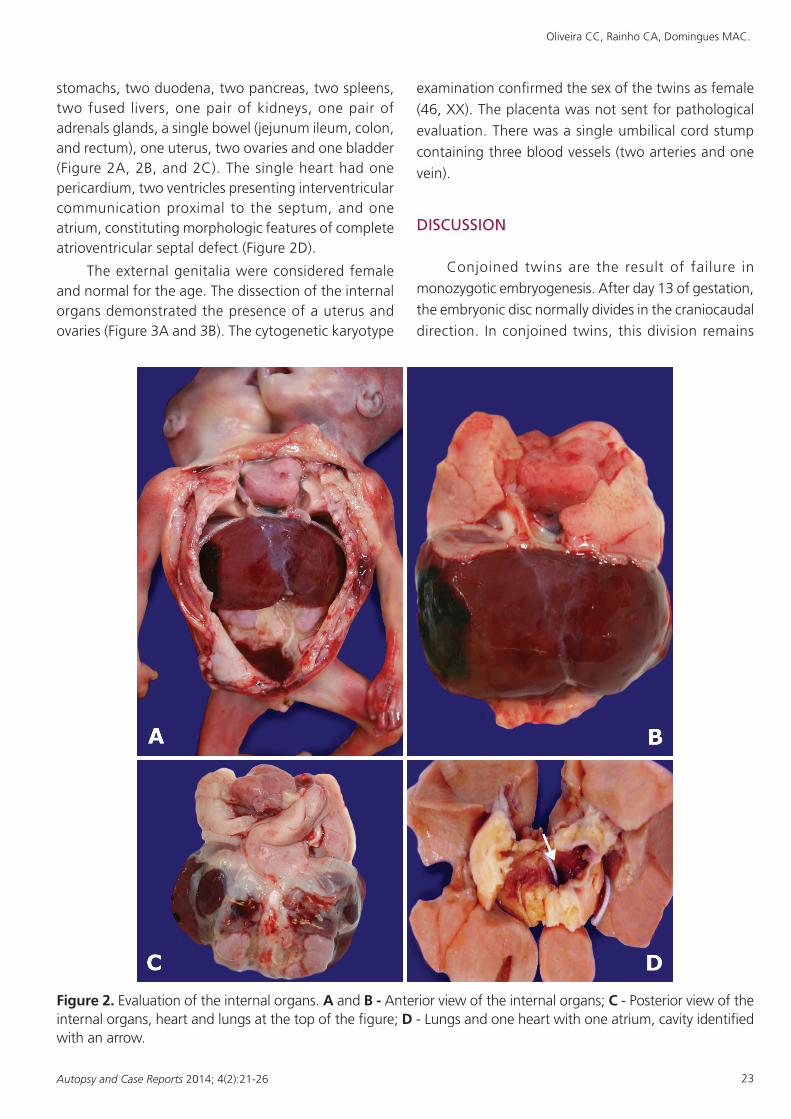
stomachs, two duodena, two pancreas, two spleens, two fused livers, one pair of kidneys, one pair of adrenals glands, a single bowel (jejunum ileum, colon, and rectum), one uterus, two ovaries and one bladder (Figure 2A, 2B, and 2C). The single heart had one pericardium, two ventricles presenting interventricular communication proximal to the septum, and one atrium, constituting morphologic features of complete atrioventricular septal defect (Figure 2D).
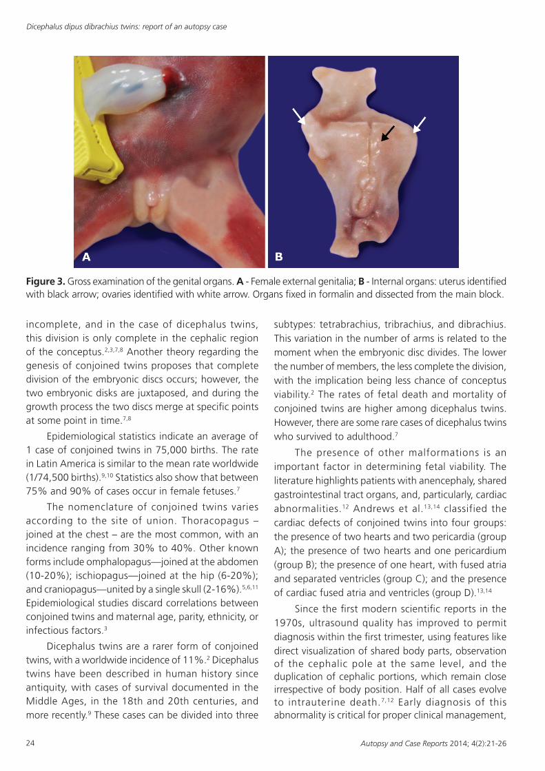
The external genitalia were considered female and normal for the age. The dissection of the internal organs demonstrated the presence of a uterus and ovaries (Figure 3A and 3B). The cytogenetic karyotype
examination confirmed the sex of the twins as female
(46, XX). The placenta was not sent for pathological
evaluation. There was a single umbilical cord stump
containing three blood vessels (two arteries and one
vein).
DISCUSSION
Conjoined twins are the result of failure in
monozygotic embryogenesis. After day 13 of gestation,
the embryonic disc normally divides in the craniocaudal
direction. In conjoined twins, this division remains
Figure 2. Evaluation of the internal organs. A and B - Anterior view of the internal organs; C - Posterior view of the internal organs, heart and lungs at the top of the figure; D - Lungs and one heart with one atrium, cavity identified with an arrow.
Autopsy and Case Reports 2014; 4(2):21-26
Dicephalus dipus dibrachius twins: report of an autopsy case
24
incomplete, and in the case of dicephalus twins, this division is only complete in the cephalic region of the conceptus.2,3,7,8 Another theory regarding the genesis of conjoined twins proposes that complete division of the embryonic discs occurs; however, the two embryonic disks are juxtaposed, and during the growth process the two discs merge at specific points at some point in time.7,8
Epidemiological statistics indicate an average of 1 case of conjoined twins in 75,000 births. The rate in Latin America is similar to the mean rate worldwide (1/74,500 births).9,10 Statistics also show that between 75% and 90% of cases occur in female fetuses.7
The nomenclature of conjoined twins varies according to the site of union. Thoracopagus – joined at the chest – are the most common, with an incidence ranging from 30% to 40%. Other known forms include omphalopagus—joined at the abdomen (10-20%); ischiopagus—joined at the hip (6-20%); and craniopagus—united by a single skull (2-16%).5,6,11 Epidemiological studies discard correlations between conjoined twins and maternal age, parity, ethnicity, or infectious factors.3
Dicephalus twins are a rarer form of conjoined twins, with a worldwide incidence of 11%.2 Dicephalus twins have been described in human history since antiquity, with cases of survival documented in the Middle Ages, in the 18th and 20th centuries, and more recently.9 These cases can be divided into three
subtypes: tetrabrachius, tribrachius, and dibrachius. This variation in the number of arms is related to the moment when the embryonic disc divides. The lower the number of members, the less complete the division, with the implication being less chance of conceptus viability.2 The rates of fetal death and mortality of conjoined twins are higher among dicephalus twins. However, there are some rare cases of dicephalus twins who survived to adulthood.7
The presence of other malformations is an important factor in determining fetal viability. The literature highlights patients with anencephaly, shared gastrointestinal tract organs, and, particularly, cardiac abnormalities.12 Andrews et al.13,14 classified the cardiac defects of conjoined twins into four groups: the presence of two hearts and two pericardia (group A); the presence of two hearts and one pericardium (group B); the presence of one heart, with fused atria and separated ventricles (group C); and the presence of cardiac fused atria and ventricles (group D).13,14
Since the first modern scientific reports in the 1970s, ultrasound quality has improved to permit diagnosis within the first trimester, using features like direct visualization of shared body parts, observation of the cephalic pole at the same level, and the duplication of cephalic portions, which remain close irrespective of body position. Half of all cases evolve to intrauterine death.7,12 Early diagnosis of this abnormality is critical for proper clinical management,
Figure 3. Gross examination of the genital organs. A - Female external genitalia; B - Internal organs: uterus identified with black arrow; ovaries identified with white arrow. Organs fixed in formalin and dissected from the main block.

Autopsy and Case Reports 2014; 4(2):21-26
Oliveira CC, Rainho CA, Domingues MAC.
25
because resolution of the pregnancy at or near term is difficult; it subjects the mother to greater risks and can compromise her reproductive health.15 According to Berezowski et al.,15 ideally, conjoined twins should be included in legal requests for early termination and pregnancy management.15 Even in cases in which separation is possible, the situation should be analyzed in depth by medical teams to determine the viability and feasibility of separating the twins. Lopes et al.16 and Brizot et al.17 have presented Brazilian historical series of conjoined twins. According to these authors, conjoined twin pregnancies should be referred to tertiary health centers because the possibility of specific image exams such as echocardiograpfiy.16,17 The thorough fetal evaluation can evaluate the postnatal prognosis16,17, as well as guide the multidisciplinary planning actions, and in some cases, prepare the termination of pregnancy. In Brazil, the court has granted in favor of termination of pregnancy in cases of malformation documented by ultrasound exams.18
The case described herein was detected during prenatal care and a legal request to interrupt the pregnancy was authorized. The autopsy evaluation confirmed the diagnosis of dicephalus dipus dibrachius twins, a rare form of conjoined twins. In addition, cytogenetic evaluation was performed. This analysis confirmed the sex of the conceptus and corroborated the etiopathogenic hypotheses described for this abnormality.
REFERENCES
1. Seo JW, Lee YS, Chi JG. Cross-sectional illustration on major types of conjoined twins. J Korean Med Sci. 1988;3(1):19-25. PMid:3267350
2. Bondeson J. Dicephalus conjoined twins: a historical review with emphasis on viability. J Pediatr Surg. 2001;36(9):1435-44. http://dx.doi.org/10.1053/jpsu.2001.26393. PMid:11528623
3. Daskalakis G, Pilalis A, Tourikis I, Moulopoulos G, Karamoutzos I, Antsaklis A. First trimester diagnosis of dicephalus conjoined twins. Eur J Obstet Gynecol Reprod Biol. 2004;112(1):110-3. http://dx.doi.org/10.1016/S0301-2115(03)00268-9. PMid:14687753
4. Mabogunje OA, Lawrie JH. Conjoined twins in West Africa. Arch Dis Child. 1980;55(8):626-30. http://dx.doi.org/10.1136/adc.55.8.626. PMid:7192076
5. Sp i tz L , K ie ly EM. Conjo ined twins . JAMA. 2003;289(10):1307-10. http://dx.doi.org/10.1001/jama.289.10.1307. PMid:12633195
6. Spitz L, Kiely EM. Experience in the management of conjoined twins. Br J Surg. 2002;89(9):1188-92. http://dx.doi.org/10.1046/j.1365-2168.2002.02193.x. PMid:12190687
7. Harma M, Harma M, Mil Z, Oksuzler C. Vaginal delivery of dicephalic parapagus conjoined twins: case report and literature review. Tohoku J Exp Med. 2005;205(2):179-85. http://dx.doi.org/10.1620/tjem.205.179. PMid:15673976
8. Spencer R. Conjoined twins: theoretical embryologic basis. Teratology. 1992;45(6):591-602. http://dx.doi.org/10.1002/tera.1420450604. PMid:1412053
9. Martínez-Frías ML, Bermejo E, Mendioroz J, et al. Epidemiological and clinical analysis of a consecutive series of conjoined twins in Spain. J Pediatr Surg. 2009;44(4):811-20. http://dx.doi.org/10.1016/j.jpedsurg.2008.07.002. PMid:19361646
10. Castilla EE, López-Camelo JS, Orioli IM, Sánchez O, Paz JE. The epidemiology of conjoined twins in Latin America. Acta Genet Med Gemellol (Roma). 1988;37(2):111-8. PMid:3239351
11. Chen H. Conjoined twins. In: Chen H. Atlas of genetis diagnosis and couseling. 2nd ed. New York: Springer; 2012. p. 495-505.
12. Mackenzie TC, Crombleholme TM, Johnson MP, et al. The natural history of prenatally diagnosed conjoined twins. J Pediatr Surg. 2002;37(3):303-9. http://dx.doi.org/10.1053/jpsu.2002.30830. PMid:11877639
13. Andrews RE, McMahon CJ, Yates RWM, et al. Echocardiographic assessment of conjoined twins. Heart. 2006;92(3):382-87. http://dx.doi.org/10.1136/hrt.2005.069682. PMid:16118238
14. Ambar SS, Halkati PC, Patted SV, Yavagal S. Twin heart with a fused atria and separate ventricles in conjoined twins. Ann Pediatr Cardiol. 2010;3(2):196-8. http://dx.doi.org/10.4103/0974-2069.74065. PMid:21234207
15. Berezowski AT, Duarte G, Rodrigues R, et al. [Conjoined twins: an experience of a tertiary hospital in Southeast Brazil]. Rev Bras Ginecol Obstet. 2010;32(2):61-5. [Portuguese.]. PMid:20305942
16. Lopes LM, Brizot ML, Schultz R, et al. Twenty-five years of fetal echocardiography in conjoined twins: lessons learned. J Am Soc Echocardiogr. 2013;26(5):530-8. http://dx.doi.org/10.1016/j.echo.2013.02.013. PMid:23562084
17. Brizot ML, Liao AW, Lopes LM, et al. Conjoined twins pregnancies: experience with 36 cases from a single center. Prenat Diagn. 2011;31(12):1120-5. PMid:21905053
18. Duarte GA, Osis MJD, Faúndes A, Sousa MH. Brazilian abortion law: the opinion of judges and prosecutors. Rev Saúde Pública. 2010;44(3):406-20. http://dx.doi.org/10.1590/S0034-89102010005000006. [Portuguese.] PMid:20464265

Autopsy and Case Reports 2014; 4(2):21-26
Dicephalus dipus dibrachius twins: report of an autopsy case
26
Conflict of interest: None
Submitted on: May 1, 2014Accepted on: June 19, 2014
Correspondence Departamento de Patologia, Faculdade de Medicina de Botucatu da Universidade Estadual Paulista Júlio de Mesquita Filho Av. Rubião Jr, s/n – Botucatu/SP – Brazil Cep 18618-000 Phone +55 (14) 3811-6238 E-mail [email protected]