diagnostic ultrasound of the ankle and foot · diagnostic ultrasound of the ankle and foot dr....
TRANSCRIPT
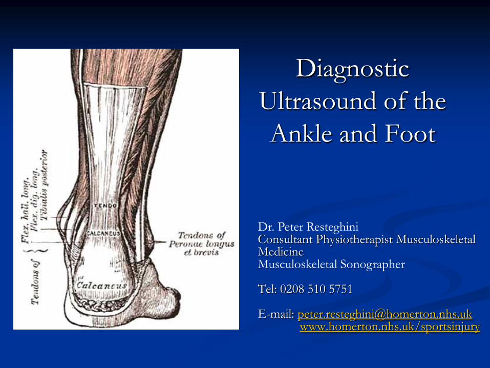
Diagnostic
Ultrasound of the
Ankle and Foot
Dr. Peter Resteghini Consultant Physiotherapist Musculoskeletal Medicine Musculoskeletal Sonographer Tel: 0208 510 5751 E-mail: [email protected] www.homerton.nhs.uk/sportsinjury
Aims
An overview of tendinopathy
Imaging of Ankle and Foot
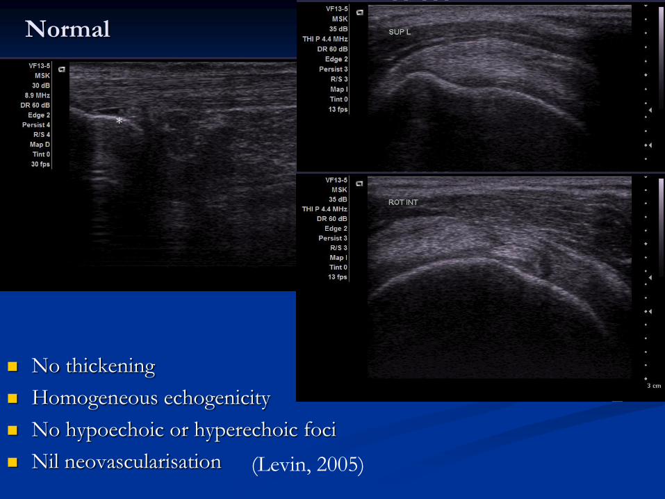
Normal
No thickening
Homogeneous echogenicity
No hypoechoic or hyperechoic foci
Nil neovascularisation (Levin, 2005)
*
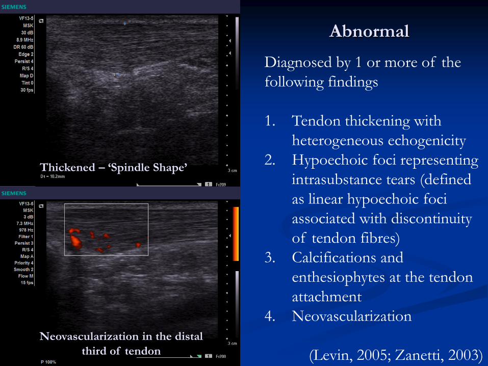
Abnormal
Neovascularization in the distal
third of tendon
Thickened – ‘Spindle Shape’
Diagnosed by 1 or more of the
following findings
1. Tendon thickening with
heterogeneous echogenicity
2. Hypoechoic foci representing
intrasubstance tears (defined
as linear hypoechoic foci
associated with discontinuity
of tendon fibres)
3. Calcifications and
enthesiophytes at the tendon
attachment
4. Neovascularization
(Levin, 2005; Zanetti, 2003)
The Ankle &
Foot

Anatomy -
Anterior
FHL Inf Ext Ret
Deep Peroneal N / Ant Tib A
EDL
Tib Ant
Superior Ext Ret
Superficial Peroneal N
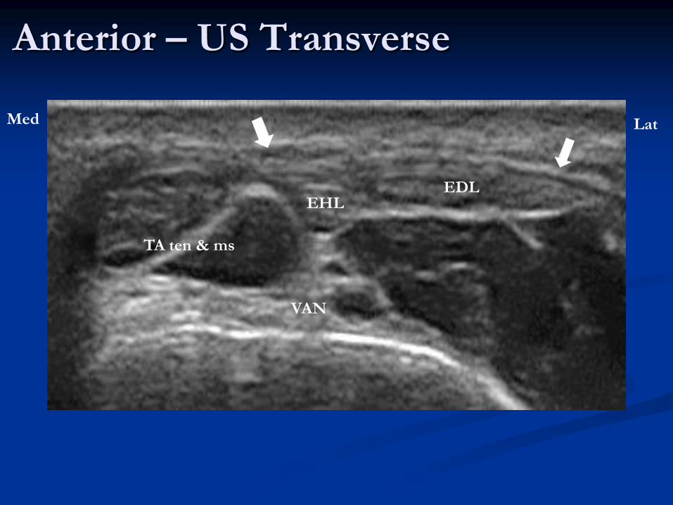
Anterior – US Transverse
Med Lat
TA ten & ms
EHL EDL
VAN
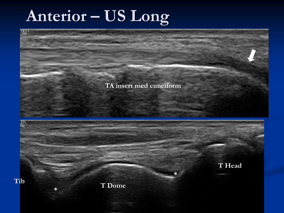
Anterior – US Long
TA insert med cuneiform
Tib T Dome
T Head
*
*
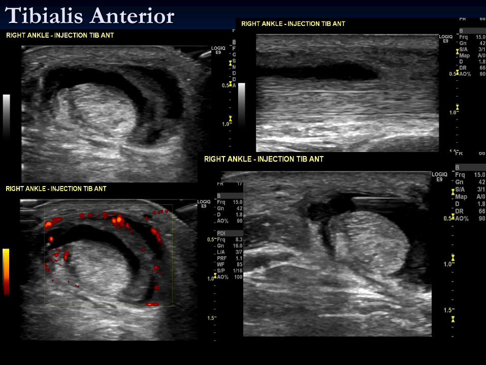
Tibialis Anterior
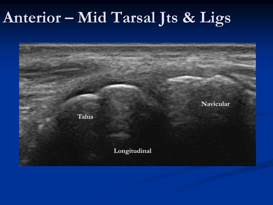
Anterior – Mid Tarsal Jts & Ligs
Longitudinal
Navicular
Talus

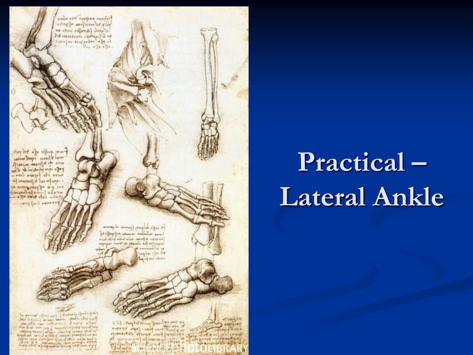
Practical –
Anterior
Ankle

Anatomy - Lateral
ATFL
CFL
Bifurcate Lig
Inf Tib
Fib Lig
Dorsal C-C Lig
EDB
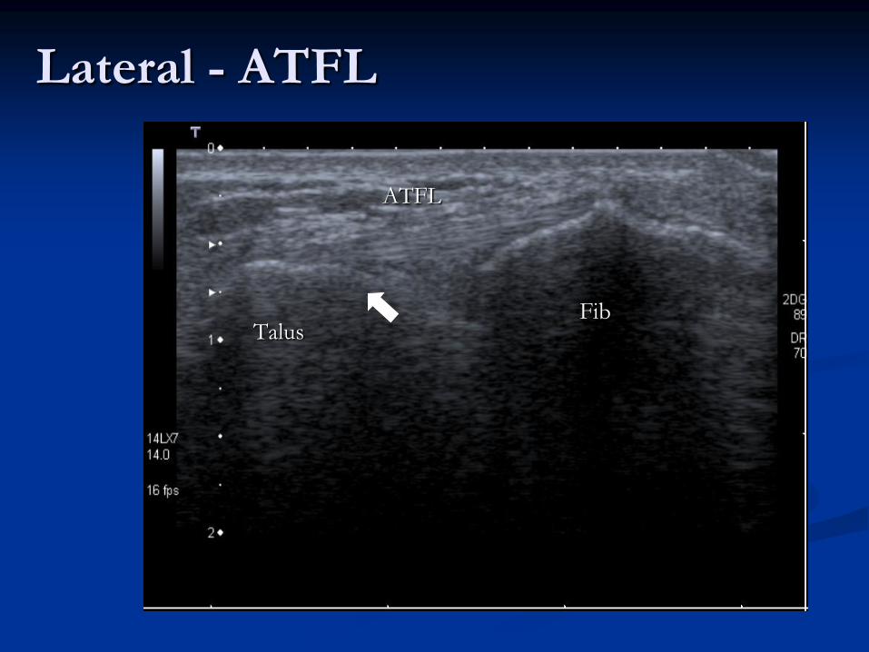
Lateral - ATFL
Fib Talus
ATFL
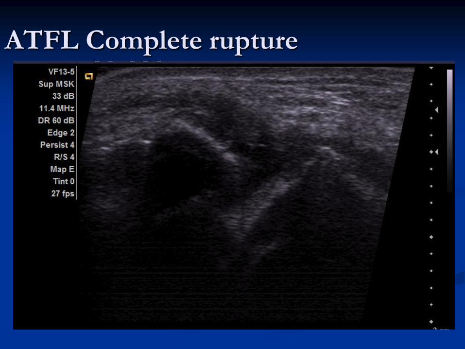
ATFL Complete rupture
ATFL rupture with +ve Sonographic Draw.avi
Image clip of +ve sonographic draw
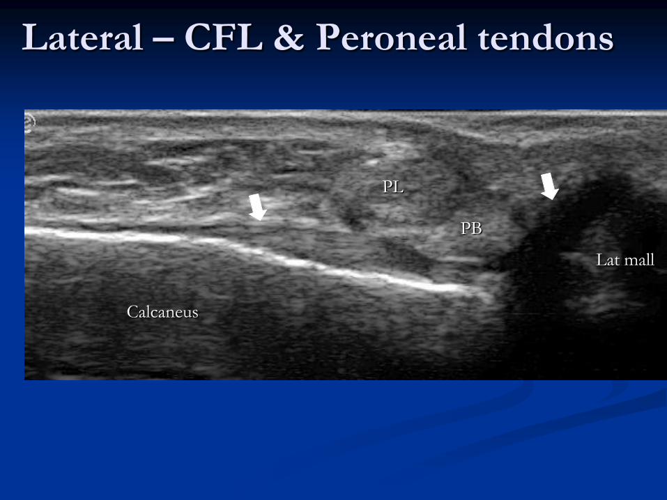
Lateral – CFL & Peroneal tendons
Lat mall
Calcaneus
PL
PB
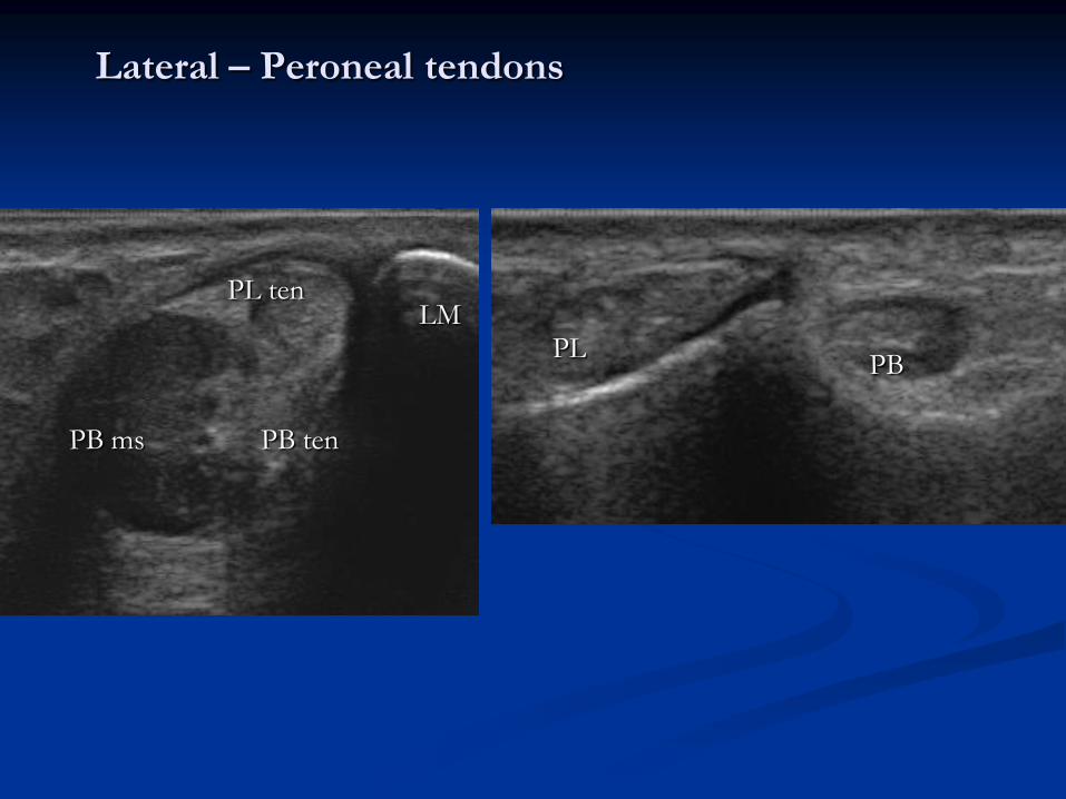
Lateral – Peroneal tendons
PB ms
LM
PB ten
PL ten
PB PL
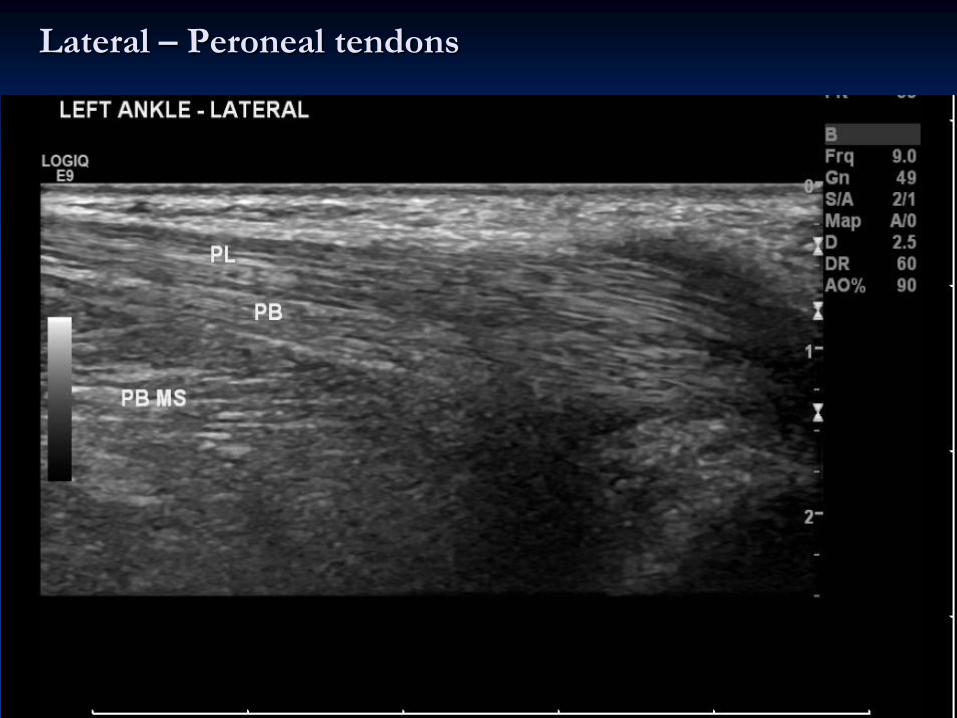
Lateral – Peroneal tendons
Practical –
Lateral Ankle

Anatomy - Medial
TA TP
FDL
FHL
FDB
AVN
Deltoid Lig
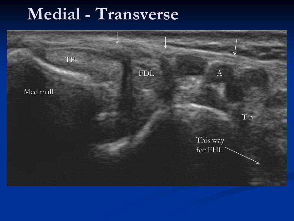
Medial - Transverse
Med mall
TP
FDL A
This way
for FHL
T n
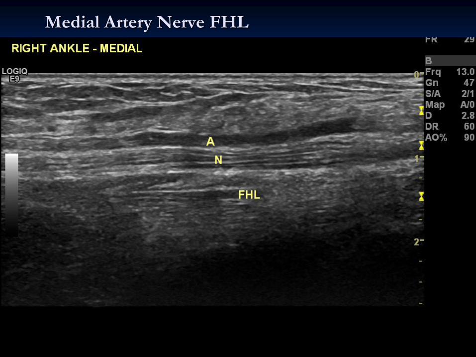
Medial Artery Nerve FHL

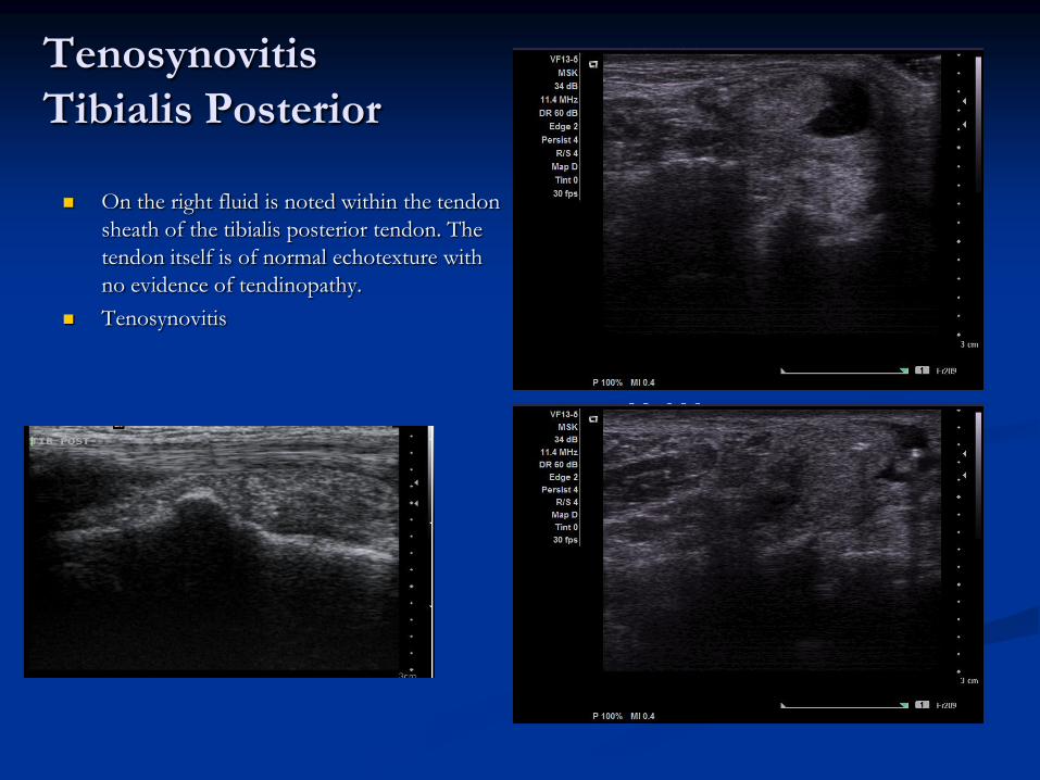
Tenosynovitis
Tibialis Posterior
On the right fluid is noted within the tendon
sheath of the tibialis posterior tendon. The
tendon itself is of normal echotexture with
no evidence of tendinopathy.
Tenosynovitis
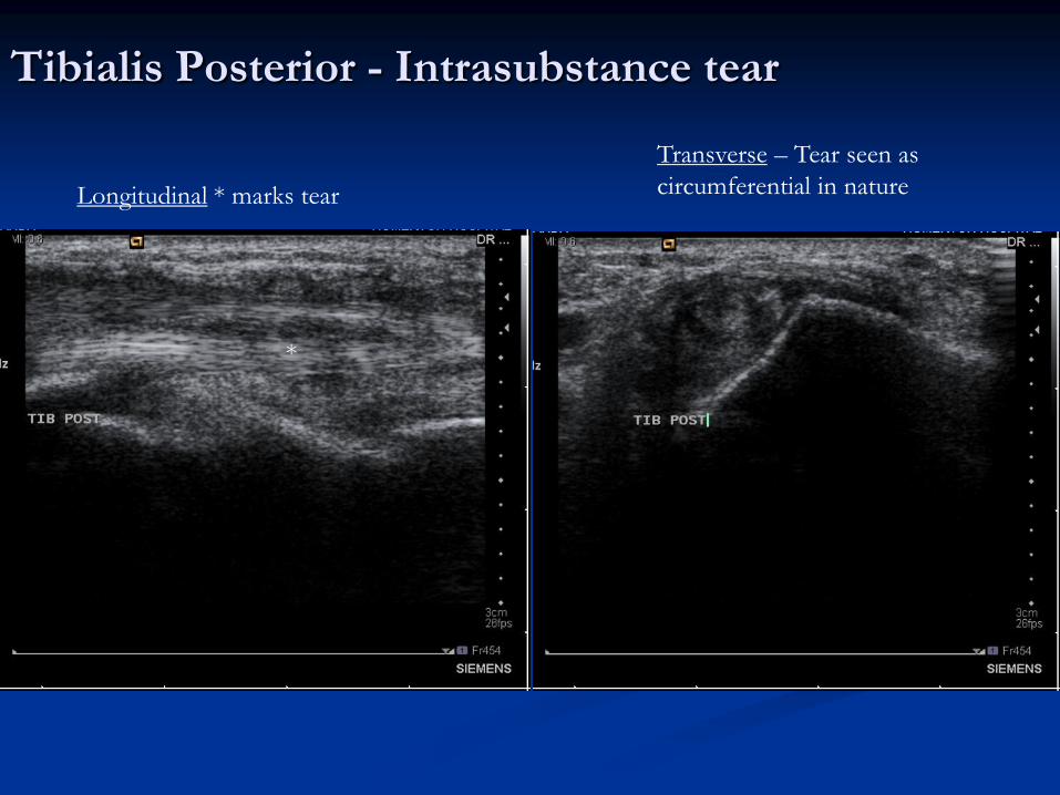
Tibialis Posterior - Intrasubstance tear
*
Longitudinal * marks tear
Transverse – Tear seen as
circumferential in nature
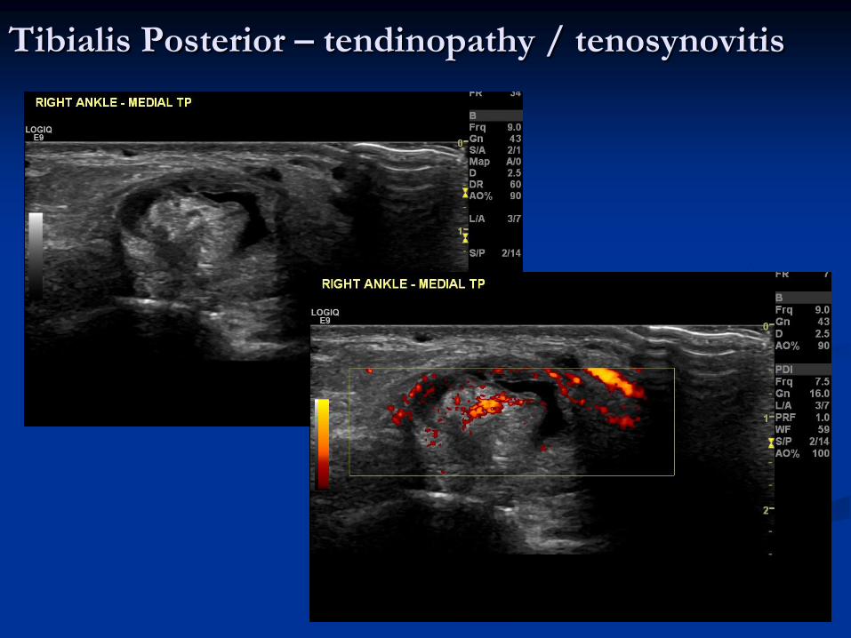
Tibialis Posterior – tendinopathy / tenosynovitis
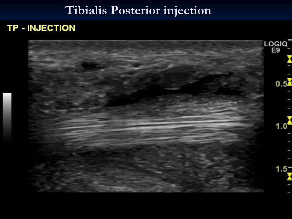
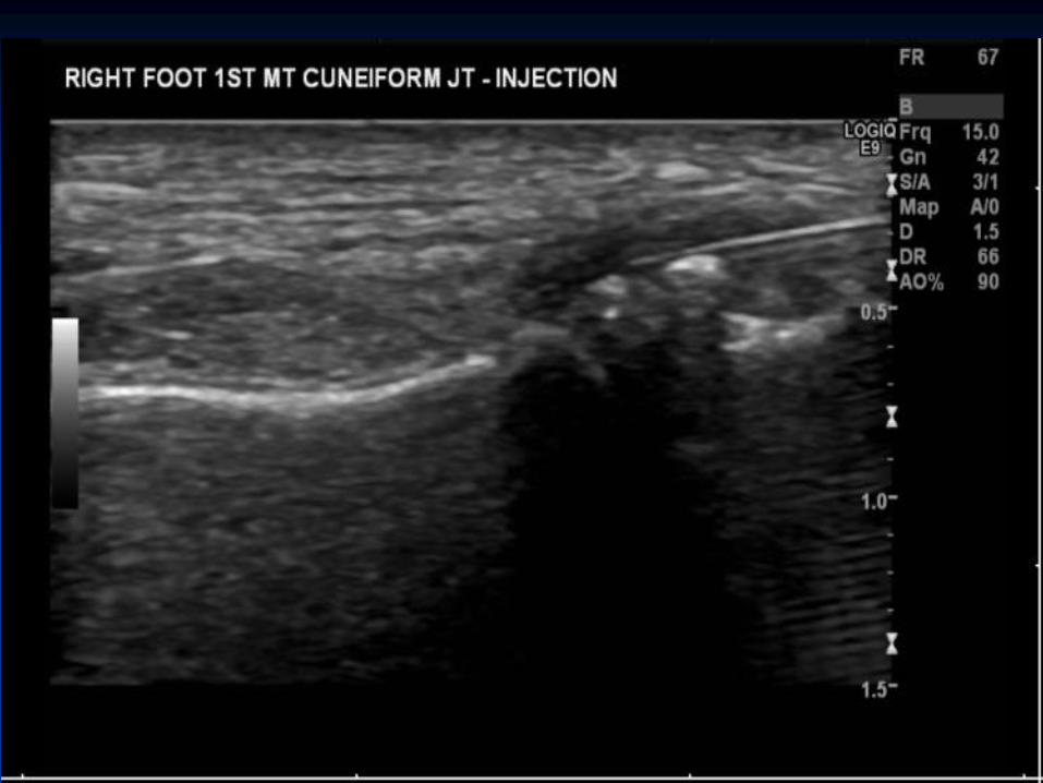
Tibialis Posterior injection
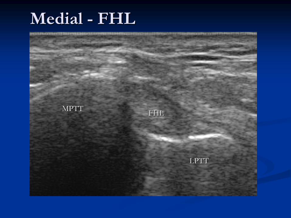
Medial - FHL
FHL MPTT
LPTT
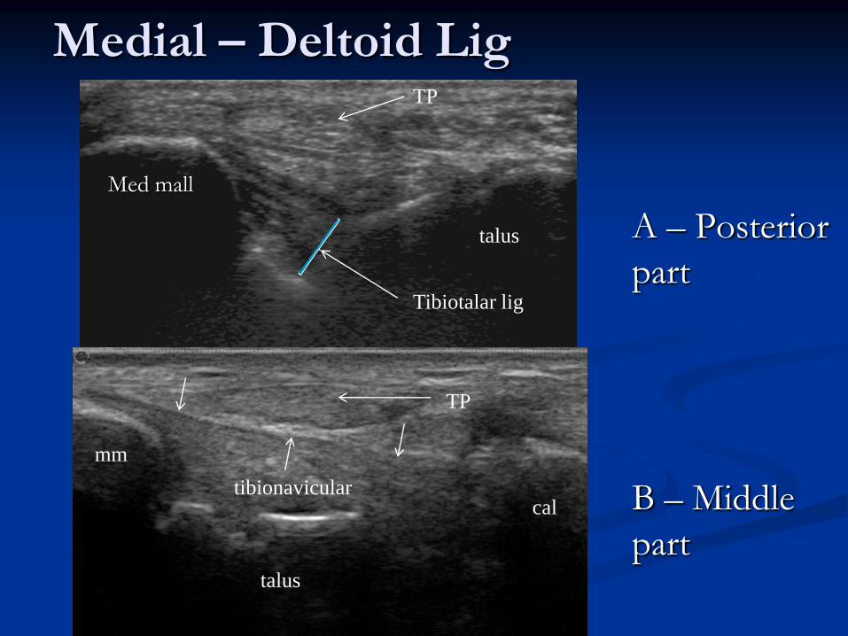
Medial – Deltoid Lig
A – Posterior
part
B – Middle
part
Med mall
talus
TP
Tibiotalar lig
mm
talus
cal
TP
tibionavicular
Practical –
Medial Ankle

Anatomy – Posterior & Superficial
Soleus
Med Gastroc Lat Gastroc
Aponeurosis of Gastroc
TA
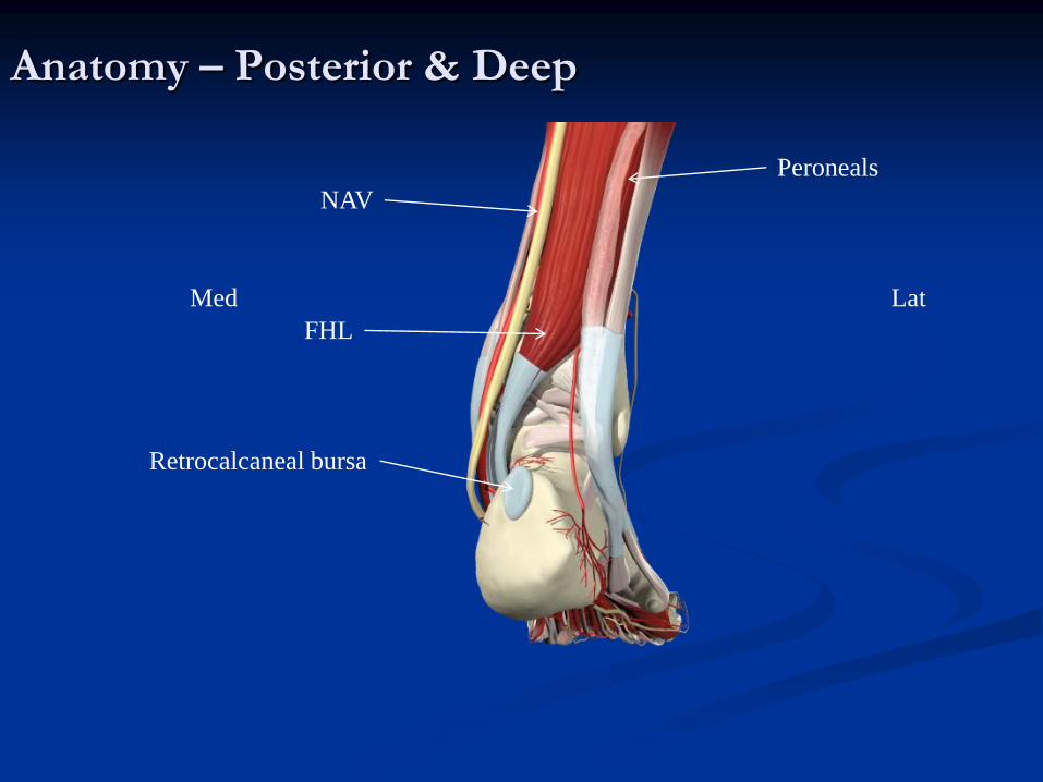
Anatomy – Posterior & Deep
Lat Med
Peroneals
FHL
NAV
Retrocalcaneal bursa

Achilles Tendon - Normal
Above Long
Right Transverse – ‘Bean shape –
wider medially’
cal Kager’s
Retrocal Bursa
Kager’s
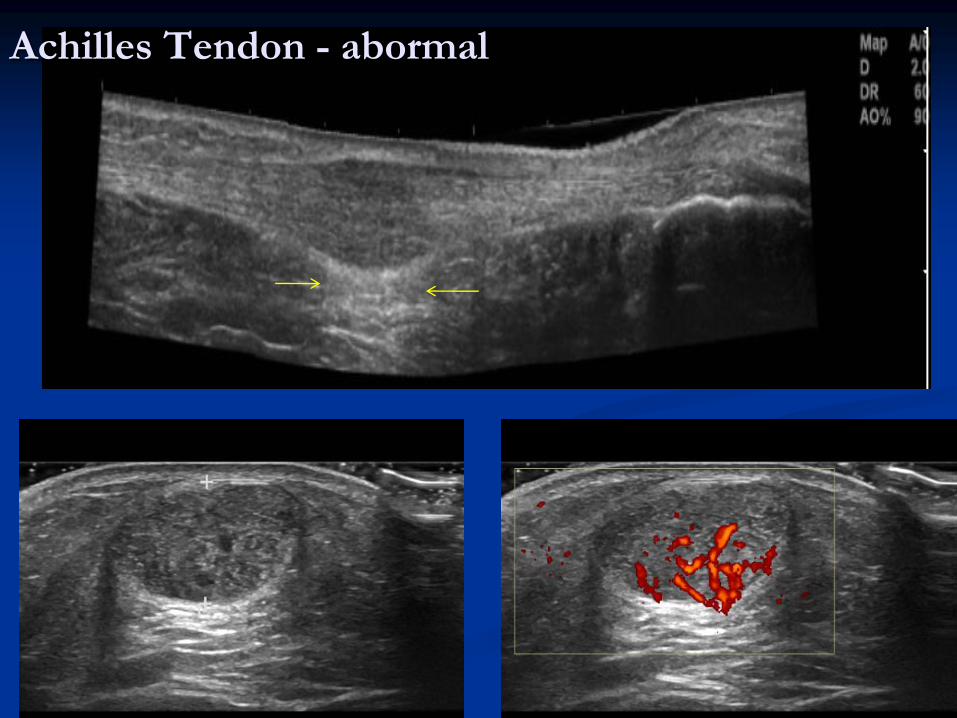
Achilles Tendon - abormal

The HVUSGI

Equipment
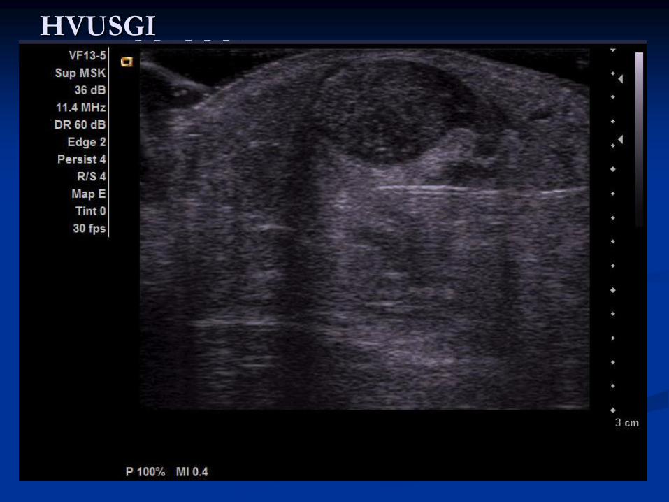
HVUSGI
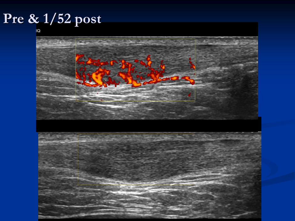
Pre & 1/52 post

Achilles tendon – Soleus insertion
Sol
FHL
TA
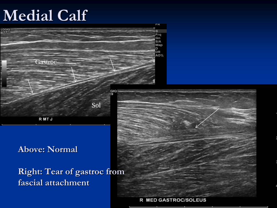
Medial Calf
Above: Normal
Right: Tear of gastroc from
fascial attachment
Sol
Gastroc
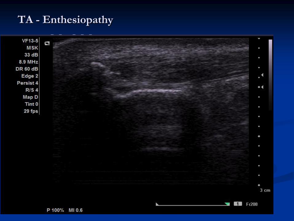
TA - Enthesiopathy
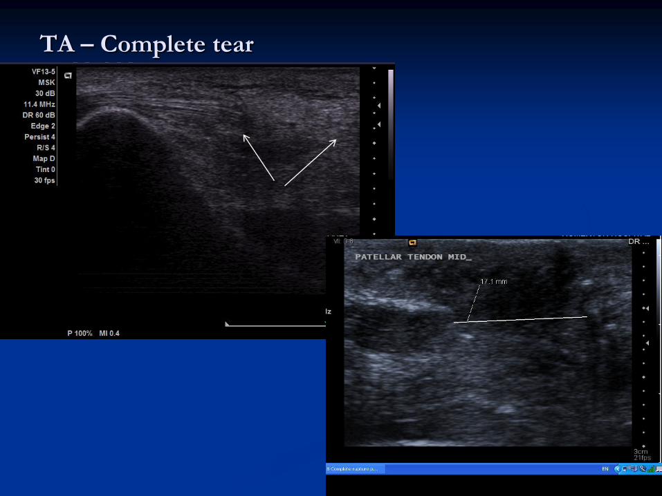
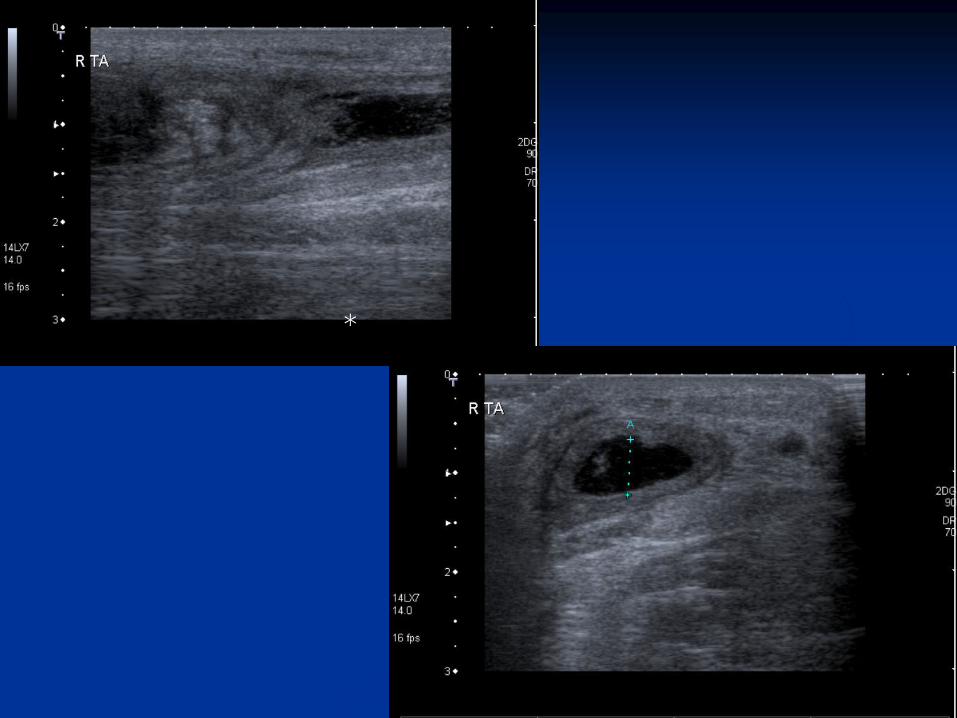
TA – Complete tear
*

Retrocalcaneal Bursitis
The Achilles tendon is intact but appears (slightly thickened). A
large retrocalcaneal bursa is demonstrated. Injected short axis
view.

Practical –
Posterior
Ankle
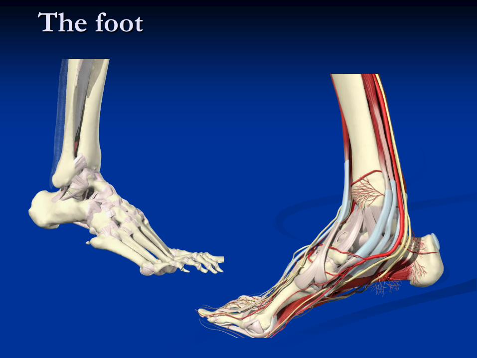
The foot
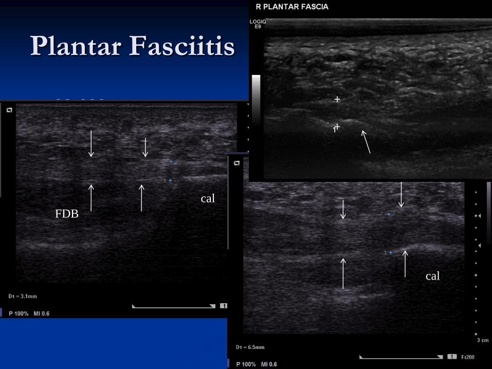
Plantar Fasciitis
cal
cal
FDB

Plantar Fasciitis
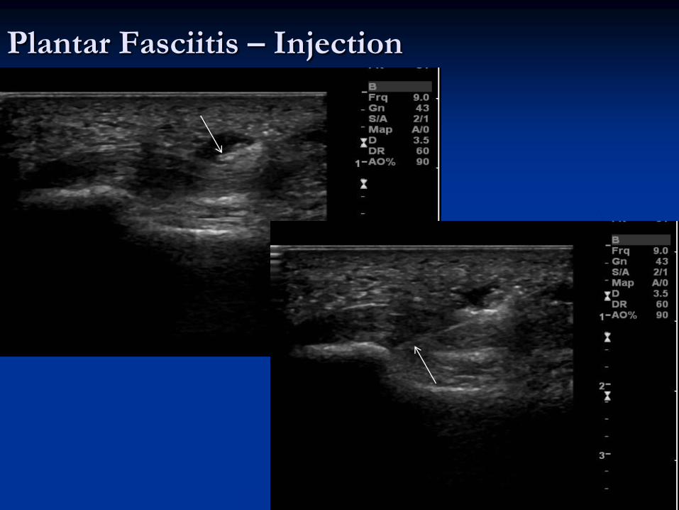
Plantar Fasciitis – Injection
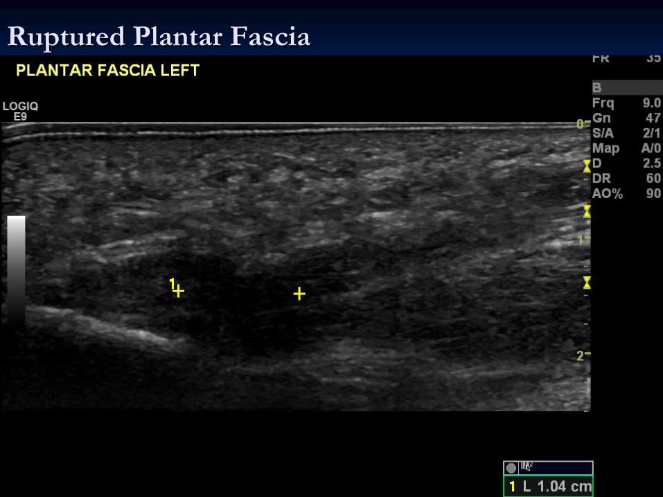
Ruptured Plantar Fascia
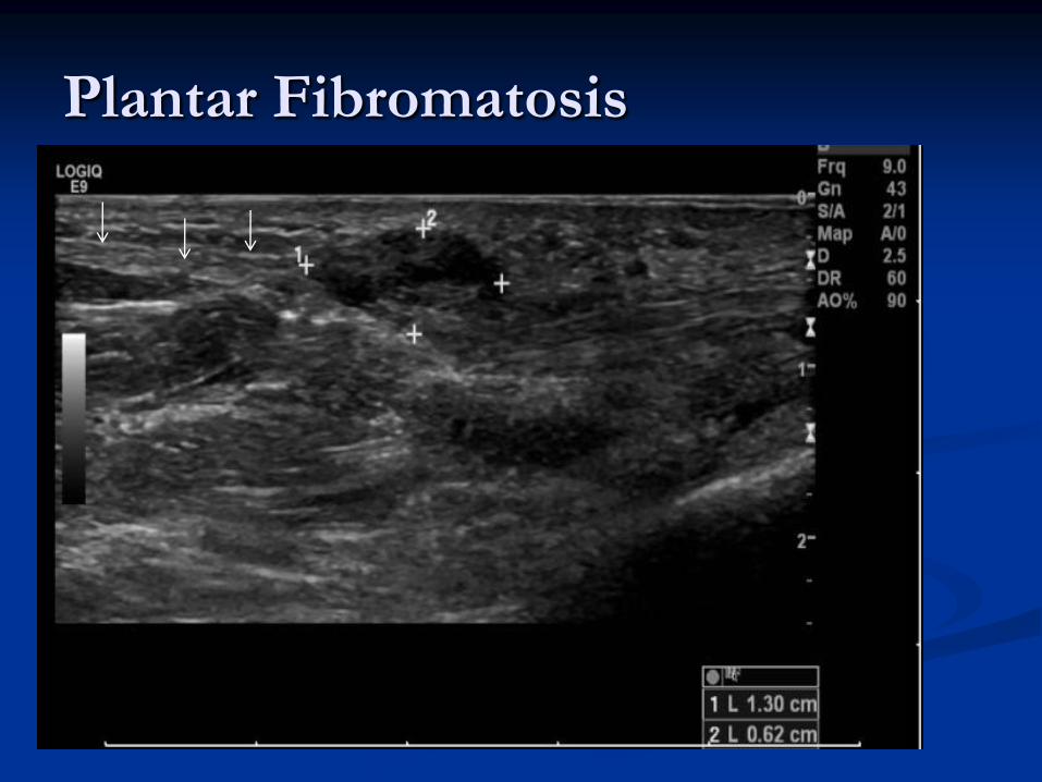
Plantar Fibromatosis
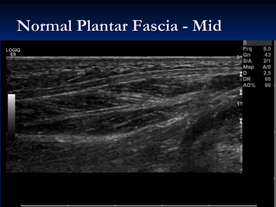
Normal Plantar Fascia - Mid

Mortons Neuroma
Transverse view: Hypoechoic focus between 3rd and 4th interspace.
A +ve Mulder’s click was present

Mortons Neuroma Longitudinal view of neuroma
between 3-4th MT heads

Aspiration / rupture ganglion foot

Practical –
Foot