diagnostic accuracy of pit pattern and vascular pattern in ...diagnostic accuracy of pit pattern and...
TRANSCRIPT
Diagnostic accuracy of pit pattern and vascular pattern in colorectal lesions
Digestive Disease Center, Showa University Northern Yokohama Hospital Department of Pathology ♯
Yoshiki Wada, Shin-ei Kudo, Hiroshi Kashida, Masashi Misawa, Takemasa Hayashi, Toshihisa Hosoya, Kunihiko Wakamura,
Nobunao Ikehara, Shigeharu Hamatani#

Background • Pit pattern analysis has been reported as useful for predicting the colorectal lesion’s histological nature. • Recently a new method of image-enhanced endoscopy called narrow band imaging (NBI) has been developed. The benefits of NBI over chromoendoscopy are that it is easier and time-saving.
Aim • The aim of this prospective study is to compare the usefulness of magnifying narrow band imaging (NBI) and magnifying chromoendoscopy in the diagnosis of colorectal lesions.

Subjects
• 3380 lesions (106 hyperplastic polyps, 3056 adenomas and 218 invasive cancers limited to submucosal layer) which were observed by magnifying scope with NBI and magnifying chromoendoscopy and treated endoscopically or surgically from January 2006 to June 2009.

• The scopes were CF-H260AZI (up to x 80). • The diagnosis of NBI and pit pattern was made by endoscopists who were blinded to the final pathological diagnosis. • The pit patterns were classified into I, II, III, IV, VI and VN according to Kudo’s classification. • VI pit pattern was divided into VI low-grade and VI high-grade. Our definitions for VI high-grade pit pattern were rough margin and narrowed lumen.
Methods

• We classified colonic microvascular pattern into six; normal, faint, network, dense, irregular and sparse. • Faint pattern was usually seen in hyperplastic polyps. Network and dense patterns were frequently recognized in benign adenomas. • Irregular pattern was characteristic for protruded or flat cancers, and sparse pattern was predictive for depressed cancers.

Kudo S, et al. Gastrointest Endosc 1996; 44: 8-14
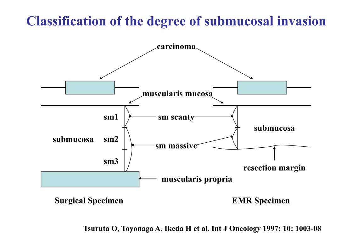
Surgical Specimen EMR Specimen
carcinoma
muscularis mucosa
sm scanty
sm massive submucosa
submucosa
resection margin muscularis propria
sm1
sm2
sm3
Tsuruta O, Toyonaga A, Ikeda H et al. Int J Oncology 1997; 10: 1003-08
Classification of the degree of submucosal invasion

Vascular Pattern Classification
Normal pattern Faint pattern Network pattern
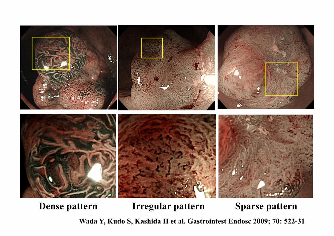
Dense pattern Irregular pattern Sparse pattern Wada Y, Kudo S, Kashida H et al. Gastrointest Endosc 2009; 70: 522-31

Pit Pattern Classification (Kudo’s Classification)
I II IIIs
IIIL IV VI VN
Kudo S et al. Endoscopy 2001; 33: 367-373
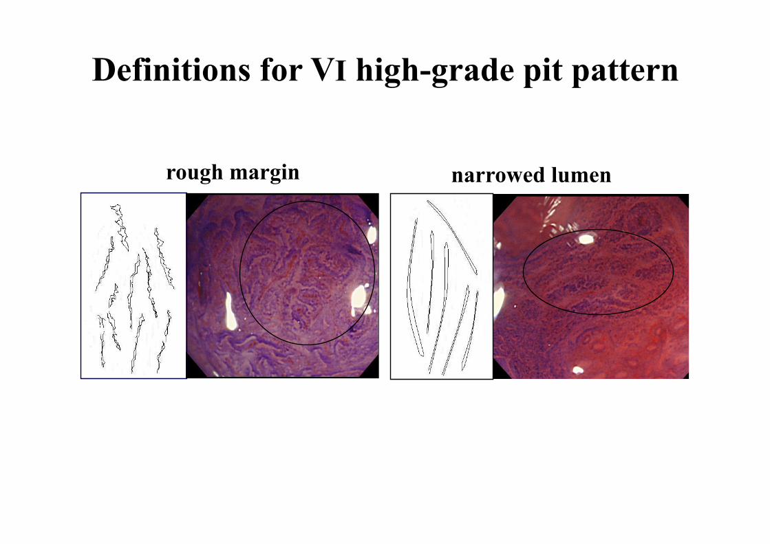
rough margin narrowed lumen
Definitions for VI high-grade pit pattern

network
dense
faint
Comparison between vascular pattern and pathological diagnosis
hyp* adenoma
vascular pattern
total pathological diagnosis
106 3056 total 3380
89
2426
531
47**
17
irregular
sparse
SMm
40
12
65
87
7
5
164
111
108
544
2481
136
* hyperplastic polyp
SMs
54
33
6
6
9
** 31 lesions were sessile serrated adenomas.
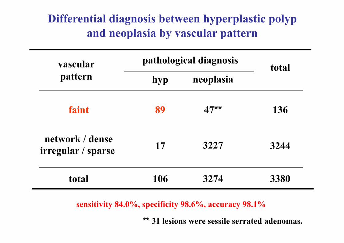
pathological diagnosis
hyp neoplasia
faint
network / dense irregular / sparse
47**
106 3274
17 3227
89
3380
136
total
total
sensitivity 84.0%, specificity 98.6%, accuracy 98.1%
3244
vascular pattern
Differential diagnosis between hyperplastic polyp and neoplasia by vascular pattern
** 31 lesions were sessile serrated adenomas.
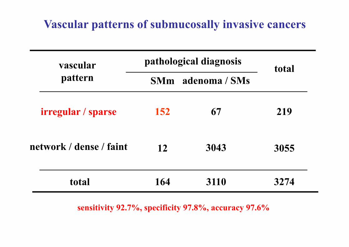
pathological diagnosis
SMm adenoma / SMs
network / dense / faint
irregular / sparse 67
164 3110
12 3043
152
3274
219
total
total
Vascular patterns of submucosally invasive cancers
sensitivity 92.7%, specificity 97.8%, accuracy 97.6%
3055
vascular pattern

1987 IIIL
IIIs II
Comparison between pit pattern and pathological diagnosis
hyp adenoma pit pattern total
pathological diagnosis
total
96
1978
35
20
9 IV
VI low-grade
SMm
788
222
6
16
807
273
35 116
VI high-grade 13 91 111 VN 51 51
SMs
12
35 7
1
106 3056 3380 164 54
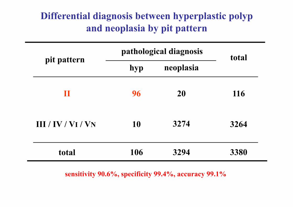
pathological diagnosis
hyp neoplasia
II
III / IV / VI / VN
20
106 3294
10 3274
96
3380
116
total
total
sensitivity 90.6%, specificity 99.4%, accuracy 99.1%
3264
pit pattern
Differential diagnosis between hyperplastic polyp and neoplasia by pit pattern

pathological diagnosis
SMm
VI low-grade / IV / III /II
VN / VI high-grade 20
164 3110
22 3090
142
3274
162
total
total
Pit patterns of submucosally invasive cancers
sensitivity 86.6%, specificity 99.4%, accuracy 98.7%
3112
pit pattern adenoma / SMs
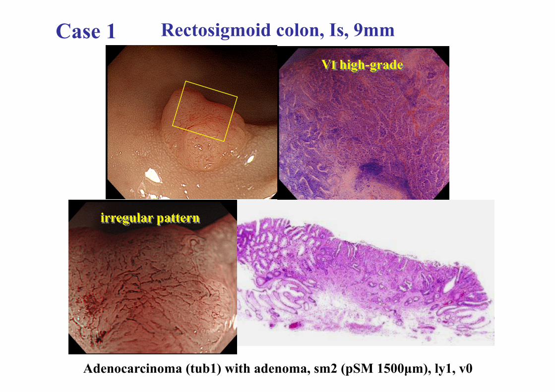
Rectosigmoid colon, Is, 9mm Case 1
irregular pattern
VI high-grade
Adenocarcinoma (tub1) with adenoma, sm2 (pSM 1500µm), ly1, v0
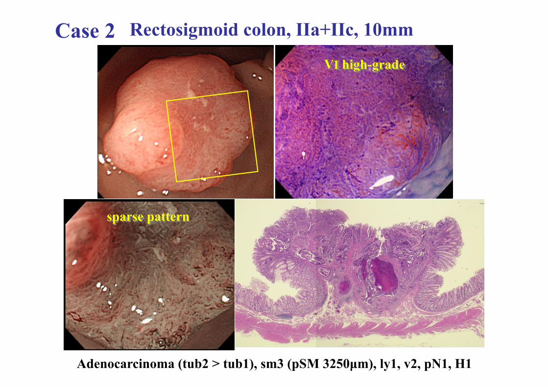
Rectosigmoid colon, IIa+IIc, 10mm Case 2
sparse pattern
VI high-grade
Adenocarcinoma (tub2 > tub1), sm3 (pSM 3250µm), ly1, v2, pN1, H1
Summary
• Both NBI and chromoendoscopy can be useful tools for distinguishing between neoplastic and non-neoplastic lesions. • In the diagnosis of submucosal cancer, pit pattern diagnosis was a little superior to vascular pattern diagnosis.
Conclusion
• It is desirable to perform not only NBI but also chromoendoscopy for distinguishing between SMm and SMs and determining treatment selection, endoscopic or surgical.
Diagnostic accuracy of pit pattern and vascular pattern in colorectal lesions
Digestive Disease Center, Showa University Northern Yokohama Hospital Department of Pathology #
Yoshiki Wada, Shin-ei Kudo, Hiroshi Kashida, Masashi Misawa, Takemasa Hayashi Toshihisa Hosoya, Kunihiko Wakamura, Nobunao Ikehara, Shigeharu Hamatani#