diagnosis, monitoring, and treatment of primary ciliary...
TRANSCRIPT
Pediatric Pulmonology 51:115–132 (2016)
State of the Art
Diagnosis, Monitoring, and Treatment of PrimaryCiliary Dyskinesia: PCD Foundation Consensus
Recommendations Based on State of the Art Review
Adam J. Shapiro, MD,1* Maimoona A. Zariwala, PhD,2 Thomas Ferkol, MD,3 Stephanie D. Davis, MD,4
Scott D. Sagel, MD, PhD,5 Sharon D. Dell, MD,6 Margaret Rosenfeld, MD,7 Kenneth N. Olivier, MD,8§
Carlos Milla, MD,9 Sam J. Daniel, MD,10 Adam J. Kimple, MD,11 Michele Manion,12
Michael R. Knowles, MD,13 and Margaret W. Leigh, MD,14
for the Genetic Disorders of Mucociliary Clearance Consortium
Summary. Primary ciliary dyskinesia (PCD) is a genetically heterogeneous, rare lung disease
resulting in chronic oto-sino-pulmonary disease in both children and adults. Many physicians
incorrectly diagnose PCD or eliminate PCD from their differential diagnosis due to inexperience
with diagnostic testing methods. Thus far, all therapies used for PCD are unproven through large
clinical trials. This review article outlines consensus recommendations from PCD physicians in
North America who have been engaged in a PCD centered research consortium for the last 10
years. These recommendations have been adopted by the governing board of the PCD
Foundation to provide guidance for PCD clinical centers for diagnostic testing, monitoring, and
appropriate short and long-term therapeutics in PCD patients. Pediatr Pulmonol. 2016;51:115–
132. � 2015 The Authors. Pediatric Pulmonology Published by Wiley Periodicals, Inc.
This is an open access article under the terms of the Creative Commons
Attribution-NonCommercial-NoDerivs License, which permits use and
distribution in anymedium, provided the original work is properly cited, the
use is non-commercial and no modifications or adaptations are made.
1Department of Pediatrics, Montreal Children’s Hospital, McGill Universi-
ty, Quebec, Canada.
2Department of Pathology and Laboratory Medicine, University of North
Carolina School of Medicine, Marsico Lung Institute, Chapel Hill, North
Carolina.
3Department of Pediatrics, Washington University School of Medicine, St.
Louis, Missouri.
4Department of Pediatrics, Riley Hospital for Children, Indiana University,
Indianapolis, Indiana.
5Department of Pediatrics, Children’s Hospital Colorado and University of
Colorado School of Medicine, Aurora, Colorado.
6Department of Pediatrics, The Hospital for Sick Children and University of
Toronto, Toronto, Ontario, Canada.
7Department of Pediatrics, Seattle Children’s Hospital and University of
Washington, Seattle, Washington.
8National Heart, Lung, and Blood Institute, Bethesda, Maryland.
9Department of Pediatrics, Stanford University, Palo Alto, California.
10Department of Otolaryngology, Montreal Children’s Hospital, McGill
University, Montreal, Quebec, Canada.
11Department of Otolaryngology—Head and Neck Surgery, University of
North Carolina School of Medicine, Chapel Hill, North Carolina.
12The PCD Foundation—President, Minneapolis, Minnesota.
13Department of Medicine, University of North Carolina, Marsico Lung
Institute, Chapel Hill, North Carolina.
14Department of Pediatrics, University of North Carolina, Marsico Lung
Institute, Chapel Hill, North Carolina.
§Supported in part by the intramural research program of the NHLBI, NIH.
Michael R. Knowles and Margaret W. Leigh are co-senior who contributed
equally to this statement as senior authors.
�Correspondence to: A.J. Shapiro, MD, Department of Pediatrics, Montreal
Children’s Hospital, McGill University, 2300 Tupper, D-380, Montreal,
Quebec, Canada, H3H 1P3. E-mail: [email protected]
Received 1 May 2015; Revised 30 June 2015; Accepted 21 August 2015.
DOI 10.1002/ppul.23304
Published online 29 September 2015 in Wiley Online Library
(wileyonlinelibrary.com).
� 2015 The Authors. Pediatric Pulmonology Published by Wiley Periodicals, Inc.

Key words: primary ciliary dyskinesia; PCD, kartagener; consensus statement; PCD
Foundation.
Funding source: National Institutes of Health (NIH), Number: U54HL096458,
5R01HL071798; The Genetic Disorders of Mucociliary Clearance (U54HL096458) is a
part of the NCATS Rare Diseases Clinical Research Network (RDCRN). RDCRN is an
initiative of the Office of Rare Diseases Research (ORDR), NCATS, funded through a
collaboration between NCATS and NHLBI; CTSA NIH/NCATS UNC ULTR000083; CTSA
NIH/NCATS Colorado UL1TR000154; Intramural Research Program of NIH/NIAID.
INTRODUCTION
Primary ciliary dyskinesia (PCD) is a geneticallyheterogeneous, rare lung disease causing chronic oto-sino-pulmonary disease and irreversible lung damage thatmay progress to respiratory failure.1–3 Recently, signifi-cant progress has been made in PCD diagnosis,4 yet fewphysicians outside of highly experienced PCD centers areskilled in recognizing the characteristic clinical pheno-type and interpreting diagnostic tests.5–9 Patients oftenreceive false-positive or false-negative PCD diagnoses, asphysicians are unaware of the pitfalls commonlyencountered with ciliary electron microscopy,10,11 PCDmolecular genetic panels,12,13 ciliary motility stud-ies,14–16 and nasal nitric oxide testing.17,18 Furthermore,PCD is often missed when respiratory symptoms arepresent in patients with other complex diseases involvingcilia, such as heterotaxy and various genetic syn-dromes.19–22 From a therapeutic perspective, there areno prospective, randomized clinical trials on monitoringor treating PCD. Thus, physicians treating PCD adapt
therapeutic approaches used for other chronic respiratorydiseases, such as cystic fibrosis (CF) and non-CFbronchiectasis. Differences in various phenotypic param-eters among PCD, CF, and non-CF bronchiectasis suggestthat extrapolating therapies may not be appropriate forPCD management in some circumstances.23–26
Because of the uncertainty surrounding diagnosis andmanagement of PCD, physicians from the GeneticDisorders of Mucociliary Clearance Consortium(GDMCC) created this consensus statement to guidenew North American PCD clinical centers endorsed bythe PCD Foundation. The GDMCC includes clinicians atnine academic centers in North America that havesystematically evaluated over 1,000 patients suspectedof having PCD and performed longitudinal studies ofpediatric patients with a confirmed diagnosis of PCD. TheGDMCC also works closely with the PCD Foundation onresearch and clinical PCD projects. This consensusstatement is evidence basedwhere possible, and addresseskey clinical PCD issues, but it is not the product ofGRADE recommendations.27 Through telephone confer-ences, email communications, and in person meetings,eight pediatric pulmonologists, two adult pulmonologists,and two otolaryngologists fromNorth America undertookto: (1) describe the PCD clinical phenotype, (2) establishstandard PCD diagnostic recommendations, (3) recom-mend PCD clinical care and long-term monitoringschedules, and (4) outline clinical therapies used tomanage PCD.After a literature review (using Pubmed andEmbase), drafts were created and circulated iteratively toparticipating physicians with discussion of feedback andsuggestions over sequential telephone conferences andelectronic communications. ParticipatingGDMCCphysi-cians and the PCD Foundation governing board unani-mously approved this consensus statement.
PCD CLINICAL PHENOTYPE
Clinical symptoms in PCD affect the entire respiratorytract; the majority of symptoms occur on a chronic, dailybasis and start soon after birth (Table 1). At least 80% ofnewborn babies with PCD develop neonatal respiratorydistress despite a full-term gestation, with increased workof breathing, tachypnea, and prevalence of upper andmiddle lobe atelectasis on chest radiographs.28 Most PCDpatients are well immediately after birth, but develop
ABBREVIATIONS:
ABPA allergic bronchopulmonary aspergillosis
CF cystic fibrosis
CFTR cystic fibrosis transmembrane regulator
CRS chronic rhinosinusitis
CT computed tomography
EM electron microscopy
ESS endoscopic sinus surgery
GDMCC Genetic Disorders of Mucociliary Clearance Consortium
IDA inner dynein arm
IF immunofluorescence testing
IVIG intravenous immunoglobulin
MTD microtubule disorganization
N-DRC nexin-dynein regulatory complex
NGS next generation sequencing
nNO nasal nitric oxide
NTM non-tuberculosis mycobacterium
ODA outer dynein arm
OME otitis media with effusion
PBB protracted bacterial bronchitis
PCD primary ciliary dyskinesia
PET pressure equalization tubes
ROM recurrent otitis media
SIT situs inversus totalis
SA situs ambiguus
TTN transient tachypnea of the newborn
116 Shapiro et al.
Pediatric Pulmonology

respiratory distress at 12–24 hr of life (as opposed to othercauses of respiratory distress in term neonates (e.g.,transient tachypnea of the newborn—TTN), which oftenpresent in the first few hours after birth). A smallproportion of PCD patients are discharged home on day 1of life but are then hospitalized with respiratory distresswithin the first few weeks of life. Often misdiagnosedwith TTN or pneumonia, PCD infants frequently requiresupplemental oxygen for days to weeks. When neonatalrespiratory distress appears, particularly with situsinversus totalis or other situs anomalies, PCD should beinvestigated.At least 80% of PCD patients also have year-round,
daily nasal congestion (or chronic sinusitis in olderchildren and adults), which appears in early infancy anddoes not resolve with changes of season or between viralinfections. Nasal polyps can occur in PCD,29 and nearlyall PCD patients demonstrate severe pansinusitis oncomputed tomography (CT) scan.4
Persistent, year-round, daily cough from early infancyis present in nearly 100% of PCD patients.4 The cough isusually wet and productive, even in infancy, yetoccasionally patients report dry cough. The cough canpartially improve with antibiotic therapy, but does notresolve with therapy or changes of season. Conversely,episodic cough alternating with symptom-free periods isunlikely to be from PCD.A spectrum of organ laterality defects occur with PCD,
including situs inversus totalis (SIT—mirror-imagearrangement) and situs ambiguus (SA—arrangementfalls somewhere between normal and mirror image;Fig. 1). SA may be associated with complex congenitalheart disease (known as heterotaxy), yet mild cardiacseptal defects can also occur with PCD. SIT occurs inslightly less than 50% of PCD patients,30 whereas SAoccurs in at least 12% of PCD.19 Subtle laterality defects(e.g., intestinal malrotation, interrupted inferior vena
cava, or polysplenia) may be undetected in PCD patientswithout further imaging studies, such as abdominalultrasound, spleen scan, or echocardiogram. In patientswith chronic oto-sino-pulmonary disease and any organlaterality or cardiac defect, PCD should be considered.Recurrent otitis media (ROM) with chronic middle ear
effusion affects at least 80% of children with PCD,particularly in the first year of life.31 Complications ofROM may include multiple sets of pressure equalizationtubes, conductive hearing loss, speech/language delay, orneed for hearing aids.32 Chronic middle ear disease isquite common in the general pediatric population;thereby, ROM alone is insufficient to warrant furtherPCD testing. The absence of ROM goes against, but doesnot rule out, a diagnosis of PCD.Recurrent pneumonia or bronchitis is common in
PCD; however, some infants will lack this history dueto frequent antibiotics for nasal discharge and otitismedia. By preschool age, up to 80% of PCD patientshave recurrent lower respiratory tract infections.4
Bronchiectasis, predominantly affecting the middleand lower lobes,33 is an age-related finding in PCD,with 50% of children having bronchiectasis by 8 yearsof age and nearly universal presence in adults.34,35 Inadults with PCD, the combination of bronchiectasis andchronic sinusitis may be the most readily identifiablePCD-related features because adults with PCD may notbe able to recall age of onset of early childhoodsymptoms.Finally, infertility occurs in nearly 100% of adult males
with PCD, while females with PCD also have reducedfertility. The structure in both sperm tails and the fimbriaeof fallopian tubes are almost identical to those inrespiratory cilia. Thus, males with PCD have diminishedfertility through reduced sperm motility,36 while femaleswith PCD have increased risk of ectopic pregnancy fromabnormal fallopian transit of oocytes.37
TABLE 1—Age-Related Prevalence of Clinical Features in Primary Ciliary Dyskinesia1
PCD clinical feature
Youngest age when
feature present in >50% of PCD
Youngest age when
feature present in >80% of PCD
Neonatal respiratory distress 12 hr of life2 24 hr of life2
Organ laterality defects (SIT or SA) Neonatal to school age —
Recurrent otitis media with effusion Infancy Infancy
Year-round, daily cough Infancy Infancy
Year-round, daily nasal congestion Infancy Infancy
Chronic pansinusitis Preschool3 School age
Recurrent lower respiratory infections Infancy Preschool
Bronchiectasis School age Adult
Male infertility — Adult
SIT, situs inversus totalis; SA, situs ambiguus.1Adapted from Knowles et al.4
2Reference.28
3Pansinusitis is seen in almost all patients with PCDwho have sinus imaging studies, but these studies are not done often in pre-school age children.
Diagnosis and Management of PCD 117
Pediatric Pulmonology

All of the above features may not be seen in eachindividual patient with PCD; however, most patients have3 or more of the above features. The combination ofmultiple distinct clinical features of PCD (neonatalrespiratory distress, chronic wet cough with recurrentlower respiratory infections and bronchiectasis, chronicnasal drainage with pansinusitis, recurrent otitis mediaparticularly in childhood, laterality defect, and maleinfertility) markedly increases the likelihood of a PCDdiagnosis.
APPROACH TO DIAGNOSING PCD
Diagnostic Tests
The first step in diagnosing PCD is evaluation forclinical features of PCD as outlined in the prior section.The diagnosis of PCD requires clinical phenotypicfeatures in conjunction with diagnostic testing. A numberof tests can be used to support the diagnosis of PCD, andoften a panel of tests are required to confirm a PCDdiagnosis (Table 2). As PCD can result from variousdefects in ciliary biogenesis, structure, function, ororganization, no single test captures all PCD defects.For instance, patients with biallelic DNAH11 mutationshave a classic clinical phenotype and low nasal nitricoxide levels, but normal electron microscopy (EM)ultrastructure with only subtle changes on ciliarywaveform analysis.10 Patients with biallelic mutationsin RSPH1 demonstrate later onset of clinical symptoms,subtle EM defects, and slight changes in ciliarywaveform, with borderline (and in some cases normal)nasal nitric oxide levels.38 Consequently, a panel of thefollowing PCD diagnostic tests are recommended, and
with a greater number of positive tests, there is a higherlikelihood of definite PCD.Diagnostic PCD algorithms will differ per patient
location (where some tests will not be readily accessible)and upon local expertise of the institution performing thePCD investigation. Furthermore, age of the patient maydictate which PCD testing should be initially pursued. Inneonates and children <5 years old, nasal nitric oxidevalues are not as reliable; thus diagnostic testing in thisage group usually includes ciliary biopsy for electronmicroscopy and/or genetic studies in North America,versus ciliary biopsy for high speed videomicroscopyanalysis in Europe. In children over 5 years old and adults,who can cooperate with the required maneuvers for nasalnitric oxide measurement, a low nasal nitric oxide valuecoupled with an appropriate clinical phenotype may beadequate for a clinical diagnosis of PCD, followed byciliary biopsy for electron microscopy or high speedvideomicroscopy and/or genetic studies, as needed.Minimal PCD diagnostic criteria have been proposedby the GDMCC (Table 3). For all patients given adiagnosis of PCD by clinicians outside of PCD Founda-tion Clinical Centers, at least one visit to a PCDFoundation Clinical Center is recommended to officiallyconfirm the diagnosis. For patients followed in centerswithout PCD expertise, a PCD Foundation ClinicalCenter referral for diagnostic investigation is highlyrecommended.
Respiratory Epithelial Biopsy With Electron Microscopy
Respiratory epithelial biopsy with EM processing forultrastructural examination of ciliary axonemes is a proventechnique for PCDdiagnosis39 and is recommended as part
Fig. 1. Examples of various laterality defects on radiology imaging in PCD. Different situs
arrangements found in PCD, including (A) a participant with situs solitus, or normal organ
arrangement, with left cardiac apex, left-sided stomachbubble, and right-sided liver; (B) a patient
with situs inversus totalis (SIT), or mirror-image organ arrangement, with right cardiac apex,
right-sided stomach bubble, and left-sided liver; (C) a patient with situs ambiguus (SA), with left
cardiac apex, right-sided stomach bubble, right-sided liver, and intestinal malrotation; This
patient also had right-sided polysplenia visualized on a CT scan. C, cardiac apex; S, stomach; L,
liver; M, intestinal malrotation. Reproduced with permission from CHEST.19
118 Shapiro et al.
Pediatric Pulmonology

of a panel of diagnostic tests for PCD.Disease causing EMdefects in the outer dynein arms,40 outer and inner dyneinarms,41 inner dynein arms with microtubule disorganiza-tion,42 radial spokes,43 or central apparatus44,45 provideconfirmation of PCD diagnosis (Fig. 2). However, EMstudies with normal ciliary ultrastructure do not rule outPCD, as certain PCD gene mutations can result in normalultrastructure,10,38,46 or subtle abnormalities (particularlythose involving the central apparatus and radial spokes)
that are not readily recognized on EM.47,48 Additionally,repeat biopsies that fail to demonstrate any respiratory ciliacould represent an oligociliary defect causing PCD.49,50
It is estimated that EM will detect approximately 70%of all PCD cases,4 but in centers inexperienced with EMprocessing and interpretation, this percentage will benotably less. Centers lacking extensive experience withciliary EM processing and interpretation should stronglyconsider referring patients to a PCD Foundation clinical
TABLE 2—Recommended Diagnostic Testing Methods for Primary Ciliary Dyskinesia
Test recommended for PCD diagnosis
Potential for false
positive results
Potential for false
negative results
Nasal nitric oxide measurement Low1 Low2
Ciliary biopsy with electron microscopy Variable3 Variable4
PCD genetic testing panels Low5 Moderate6
Functional ciliary beat/waveform analysis with high speed videomicroscopy Variable7 Moderate8
Immunofluorescence testing Unknown Unknown
1As long as cystic fibrosis has been excluded. Risk of false positive result is increased during viral respiratory infection, epistaxis, and non-atopic
sinusitis. Testing should be performed at baseline health status and repeated if there is any question about health status.2Reference.18
3The risk of false positive result is moderately increased with secondary changes from infectious processes or pollutant exposures, improper
specimen handling and processing, or inexperience with electron microscopy interpretation.4
4Several PCD-causing genetic mutations can result in normal electron microscopy10 or subtle changes which are not readily apparent.38
5Misinterpretation of genetic panel result (e.g., variants of unknown significance or single mutations in two different PCD genes interpreted as
“diagnostic”).6Genetic panel testing may miss large insertions, deletions, and mutations in novel genes, since approximately 30% of PCD do not have identifiable
mutations in the currently known PCD associated genes, but this risk should decreasewith broader range of genetic analysis provided byNGSpanels.7With a high risk for false positive results from secondary insults on a single test. To limit this risk, many centers now perform three ciliary biopsies
at separate clinical visits for repeat high speed videomicroscopy analysis.8Subtle waveform defects will be missed in centers without extensive experience.
TABLE 3—Recommended PCD Diagnostic Criteria by Age
Newborns (0–1 month of age)
Situs inversus totalis and unexplained neonatal respiratory distress at term birth plus at least one of the following:
Diagnostic ciliary ultrastructure on electron micrographs
Biallelic mutations in one PCD-associated gene
Persistent and diagnostic ciliary waveform abnormalities on high-speed videomicroscopy, on multiple occasions
Children (1 month to 5 years)
Two or more major PCD clinical criteria (see below) plus at least one of the following (nasal nitric oxide not included in this age group, since
it is not yet sufficiently tested):
Diagnostic ciliary ultrastructure on electron micrographs
Biallelic mutations in one PCD-associated gene
Persistent and diagnostic ciliary waveform abnormalities on high-speed videomicroscopy, on multiple occasions
Children 5–18 years of age and adults
Two or more major PCD clinical criteria (see below) plus at least one of the following:
Nasal nitric oxide during plateau <77 nl/min on 2 occasions, >2 months apart, with cystic fibrosis excluded
Diagnostic ciliary ultrastructure on electron micrographs
Biallelic mutations in one PCD-associated gene
Persistent and diagnostic ciliary waveform abnormalities on high-speed videomicroscopy, on multiple occasions
Major clinical criteria for PCD diagnosis�
1) Unexplained neonatal respiratory distress (at term birth) with lobar collapse and/or need for respiratory support with CPAP and/or oxygen for
>24 hr.
2) Any organ laterality defect—situs inversus totalis, situs ambiguous, or heterotaxy.
3) Daily, year-round wet cough starting in first year of life or bronchiectasis on chest CT.
4) Daily, year-round nasal congestion starting in first year of life or pansinusitis on sinus CT.�Other diagnostic possibilities should have been considered, such as cystic fibrosis and immunodeficiencies, and diagnostic tests performed to rule
out those disorders, as clinically indicated.
Diagnosis and Management of PCD 119
Pediatric Pulmonology
center for PCD investigations. At least 20–50 clear ciliarycross-sections are required for a diagnostic EM study, anddiagnostic abnormalities should be consistently demon-strated on cross sectional images from multiple differentcilia to be considered disease causing. Physicians may trynasal corticosteroids, nasal saline lavages, or systemicantibiotics for persistent nasal symptoms interfering withbiopsies, but these practices are unproven and may notimprove biopsy yield. Furthermore, it is essential thatbiopsies are collected when patients are at their baselinehealth, as secondary changes in ciliary ultrastructure canoccur during respiratory exacerbations.15 Thus, biopsiesshould be delayed until at least 2 weeks after full recoveryfrom an illness. For absence of inner dynein arms inisolation, repeat biopsy and EM studies are alwaysrequired to verify that this pathologic change persists andtherefore is more likely genetic (primary) and not fromsecondary causes.11 One may also consider repeatbiopsies to verify the universality and permanence offindings suggestive of central apparatus, radial spoke, orinner dynein arm with microtubule disorganizationdefects. Patients with EM studies consistent with PCDshould be referred to a PCD Foundation Clinical Centerfor confirmation.
Nasal Nitric Oxide Measurement
Measurement of nasal nitric oxide (nNO) by chemilu-minescence analyzer is recommended as part of a panel ofdiagnostic tests for PCD in adults and children �5 yearsold.18 This test is sensitive, rapid, non-invasive, andresults are immediately available. Nasal NO values aremore reliable in school aged children and adults becausethese patients can cooperate with blowing into a resistor.Tidal breathing techniques for nNO measurement inchildren <5 years old are currently being investigated,17
but PCD diagnostic cutoff values for tidal techniques arenot currently available. Unfortunately, chemilumines-cence devices are limited to research settings in North
America, but they are gaining acceptance as a clinical toolin various countries across Europe, through efforts by theBESTCILIA PCD consortium.51 Handheld electrochem-ical nNO analyzers are affordable and portable, but withonly limited prospective study in PCD,52,53 these devicesare not currently recommended for PCD testing.Nasal nitric oxide values are extremely low in
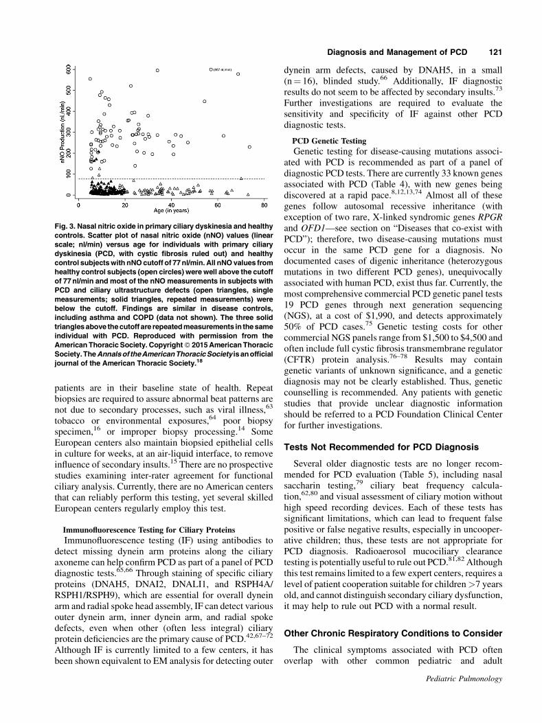
PCD.54,55 Using a nNO cutoff value <77 nl/min, onewill detect PCD, resulting from ciliary axonemal defectsor mutations in DNAH11, with sensitivity and specificityof 98% and >99%, respectively, if CF has been ruled out(Figure 3).18 Values well above this cutoff levelsignificantly decrease the likelihood of PCD. However,clinicians still must consider PCD when confronted withan appropriate clinical phenotype for PCD and nNOvalues above 77 nl/min, as forms of PCDwith nNO valuesabove this cutoff have rarely been reported.56,57 Very lownNO levels (below 77 nl/min) can occur during acute viralrespiratory infections and in approximately 30% ofpatients with cystic fibrosis; therefore, nNO testingmust be performed when the patient has fully recoveredfrom a viral illness and after diagnostic testing to rule outcystic fibrosis.58 Other conditions can also result in nNOlevels below PCD cutoff values (i.e., HIV,59 panbron-chiolitis,60 non-atopic sinusitis61). Lastly, nNO deviceoperators must be well trained and use standard operatingprotocols to avoid false results.18
Functional Ciliary Beat/Waveform Analysis With High
Speed Videomicroscopy
Ciliary biopsy with examination of cilia waveform byhigh speed videomicroscopy can provide confirmation ofPCD, and this test is recommended as part of a panel ofPCD diagnostic tests, but only in centers highlyexperienced with this technology.62 Functional ciliaryanalysis is difficult to perform correctly, and considerableexperience is necessary to avoid false-positive and false-negative results. Biopsies should only be performed when
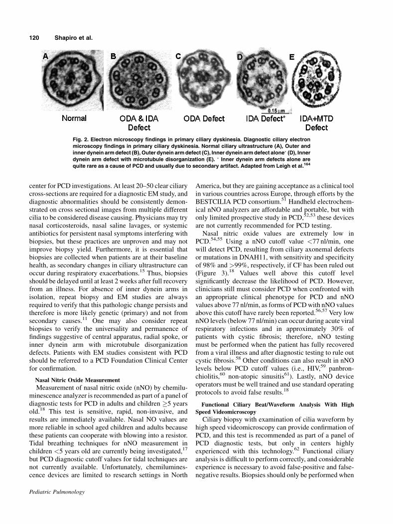
Fig. 2. Electron microscopy findings in primary ciliary dyskinesia. Diagnostic ciliary electron
microscopy findings in primary ciliary dyskinesia. Normal ciliary ultrastructure (A), Outer and
inner dynein armdefect (B), Outer dynein armdefect (C), Inner dynein armdefect alone� (D), Innerdynein arm defect with microtubule disorganization (E). � Inner dynein arm defects alone are
quite rare as a cause of PCD and usually due to secondary artifact. Adapted from Leigh et al.164
120 Shapiro et al.
Pediatric Pulmonology
patients are in their baseline state of health. Repeatbiopsies are required to assure abnormal beat patterns arenot due to secondary processes, such as viral illness,63
tobacco or environmental exposures,64 poor biopsyspecimen,16 or improper biopsy processing.14 SomeEuropean centers also maintain biopsied epithelial cellsin culture for weeks, at an air-liquid interface, to removeinfluence of secondary insults.15 There are no prospectivestudies examining inter-rater agreement for functionalciliary analysis. Currently, there are no American centersthat can reliably perform this testing, yet several skilledEuropean centers regularly employ this test.
Immunofluorescence Testing for Ciliary Proteins
Immunofluorescence testing (IF) using antibodies todetect missing dynein arm proteins along the ciliaryaxoneme can help confirm PCD as part of a panel of PCDdiagnostic tests.65,66 Through staining of specific ciliaryproteins (DNAH5, DNAI2, DNALI1, and RSPH4A/RSPH1/RSPH9), which are essential for overall dyneinarm and radial spoke head assembly, IF can detect variousouter dynein arm, inner dynein arm, and radial spokedefects, even when other (often less integral) ciliaryprotein deficiencies are the primary cause of PCD.42,67–72
Although IF is currently limited to a few centers, it hasbeen shown equivalent to EM analysis for detecting outer
dynein arm defects, caused by DNAH5, in a small(n¼ 16), blinded study.66 Additionally, IF diagnosticresults do not seem to be affected by secondary insults.73
Further investigations are required to evaluate thesensitivity and specificity of IF against other PCDdiagnostic tests.
PCD Genetic Testing
Genetic testing for disease-causing mutations associ-ated with PCD is recommended as part of a panel ofdiagnostic PCD tests. There are currently 33 known genesassociated with PCD (Table 4), with new genes beingdiscovered at a rapid pace.8,12,13,74 Almost all of thesegenes follow autosomal recessive inheritance (withexception of two rare, X-linked syndromic genes RPGRand OFD1—see section on “Diseases that co-exist withPCD”); therefore, two disease-causing mutations mustoccur in the same PCD gene for a diagnosis. Nodocumented cases of digenic inheritance (heterozygousmutations in two different PCD genes), unequivocallyassociated with human PCD, exist thus far. Currently, themost comprehensive commercial PCD genetic panel tests19 PCD genes through next generation sequencing(NGS), at a cost of $1,990, and detects approximately50% of PCD cases.75 Genetic testing costs for othercommercial NGS panels range from $1,500 to $4,500 andoften include full cystic fibrosis transmembrane regulator(CFTR) protein analysis.76–78 Results may containgenetic variants of unknown significance, and a geneticdiagnosis may not be clearly established. Thus, geneticcounselling is recommended. Any patients with geneticstudies that provide unclear diagnostic informationshould be referred to a PCD Foundation Clinical Centerfor further investigations.
Tests Not Recommended for PCD Diagnosis
Several older diagnostic tests are no longer recom-mended for PCD evaluation (Table 5), including nasalsaccharin testing,79 ciliary beat frequency calcula-tion,62,80 and visual assessment of ciliary motion withouthigh speed recording devices. Each of these tests hassignificant limitations, which can lead to frequent falsepositive or false negative results, especially in uncooper-ative children; thus, these tests are not appropriate forPCD diagnosis. Radioaerosol mucociliary clearancetesting is potentially useful to rule out PCD.81,82 Althoughthis test remains limited to a few expert centers, requires alevel of patient cooperation suitable for children>7 yearsold, and cannot distinguish secondary ciliary dysfunction,it may help to rule out PCD with a normal result.
Other Chronic Respiratory Conditions to Consider
The clinical symptoms associated with PCD oftenoverlap with other common pediatric and adult
Fig. 3. Nasal nitric oxide in primary ciliary dyskinesia and healthy
controls. Scatter plot of nasal nitric oxide (nNO) values (linear
scale; nl/min) versus age for individuals with primary ciliary
dyskinesia (PCD, with cystic fibrosis ruled out) and healthy
control subjectswith nNOcutoff of 77nl/min. All nNOvalues from
healthy control subjects (open circles) were well above the cutoff
of 77nl/min and most of the nNOmeasurements in subjects with
PCD and ciliary ultrastructure defects (open triangles, single
measurements; solid triangles, repeated measurements) were
below the cutoff. Findings are similar in disease controls,
including asthma and COPD (data not shown). The three solid
trianglesabove thecutoff are repeatedmeasurements in thesame
individual with PCD. Reproduced with permission from the
AmericanThoracic Society. Copyright� 2015AmericanThoracic
Society.TheAnnalsof theAmericanThoracicSociety isanofficial
journal of the American Thoracic Society.18
Diagnosis and Management of PCD 121
Pediatric Pulmonology
respiratory diseases (Table 6). Each of these otherdiseases should be considered in patients with chronicoto-sino-pulmonary symptoms; however, investigationsshould only be pursued when the clinical picture suggeststheir presence. Thus, PCD is not a diagnosis of exclusion.
Sweat testing or cystic fibrosis genetic testing arerecommended when evaluating patients for PCD, as bothdiseases can present with similar phenotypes83 andproduce nNO levels below the PCD diagnostic cutoff of77 nl/min.58 Immunodeficiency can also present similarly
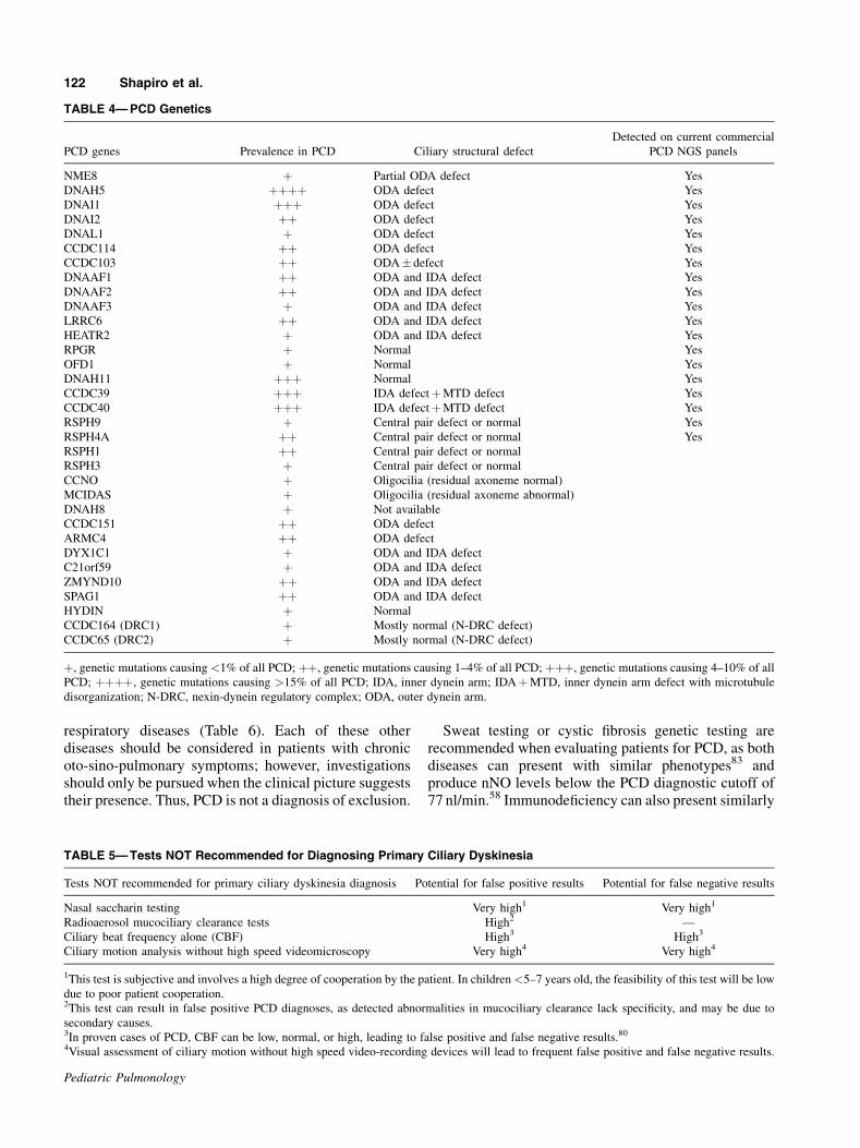
TABLE 4—PCD Genetics
PCD genes Prevalence in PCD Ciliary structural defect
Detected on current commercial
PCD NGS panels
NME8 þ Partial ODA defect Yes
DNAH5 þþþþ ODA defect Yes
DNAI1 þþþ ODA defect Yes
DNAI2 þþ ODA defect Yes
DNAL1 þ ODA defect Yes
CCDC114 þþ ODA defect Yes
CCDC103 þþ ODA� defect Yes
DNAAF1 þþ ODA and IDA defect Yes
DNAAF2 þþ ODA and IDA defect Yes
DNAAF3 þ ODA and IDA defect Yes
LRRC6 þþ ODA and IDA defect Yes
HEATR2 þ ODA and IDA defect Yes
RPGR þ Normal Yes
OFD1 þ Normal Yes
DNAH11 þþþ Normal Yes
CCDC39 þþþ IDA defectþMTD defect Yes
CCDC40 þþþ IDA defectþMTD defect Yes
RSPH9 þ Central pair defect or normal Yes
RSPH4A þþ Central pair defect or normal Yes
RSPH1 þþ Central pair defect or normal
RSPH3 þ Central pair defect or normal
CCNO þ Oligocilia (residual axoneme normal)
MCIDAS þ Oligocilia (residual axoneme abnormal)
DNAH8 þ Not available
CCDC151 þþ ODA defect
ARMC4 þþ ODA defect
DYX1C1 þ ODA and IDA defect
C21orf59 þ ODA and IDA defect
ZMYND10 þþ ODA and IDA defect
SPAG1 þþ ODA and IDA defect
HYDIN þ Normal
CCDC164 (DRC1) þ Mostly normal (N-DRC defect)
CCDC65 (DRC2) þ Mostly normal (N-DRC defect)
þ, genetic mutations causing <1% of all PCD; þþ, genetic mutations causing 1–4% of all PCD; þþþ, genetic mutations causing 4–10% of all
PCD; þþþþ, genetic mutations causing >15% of all PCD; IDA, inner dynein arm; IDAþMTD, inner dynein arm defect with microtubule
disorganization; N-DRC, nexin-dynein regulatory complex; ODA, outer dynein arm.
TABLE 5—Tests NOT Recommended for Diagnosing Primary Ciliary Dyskinesia
Tests NOT recommended for primary ciliary dyskinesia diagnosis Potential for false positive results Potential for false negative results
Nasal saccharin testing Very high1 Very high1
Radioaerosol mucociliary clearance tests High2 —
Ciliary beat frequency alone (CBF) High3 High3
Ciliary motion analysis without high speed videomicroscopy Very high4 Very high4
1This test is subjective and involves a high degree of cooperation by the patient. In children <5–7 years old, the feasibility of this test will be low
due to poor patient cooperation.2This test can result in false positive PCD diagnoses, as detected abnormalities in mucociliary clearance lack specificity, and may be due to
secondary causes.3In proven cases of PCD, CBF can be low, normal, or high, leading to false positive and false negative results.80
4Visual assessment of ciliary motion without high speed video-recording devices will lead to frequent false positive and false negative results.
122 Shapiro et al.
Pediatric Pulmonology

to PCD,84 and in patients with suspected PCD, laboratorystudies investigating immunodeficiency are necessary.Preliminary study of nNO in certain humoral immuno-deficiencies has shown normal values well above 77 nl/min,85 but further study is required to know if all forms ofimmunodeficiency produce normal nNO levels.Pulmonary aspiration, with or without gastroesopha-
geal reflux, can cause chronic respiratory symptoms inadults and children, including cough, wheeze, bronchitis,or pneumonia.86,87 Thus in patients with possible PCD, athorough feeding history is essential. A history of chroniccough from asthma can also resemble PCD in youngchildren, especially with frequent viral infections fromdaycare exposures. Additionally, chronic nasal conges-tion from allergic rhinitis can seem similar to PCDrhinosinusitis. However, PCD nasal disease is presentyear-round and does not resolve with seasonal change, asoften occurs with allergic rhinitis. Lastly, protractedbacterial bronchitis (PBB) is a disorder of preschool agedchildren causing >3 weeks of wet cough with lowerairway bacterial infection and airway neutrophilia.88 Ingeneral though, the characteristic, year-round, daily, oftenwet or productive cough of children with PCD usuallydistinguishes them from these other conditions.
Diseases that Co-Exist With PCD
PCD can rarely co-exist with other rare disorders(Table 7). Retinitis Pigmentosa (an inherited cause ofblindness from retinal ciliary dysfunction) and Orofacio-digital Syndrome (including mental retardation, cranio-facial abnormalities, macrocephaly, digital anomalies,and cystic kidneys) are X-linked disorders involvingciliary genes, RPGR and OFD1, respectively.21,89
Although these account for a very small minority ofPCD cases, there may be further overlap of retinal andrespiratory cilia.90,91 Thus, retinal examination is
recommended in individuals with PCD due to genemutations in RPGR, clinical visual disturbances, or afamily history of Retinitis Pigmentosa, whereas PCDpatients with OFD1 phenotypes should be referred forgenetic consultation.Various diseases caused by genetic disorders of non-
motile cilia can result in cystic kidneys, cystic orcholestatic liver, skeletal malformations, developmentaldelay, hydrocephalus, blindness, or deafness. Theseinclude Joubert Syndrome, Bardet–Biedl syndrome,Usher Syndrome, Jeune Syndrome, polycystic kidneydisease, and others. The overlap of these non-motileciliopathies with respiratory cilia dysfunction is unusual,and poorly understood at present,90,92,93 but increasedrates of bronchiectasis are found in polycystic kidneydisease.94 Therefore, consultation with a geneticist orother subspecialists is recommended when patients withpossible PCD have features of non-motile ciliarydysfunction.PCD can also co-exist with other rare diseases through
close proximity of disease causing mutations at the samechromosomal locus (Table 7). Cri du Chat syndrome canoccur with PCD due to a large deletion on chromosome 5pand a point mutation in DNAH5 on the remainingchromosome.22 Glanzmann Thrombasthenia (associatedwith ITGB3) can occur with PCD (associated withCCDC103) through mutations in the neighboring geneson chromosome 17.95 Alternatively, PCD can co-existwith other rare diseases through disease-causing muta-tions which are not in close genetic proximity; such ascystic fibrosis due to mutations in CFTR (Chr7q) withPCD due to mutations in DNAH11 (Chr7p),96 and MillerSyndrome due to mutations in DHODH (chr 16) withPCD due to mutations in DNAH5 (Chr5).97
Recent publications have also shown respiratory ciliarydysfunction in patients with mild forms of congenitalheart disease, notmeeting cardiology definitions for SA or
TABLE 6—Other Chronic Respiratory Conditions to Consider When Considering a Diagnosis of PCD
Chronic condition Methods of evaluation
Cystic fibrosis Sweat chloride testing or cystic fibrosis genetic testing
Immunodeficiency Quantitative measurement of immunoglobulins, lymphocytes, complement levels, antibody responses to vaccines,
and complete blood counts. Consultation with a board certified Immunologist is also recommended
Asthma Clinical history, pulmonary function testing, and asthma medication trials. Although one normally expects asthma-
related cough to be dry in nature, it can seem wet to parents when accompanied by viral respiratory infections.
Obstructive defects on pulmonary function testing can be seen with both PCD and asthma, and bronchodilator
responsiveness is not exclusive to asthma and does not exclude a diagnosis of PCD
Pulmonary aspiration Clinical feeding history followed by swallowing assessment and intervention only when pulmonary aspiration seems
likely
Allergic rhinitis Clinical history of seasonal symptoms, allergy testing, and trials of nasal corticosteroids and antihistamines, which
should greatly improve allergic rhinitis symptoms. PCD nasal disease shows minimal (if any) improvement with
these interventions
Protracted bacterial
bronchitis
Clinical history of 3 weeks of wet cough in pre-school aged children, with resolution of cough after 14 days of
amoxicillin plus clavulinic acid.122The cough usually does not return after a subsequent 2-week period off
antibiotics
Diagnosis and Management of PCD 123
Pediatric Pulmonology

heterotaxy.98 Thus, physicians should ask about chronicoto-sino-pulmonary symptoms in all patients withcongenital heart disease to screen for possible PCD andtest as indicated.
CLINICAL CARE AND LONG-TERM MONITORING
Pulmonary Care and Monitoring
Long-term follow-up should be in a PCD Foundationclinical center or an accredited cystic fibrosis center thathas a comprehensive, multidisciplinary team approach tocare. Outpatient visits with a pulmonologist experiencedin management of chronic suppurative lung disease, suchas cystic fibrosis, are recommended 2–4 times annually(Table 8). Surveillance cultures of expectorated sputum ororopharyngeal cough swabs are recommended two to fourtimes annually in all PCD patients.1 Although the mostcommon airway pathogens in children with PCD areStreptococcus pneumoniae, Haemophilus influenzae, andMoraxella catarrhalis, surveillance cultures should beprocessed in the same manner as cystic fibrosis cultures,
including examination for Pseudomonas aeruginosa andother Gram negative organisms, as well as non-tuberculosis mycobacterial (NTM) organisms.1,4 Cultureresults should guide antibiotic therapy during futurerespiratory exacerbations. When PCD patients are notresponding to culture-directed antibiotics, physiciansshould consider additional NTM and fungal cultures,allergic bronchopulmonary aspergillosis testing (ABPA)testing (IgE levels� evidence of aspergillus specificity)and bronchoscopy with bronchoalveolar lavage fluidcultures to guide antimicrobial therapy.Spirometry using ATS/ERS criteria99 is suggested two
to four times annually to follow disease progression inPCD. Although spirometry may not be the most sensitivetest of pulmonary function in PCD, it is the most availabletesting method in pediatric and adult centers. With furthervalidation, other tests of pulmonary function, such asmultiple breath washout, may be useful in PCD.100,101
Chest radiography should be performed at diagnosisand during respiratory exacerbations, as indicated.Otherwise, chest radiography should be performed every
TABLE 7—Other Diseases Co-Segregating With PCD
Associated rare disorder Level of PCD association Method of PCD overlap Specific gene affected
Situs ambiguus and heterotaxy At least 12% of PCD Shared common genes Any PCD gene encoding for ODA, IDA,
or ODAþ IDA proteins (ex: DNAH5,
DNAH11, CCDC39/40, LRRC6,
DNAAF1/2/3)19
Retinitis pigmentosa Multiple unrelated cases
reported; <1% of PCD
Shared common gene mutation RPGR21,91,161,162
Orofaciodigital syndrome 1 sibling-pair reported; Shared common gene mutation OFD189
(sibling males with mental
retardation and macrocephaly)
<1% of PCD
Cri du chat syndrome 2 unrelated cases reported Mutation in close proximity
to PCD gene
Chr 5p deletion including DNAH522
Glanzmann thrombasthenia 1 sibling-pair reported Mutation in close proximity
to PCD gene
Chr 17q haplotype that includes
CCDC103 and ITGB395
Cystic fibrosis 1 case reported Mutations in two different genes Chr 7 including region with DNAH11
and CFTR via uniparental isodisomy96
Miller syndrome 1 sibling-pair reported Mutations in two different genes Biallelic mutations in DNAH5 (Chr 5)
for PCD and DHODH (Chr 16) for
Miller syndrome97
Common variable
immunodeficiency
1 sibling-pair and 2
unrelated cases reported
Unknown 1 sibling-pair with homozygous
DNAH11 mutations and low IgM and
S.pneumoniae titers after booster.157 2
unrelated cases; 1 with outer dynein
arm defect and low IgG titers157, and 1
with Kartagener Syndrome, abnormal
ciliary ultrastructure, and low IgG,
IgM, Tetanus and S.pneumoniae titers
after booster.158
Polycystic kidney disease 1 case reported Unknown Unknown92
Familial mediterranean fever 1 case reported Unknown MEFV-R202Q polymorphism on Chr
16p13.3 and unknown PCD gene163
Other non-motile ciliopathies No definite cases reported Unknown Unknown
(Joubert, Bardet-biedl, Usher,
Jeune syndromes, and others)
124 Shapiro et al.
Pediatric Pulmonology

2–4 years in stable patients, in order to monitor diseaseprogression. The decision to use serial CT scans formonitoring PCD disease progression should be decidedon a case by case basis, and the lowest possible radiationdoses should be used. However, a chest CT scan isgenerally recommended at least once after diagnosis todetect bronchiectasis, which may encourage bettercompliance to airway clearance in patients and parentswho are aware of this finding. Chest CT can be consideredwhen children are old enough to cooperate (and avoidsedation), and images will be of sufficient quality todiagnose bronchiectasis, or sooner depending on clinicalsymptoms.33,34 Some centers perform chest CT scans onPCD patients every 5 years, but there is no evidence thatthis improves clinical outcomes,102 and cumulativeradiation doses need to be considered for PCD patients.Infection control policy is essential for clinical care in
PCD, and general hospital infection control policiesshould be followed where PCD patients receive care.Patientswith resistant organisms on sputumculture shouldbe specifically targeted for infection control in all clinicalareas. Although there is no evidence for cross contamina-tion of respiratory organisms among PCD patients, it islogical to assume this may occur, as it does in similardiseases.103More stringent infection control policies havethe potential to cause psychosocial harm to patients andfamilies,104 and thus should be avoided in PCD. However,this recommendation may be adjusted if there is clearevidence for risks that outweigh potential harm.
Otolaryngology Care and Monitoring
Pediatric PCD patients should visit a pediatric otolaryn-gologist at least once to twice annually,while adult patientsshould have otolaryngology care, as needed. An initialaudiology assessment in all PCD patients is suggested atdiagnosis, with subsequent evaluations coordinatedthrough their otolaryngologist. The major otolaryngologyconcern in PCD patients is the nearly universal conductivehearing loss due to persistent otitis media with effusion(OME).105 Hearing abnormalities often improve inadolescence, but in some cases, continue into adulthood.Pressure equalization tubes (PET) are advocated forchildren with PCD who have hearing deficits or speechdelay and middle ear effusions. Although severalsystematic reviews have cast doubt on the utility of PETin OME,106,107 these studies are not necessarily generaliz-able to a PCD population, where individuals are expectedto have greater portion of their prelingual life withconductive hearing loss. In studies assessing hearing inchildren with PCD post-PET placement, hearing normal-ized in 80–100% of participants.32,108,109 In another studyexamining surgical treatment with PET versus medicalmanagement alone in PCD, children with PET had largerhearing improvements post-operatively than those treatedwith medical therapy.108
All patients undergoing PET insertion should becounselled on the likelihood of multiple insertions, post-operative otorrhea, and the possibility of a permanent
TABLE 8—Suggested Schedule of Investigations and Clinical Care in Primary Ciliary Dyskinesia
Clinical visits
Pulmonology: 2–4 times/year
Otolaryngology: 1-2 time/year in children, as needed in adults
Audiology: at diagnosis and as needed per otolaryngology
Reproductive medicine: As clinically needed
Long-term surveillance
Chest radiography: every 2–4 years
Chest computed tomography: consider at least once after 5–7 years old (when sedation not required and images are of highest quality)1
Airway microbiology cultures: 2–4 times/year
Non-tuberculosis mycobacterial cultures: every 2 years (and with unexplained clinical decline)
Pulmonary function testing: 2–4 times/year
ABPA testing: IgE levels� evidence of aspergillus specificity at diagnosis, with new onset wheezing, unexplained clinical decline
Preventative therapies
Airway clearance: daily
Nasal sinus lavage: daily (when pertinent)
Standard vaccinations: per local schedule
Influenza vaccine: annually2
13-valent pneumococcal vaccine: per ACIP guidelines3
23-valent pneumococcal vaccine: per ACIP guidelines4
RSV immunoprophylaxis: consider monthly in first winter5
1And as clinically indicated on a case by case basis.2After 6 months old, including household members.3ACIP guidelines.4ACIP guidelines.5Specifically consider in infants with complicated respiratory courses, including prematurity, prolonged mechanical ventilation, prolonged need
for supplemental oxygen, need for home supplemental oxygen, or frequent respiratory illnesses.
Diagnosis and Management of PCD 125
Pediatric Pulmonology

tympanic membrane perforation (up to 50% in onestudy).110 Additionally, patients with PET are typicallyseen by their otolarygologist every 3–6 months while thetubes remain in place.107 Although some physicians avoidPET in PCD for fear of prolonged post-operative otorrhea,studies show that post-operative otorrhea in PCD is noworse than the general population111 and is easilycontrolled with topical therapies.109 Persistent otorrheacan be attributed to biofilm formation, especially inchildren with longer lasting PET112; however, given thepoor eustachian tube function andmultiple PET insertions,acquired cholesteatoma should also be considered as apotential cause of persistent otorrhea in PCD.Otolaryngologists should also monitor for chronic
rhinosinusitis (CRS) in PCD patients. CRS is estimated toaffect over 50% of patients with PCD31 and nasalendoscopy (as permitted by age) can be used to identifypolyps which may be exacerbating already poor muco-ciliary clearance. Nasal polyposis has been observed in upto 15% of PCD patients.29,113 Although CRS is notgenerally life threatening, it substantially affects qualityof life.114 Daily saline irrigation has been demonstrated assafe and beneficial in patients with CRS.115 Anecdotally,in PCD patients, saline nasal irrigations are beneficial, butstudies demonstrating their efficacy are lacking. Given theminimal side effect profile and likelihood for benefit,nasal irrigations are generally encouraged for symptom-atic CRS relief in PCD. The effects of saline irrigation arelikely increased after functional endoscopic sinus surgery(ESS), as the saline solution will more easily reach thesinus mucosa through post-surgical ostia. Thus, ESS isoften performed in PCD patients and may improve lowerrespiratory tract disease in some patients.116 Antibioticsand nasal steroids may be used in acute on chronicexacerbations of rhinosinusitis; however, a recent reviewshowed lack of consensus on the treatment of CRS inchildren with PCD,113 and there are no randomized,controlled, or long-term prospective CRS studies in PCD.
PRINCIPLES OF TREATMENT
Routine Therapies in PCD
Airway clearance through daily chest physiotherapy ishighly recommended in PCD.117 Unlike cystic fibrosis,cough clearance is preserved in PCD.118 Thus, airwayclearance is expected to be quite beneficial in PCD andshould be a cornerstone of long-term therapy. Dailycardiovascular exercise should also be strongly encour-aged, as poor exercise capacity is linked to decreasedpulmonary function in PCD,119 and exercise may improvemucus clearance.120
Antibiotics should be given for acute respiratoryexacerbations in PCD. Acute changes in cough, sputumproduction, respiratory rate, or work of breathing arelikely reliable markers of a respiratory exacerbation in
PCD (as demonstrated in non-CF bronchiectasis121), andoral antibiotics are recommended for mild exacerbations.Most physicians use a broad-spectrum oral antibiotic(amoxicillin plus clavulinic acid or an equivalentcephalosporin) to target the common respiratory patho-gens in children with PCD. Typically, at least 2–3 weeksof oral antibiotics are recommended in PCD, based uponother disorders with similar pathophysiology (protractedbacterial bronchitis,122 cystic fibrosis,123 and non-CFbronchiectasis124). More severe exacerbations, or thosefailing oral therapy, may require parenteral antibiotics.Antibiotic choice should be guided by past respiratorycultures. Despite a lack of published evidence, inhaledantibiotics are also an option for acute PCD respiratoryexacerbations, but these are usually reserved for patientswith Pseudomonas aeruginosa infection. Eradication ofinitial positive Pseudomonas airway culture also seemsprudent in PCD, although no evidence supports thispractice. Non-CF bronchiectasis guidelines make similarsuggestions for Pseudomonas eradication.125,126 Al-though Burkholderia cepacia has not been reported inPCD, recovery of this organism should prompt eradica-tion practices.Finally, PCD patients should receive recommended
vaccinations per local schedules. Annual influenza127 andpneumococcal vaccinations (per the Advisory Committeeon Immunization Practices)128,129 are recommended inPCD. In the first year of life, monthly (seasonal)immunoprophylaxis against respiratory synctial viruscan be considered for infants with PCD, and morespecifically for infants with complicated respiratorycourses requiring prolonged oxygen supplementation.
Therapies to Consider on a Case by Case Basis inPCD
Chronic suppressive inhaled antibiotics can be used onan individual basis in PCD patients. Inhaled aminoglyco-side and beta-lactam antibiotics are recommended forchronic respiratory infections (particularly those associat-ed with Pseudomonas aeruginosa) in non-CF bronchiec-tasis,125,130,131 and several months of inhaledaminoglycosides or colistin in Pseudomonas colonizedadults with non-CF bronchiectasis result in decreasedhospitalization and improved respiratory symptoms.132–134
However, there are no studies of inhaled antibiotics inchildren with non-CF bronchiectasis or PCD.Chronic suppressive oral antibiotics, including trimeth-
oprim-sulfamethoxazole, macrolides, or other agents, canbe used on a case by case basis in PCD. Chronicmacrolidetherapy in PCD is currently under prospective investiga-tion by the BESTCILIA consortium in Europe.51 Whenusing chronic macrolide therapy, sputum culture surveil-lance for non-tuberculous mycobacterium infection isindicated.135 Prospective clinical study of chronic
126 Shapiro et al.
Pediatric Pulmonology

macrolides in adults and children with non-CF bronchi-ectasis shows decreased respiratory exacerbations andimproved lung function,136–138 but increased emergenceof macrolide resistant respiratory organisms. The long-term significance of macrolide resistance is unclear.139
Small case reports of chronic macrolide therapy in PCDalso demonstrate some benefits, although not as robust asthose in non-CF bronchiectasis.140–143 Remote studies ontrimethoprim-sulfamethoxazole in chronic bronchitis alsosuggest benefit, but this agent has not been studied inPCD.144,145
Inhaled hyperosmolar agents can be used on a case-by-case basis in PCD. These agents promote cough clearanceand alter mucus rheology to favor increased coughclearance. However, a recent meta-analysis reportedunclear long-term benefits of hyperosmolar agents in non-CF bronchiectasis.146 Hypertonic saline (3% to 7%concentration) has not been studied in PCD. Trialscomparing inhaled hypertonic saline to isotonic salineshow limited positive effects in non-CF bronchiectasis.147
When physicians use inhaled hypertonic saline in PCD, itis essential that they instruct patients in proper equipmentsterilization. Inhaled dry powder mannitol has also beenstudied in non-CF bronchiectasis, but outcomes areinconclusive.146,148 Mannitol has not been studied inPCD.DNase (dornase-alfa or Pulmozyme1) can be used on
an individual basis in PCD. Although there are noprospective trials of DNase in PCD, studies of DNase inadults with non-CF bronchiectasis show no clinicalbenefits in one study149 and increased frequency ofrespiratory exacerbations with worsened lung function inanother study.150 Several case reports of DNase in PCDsuggest possible benefit when used for both short andlong-term periods.151–153 Larger, prospective clinicalstudies of DNase in children and young adults withPCD are required before the potential negative effects ofthis medication can be dismissed.Lastly, inhaled bronchodilators can be used on a case-
by-case basis in PCD. In limited study, long-actingbronchodilators (with inhaled corticosteroids) in non-CFbronchiectasis do not show clinical efficacy. In PCD,bronchodilators show mixed results, with one studydemonstrating significant improvement in lung functionafter a single bronchodilator dose,154 whereas anotherstudy showed unchanged lung function after 6 weeks ofregular bronchodilators.155
Therapies Not Routinely Recommended in PCD
Inhaled corticosteroids are not routinely recommend inPCD and should be reserved for PCD patients withassociated asthma or airway reactivity. Inhaled cortico-steroids are also discouraged in non-CF bronchiectasiswithout airway reactivity.156 Similarly, intravenous
immunoglobulin (IVIG) is not recommended for routineuse in patients with PCD. Immunodeficiency rarely existswith PCD,157,158 and most PCD patients have normalimmune function. PCD patients with documenteddysfunction of vaccine responses or other aspects ofhumoral immunity may benefit from IVIG therapy.Isolated IgA or IgG subclass disorders do not justifyIVIG therapy.Lobectomy is not routinely suggested as therapy in
PCD. The decision to perform lobectomy in PCD requiresmulti-disciplinary discussion between pulmonologists,intensivists, and surgeons. In the post-operative period,airway clearance is limited by pain and immobility, andPCD patients are at risk of pulmonary deterioration.Although lobectomy may be beneficial in rare cases ofPCD with severe, localized bronchiectasis, it should beconsidered with caution. Similarly, lung transplantationcan be considered in PCD patients with advancedpulmonary disease, but situs anomalies may surgicallycomplicate this procedure.159,160
Summary
PCD is a rare disorder; consequently, only a limitednumber of centers have extensive experience in thediagnosis and management of PCD. Research over thepast decade has led to a revolution in diagnosticapproaches, including nNO and genetic testing. Never-theless, many PCD patients are still undiagnosed ormisdiagnosed. To date, only limited studies haveaddressed management of PCD, and there have been nolarge, randomized clinical trials to direct therapy.Therefore, this review article includes consensus recom-mendations from PCD physicians in North America fordiagnosis, monitoring and management of PCD.
ACKNOWLEDGMENTS
The authors thank the US PCD Foundation and otherinvestigators and coordinators of the Genetic Disorders ofMucociliary Clearance Consortium, including Lou Ep-person, RN (Indiana University), Jane Quante, RN, BS(Washington University in St. Louis), Carol Kopecky,Shelley Mann and Donna Parker (Children’s HospitalColorado), Jacquelyn Zirbes, DNP, RN, CNP, CCRC(Stanford University), Robert Johnson, MS (SeattleChildren’s Hospital),MelodyMikki, RN, BScN (Hospitalfor Sick Children), Hoon Chang, IRTA Fellow (NationalInstitutes of Health). Thanks to Rosalie Helfrich(DMCC). Lastly, the authors thank site directors of thePCD Foundation Clinical Center Network, George M.Solomon, MD (University of Alabama—Birmingham),Maureen Josephson, MD (Children’s Hospital of Phila-delphia), Benjamin Gaston, MD (Case Western ReserveUniversity), and Umakanth Kkatwa, MD and KenanHaver, MD (Boston Children’s Hospital), who reviewed
Diagnosis and Management of PCD 127
Pediatric Pulmonology

drafts of this document and provided input in areasneeding clarification.
REFERENCES
1. Davis SD, Ferkol TW, Rosenfeld M, Lee HS, Dell SD, Sagel SD,
Milla C, Zariwala MA, Pittman JE, Shapiro AJ, et al. Clinical
features of childhood primary ciliary dyskinesia by genotype and
ultrastructural phenotype. Am J Respir Crit Care Med
2015;191:316–324.
2. Magnin ML, Cros P, Beydon N, Mahloul M, Tamalet A,
Escudier E, Clement A, Le Pointe HD, Blanchon S. Longitudinal
lung function and structural changes in children with primary
ciliary dyskinesia. Pediatr Pulmonol 2012;47:816–825.
3. Marthin JK, Petersen N, Skovgaard LT, Nielsen KG. Lung
function in patients with primary ciliary dyskinesia: a cross-
sectional and 3-decade longitudinal study. Am J Respir Crit Care
Med 2010;181:1262–1268.
4. Knowles MR, Daniels LA, Davis SD, Zariwala MA, Leigh MW.
Primary ciliary dyskinesia. Recent advances in diagnostics,
genetics, and characterization of clinical disease. Am J Respir
Crit Care Med 2013;188:913–922.
5. Leigh MW, O’Callaghan C, Knowles MR. The challenges of
diagnosing primary ciliary dyskinesia. Proc Am Thorac Soc
2011;8:434–437.
6. Hosie P, Fitzgerald DA, Jaffe A, Birman CS, Morgan L. Primary
ciliary dyskinesia: overlooked and undertreated in children.
J Paediatr Child Health 2014;50:952–958.
7. Lucas JS, Leigh MW. Diagnosis of primary ciliary dyskinesia:
searching for a gold standard. Eur Respir J 2014;44:1418–1422.
8. Zariwala MA, Knowles MR, Leigh MW. Primary ciliary
dyskinesia. 2007 Jan24 [updated 2015 Sep 3] In: Pagon RA,
AdamMP, Amemiya A, Bird TD, et al., editors. GeneReviewsTM.
Seattle (WA): University of Washington, Seattle; 1993–2015.
Available from: http://www.ncbi.nlm.nih.gov/books/NBK1122/.)
9. Boon M, Jorissen M, Proesmans M, De Boeck K. Primary ciliary
dyskinesia, an orphan disease. Eur J Pediatr 2013;172:151–162.
10. Knowles MR, Leigh MW, Carson JL, Davis SD, Dell SD,
Ferkol TW, Olivier KN, Sagel SD, Rosenfeld M, Burns KA, et al.
Mutations of DNAH11 in patients with primary ciliary dyskinesia
with normal ciliary ultrastructure. Thorax 2012;67:433–441.
11. O’Callaghan C, Rutman A,Williams GM, Hirst RA. Inner dynein
arm defects causing primary ciliary dyskinesia: repeat testing
required. Eur Respir J 2011;38:603–607.
12. Kim RH, A Hall D, Cutz E, Knowles MR, Nelligan KA,
Nykamp K, ZariwalaMA, Dell SD. The role of molecular genetic
analysis in the diagnosis of primary ciliary dyskinesia. Ann Am
Thorac Soc 2014;11:351–359.
13. Berg JS, Evans JP, Leigh MW, Omran H, Bizon C, Mane K,
Knowles MR, Weck KE, Zariwala MA. Next generation
massively parallel sequencing of targeted exomes to identify
genetic mutations in primary ciliary dyskinesia: implications for
application to clinical testing. Genet Med 2011;13:218–229.
14. Jackson CL, Goggin PM, Lucas JS. Ciliary beat pattern analysis
below 37 degrees C may increase risk of primary ciliary
dyskinesia misdiagnosis. Chest 2012;142:543–544; author reply
544–5.
15. Hirst RA, Rutman A, Williams G, O’Callaghan C. Ciliated air-
liquid cultures as an aid to diagnostic testing of primary ciliary
dyskinesia. Chest 2010;138:1441–1447.
16. Thomas B, Rutman A, O’Callaghan C. Disrupted ciliated
epithelium shows slower ciliary beat frequency and increased
dyskinesia. Eur Respir J 2009;34:401–404.
17. Mateos-Corral D, Coombs R, Grasemann H, Ratjen F, Dell SD.
Diagnostic value of nasal nitric oxide measured with non-velum
closure techniques for children with primary ciliary dyskinesia.
J Pediatr 2011;159:420–424.
18. Leigh MW, Hazucha MJ, Chawla KK, Baker BR, Shapiro AJ,
Brown DE, Lavange LM, Horton BJ, Qaqish B, Carson JL, et al.
Standardizing nasal nitric oxide measurement as a test for
primary ciliary dyskinesia. Ann Am Thorac Soc 2013;10:
574–581.
19. Shapiro AJ, Davis SD, Ferkol T, Dell SD, Rosenfeld M,
Olivier KN, Sagel SD, Milla C, Zariwala MA, Wolf W, et al.
Laterality defects other than situs inversus totalis in primary
ciliary dyskinesia: insights into situs ambiguus and heterotaxy.
Chest 2014. 146:1176–86.
20. Shapiro AJ, Tolleson-Rinehart S, Zariwala MA, Knowles MR,
Leigh MW. The prevalence of clinical features associated with
primary ciliary dyskinesia in a heterotaxy population: results of a
web-based survey. Cardiol Young 2014;25:752–9.
21. Moore A, Escudier E, Roger G, Tamalet A, Pelosse B, Marlin S,
Cl�ement A, Geremek M, Delaisi B, Bridoux AM, et al. RPGR is
mutated in patients with a complex X linked phenotype
combining primary ciliary dyskinesia and retinitis pigmentosa.
J Med Genet 2006;43:326–333.
22. Shapiro AJ, Weck KE, Chao KC, Rosenfeld M, Nygren AO,
Knowles MR, Leigh MW, Zariwala MA. Cri du chat syndrome
and primary ciliary dyskinesia: a common genetic cause on
chromosome 5p. J Pediatr 2014;165:858–861.
23. Lucas JS, Carroll M. Primary ciliary dyskinesia and cystic
fibrosis: different diseases require different treatment. Chest
2014;145:674–676.
24. Cohen-Cymberknoh M, Simanovsky N, Hiller N, Gileles
Hillel A, ShoseyovD, KeremE. Differences in disease expression
between primary ciliary dyskinesia and cystic fibrosis with and
without pancreatic insufficiency. Chest 2014;145:738–744.
25. Paff T, van der Schee MP, Daniels JM, Pals G, Postmus PE,
Sterk PJ, Haarman EG. Exhaled molecular profiles in the
assessment of cystic fibrosis and primary ciliary dyskinesia.
J Cyst Fibros 2013;12:454–460.
26. Horvath I, Loukides S, Wodehouse T, Csiszer E, Cole PJ,
Kharitonov SA, Barnes PJ. Comparison of exhaled and nasal
nitric oxide and exhaled carbonmonoxide levels in bronchiectatic
patients with and without primary ciliary dyskinesia. Thorax
2003;58:68–72.
27. Guyatt GH, Oxman AD, Vist GE, Kunz R, Falck-Ytter Y, Alonso-
Coello P, Schunemann HJ, Group GW. GRADE: an emerging
consensus on rating quality of evidence and strength of
recommendations. BMJ 2008;336:924–926.
28. Mullowney T, Manson D, Kim R, Stephens D, Shah V, Dell S.
Primary ciliary dyskinesia and neonatal respiratory distress.
Pediatrics 2014;134:1160–1166.
29. Campbell R. Managing upper respiratory tract complications of
primary ciliary dyskinesia in children. Curr Opin Allergy Clin
Immunol 2012;12:32–38.
30. Afzelius BA. A human syndrome caused by immotile cilia.
Science 1976;193:317–319.
31. Sommer JU, Schafer K, Omran H, Olbrich H, Wallmeier J,
Blum A, Hormann K, Stuck BA. ENT manifestations in patients
with primary ciliary dyskinesia: prevalence and significance of
otorhinolaryngologic co-morbidities. Eur Arch Otorhinolaryngol
2011;268:383–388.
32. el-Sayed Y, al-Sarhani A, al-Essa AR. Otological manifestations
of primary ciliary dyskinesia. Clin Otolaryngol Allied Sci
1997;22:266–270.
33. Kennedy MP, Noone PG, Leigh MW, Zariwala MA, Minnix SL,
Knowles MR, Molina PL. High-resolution CT of patients with
primary ciliary dyskinesia. AJR Am J Roentgenol 2007;188:
1232–1238.
128 Shapiro et al.
Pediatric Pulmonology

34. Brown DE, Pittman JE, Leigh MW, Fordham L, Davis SD. Early
lung disease in young children with primary ciliary dyskinesia.
Pediatr Pulmonol 2008;43:514–516.
35. Noone PG, Leigh MW, Sannuti A, Minnix SL, Carson JL,
Hazucha M, Zariwala MA, Knowles MR. Primary ciliary
dyskinesia: diagnostic and phenotypic features. Am J Respir
Crit Care Med 2004;169:459–467.
36. Munro NC, Currie DC, Lindsay KS, Ryder TA, Rutman A,
Dewar A, Greenstone MA, Hendry WF, Cole PJ. Fertility in men
with primary ciliary dyskinesia presenting with respiratory
infection. Thorax 1994;49:684–687.
37. McComb P, Langley L, VillalonM, Verdugo P. The oviductal cilia
and Kartagener’s syndrome. Fertil Steril 1986;46:412–416.
38. Knowles MR, Ostrowski LE, Leigh MW, Sears PR, Davis SD,
Wolf WE, Hazucha MJ, Carson JL, Olivier KN, Sagel SD, et al.
Mutations in RSPH1 cause primary ciliary dyskinesia with a
unique clinical and ciliary phenotype. Am J Respir Crit CareMed
2014;189:707–717.
39. Olin JT, Burns K, Carson JL, Metjian H, Atkinson JJ, Davis SD,
Dell SD, Ferkol TW,Milla CE, Olivier KN, et al. Diagnostic yield
of nasal scrape biopsies in primary ciliary dyskinesia: a
multicenter experience. Pediatr Pulmonol 2011; 46:483-8.
40. de Iongh RU, Rutland J. Ciliary defects in healthy subjects,
bronchiectasis, and primary ciliary dyskinesia. Am J Respir Crit
Care Med 1995;151:1559–1567.
41. Escudier E, Couprie M, Duriez B, Roudot-Thoraval F,
Millepied MC, Pruliere-Escabasse V, Labatte L, Coste A.
Computer-assisted analysis helps detect inner dynein arm
abnormalities. Am J Respir Crit Care Med 2002;166:1257–1262.
42. Antony D, Becker-Heck A, Zariwala MA, Schmidts M,
Onoufriadis A, Forouhan M, Wilson R, Taylor-Cox T,
Dewar A, Jackson C, et al. Mutations in CCDC39 and
CCDC40 are the major cause of primary ciliary dyskinesia
with axonemal disorganization and absent inner dynein arms.
Hum Mutat 2013;34:462–472.
43. Castleman VH, Romio L, Chodhari R, Hirst RA, de Castro SC,
Parker KA, Ybot-Gonzalez P, Emes RD, Wilson SW, Wallis C,
et al. Mutations in radial spoke head protein genes RSPH9
and RSPH4A cause primary ciliary dyskinesia with central-
microtubular-pair abnormalities. Am J Hum Genet 2009;84:
197–209.
44. Onoufriadis A, Shoemark A, Schmidts M, Patel M, Jimenez G,
Liu H, Thomas B, Dixon M, Hirst RA, Rutman A, et al. Targeted
NGS gene panel identifies mutations in RSPH1 causing primary
ciliary dyskinesia and a common mechanism for ciliary central
pair agenesis due to radial spoke defects. Hum Mol Genet
2014;23:3362–3374.
45. Jeanson L, Copin B, Papon JF, Dastot-Le Moal F, Duquesnoy P,
Montantin G, Cadranel J, Corvol H, Coste A, Desir J, et al.
RSPH3 mutations cause primary ciliary dyskinesia with central-
complex defects and a near absence of radial spokes. Am J Hum
Genet 2015; 97:153-62.
46. Horani A, Brody SL, Ferkol TW, Shoseyov D, Wasserman MG,
Ta-shma A, Wilson KS, Bayly PV, Amirav I, Cohen-
Cymberknoh M, et al. CCDC65 mutation causes primary ciliary
dyskinesia with normal ultrastructure and hyperkinetic cilia.
PLoS ONE 2013;8:e72299.
47. Olbrich H, Schmidts M, Werner C, Onoufriadis A, Loges NT,
Raidt J, Banki NF, Shoemark A, Burgoyne T, Al Turki S, et al.
Recessive HYDIN mutations cause primary ciliary dyskinesia
without randomization of left-right body asymmetry. Am J Hum
Genet 2012;91:672–684.
48. Wirschell M, Olbrich H, Werner C, Tritschler D, Bower R,
Sale WS, Loges NT, Pennekamp P, Lindberg S, Stenram U, et al.
The nexin-dynein regulatory complex subunit DRC1 is essential
for motile cilia function in algae and humans. Nat Genet
2013;45:262–268.
49. Wallmeier J, Al-Mutairi DA, Chen CT, Loges NT, Pennekamp P,
Menchen T, Ma L, Shamseldin HE, Olbrich H, Dougherty GW,
et al. Mutations in CCNO result in congenital mucociliary
clearance disorder with reduced generation of multiple motile
cilia. Nat Genet 2014;46:646–651.
50. Boon M, Wallmeier J, Ma L, Loges NT, Jaspers M, Olbrich H,
Dougherty GW, Raidt J, Werner C, Amirav I, et al. MCIDAS
mutations result in a mucociliary clearance disorder with reduced
generation of multiple motile cilia. Nat Commun 2014;5:4418.
51. Werner C, Onnebrink JG, Omran H. Diagnosis and management
of primary ciliary dyskinesia. Cilia 2015;4:2.
52. Marthin JK, Nielsen KG. Hand-held tidal breathing nasal nitric
oxide measurement-a promising targeted case-finding tool for the
diagnosis of primary ciliary dyskinesia. PLoS ONE 2013;8:
e57262.
53. Harris A, Bhullar E, Gove K, Joslin R, Pelling J, Evans HJ,
Walker WT, Lucas JS. Validation of a portable nitric oxide
analyzer for screening in primary ciliary dyskinesias. BMC Pulm
Med 2014;14:18.
54. Lundberg JO, Weitzberg E, Nordvall SL, Kuylenstierna R,
Lundberg JM, Alving K. Primarily nasal origin of exhaled nitric
oxide and absence in Kartagener’s syndrome. Eur Respir J
1994;7:1501–1504.
55. Collins SA, Gove K, Walker W, Lucas JS. Nasal nitric oxide
screening for primary ciliary dyskinesia: systematic review and
meta-analysis. Eur Respir J 2014;44:1589–1599.
56. Pifferi M, Caramella D, Cangiotti AM, Ragazzo V, Macchia P,
Boner AL. Nasal nitric oxide in atypical primary ciliary
dyskinesia. Chest 2007;131:870–873.
57. Marthin JK, NielsenKG. Choice of nasal nitric oxide technique as
first-line test for primary ciliary dyskinesia. Eur Respir J
2011;37:559–565.
58. Balfour-Lynn IM, Laverty A, Dinwiddie R. Reduced upper
airway nitric oxide in cystic fibrosis. Arch Dis Child 1996;75:
319–322.
59. Palm J, Lidman C, Graf P, Alving K, Lundberg J. Nasal nitric
oxide is reduced in patients with HIV. Acta Otolaryngol
2000;120:420–423.
60. Nakano H, Ide H, Imada M, Osanai S, Takahashi T, Kikuchi K,
Iwamoto J. Reduced nasal nitric oxide in diffuse panbronchiolitis.
Am J Respir Crit Care Med 2000;162:2218–2220.
61. Arnal JF, Flores P, Rami J, Murris-Espin M, Bremont F,
Pasto IAM, Serrano E, Didier A. Nasal nitric oxide concentration
in paranasal sinus inflammatory diseases. Eur Respir J 1999;13:
307–312.
62. Stannard WA, Chilvers MA, Rutman AR, Williams CD,
O’Callaghan C. Diagnostic testing of patients suspected of
primary ciliary dyskinesia. Am J Respir Crit Care Med
2010;181:307–314.
63. Chilvers MA, McKean M, Rutman A, Myint BS, Silverman M,
O’Callaghan C. The effects of coronavirus on human nasal
ciliated respiratory epithelium. Eur Respir J 2001;18:965–970.
64. Tilley AE, Walters MS, Shaykhiev R, Crystal RG. Cilia
dysfunction in lung disease. Annu Rev Physiol 2015;77:379–406.
65. Omran H, Loges NT. Immunofluorescence staining of ciliated
respiratory epithelial cells. Methods Cell Biol 2009;91:123–133.
66. Fliegauf M, Olbrich H, Horvath J, Wildhaber JH, Zariwala MA,
Kennedy M, Knowles MR, Omran H. Mislocalization of DNAH5
and DNAH9 in respiratory cells from patients with primary
ciliary dyskinesia. Am J Respir Crit Care Med 2005;171:
1343–1349.
67. Omran H, Kobayashi D, Olbrich H, Tsukahara T, Loges NT,
Hagiwara H, Zhang Q, Leblond G, O’Toole E, Hara C, et al. Ktu/
Diagnosis and Management of PCD 129
Pediatric Pulmonology
PF13 is required for cytoplasmic pre-assembly of axonemal
dyneins. Nature 2008;456:611–616.
68. Frommer A, Hjeij R, Loges NT, Edelbusch C, Jahnke C, Raidt J,
Werner C, Wallmeier J, Grosse-Onnebrink J, Olbrich H, et al.
Immunofluorescence analysis and diagnosis of primary ciliary
dyskinesia with radial spoke defects. Am J Respir Cell Mol Biol
2015. [Epub ahead of print].
69. Hjeij R, Onoufriadis A, Watson CM, Slagle CE, Klena NT,
Dougherty GW, Kurkowiak M, Loges NT, Diggle CP,
Morante NF, et al. CCDC151 mutations cause primary ciliary
dyskinesia by disruption of the outer dynein arm docking
complex formation. Am J Hum Genet 2014;95:257–274.
70. Loges NT, Olbrich H, Fenske L, Mussaffi H, Horvath J,
Fliegauf M, Kuhl H, Baktai G, Peterffy E, Chodhari R, et al.
DNAI2 mutations cause primary ciliary dyskinesia with
defects in the outer dynein arm. Am J Hum Genet 2008;83:
547–558.
71. Tarkar A, Loges NT, Slagle CE, Francis R, Dougherty GW,
Tamayo JV, Shook B, Cantino M, Schwartz D, Jahnke C, et al.
DYX1C1 is required for axonemal dynein assembly and ciliary
motility. Nat Genet 2013;45:995–1003.
72. Knowles MR, Ostrowski LE, Loges NT, Hurd T, Leigh MW,
Huang L, Wolf WE, Carson JL, Hazucha MJ, Yin W, et al.
Mutations in SPAG1 cause primary ciliary dyskinesia associated
with defective outer and inner dynein arms. Am J Hum Genet
2013;93:711–720.
73. Olbrich H, Horvath J, Fekete A, Loges NT, Storm van’s
Gravesande K, Blum A, Hormann K, Omran H. Axonemal
localization of the dynein component DNAH5 is not altered in
secondary ciliary dyskinesia. Pediatr Res 2006;59:418–422.
74. Horani A, Brody SL, Ferkol TW. Picking up speed: advances in
the genetics of primary ciliary dyskinesia. Pediatr Res 2014;
75:158–64.
75. Prevention Genetics. 2015 April 1. Primary ciliary dyskinesia
(PCD)/immotile cilia syndrome nextgen sequencing (NGS)
panel. https://www.preventiongenetics.com/clinical-dna-testing/
test/primary-ciliary-dyskinesia-pcdimmotile-cilia-syndrome-
nextgen-sequencing-panel/1833/. Accessed 2015 April 1.
76. Ambry Genetics. 2015 April 1. Primary ciliary dyskinesia testing.
http://www.ambrygen.com/tests/primary-ciliary-dyskinesia-
testing. Accessed 2015 April 1.
77. Invitae Genetics. 2015April 1. Primary ciliary dyskinesia. https://
www.invitae.com/en/physician/condition-detail/CND0072/. Ac-
cessed 2015 April 1.
78. Partners HealthCare. 2015 April 1. Bronchiectasis panel (17
Genes) test details. http://personalizedmedicine.partners.org/
Laboratory-For-Molecular-Medicine/Tests/Pulmonary-Disease/
Bronchiectasis-Panel.aspx. Accessed 2015 April 1.
79. Canciani M, Barlocco EG, Mastella G, de Santi MM, Gardi C,
Lungarella G. The saccharin method for testing mucociliary
function in patients suspected of having primary ciliary
dyskinesia. Pediatr Pulmonol 1988;5:210–214.
80. Raidt J, Wallmeier J, Hjeij R, Onnebrink JG, Pennekamp P,
Loges NT, Olbrich H, Haffner K, Dougherty GW, Omran H, et al.
Ciliary beat pattern and frequency in genetic variants of primary
ciliary dyskinesia. Eur Respir J 2014;44:1579–1588.
81. De Boeck K, Proesmans M, Mortelmans L, Van Billoen B,
Willems T, Jorissen M. Mucociliary transport using 99mTc-
albumin colloid: a reliable screening test for primary ciliary
dyskinesia. Thorax 2005;60:414–417.
82. Marthin JK, Mortensen J, Pressler T, Nielsen KG. Pulmonary
radioaerosol mucociliary clearance in diagnosis of primary
ciliary dyskinesia. Chest 2007;132:966–976.
83. Hamosh A, Corey M. Correlation between genotype and
phenotype in patients with cystic fibrosis. The cystic fibrosis
genotype-phenotype consortium. N Engl J Med 1993;329:
1308–1313.
84. Kesson AM, Kakakios A. Immunocompromised children:
conditions and infectious agents. Paediatr Respir Rev 2007;8:
231–239.
85. Boon M, Meyts I, Proesmans M, Vermeulen FL, Jorissen M, De
Boeck K. Diagnostic accuracy of nitric oxide measurements to
detect primary ciliary dyskinesia. Eur J Clin Invest 2014;44:
477–485.
86. Platzker A. Gastroesophageal reflux and aspiration syndromes.
In: Chernick VB, TF, Wilmott, RW, Bush, A, editor. Kendig’s
Disorders of the Respiratory Tract in Children (Seventh Edition)
Philadelphia: Saunders Elsevier; 2006. pp 592–609.
87. Cardasis JJ, MacMahon H, Husain AN. The spectrum of lung
disease due to chronic occult aspiration. Ann Am Thorac Soc
2014;11:865–873.
88. Wurzel DF, Marchant JM, Yerkovich ST, Upham JW,
Mackay IM, Masters IB, Chang AB. Prospective characterization
of protracted bacterial bronchitis in children. Chest 2014;145:
1271–1278.
89. Budny B, Chen W, Omran H, Fliegauf M, Tzschach A,
Wisniewska M, Jensen LR, Raynaud M, Shoichet SA,
Badura M, et al. A novel X-linked recessive mental retardation
syndrome comprising macrocephaly and ciliary dysfunction is
allelic to oral-facial-digital type I syndrome. Hum Genet
2006;120:171–178.
90. Tosi GM, de Santi MM, Pradal U, Braggion C, Luzi P.
Clinicopathologic reports, case reports, and small case series:
usher syndrome type 1 associated with primary ciliary aplasia.
Arch Ophthalmol 2003;121:407–408.
91. Bukowy-Bieryłło Z, Zie ̨tkiewicz E, Loges NT, Wittmer M,
Geremek M, Olbrich H, Fliegauf M, Voelkel K, Rutkiewicz E,
Rutland J, et al. RPGRmutations might cause reduced orientation
of respiratory cilia. Pediatr Pulmonol 2013;48:352–363.
92. Saeki H, Kondo S, Morita T, Sasagawa I, Ishizuka G, Koizumi Y.
Immotile cilia syndrome associated with polycystic kidney.
J Urol 1984;132:1165–1166.
93. Papon JF, Perrault I, Coste A, Louis B, Gerard X, Hanein S, Fares-
Taie L, Gerber S, Defoort-Dhellemmes S, Vojtek AM, et al.
Abnormal respiratory cilia in non-syndromic Leber congenital
amaurosis with CEP290 mutations. J Med Genet 2010;47:
829–834.
94. Driscoll JA, Bhalla S, Liapis H, Ibricevic A, Brody SL.
Autosomal dominant polycystic kidney disease is associated
with an increased prevalence of radiographic bronchiectasis.
Chest 2008;133:1181–1188.
95. D’Andrea G, Schiavulli M, Dimatteo C, Santacroce R, Guerra E,
Longo VA, Grandone E, Margaglione M. Homozygosity by
descent of a 3Mb chromosome 17 haplotype causes coinheritance
of Glanzmann thrombasthenia and primary ciliary dyskinesia.
Blood 2013;122:4289–4291.
96. Bartoloni L, Blouin JL, Pan Y, Gehrig C, Maiti AK,
Scamuffa N, Rossier C, Jorissen M, Armengot M, Meeks M,
et al. Mutations in the DNAH11 (axonemal heavy chain dynein
type 11) gene cause one form of situs inversus totalis and most
likely primary ciliary dyskinesia. Proc Natl Acad Sci USA
2002;99:10282–10286.
97. Roach JC, GlusmanG, Smit AF, Huff CD, Hubley R, Shannon PT,
Rowen L, Pant KP, Goodman N, Bamshad M, et al. Analysis of
genetic inheritance in a family quartet by whole-genome
sequencing. Science 2010;328:636–639.
98. Garrod AS, Zahid M, Tian X, Francis RJ, Khalifa O, Devine W,
Gabriel GC, Leatherbury L, Lo CW. Airway ciliary dysfunction
and sinopulmonary symptoms in patients with congenital heart
disease. Ann Am Thorac Soc 2014;11:1426–1432.
130 Shapiro et al.
Pediatric Pulmonology

99. Miller MR, Hankinson J, Brusasco V, Burgos F, Casaburi R,
Coates A, Crapo R, Enright P, van der Grinten CP, Gustafsson P,
et al. Standardisation of spirometry. Eur Respir J 2005;26:
319–338.
100. BoonM, Vermeulen FL, GysemansW, ProesmansM, JorissenM,
De Boeck K. Lung structure-function correlation in patients with
primary ciliary dyskinesia. Thorax 2015;70:339–345.
101. Green K, Buchvald FF, Marthin JK, Hanel B, Gustafsson PM,
Nielsen KG. Ventilation inhomogeneity in children with primary
ciliary dyskinesia. Thorax 2012;67:49–53.
102. Fauroux B, Tamalet A, Clement A. Management of primary
ciliary dyskinesia: the lower airways. Paediatr Respir Rev
2009;10:55–57.
103. Zuckerman JB, Zuaro DE, Prato BS, Ruoff KL, Sawicki RW,
Quinton HB, Saiman L, Infection Control Study G. Bacterial
contamination of cystic fibrosis clinics. J Cyst Fibros 2009;8:
186–192.
104. Russo K, Donnelly M, Reid AJ. Segregation-the perspectives of
young patients and their parents. J Cyst Fibros 2006;5:93–99.
105. Turner JA, Corkey CW, Lee JY, Levison H, Sturgess J. Clinical
expressions of immotile cilia syndrome. Pediatrics 1981;67:
805–810.
106. Wallace IF, Berkman ND, Lohr KN, Harrison MF, Kimple AJ,
Steiner MJ. Surgical treatments for otitis media with effusion: a
systematic review. Pediatrics 2014;133:296–311.
107. Browning GG, Rovers MM, Williamson I, Lous J, Burton MJ.
Grommets (ventilation tubes) for hearing loss associated with
otitis media with effusion in children. Cochrane Database Syst
Rev. 2010 Oct 6;(10):CD001801. doi: 10.1002/14651858.
CD001801.pub3
108. Hadfield PJ, Rowe-Jones JM, Bush A, Mackay IS. Treatment of
otitis media with effusion in children with primary ciliary
dyskinesia. Clin Otolaryngol Allied Sci 1997;22:302–306.
109. Wolter NE, Dell SD, James AL, Campisi P.Middle ear ventilation
in children with primary ciliary dyskinesia. Int J Pediatr
Otorhinolaryngol 2012;76:1565–1568.
110. Pruliere-Escabasse V, Coste A, Chauvin P, Fauroux B, Tamalet A,
Garabedian EN, Escudier E, Roger G. Otologic features in
children with primary ciliary dyskinesia. Arch Otolaryngol Head
Neck Surg 2010;136:1121–1126.
111. Campbell RG, Birman CS, Morgan L. Management of otitis
media with effusion in children with primary ciliary dyskinesia:
a literature review. Int J Pediatr Otorhinolaryngol 2009;73:
1630–1638.
112. Jang CH, Cho YB, Choi CH. Structural features of tympanos-
tomy tube biofilm formation in ciprofloxacin-resistant Pseudo-
monas otorrhea. Int J Pediatr Otorhinolaryngol 2007;71:
591–595.
113. Mener DJ, Lin SY, Ishman SL, Boss EF. Treatment and outcomes
of chronic rhinosinusitis in children with primary ciliary
dyskinesia: where is the evidence? A qualitative systematic
review. Int Forum Allergy Rhinol 2013;3:986–991.
114. Rollin M, Seymour K, Hariri M, Harcourt J. Rhinosinusitis,
smptomatology & absence of polyposis in children with primary
ciliary dyskinesia. Rhinology 2009;47:75–78.
115. Harvey R, Hannan SA, Badia L, Scadding G. Nasal saline
irrigations for the symptoms of chronic rhinosinusitis. Cochrane
Database Syst Rev 2007 Jul 18;(3):CD006394.
116. Parsons DS, Greene BA. A treatment for primary ciliary
dyskinesia: efficacy of functional endoscopic sinus surgery.
Laryngoscope 1993;103:1269–1272.
117. Gremmo ML, Guenza MC. Positive expiratory pressure
in the physiotherapeutic management of primary ciliary
dyskinesia in paediatric age. Monaldi Arch Chest Dis 1999;
54:255–257.
118. Moller W, Haussinger K, Ziegler-Heitbrock L, Heyder J.
Mucociliary and long-term particle clearance in airways of
patients with immotile cilia. Respir Res 2006;7:10.
119. Madsen A, Green K, Buchvald F, Hanel B, Nielsen KG. Aerobic
fitness in children and young adults with primary ciliary
dyskinesia. PLoS ONE 2013;8:e71409.
120. Phillips GE, Thomas S, Heather S, Bush A. Airway response of
children with primary ciliary dyskinesia to exercise and beta2-
agonist challenge. Eur Respir J 1998;11:1389–1391.
121. Kapur N, Masters IB, Morris PS, Galligan J, Ware R, Chang AB.
Defining pulmonary exacerbation in children with non-cystic
fibrosis bronchiectasis. Pediatr Pulmonol 2012;47:68–75.
122. Marchant J, Masters IB, Champion A, Petsky H, Chang AB.
Randomised controlled trial of amoxycillin clavulanate in
children with chronic wet cough. Thorax 2012;67:689–693.
123. Bhatt JM. Treatment of pulmonary exacerbations in cystic
fibrosis. Eur Respir Rev 2013;22:205–216.
124. Hill AT, Pasteur M, Cornford C, Welham S, Bilton D. Primary
care summary of the British Thoracic Society Guideline on the
management of non-cystic fibrosis bronchiectasis. Prim Care
Respir J 2011;20:135–140.
125. Pasteur MC, Bilton D, Hill AT. British thoracic society
bronchiectasis non CFGG. British Thoracic Society guideline
for non-CF bronchiectasis. Thorax 2010;65:i1–58.
126. White L, Mirrani G, Grover M, Rollason J, Malin A,
Suntharalingam J. Outcomes of Pseudomonas eradication
therapy in patients with non-cystic fibrosis bronchiectasis. Respir
Med 2012;106:356–360.
127. Chang CC, Morris PS, Chang AB. Influenza vaccine for children
and adults with bronchiectasis. Cochrane Database Syst Rev 2007
Jul 18;(3):CD006218.
128. Nuorti JP, Whitney CG. Centers for Disease C, Prevention.
Prevention of pneumococcal disease among infants and chil-
dren—use of 13-valent pneumococcal conjugate vaccine and 23-
valent pneumococcal polysaccharide vaccine—recommenda-
tions of the Advisory Committee on Immunization Practices
(ACIP). MMWR Recomm Rep 2010;59:1–18.
129. Chang CC, Singleton RJ, Morris PS, Chang AB. Pneumococcal
vaccines for children and adults with bronchiectasis. Cochrane
Database Syst Rev 2009 Apr 15;(2):CD006316. doi: 10.1002/
14651858.CD006316.pub3
130. Quon BS, Goss CH, Ramsey BW. Inhaled antibiotics for lower
airway infections. Ann Am Thorac Soc 2014;11:425–434.
131. Brodt AM, Stovold E, Zhang L. Inhaled antibiotics for stable non-
cystic fibrosis bronchiectasis: a systematic review. Eur Respir J
2014;44:382–393.
132. Drobnic ME, Sune P, Montoro JB, Ferrer A, Orriols R. Inhaled
tobramycin in non-cystic fibrosis patients with bronchiectasis and
chronic bronchial infection with Pseudomonas aeruginosa. Ann
Pharmacother 2005;39:39–44.
133. Scheinberg P, Shore E. A pilot study of the safety and efficacy of
tobramycin solution for inhalation in patients with severe
bronchiectasis. Chest 2005;127:1420–1426.
134. Steinfort DP, Steinfort C. Effect of long-term nebulized colistin
on lung function and quality of life in patients with chronic
bronchial sepsis. Intern Med J 2007;37:495–498.
135. RennaM, Schaffner C, BrownK, Shang S, TamayoMH,Hegyi K,
Grimsey NJ, Cusens D, Coulter S, Cooper J, et al. Azithromycin
blocks autophagy and may predispose cystic fibrosis patients to
mycobacterial infection. J Clin Invest 2011;121:3554–3563.
136. Altenburg J, de Graaff CS, Stienstra Y, Sloos JH, van Haren EH,
Koppers RJ, van der Werf TS, Boersma WG. Effect of
azithromycin maintenance treatment on infectious exacerbations
among patients with non-cystic fibrosis bronchiectasis: the BAT
randomized controlled trial. JAMA 2013;309:1251–1259.
Diagnosis and Management of PCD 131
Pediatric Pulmonology
137. Serisier DJ, Martin ML, McGuckin MA, Lourie R, Chen AC,
Brain B, Biga S, Schlebusch S, Dash P, Bowler SD. Effect of long-
term, low-dose erythromycin on pulmonary exacerbations among
patients with non-cystic fibrosis bronchiectasis: the BLESS
randomized controlled trial. JAMA 2013;309:1260–1267.
138. Valery PC, Morris PS, Byrnes CA, Grimwood K, Torzillo PJ,
Bauert PA, Masters IB, Diaz A, McCallum GB, Mobberley C,
et al. Long-term azithromycin for Indigenous children with non-
cystic-fibrosis bronchiectasis or chronic suppurative lung disease
(Bronchiectasis Intervention Study): a multicentre, double-blind,
randomised controlled trial. Lancet Respir Med 2013;1:610–620.
139. Elborn JS, Tunney MM. Macrolides and bronchiectasis: clinical
benefit with a resistance price. JAMA 2013;309:1295–1296.
140. Kido T, Yatera K, Yamasaki K, Nagata S, Choujin Y, Yamaga C,
Hara K, Ishimoto H, Hisaoka M, Mukae H. Two cases of primary
ciliary dyskinesia with different responses to macrolide treat-
ment. Intern Med 2012;51:1093–1098.
141. Yoshioka D, Sakamoto N, Ishimatsu Y, Kakugawa T, Ishii H,
Mukae H, Kadota J, Kohno S. Primary ciliary dyskinesia that
responded to long-term, low-dose clarithromycin. Intern Med
2010;49:1437–1440.
142. Itoh M, Kishi K, Nakamura H, Hatao H, Kioi K, Sudou A,
Kobayasi K, Tuchida F, Adachi H, Yagyuu H, et al. A case of
immotile-dyskinetic cilia syndrome responding to clenbuterol
hydrochloride and azithromycin. Nihon Kokyuki Gakkai Zasshi
2002;40:617–621.
143. Nishi K, Mizuguchi M, Tachibana H, Ooka T, Amemiya T,
Myou S, Fujimura M, Matsuda T. Effect of clarithromycin on
symptoms and mucociliary transport in patients with sino-
bronchial syndrome. Nihon Kyobu Shikkan Gakkai Zasshi
1995;33:1392–1400.
144. Pines A. Trimethoprim-sulfamethoxazole in the treatment and
prevention of purulent exacerbations of chronic bronchitis.
J Infect Dis 1973;128:706–709.
145. Jordan GW, Krajden SF, Hoeprich PD, Wong GA, Peirce TH,
Rausch DC. Trimethoprim-sulfamethoxazole in chronic bronchi-
tis. Can Med Assoc J 1975;112:91–95.
146. Hart A, Sugumar K, Milan SJ, Fowler SJ, Crossingham I. Inhaled
hyperosmolar agents for bronchiectasis. Cochrane Database Syst
Rev 2014;5:CD002996.
147. Nicolson CH, Stirling RG, Borg BM, Button BM, Wilson JW,
Holland AE. The long term effect of inhaled hypertonic saline 6%
in non-cystic fibrosis bronchiectasis. Respir Med 2012;106:
661–667.
148. Bilton D, Daviskas E, Anderson SD, Kolbe J, King G, Stirling RG,
Thompson BR, Milne D, Charlton B, Investigators B. Phase 3
randomized study of the efficacy and safety of inhaled dry powder
mannitol for the symptomatic treatment of non-cystic fibrosis
bronchiectasis. Chest 2013;144:215–225.
149. Wilkinson M, Sugumar K, Milan SJ, Hart A, Crockett A,
Crossingham I, . Mucolytics for bronchiectasis. Cochrane
Database Syst Rev 2014;5:CD001289.
150. O’Donnell AE, Barker AF, Ilowite JS, Fick RB. Treatment
of idiopathic bronchiectasis with aerosolized recombinant
human DNase I. rhDNase Study Group. Chest 1998;113:
1329–1334.
151. El-Abiad NM, Clifton S, Nasr SZ. Long-term use of nebulized
human recombinant DNase1 in two siblings with primary ciliary
dyskinesia. Respir Med 2007;101:2224–2226.
152. ten Berge M, Brinkhorst G, Kroon AA, de Jongste JC. DNase
treatment in primary ciliary dyskinesia?assessment by nocturnal
pulse oximetry. Pediatr Pulmonol 1999;27:59–61.
153. DesaiM,Weller PH, Spencer DA. Clinical benefit from nebulized
human recombinant DNase in Kartagener’s syndrome. Pediatr
Pulmonol 1995;20:307–308.
154. Hellinckx J, DemedtsM, DeBoeckK. Primary ciliary dyskinesia:
evolution of pulmonary function. Eur J Pediatr 1998;157:
422–426.
155. Koh YY, Park Y, Jeong JH, Kim CK, Min YG, Chi JG. The effect
of regular salbutamol on lung function and bronchial responsive-
ness in patients with primary ciliary dyskinesia. Chest 2000;
117:427–433.
156. Goyal V, Chang AB. Combination inhaled corticosteroids and
long-acting beta2-agonists for children and adults with bronchi-
ectasis. Cochrane Database Syst Rev 2014;6:CD010327.
157. Boon M, De Boeck K, Jorissen M, Meyts I. Primary ciliary
dyskinesia and humoral immunodeficiency-is there a missing
link? Respir Med 2014;108:931–934.
158. Skorpinski EW, Kung SJ, Yousef E, McGeady SJ. Diagnosis of
common variable immunodeficiency in a patient with primary
ciliary dyskinesia. Pediatrics 2007;119:e1203–e1205.
159. Macchiarini P, Chapelier A, Vouhe P, Cerrina J, Ladurie FL,
Parquin F, Brenot F, Simonneau G, Dartevelle P. Double lung
transplantation in situs inversus with Kartagener’s syndrome.
Paris-Sud University Lung Transplant Group. J Thorac Cardio-
vasc Surg 1994;108:86–91.
160. Deuse T, Reitz BA. Heart-lung transplantation in situs inversus
totalis. Ann Thorac Surg 2009;88:1002–1003.
161. Zito I, Downes SM, Patel RJ, Cheetham ME, Ebenezer ND,
Jenkins SA, Bhattacharya SS, Webster AR, Holder GE, Bird AC,
et al. RPGR mutation associated with retinitis pigmentosa,
impaired hearing, and sinorespiratory infections. J Med Genet
2003;40:609–615.
162. Ohga H, Suzuki T, Fujiwara H, Furutani A, Koga H. A case of
immotile cilia syndrome accompanied by retinitis pigmentosa.
Nihon Ganka Gakkai Zasshi 1991;95:795–801.
163. Cetin D, Genc Cetin B, Senturk T, Sahin Cildag S, Yilmaz
Akdam I. Coexistence of two rare genetic disorders: Kartagener
syndrome and familial Mediterranean fever. Mod Rheumatol
2015;25:312–314.
164. Leigh MW, Pittman JE, Carson JL, Ferkol TW, Dell SD,
Davis SD, Knowles MR, Zariwala MA. Clinical and genetic
aspects of primary ciliary dyskinesia/Kartagener syndrome.
Genet Med 2009;11:473–487.
132 Shapiro et al.
Pediatric Pulmonology