diagnosis and management of gastroesophageal reflux...
TRANSCRIPT

Diagnosis and Management of Gastroesophageal Reflux Disease
Gastroenterology Research and Practice
Guest Editors: Ping-I Hsu, Nayoung Kim, Khean Lee Goh, and Deng-Chyang Wu

Diagnosis and Management of GastroesophagealReflux Disease

Gastroenterology Research and Practice
Diagnosis and Management of GastroesophagealReflux Disease
Guest Editors: Ping-I Hsu, Nayoung Kim, Khean Lee Goh,and Deng-Chyang Wu

Copyright © 2013 Hindawi Publishing Corporation. All rights reserved.
This is a special issue published in “Gastroenterology Research and Practice.” All articles are open access articles distributed under theCreative Commons Attribution License, which permits unrestricted use, distribution, and reproduction in any medium, provided theoriginal work is properly cited.

Editorial Board
Firas H. Al-Kawas, USAGianfranco D. Alpini, USAAkira Andoh, JapanEverson Artifon, BrazilMala Banerjee, IndiaRamon Bataller, SpainEdmund J. Bini, USAE. Bjornsson, SwedenSedat Boyacioglu, TurkeyDavid A. A. Brenner, USAPeter Bytzer, DenmarkAntoni Castells, SpainPierre-Alain Clavien, SwitzerlandVito D. Corleto, ItalyGianfranco F. Delle Fave, ItalyCataldo Doria, USAPeter V. Draganov, USAR. Eliakim, IsraelMohamad A. Eloubeidi, USAPaul Enck, GermanyMaria Eugenicos, UKD. Fan, ChinaFabio Farinati, ItalyR. Fass, USADavide Festi, Italy
Alfred Gangl, AustriaK. Geboes, BelgiumEdoardo Giovanni Giannini, ItalyPaolo Gionchetti, ItalyGuillermo A. Gomez, USABob Grover, UKB. J. Hoffman, USAJan D. Huizinga, CanadaHaruhiro Inoue, JapanMichel Kahaleh, USAVikram Kate, IndiaJohn Kellow, AustraliaSpiros D. Ladas, GreeceGreger Lindberg, SwedenLawrence L. Lumeng, USAAriane Mallat, FranceNirmal S. Mann, USAGerassimos Mantzaris, GreeceFabio Marra, ItalySergio Morini, ItalyBjørn Moum, NorwayZeynel Mungan, TurkeyRobert Odze, USAStephen O’Keefe, USAJohn N. Plevris, UK
Massimo Raimondo, USAJ. F. Rey, FranceLorenzo Rossaro, USAMuhammadWasif Saif, USAHirozumi Sawai, JapanHakan Senturk, TurkeyOrhan Sezgin, TurkeyEldon A. Shaffer, CanadaMatthew Shale, UKPrateek Sharma, USABo Shen, USAStuart Sherman, USADavor Stimac, CroatiaM. Storr, CanadaAndrewThillainayagam, UKH. Tilg, AustriaVasundhara Tolia, USAkeith Tolman, USAChristian Trautwein, GermanyDino Vaira, ItalyDavid Hoffman VanThiel, USATakuya Watanabe, JapanPeter James Whorwell, UKYoshio Yamaoka, USA

Contents
Diagnosis and Management of Gastroesophageal Reflux Disease, Ping-I Hsu, Nayoung Kim,Khean Lee Goh, and Deng-Chyang WuVolume 2013, Article ID 709620, 2 pages
Stretta Radiofrequency Treatment for GERD: A Safe and Effective Modality, Mark Franciosa,George Triadafilopoulos, and Hiroshi MashimoVolume 2013, Article ID 783815, 8 pages
The Frequencies of Gastroesophageal and Extragastroesophageal Symptoms in Patients with MildErosive Esophagitis, Severe Erosive Esophagitis, and Barrett’s Esophagus in Taiwan, Sung-Shuo Kao,Wen-Chih Chen, Ping-I Hsu, Seng-Kee Chuah, Ching-Liang Lu, Kwok-Hung Lai, Feng-Woei Tsai,Chun-Chao Chang, and Wei-Chen TaiVolume 2013, Article ID 480325, 6 pages
Current Advances in the Diagnosis and Treatment of Nonerosive Reflux Disease,Chien-Lin Chen and Ping-I HsuVolume 2013, Article ID 653989, 8 pages
Antireflux EndoluminalTherapies: Past and Present, Kuo Chao Yew and Seng-Kee ChuahVolume 2013, Article ID 481417, 6 pages
Current Pharmacological Management of Gastroesophageal Reflux Disease, Yao-Kuang Wang,Wen-Hung Hsu, Sophie S. W. Wang, Chien-Yu Lu, Fu-Chen Kuo, Yu-Chung Su, Sheau-Fang Yang,Chiao-Yun Chen, Deng-Chyang Wu, and Chao-Hung KuoVolume 2013, Article ID 983653, 12 pages
PharmacologicalTherapy of Gastroesophageal Reflux in Preterm Infants, Luigi Corvaglia,Caterina Monari, Silvia Martini, Arianna Aceti, and Giacomo FaldellaVolume 2013, Article ID 714564, 12 pages
Surgical Management of Pediatric Gastroesophageal Reflux Disease,Hope T. Jackson and Timothy D. KaneVolume 2013, Article ID 863527, 8 pages
Duodenal Tube Feeding: An Alternative Approach for Effectively PromotingWeight Gain in Childrenwith Gastroesophageal Reflux and Congenital Heart Disease, Seiko Kuwata, Yoichi Iwamoto,Hirotaka Ishido, Mio Taketadu, Masanori Tamura, and Hideaki SenzakiVolume 2013, Article ID 181604, 4 pages
Changes in Ghrelin-Related Factors in Gastroesophageal Reflux Disease in Rats, Miwa Nahata,Yayoi Saegusa, Yumi Harada, Naoko Tsuchiya, Tomohisa Hattori, and Hiroshi TakedaVolume 2013, Article ID 504816, 8 pages

Hindawi Publishing CorporationGastroenterology Research and PracticeVolume 2013, Article ID 709620, 2 pageshttp://dx.doi.org/10.1155/2013/709620
EditorialDiagnosis and Management of Gastroesophageal Reflux Disease
Ping-I Hsu,1 Nayoung Kim,2 Khean Lee Goh,3 and Deng-Chyang Wu4
1 Division of Gastroenterology, Department of Internal Medicine, Kaohsiung Veterans General Hospital,National Yang-Ming University, Kaohsiung 813, Taiwan
2Division of Gastroenterology, Department of Internal Medicine, Seoul National University Bundang Hospital,Seoul National University, Republic of Korea
3 Department of Medicine, University of Malaysia, Kuala Lumpur, Malaysia4Division of Gastroenterology, Department of Internal Medicine, Kaohsiung Medical University Hospital, Kaohsiung 807, Taiwan
Correspondence should be addressed to Ping-I Hsu; [email protected]
Received 25 September 2013; Accepted 25 September 2013
Copyright © 2013 Ping-I Hsu et al. This is an open access article distributed under the Creative Commons Attribution License,which permits unrestricted use, distribution, and reproduction in any medium, provided the original work is properly cited.
Gastroesophageal reflux disease (GERD) is one of the mostcommon disorders in medical practice. It is the mostcommon gastrointestinal diagnosis recorded during visitsto outpatient clinics in the United States. Apart from theeconomic burden of the disease and its impact on qualityof life, GERD is the most common predisposing factor foresophageal adenocarcinoma [1].
Recently, many important issues have emerged regardingthe classification, pathogenesis, natural history, and treat-ment of GERD.Although use of proton-pump inhibitor (PPI)is the treatment of choice forGERD, approximately, one-thirdof patients with GERD fail to response symptomatically to astandard-dose proton-pump inhibitor (PPI), either partiallyor completely [2]. Additionally, most GERD patients needlong-term treatment for frequent relapses after discontinuingacid inhibition therapy. This has led to great interest in newendoscopic therapies for the treatment of this disease. Withregard to the diagnosis of GERD, patients with refractoryreflux symptoms and normal upper endoscopy are moredifficult to diagnose and treat. Combined 24-hour pH andimpedance monitoring allows classifying the patients ashaving true nonerosive reflux disease (NERD), hypersensitiveesophagus, or functional heartburn and is helpful for furthermanagement of the patients [3].
The main focus of this special issue is on recent advancesin the treatment of erosive esophagitis, NERD and Barrett’sesophagus. In addition, the emerging diagnostic methods,pharmacological treatments, and endoscopic therapies forGERD are also discussed.
The paper entitled “The frequencies of gastroesophagealand extragastroesophageal symptoms in patients with milderosive esophagitis, severe erosive esophagitis, and Barrett’sesophagus, in Taiwan” is the first work simultaneously assess-ing the differences in reflux symptom profiles among thethree different categories of GERD.The data showed that thefrequencies of some esophageal and extraesophageal symp-toms in patients with Los Angeles grade A/B erosive esoph-agitis were higher than those in patients with Los Angelesgrade C/D erosive esophagitis and Barrett’s esophagus.
In the paper entitled “Current pharmacological manage-ment of gastroesophageal reflux disease,” Y.-K. Wang et al.present the current and developing therapeutic agents forGERD treatment. The efficacies of PPIs and potassium-competitive acid blocker in GERD therapy are well reviewed.Additionally, the article summarizes the development ofnovel therapeutic agents focusing on the underlying mech-anisms of GERD.
In the paper entitled “Pharmacological therapy of gastroe-sophageal reflux in preterm infants,” L. Corvaglia et al. reviewthe pathogenesis, presentation, diagnosis, and treatmentof gastroesophageal reflux in preterm infants. A stepwiseapproach is advisable for the treatment of gastroesophagealreflux in preterm infants, firstly, promoting nonpharmaco-logical interventions and secondly, limiting drugs to selectedinfants unresponsive to the conservative measures or whoare suffering from severe gastroesophageal refluxwith clinicalcomplications.

2 Gastroenterology Research and Practice
In the paper entitled “Stretta radiofrequency treatment forGERD: a safe and effective modality,” M. Franciosa et al. focuson the safety, efficacy, and durability of the Stretta radiofre-quency treatment for GERD therapy. The novel endoscopictreatment reduces esophageal acid exposure, decreases thefrequency of transient lower esophageal relaxation, decreasesmedication use and improves quality of life inGERDpatients.
In the paper entitled “Duodenal tube feeding: an alterna-tive approach for effectively promoting weight gain in childrenwith gastroesophageal reflux and congenital heart disease,” S.Kuwata et al. showed that duodenal tube feeding improvesthe weight gain of infants with gastroesophageal reflux whoneed treatment for congenital-heart-disease-associated heartfailure.
In the paper entitled “Changes in ghrelin-related factorsin gastroesophageal reflux disease in rats,” M. Nahata et al.examined gastrointestinal hormone profiles and functionalchanges in ratswithGERD.The results suggest that aberrantlyincreased secretion of peripheral ghrelin and decreasedghrelin responsiveness may occur in GERD rats.
In the paper entitled “Surgical management of pediatricgastroesophageal reflux disease,” H. T. Jackson and T. D.Kane review the clinical presentation of GERD in pediatricpopulation and discuss the options for surgical managementand outcome in these patients.
In the paper entitled “Current advances in the diagnosisand treatment of nonerosive reflux disease,” C. L. Chen and P. I.Hsu, review the literature about the pathogenesis, natural his-tory, diagnosis and treatment of NERD. The authors suggestthat a combination of 24-hour esophageal impedance and pHmonitoring is indicated to differentiate acid-reflux-relatedNERD, weakly acid reflux-related NERD (hypersensitiveesophagus), nonacid-reflux-related NERD, and functionalheartburn in patients with poor response to appropriate PPItreatment.
In the paper entitled “Antireflux endoluminal therapies:past and present,” K. C. Yew et al. and S.-K. Chuah review,highlight, and discuss three commonly employed antirefluxendoluminal procedures: fundoplication or suturing tech-niques (EndoCinch, NDO, EsophyX), intramural injection orimplant techniques (enhancing LES volume and/or strength-ening compliance of the LES-EnteryX, Gatekeeper), andradiofrequency ablation of lower esophageal sphincter andcardia (the Stretta system).
Ping-I HsuNayoung Kim
Khean Lee GohDeng-Chyang Wu
References
[1] J. Lagergren, R. Bergstrom, A. Lindgren, and O. Nyren, “Symp-tomatic gastroesophageal reflux as a risk factor for esophagealadenocarcinoma,” The New England Journal of Medicine, vol.340, no. 11, pp. 825–831, 1999.
[2] R. Carlsson, J. Dent, R. Watts et al., “Gastro-oesophageal refluxdisease in primary care: an international study of differenttreatment strategies with omeprazole,” European Journal of
Gastroenterology and Hepatology, vol. 10, no. 2, pp. 119–124,1998.
[3] E. Savarino, P. Zentilin, R. Tutuian et al., “The role of nonacidreflux in NERD: lessons learned from impedance-pH mon-itoring in 150 patients off therapy,” The American Journal ofGastroenterology, vol. 103, no. 11, pp. 2685–2693, 2008.

Hindawi Publishing CorporationGastroenterology Research and PracticeVolume 2013, Article ID 783815, 8 pageshttp://dx.doi.org/10.1155/2013/783815
Review ArticleStretta Radiofrequency Treatment for GERD:A Safe and Effective Modality
Mark Franciosa,1 George Triadafilopoulos,2 and Hiroshi Mashimo1
1 Center for Swallowing and Motility Disorders, VA Boston Healthcare System, Harvard Medical School, Boston, MA 02132, USA2Division of Gastroenterology and Hepatology, Stanford University School of Medicine, Stanford, CA 94040, USA
Correspondence should be addressed to Hiroshi Mashimo; [email protected]
Received 24 March 2013; Revised 17 June 2013; Accepted 3 July 2013
Academic Editor: Deng-Chyang Wu
Copyright © 2013 Mark Franciosa et al.This is an open access article distributed under the Creative Commons Attribution License,which permits unrestricted use, distribution, and reproduction in any medium, provided the original work is properly cited.
Gastroesophageal reflux disease is one of the leading gastrointestinal disorders. Current treatments include lifestyle modifications,pharmacological therapies, surgical fundoplications, and, more recently, endoscopic procedures. The rising concern of long-term side effects of the popular proton-pump inhibitors and the more recent evidence raising doubts about the durability offundoplication have spurred reinterest in endoscopic procedures to treat reflux disorders. In the aftermath of several innovativeantireflux procedures that were introduced and failed clinically or financially over the past decade, there is lingering confusionregarding the merits of the presently available interventions. This paper focuses on one endoscopic procedure, Stretta, whichnow enjoys the longest experience, a recent meta-analysis, and robust data supporting its safety, efficacy, and durability. Strettareduces esophageal acid exposure, decreases the frequency of transient lower esophageal relaxation, increases patient satisfaction,decreases medication use, and improves quality of life. As such, this procedure remains a valuable nonsurgical treatment option inthe management of gastroesophageal reflux disease.
1. The Burden of GastroesophagealReflux Disease
Gastroesophageal reflux disease (GERD) is themost commondigestive disorder affecting one third of the populationworldwide and resulting in 4 to 5 million physician visitsannually. It results primarily from the loss of an effectiveantireflux barrier against the retrograde movement of gastriccontents into the distal esophagus. The average incrementalcost in the United States to an employer for an employeewith GERD in 2007 was estimated to be $ 3,355 per yearincludingmedical costs, prescription drug costs, and indirectcosts such as absenteeism and disability [1]. Furthermore,a significant financial burden on medical care comes fromhospital admissions due to acid-induced noncardiac chestpain. Uncontrolled GERD results in a significant reduction inquality and productivity at work. GERD is also a risk factorfor esophageal adenocarcinoma that is becoming increasingly
prevalent and has the fastest rising incidence of any cancer[2]. The current treatment for GERD consists of lifestylemodifications, pharmacological therapies, endoscopic proce-dures, and surgical interventions. The initial management ofGERD includes lifestyle modifications, such as elevating thehead of the bed, dietary modifications, restricting alcohol,and managing obesity. Pharmacological management typi-cally consists of the use of H2 blockers and, in most cases,proton-pump inhibitors (PPIs). Although medical therapywith PPIs is effective in most patients, there are increasingconcerns regarding the long-term use of these drugs. Theseinclude interaction with a number of cardiac medicationssuch as clopidogrel [3], association with osteoporotic frac-tures [4], hospital-acquired diarrhea and pneumonia, hypo-magnesemia, and vitamin B12malabsorption [5]. In addition,prolonged PPIs use has been associated with chronic atrophicgastritis in patients infected with H. pylori [6]. In the recentyears, a significant number of patients with GERD are found

2 Gastroenterology Research and Practice
to be refractory to PPIs therapy despite even twice daily useof these drugs [7]. Surgical options for GERD also have theirlimitations including increased costs, hospitalization, up to10% complication rate, and 28-day recovery [8]. Furthermore,the durability and side effects of fundoplication have fallenshort of expectations. Recent 5-year data from the LOTUStrial suggests that 15%–20% of those who have undergonefundoplication may have GERD symptoms [9].
2. Advent of Nonsurgical Antireflux Devices
Since the early 2000’s, several devices have been developed forthe endoscopic treatment of GERD, using approaches such assewing, transmural fasteners, endoscopic staplers, and ther-mal treatment using radiofrequency energy. Other devicesinvolving injection, Enteryx (Boston Scientific, Boston, MA,USA) or implantation of foreign materials, Gatekeeper refluxrepair system (Medtronic, Inc., Minneapolis, MN, USA)at the esophageal junction are no longer used. Devicesthat are currently commercially available for the endoscopictreatment of GERD in the United States include the follow-ing: EndoCinch (C. R. Bard, Inc., Murray Hill, NJ, USA);EsophyX (EndoGastric Solutions, Redwood City, CA, USA);Stretta (MederiTherapeutics, Greenwich, CT, USA); and SRSEndoscope (Medigus,Omer, Israel).These are summarized inTable 1.Of these, Stretta, which applies radiofrequency energyto the lower esophageal sphincter (LES), has the longestexperience in the treatment of GERD.
3. What Is Stretta?
The Stretta procedure involves the application of controlledradiofrequency (RF) energy to the LES region.Theprocedure,approved by the Food andDrugAdministration in theUnitedStates in 2000, uses a flexible catheter with a balloon-basketassembly and nickel-titanium needle electrodes to deliverthe radiofrequency energy into the esophageal wall and LEScomplex, while irrigating the overlying mucosa to preventheat injury. Figure 1 illustrates the established mechanisms ofaction of Stretta.
Initial animal studies used porcine and canine modelsand showed a thickening of the LES, decreased transientlower esophageal relaxations (TLESRs), and decreased refluxevents [10].Multiple studies have demonstrated the safety andefficacy of the Stretta procedure for GERD therapy. Somestudies had mixed results of its effectiveness and durability[11]. Despite four randomized clinical trials, more than 60prospective trials and more than 800 patients followed post-Stretta procedure for 12 to 48 months, and there remainunanswered questions, overstated myths, and underappre-ciated realities about options in management of GERD.Such questions include whether PPIs are truly effective andsafe, whether Stretta causes a stricture or neurolysis of theLES, whether Stretta effectively decreases acid exposure andimproves symptoms and quality of life, and whether theimprovements are durable over time. In this paper we addressthese questions and conclude that Stretta is a safe and effective
alternative to medical management or surgical managementin selected patients.
4. Myths about Stretta
4.1. Myth: Proton-Pump Inhibitors Effectively Control Symp-toms in All Patients with GERD. PPIs comprise a class ofdrugs widely used for the treatment of GERD. Their mecha-nism of action involves inhibition of theH-KATPase enzymethat is present in gastricmucosal parietal cells.This enzyme isresponsible for the secretion of hydrogen ions in exchange forpotassium in the gastric lumen, and its inhibition decreasesgastric acidity. First introduced in the late 1980’s, PPIs werethe most potent inhibitors of gastric acid secretion available,with efficacy superior to histamine-2 receptor antagonists.Because they effectively alleviate gastric-peptic symptomsand facilitate healing of inflamed or ulcerated mucosa, cur-rent guidelines recommend their use for the treatment ofGERD. PPIs are alsowell tolerated, with side effects occurringat a rate of 1%–3% and with no significant differences amongthe various agents. Such side effects most commonly includeheadaches, nausea, abdominal pain, constipation, flatulence,diarrhea, rash, and dizziness. However, over the past decade,an increasing number of studies has shown that GERDsymptom control is not as optimal as originally thoughtand marketed. A post hoc analysis of 5,794 patients fromfour randomized double-blind studies revealed that partialheartburn relief was experienced with the use of PPIs in19.9% of patients with nonerosive reflux disease and in 14% ofpatients with reflux esophagitis [7]. Another study reportedthat only 61% of patients on PPIs with nonerosive esophagealreflux disease experienced resolution of heartburn [12].
4.2. Myth: PPIs Use Is Safe. Over the past decade, sev-eral potential adverse effects of long-term PPIs use hadgenerated great concerns: B12 deficiency; iron deficiency;hypomagnesemia; increased susceptibility to pneumonia;enteric infections; fractures; hypergastrinemia; and drug-drug interactions [4]. This has led many patients with GERDeither to self-discontinue therapy resulting in symptomaticrecurrence or to solicit alternative methods to control theirsymptoms. Miyamoto and colleagues followed a cohort of 44patients over 5 years and found that only 77% had improve-ment in their reflux symptoms [13]. Lundell and colleaguesfollowed a cohort of 53 patients randomized to PPIs versusfundoplication; only 45% had continuous remission up to 12years after randomization to the PPIs arm [14].
4.3. Myth: Fundoplication Effectively Controls Reflux Symp-toms. Fundoplication as ameans of controllingGERD symp-toms over a sustained period of time has shown poor results.Lundell and colleagues followed a cohort of 144 patients for 7years after fundoplication examining for recurrence of GERDsymptoms and the need to resume medical management ofreflux symptoms. They found that 34% had symptomaticrelapse, and many of them required medical management[15]. Smith and colleagues followed a cohort of 1892 patientsfor 10 years post fundoplication and found that 17% had

Gastroenterology Research and Practice 3
Table 1: Overview of treatments for GERD.
Procedures Anesthesia CostNumber of
casesworldwide
Years ofexperience
Number ofcenters usingthe device
FDA-reportedadverse events
Stretta Conscious sedation $2000–3,500 per case 15,000 13 125 29EsophyX General gnesthesia $7,000 per case 11,000 7 200 2Medigus General anesthesia $3,200 per case >100 2 2 0Linx General anesthesia $12,000 per case 1000 5 70 0
Figure 1: Stretta radiofrequency treatment mechanism of action(with the permission of Mederi Therapeutics, copyright 2013).
resumed using antisecretory medications [16]. Spechler andcolleagues followed a cohort of 38 patients for 10 years afterfundoplication and found that 62% were using antisecretorymedications [17]. Oelschlager and colleagues followed acohort of 289 patients for 5 years and found that 61% of themwere taking some forms of antacid [18].
4.4. Myth: Stretta Causes Distal Esophageal Strictures.Although the exact mechanism of action of Stretta inrelieving symptoms of acid reflux is unknown, one potentialmechanism is that it decreases the number of TLESRs[19]. The latest theory suggests that this is accomplishedby a structural rearrangement of the smooth muscle andredistribution of the interstitial cells of Cajal in the smoothmuscle of the LES [20]. Stretta was designed to minimizedamage to the esophagus. The four-channel radiofrequency(RF) generator and catheter system delivers pure sine-waveenergy (465 kHz, 2 to 5watts per channel, and 80 voltsmaximum at 100 to 800 ohms). Each needle tip incorporatesa thermocouple that automatically modulates power outputto maintain a desired target (muscle) tissue temperature.Maintaining lesion temperatures below 50∘C minimizesthe collateral tissue damage due to vaporization and highimpedance values. Temperature is similarly monitored with athermocouple at each needle base, and power delivery ceasesif the mucosal temperature exceeds 50∘C or if impedanceexceeds 1000mOhms [19]. Maintaining tight temperaturecontrol prevents mucosal damage to the distal esophagusand gastroesophageal junction thus preventing stricture
formation. A recent double-blind sham-controlled studyof 22 patients showed that administration of sildenafil,an esophageal smooth muscle relaxant, normalized thegastroesophageal junction compliance to pre-Stretta levels,arguing against GE junction fibrosis as an underlyingmechanism [19].
4.5. Myth: Stretta Causes Neurolysis in the Distal Esophagus.DiBaise and colleagues followed a cohort of 18 patients6 months after Stretta and found no adverse effects onabdominal vagal function and no significant changes in anyesophageal motility parameter; however, a trend was notedtoward a reduction in the number of TLESRs induced bygastric air distension (3.5/h versus 1.0/h; 𝑃 = 0.13). Nodetrimental effects on peristalsis or swallow-induced LESrelaxation pressure were seen [21]. Arts and colleagues alsofollowed a cohort of 13 patients for 6 months after Stretta andfound that esophageal peristalsis (low-amplitude peristalsisin the same three patients), resting LES pressure (18.2 ±2.0mmHg; NS), and swallow-induced relaxations were notsignificantly altered by the radiofrequency energy deliveryprocedure, which also argues against the theory of neurolysis[22].
4.6. Myth: Stretta Does Not Decrease Esophageal Acid Expo-sure. Several studies have shown a decrease in esophagealacid exposure after Stretta. Arts and colleagues followed acohort of 13 patients over 6 months, and all patients under-went repeat pH monitoring 6 months after the procedure.One measurement was technically inadequate and not inter-pretable. In the evaluable patients, esophageal pHmonitoringwas significantly improved, from 11.6%±1.6% to 8.5%±1.8%of the time at pH < 4 (𝑃 < 0.05) (Figure 2). Normalizationof the pH monitoring (<4% of the time at pH < 4) occurredin only three patients. The DeMeester score showed a similarimprovement, from 46.8 ± 7.3 to 35.6 ± 6.7 (𝑃 = 0.01)[21]. Aziz and colleagues showed similar results from theirprospective randomized sham study of 36 patients, whichshowed significant reduction in esophageal acid exposure[23]. Not all studies have come to the same conclusion.DiBaise and colleagues followed a cohort of 18 patients afterStretta for 6 months and found that there were no adverseeffects on vagal function and esophageal motility. Therewere an improvement in symptoms, a decreased antaciduse, and decreased TLESRs, but no significant differencein esophageal acid exposure [21]. Even though decrease inacid exposure was not achieved in this study, Stretta did

4 Gastroenterology Research and Practice
0
2
4
6
8
10
12
14
Baseline 6 months
Figure 2: Acid exposure (% pH < 4.0) before and after Stretta.
accomplish the primary goals of GERD treatment which areto improve symptoms, improve quality of life, and decreasemedication use. Although this study did not show decreasedacid exposure, there are multiple other studies that did showa decrease, and it is important to look at the entire bodyof research showing, in many cases, improvement in acidexposure.
The recently published meta-analysis by Perry and col-leagues evaluated 18 studies and 1441 patients and showeda significant reduction in esophageal acid exposure afterStretta. Preprocedure and postprocedure esophageal pHstudies were documented in 11 of the 20 studies. TheDeMeester score improved from 44.37 ± 93 before Strettato 28.53 ± 33.4 after Stretta over an average period of 13.1months in 267 patients across 7 studies (𝑃 = 0.0074).The esophageal acid exposure was reported in 11 studiescomprising of 364 patients over a mean follow-up periodof 11.9 months. Esophageal acid exposure decreased from amean of 10.29% ± 17.8% to 6.51% ± 12.5% (𝑃 = 0.0003) [11].
4.7. Myth: Stretta Relieves GERD Symptoms by Placebo Effect.Due to the lack of certainty around the mechanism by whichStretta relieves GERD symptoms, there is a misconceptionthat Stretta works by placebo effect. Arts and colleaguesperformed a double-blind sham-controlled study showingthat Stretta decreases LES compliance, which likely mitigatesinappropriate LES relaxations, the most common underlyingcause of GERD. This study also showed that there was asignificant improvement in symptoms as compared withthe patients who received the sham procedure [19]. Furtherevidences that Stretta does not work by placebo effect arethe studies showing decreased esophageal acid exposure afterStretta [22, 23]; see Table 2 for the summary of the mythsabout Stretta.
5. Realities of Stretta
5.1. Reality: Stretta Improves Quality of Life and PatientSatisfaction. There have been numerous studies showing thatpatients treated with Stretta have a significant improvementin quality of life. In the meta-analysis by Perry, 18 studiescontaining 433 patients evaluated the effect of treatment onpatient quality of life (QoL) using the GERD-HRQL scalewith an average follow-up interval of 19.8 months. The QoL
scores improved from 26.11 ± 27.2 at baseline to 9.25 ± 23.7after treatment (𝑃 = 0.0001). QoL scores were collected from4 studies comprising 250 patients and were improved from3.3 ± 5.9 to 4.97 ± 4.9 at a mean follow-up interval of 25.2months (𝑃 = 0.001). SF-36 was utilized to assess global QoLof the patient population in 6 studies. A total of 299 patientsresponded to the SF-36 physical form, during a mean follow-up period of 9.5 months, demonstrating an improvementfrom 36.45 ± 51.6 at baseline to 46.12 ± 61.9 after procedure(𝑃 = 0.0001). Two hundred sixty-four patients in 5 of the 6studies responded to the SF-36 mental form demonstratingimprovement from 46.79 ± 20.5 to 55.16 ± 17.6 at 10-monthfollowup (𝑃 = 0.0015) [11] (Figure 3).
5.2. Reality: Stretta Decreases Acid Reflux Symptoms andMedication Use. There have been several studies showinga significant decrease in medication use after Stretta. Tri-adafilopoulos and colleagues conducted a nonrandomized,prospective, and multicenter study that included 118 patientstreated with Stretta for GERD. Follow-up information wasavailable for 94 patients (80%) at 12 months; the proportionof patients requiring PPIs fell from 88% to 30%. There wasalso an improvement in quality of life scores and reductionin esophageal acid exposure [20]. In another trial by Liu andcolleagues of 90 patients with nonerosive or mildly erosivedisease, the onset of GERD symptom relief after Stretta wasless than two months in 70.0% and two-to-six months in16.7%, while there was a significant improvement in GERDsymptoms and patient satisfaction (Figure 4). Medicationusage decreased significantly from 100% of patients on PPIstherapy at baseline to 76.7% of patients showing eliminationof medication use or only as-needed use of antacids/H2-receptor antagonists at 12 months [24]. Dughera and col-leagues reported similar results in 48-month follow-up datafor 56 out of 69 patients who were treated with Stretta. RFtreatment significantly improved heartburn scores, GERD-related quality of life scores, and general quality of life scoresat 24 and 48 months in 52 out of 56 patients (92.8%). At 48months, 41 out of 56 patients (72.3%)were completely off PPIs(Figure 5). Morbidity was minimal, except for one patientwho developed transient gastroparesis [25].
5.3. Reality: Stretta Is Safe. The recently published meta-analysis by Perry revealed that the most common compli-cations encountered after the Stretta procedure were gas-troparesis and erosive esophagitis. These are known to betransient and reversible. Early reports of esophageal perfo-rations were attributed to operators’ inexperience, and nosuch grave complications have been reported since then [10].In a study of 77 patients who had the Stretta procedure,none had esophageal perforation, dysphasia, or severe gasbloating or stricture, documenting low complication ratesfor mild fever (2/24:8%), pneumonia (1/24:4%), transientdysphasia (3/24:12.5%), abdominal pain (2/24:8%) and 0%mortality [26]. Complication rates compare favorably withthose of surgical interventions that appear to be around 4%,for laparoscopic procedures and 9% for open fundoplications[27]. There have been only 29 adverse events for more

Gastroenterology Research and Practice 5
Table 2: Summary of myths and realities concerning GERD treatment.
Myths RealitiesProton-pump inhibitors effectively control symptomsin all patients with GERD. Stretta improves quality of life and patient satisfaction.
Proton-pump inhibitor use is safe. Stretta decreases acid reflux symptoms and medicationuse.
Fundoplication effectively controls reflux symptoms. Stretta is not for every patient with GERD.Stretta causes distal esophageal strictures. Stretta is safe.Stretta causes neurolysis in the distal esophagus. Stretta is durable.Stretta does not decrease esophageal acid exposure. Stretta improves gastric emptying.Stretta works by placebo effect. Stretta has limitations.
0
10
20
30
40
50
60
GERD-HRQL
PrePost
physical SF-36SF-36 mental
Figure 3: Scores before and after Stretta.
Baseline 1 month 3 months 6 months02468
101214
Figure 4: Reflux symptoms 6 months after Stretta.
than 15,000 preformed procedures reported to the FDAwith the last being in 2005. There have been no adverseevents reported since the latest upgrades of the Stretta devicein 2005. The upgrades include more sensitive temperaturecontrols, easier user interface, and newer ablation prongs.
5.4. Reality: Stretta Is Durable. There have been severallong-term studies examining the durability of Stretta. Oneof the longest follow-up studies has been that by Noarand colleagues who showed that, in 109 patients with 48months of followup, 75% of patients showed statisticallysignificant reduction in PPIs usage, and there were significant
100
0
25
50
75
Baseline PPI use 48 months PPIuse
48 monthsantacid use
Patie
nts (
%)
Figure 5: PPI and antacid use 48 months after Stretta.
improvements in patient satisfaction and heartburn scores[28]. Another study by Reymunde and colleagues followed acohort of 83 patients for 48months and found statistically sig-nificant improvement in GERD symptom scores and GERD-QoL scores, besides reporting that daily medication use wasneeded by only 13.6%of patients at 48months, comparedwith100% prior to treatment [29] (Figure 6). Recently, Dugherareported on 56 patients who also reached 48 months offollowup and had significantly improved heartburn scores,GERD-specific QoL scores, and general QoL scores at 24 and48 months in 52 (93%) of patients. At 48 months, 41 patients(72%) were completely off PPIs [25]. At 8 years, 60% ofavailable patientswere still not using PPIs [30].This comparesfavorably with outcomes after fundoplication, showing thatnearly 60% undergoing surgery were back on PPIs after 8years.
5.5. Reality: Stretta Improves Gastric Emptying. Growingclinical evidence shows that delayed gastric emptying (gas-troparesis) may be a factor associated with severe reflux,dyspepsia, or both. Gastroparesis, concomitant in 25% ofpatients with gastroesophageal reflux disease (GERD), hasbeen shown to improve after Stretta. Radiofrequency treat-ment for GERD may potentially correct GERD-associatedgastroparesis and resultant reflux failures despite the twicedaily use of PPIs. Noar and colleagues showed that at 6months after Stretta procedure gastric emptying scores hadimproved significantly, with the percentage of solid foodemptied at 90 min improving from 41% to 66% (𝑃 < 0.0001)and at 120min improving from 55% to 84%. Significantimprovements were seen at all time intervals. Overall, 23

6 Gastroenterology Research and Practice
Baseline 24 months 48 months
HeartburnGERD-HRQL
SF-mentalSF-physical
0
10
20
30
40
50
60
Figure 6: Symptoms and quality of life after Stretta.
patients (74%) experienced normalization of gastric empty-ing, and 4 patients improved but remained abnormal. Fourpatients showed no improvement on their gastric emptyingscans, with one patient electing to undergo a Nissen pro-cedure. All of the patients had a 1-year symptom follow-up assessment, which showed significant improvements inGERD-related quality of life, dyspepsia, and heartburn scores[31].
5.6. Reality: Stretta Has Limitations. One of the limitations ofStretta is that it has not proven to be cost effective. In a studybyComay et al., which followed a cohort of patients for 5 yearsafter being randomized to either once daily PPIs therapy,fundoplication, or Stretta, this cohort was evaluated forquality-adjusted life years, symptom-free months, and costeffectiveness. Their results showed that the PPIs procedurewas the most cost effective strategy depending on the priceof omeprazole per pill. If the price of omeprazole was over$2.00 per pill, then Stretta was deemed themost cost-effectiveof the three strategies.The costs in this study were reported inCanadian dollars and based on costs in the Canadian healthsystem.The estimated cost in this study of 5 years of PPIs usewas $2394.10, the cost of Stretta was $3,239.30, and the costof fundoplication was $7394.70 [32].There is great variabilityin the cost of PPIs in the United States. At the time of thispublication, the average retail price per pill in one majorpharmacy chain was for $2.63 per pill omeprazole, for $4per pill pantoprazole, and for $8.30 per pill esomeprazole.The hidden cost the patient must also take into considerationis the increasing number of side effects of PPIs that arebeing reported and the increasing appreciation of treatmentfailures [12]. Although Stretta has been associated with 29complications in over 15,000 cases, including 5 esophagealperforations early in its launch, no serious adverse eventshave been experienced since the modified generator andcatheter in 2011 under Mederi Therapeutics were used.
Gastroparesis is a side effect of Stretta. Dughera andcolleagues found that only 1 out of 56 patients treated withStretta developed gastroparesis, and this resolved in 8 weeks[25]. Noar and colleagues showed that Stretta improvedgastroparesis in a study where they followed a cohort of 31patients with gastroparesis 6 months after Stretta and foundthat 74% of patients have normalization of gastric emptying[31]. There have been more frequent cases of postsurgicalgastroparesis that develops after surgical fundoplication forGERD. It is estimated that 4% to 40% of patients whoundergo laparoscopic fundoplication develop intraoperativevagal damage to some degree [33].
5.7. Reality: Stretta Is Not for Every Patient with GERD.Stretta is ideal for patients with heartburn or regurgitation,patients who have adequate esophageal peristalsis, who haveunsatisfactory GERD control with PPIs therapy, patients whohave 24-hour pH monitoring demonstrating pathologic acidreflux, and patients who have nonerosive reflux disease orgrade A or B esophagitis.The patients who are not consideredgood candidates for the Stretta procedure include thosepatients with a greater than 2 cm long hiatal hernia, patientswho have significant dysphagia, patients who have grade C orD esophagitis, and patients who have inadequate esophagealperistalsis and incomplete LES relaxation with swallowing[34]. Thus, careful patient selection is important to assurebenefit from this as well as other comparable procedures; seeTable 2 for the summary of the realities of Stretta.
6. Conclusions
In this paper, several randomized and prospective long-termstudies have been presented that address concerns about thesafety, tolerability, efficacy, and durability of Stretta that maymake Stretta a more desirable treatment option than chronicPPI use or fundoplication in selected patients.
Conflict of Interests
Dr. Franciosa, Dr. Triadafilopoulos, and Dr. Mashimo do nothave any conflict of interests to report with this paper.
Authors’ Contribution
M. Franciosa, G. Triadafilopoulos, H. Mashimo all con-tributed equally to this work.
References
[1] V. V. Goff,Managing Esophageal Reflux Disease, National Busi-ness Group onHealth. Guide for Employers Using ComparativeEffectiveness Research, 2011.
[2] J. Lagergren, R. Bergstrom, A. Lindgren, and O. Nyren, “Symp-tomatic gastroesophageal reflux as a risk factor for esophagealadenocarcinoma,” The New England Journal of Medicine, vol.340, pp. 825–831, 1999.
[3] S. Banerjee, R. A. Weideman, M. W. Weideman et al., “Effectof concomitant use of clopidogrel and proton pump inhibitors

Gastroenterology Research and Practice 7
after percutaneous coronary intervention,” The American Jour-nal of Cardiology, vol. 107, no. 6, pp. 871–878, 2011.
[4] C. M. Girgis, D. Sher, and M. J. Seibel, “Atypical femoralfractures and bisphosphonate use,”The New England Journal ofMedicine, vol. 362, no. 19, pp. 1848–1849, 2010.
[5] J. J. Heidelbaugh, K. L. Goldberg, and J. M. Inadomi, “Adverserisks associated with proton pump inhibitors: a systematicreview,”Gastroenterology andHepatology, vol. 5, no. 10, pp. 725–734, 2009.
[6] C. Aguirre, G. Ruiz-Irastorza, and M. V. Egurbide, “Gastroe-sophageal reflux disease,”TheNew England Journal of Medicine,vol. 360, no. 7, pp. 729–730, 2009.
[7] P. Bytzer, S. V. van Zanten, H. Mattsson, and B. Werners-son, “Partial symptom-response to proton pump inhibitors inpatientswith non-erosive reflux disease or reflux oesophagitis—a post hoc analysis of 5796 patients,” Alimentary PharmacologyandTherapeutics, vol. 36, no. 7, pp. 635–643, 2012.
[8] T. Lind, “Changing surgical principles for gastro-oesophagealreflux disease—is laparoscopic fundoplication justified in thelight of surgical complications?” European Journal of Surgery,supplement 1, no. 585, pp. 31–33, 2000.
[9] J. P. Galmiche, J. Hatlebakk, S. Attwood et al., “Laparo-scopic antireflux surgery vs esomeprazole treatment for chronicGERD: the LOTUS randomized clinical trial,”The Journal of theAmerican Medical Association, vol. 305, no. 19, pp. 1969–1977,2011.
[10] D. S. Utley, “The Stretta procedure: device, technique, and pre-clinical study data,” Gastrointestinal Endoscopy Clinics of NorthAmerica, vol. 13, no. 1, pp. 135–145, 2003.
[11] K. A. Perry, A. Banerjee, and W. S. Melvin, “Radiofrequencyenergy delivery to the lower esophageal sphincter reducesesophageal acid exposure and improves GERD symptoms: asystematic review and meta-analysis,” Surgical Laparoscopy,Endoscopy & Percutaneous Techniques, vol. 22, no. 4, pp. 283–288, 2012.
[12] B. van Pinxteren, K. E. Sigterman, P. Bonis, J. Lau, and M. E.Numans, “Short-term treatment with proton pump inhibitors,H2-receptor antagonists andprokinetics for gastro-oesophagealreflux disease-like symptoms and endoscopy negative refluxdisease,” Cochrane Database of Systematic Reviews, vol. 11,Article ID CD002095, 2010.
[13] M. Miyamoto, K. Haruma, M. Kuwabara, M. Nagano, T.Okamoto, and M. Tanaka, “Long-term gastroesophageal refluxdisease therapy improves reflux symptoms in elderly patients:five-year prospective study in community medicine,” Journal ofGastroenterology and Hepatology, vol. 22, no. 5, pp. 639–644,2007.
[14] L. Lundell, P. Miettinen, H. E. Myrvold et al., “Comparison ofoutcomes twelve years after antireflux surgery or omeprazolemaintenance therapy for reflux esophagitis,” Clinical Gastroen-terology and Hepatology, vol. 7, no. 12, pp. 1292–1298, 2009.
[15] L. Lundell, P. Miettinen, H. E. Myrvold et al., “Seven-yearfollow-up of a randomized clinical trial comparing proton-pump inhibition with surgical therapy for reflux oesophagitis,”British Journal of Surgery, vol. 94, no. 2, pp. 198–203, 2007.
[16] C. D. Smith, D. A. McClusky, M. A. Rajad et al., “Whenfundoplication fails: redo?” Annals of Surgery, vol. 241, no. 6,pp. 861–871, 2005.
[17] S. J. Spechler, E. Lee, D. Ahnen et al., “Long-term outcomeof medical and surgical therapies for gastroesophageal refluxdisease: follow-up of a randomized controlled trial,”The Journal
of the American Medical Association, vol. 285, no. 18, pp. 2331–2338, 2001.
[18] B. K. Oelschlager, E. Quiroga, J. D. Parra, M. Cahill, N. Polissar,and C. A. Pellegrini, “Long-term outcomes after laparoscopicantireflux surgery,” The American Journal of Gastroenterology,vol. 103, no. 2, pp. 280–287, 2008.
[19] J. Arts, R. Bisschops, K. Blondeau et al., “A double-blind sham-controlled study of the effect of radiofrequency energy onsymptoms and distensibility of the gastro-esophageal junctionin GERD,” The American Journal of Gastroenterology, vol. 107,no. 2, pp. 222–230, 2012.
[20] R. M. Herman, D. Wojtysiak, R. Jansuz et al., “Interstitial Cellsof Cajal (ICC) and Smooth Muscle Actin (SMA) activity afternon-ablative radiofrequency energy application to the InternalAnal Sphincter (IAS),” inPoster Presentation atDigestiveDiseaseWeek Conference, 2013.
[21] J. K. DiBaise, R. E. Brand, and E. M. M. Quigley, “Endoluminaldelivery of radiofrequency energy to the gastroesophageal junc-tion in uncomplicatedGERD: efficacy and potentialmechanismof action,”TheAmerican Journal of Gastroenterology, vol. 97, no.4, pp. 833–842, 2002.
[22] J. Arts, D. Sifrim, P. Rutgeerts, A. Lerut, J. Janssens, and J.Tack, “Influence of radiofrequency energy delivery at the gas-troesophageal junction (the Stretta procedure) on symptoms,acid exposure, and esophageal sensitivity to acid perfusion ingastroesophagal reflux disease,” Digestive Diseases and Sciences,vol. 52, no. 9, pp. 2170–2177, 2007.
[23] A. M. Abdel Aziz, H. R. El-Khayat, A. Sadek et al., “Aprospective randomized trial of sham, single-dose Stretta,and double-dose Stretta for the treatment of gastroesophagealreflux disease,” Surgical Endoscopy and Other InterventionalTechniques, vol. 24, no. 4, pp. 818–825, 2010.
[24] H. F. Liu, J. G. Zhang, J. Li, X. G. Chen, and W.-A. Wang,“Improvement of clinical parameters in patients with gastroe-sophageal reflux disease after radiofrequency energy delivery,”World Journal of Gastroenterology, vol. 17, no. 39, pp. 4429–4433,2011.
[25] L. Dughera, M. Navino, P. Cassolino et al., “Long-term resultsof radiofrequency energy delivery for the treatment of GERD:results of a prospective 48-month study,” Diagnostic andThera-peutic Endoscopy, vol. 2011, Article ID 507157, 6 pages, 2011.
[26] R. E. Lutfi, A. Torquati, J. Kaiser, M. Holzman, and W. O.Richards, “Three year’s experience with the Stretta procedure:did it really make a difference?” Surgical Endoscopy and OtherInterventional Techniques, vol. 19, no. 2, pp. 289–295, 2005.
[27] M. J. Peters, A. Mukhtar, R. M. Yunus et al., “Meta-analysisof randomized clinical trials comparing open and laparoscopicanti-reflux surgery,” The American Journal of Gastroenterology,vol. 104, no. 6, pp. 1548–1561, 2009.
[28] M. D. Noar and S. Lotfi-Emran, “Sustained improvement insymptoms of GERD and antisecretory drug use: 4-year follow-up of the Stretta procedure,”Gastrointestinal Endoscopy, vol. 65,no. 3, pp. 367–372, 2007.
[29] A. Reymunde and N. Santiago, “Long-term results of radiofre-quency energy delivery for the treatment of GERD: sustainedimprovements in symptoms, quality of life, and drug use at 4-year follow-up,” Gastrointestinal Endoscopy, vol. 65, no. 3, pp.361–366, 2007.
[30] L. Dughera, “Stretta effectiveness for GERD proven “evenlonger” term,” in Poster, and Featured in Endotherapy for GERDSession (OESO ’12), 2012.

8 Gastroenterology Research and Practice
[31] M.D. Noar and E. Noar, “Gastroparesis associated with gastroe-sophageal reflux disease and corresponding reflux symptomsmay be corrected by radiofrequency ablation of the cardiaand esophagogastric junction,” Surgical Endoscopy and OtherInterventional Techniques, vol. 22, no. 11, pp. 2440–2444, 2008.
[32] D. Comay, V. Adam, E. B. da Silveira, W. Kennedy, S. Mayrand,and A. N. Barkun, “The Stretta procedure versus proton pumpinhibitors and laparoscopic Nissen fundoplication in the man-agement of gastroesophageal reflux disease: a cost-effectivenessanalysis,” Canadian Journal of Gastroenterology, vol. 22, no. 6,pp. 552–558, 2008.
[33] T. L. Trus, T. Bax, W. S. Richardson et al., “Complications oflaparoscopic paraesophageal hernia repair,” Journal of Gastroin-testinal Surgery, vol. 1, no. 3, pp. 221–228, 1997.
[34] G. Triadafilopoulos, “Stretta: an effective, minimally invasivetreatment for gastroesophageal reflux disease,” The AmericanJournal of Medicine, vol. 115, supplement 1, no. 3, pp. 192–200,2003.

Hindawi Publishing CorporationGastroenterology Research and PracticeVolume 2013, Article ID 480325, 6 pageshttp://dx.doi.org/10.1155/2013/480325
Clinical StudyThe Frequencies of Gastroesophageal and ExtragastroesophagealSymptoms in Patients with Mild Erosive Esophagitis,Severe Erosive Esophagitis, and Barrett’s Esophagus in Taiwan
Sung-Shuo Kao,1 Wen-Chih Chen,1 Ping-I Hsu,1,2 Seng-Kee Chuah,3 Ching-Liang Lu,4
Kwok-Hung Lai,1,5 Feng-Woei Tsai,1 Chun-Chao Chang,6 and Wei-Chen Tai3
1 Division of Gastroenterology, Department of Internal Medicine, Kaohsiung Veterans General Hospital, Kaohsiung, Taiwan2National Yang-Ming University, Taipei, Taiwan3Division of Hepato-Gastroenterology, Department of Internal Medicine, Kaohsiung Chang Gung Memorial Hospitaland Chang Gung University College of Medicine, Kaohsiung City 833, Taiwan
4Division of Gastroenterology, Department of Internal Medicine, Taipei Veterans General Hospital, Taipei, Taiwan5Department of Medical Education and Research, Kaohsiung Veterans General Hospital, Kaohsiung, Taiwan6Department of General Medicine, School of Medicine, College of Medicine, Taipei Medical University, Taipei, Taiwan
Correspondence should be addressed to Wei-Chen Tai; [email protected]
Received 29 May 2013; Accepted 5 July 2013
Academic Editor: Deng-Chyang Wu
Copyright © 2013 Sung-Shuo Kao et al.This is an open access article distributed under the Creative Commons Attribution License,which permits unrestricted use, distribution, and reproduction in any medium, provided the original work is properly cited.
Background. Gastroesophageal reflux disease (GERD) may present with gastroesophageal and extraesophageal symptoms. Curre-ntly, the frequencies of gastroesophageal and extragastroesophageal symptoms in Asian patients with different categories of GERDremain unclear.Aim. To investigate the frequencies of gastroesophageal and extragastroesophageal symptoms in patients with milderosive esophagitis, severe erosive esophagitis, and Barrett’s esophagus of GERD.Methods. The symptoms of symptomatic subjectswith (1) Los Angeles grade A/B erosive esophagitis, (2) Los Angeles grade C/D erosive esophagitis, and (3) Barrett’s esophagusproven by endoscopy were prospectively assessed by a standard questionnaire for gastroesophageal and extragastroesophagealsymptoms. The frequencies of the symptoms were compared by Chi-square test. Result. Six hundred and twenty-five patients(LA grade A/B: 534 patients; LA grade C/D: 37 patients; Barrett’s esophagus: 54 patients) were assessed for gastroesophageal andextragastroesophageal symptoms. Patients with Los Angeles grade A/B erosive esophagitis had higher frequencies of symptomsincluding epigastric pain, epigastric fullness, dysphagia, and throat cleaning than patients with Los Angeles grade C/D erosiveesophagitis. Patients with Los Angeles grade A/B erosive esophagitis also had higher frequencies of symptoms including acidregurgitation, epigastric acidity, regurgitation of food, nausea, vomiting, epigastric fullness, dysphagia, foreign body sensation ofthroat, throat cleaning, and cough than patients with Barrett’s esophagus. Conclusion. The frequencies of some esophageal andextraesophageal symptoms in patients with Los Angeles grade A/B erosive esophagitis were higher than those in patients with LosAngeles grade C/D erosive esophagitis and Barrett’s esophagus. The causes of different symptom profiles in different categories ofGERD patients merit further investigations.
1. IntroductionThe Montreal Definition and Classification of Gastroe-sophageal Reflux Disease defines GERD as a condition whichdevelops when the reflux of stomach contents causes trouble-some symptoms and/or complications [1]. Gastroesophagealreflux occurs when there is a transient decrease in tensionin the lower esophageal sphincter, allowing gastric contents
to leak into the esophagus [2]. In most people with GERD,gastric juice reflux causes heartburn, as a painful or burningsensation in the esophagus, but regurgitation of digestivejuices is also common [3]. Other than two classic refluxsymptoms above, dysphagia is reported by more than 30%of individuals with GERD [4]. Less common symptomsassociated with GERD include water brash, burping, hiccups,

2 Gastroenterology Research and Practice
nausea, and vomiting [5]. Gastroesophageal reflux may alsobe associated with manifestations affecting a wide range ofextraesophageal tissues and organ systems. In the large Ger-man ProGERD study of patients presenting with heartburn,nearly one-third had extraesophageal reflux disorders atbaseline. Common extraesophageal manifestations in GERDpatients were chronic cough, laryngeal disorders, and asthma[6]. Some patients with GERD, however, are asymptomatic[7]. This is particularly true in the older adults, perhapsbecause of decreased acidity of the reflux material in someor decreased pain perception in others [8].
Although patients with Los Angeles grade C/D erosiveesophagitis and Barrett’s esophagus have more frequencies ofacidic reflux episodes than those with LA grade A/B erosiveesophagitis [9], the intensity and frequency of reflux symp-toms are poor predictors of the presence of severe esophagitis.In a study investigating over 4000 patients with esophagitis,the percentage of patients with moderate or severe heartburnwas comparable across all grades of disease [10]. Anotherstudy comparing the spectrum of heartburn severity in thosewith and without underlying esophagitis is similar, with over60% of patients in both groups experiencing moderate orsevere heartburn [11]. Additionally, an international, mul-ticenter study revealed that the gastrointestinal symptompatterns were similar in patients with erosive and nonero-sive esophagitis [12]. Another Chinese study also pointedout symptom resolution not predicting healing of erosiveesophagitis [13]. These results may reflect the phenomenonthat acid exposure is related to the severity of esophagi-tis but does not completely correlate with the severity ofsymptoms.
Barrett’s esophagus, the normal squamous epitheliumin the distal esophagus replaced by columnar epithelium,is considered one of the most important complications ofgastroesophageal reflux disease [14]. There is controversy asto whether GERD exists as a spectrum of disease severityor as a categorical disease in three distinct groups, includingBarrett’s esophagus. In a prevalence study in Sweden, Barrett’sesophagus was found in 1.6% of the general adult population,of which 56.3% had reflux symptoms [15].Many patients withshort-segment Barrett’s esophagus have no GERD symptomsand no endoscopic signs of esophagitis in another study [16].Bredenoord et al. discovered that patients with LA grade C/Dreflux esophagitis and those with Barrett’s esophagus havehigh total number of reflux episodes, but patients with LAgrade C/D have higher percentage of reflux episodes reachingthe proximal esophagus than those with Barrett’s esophagus[9]. This might explain their low sensitivity to reflux inpatients with Barrett’s esophagus.
Past studies regarding the prevalence of GERD symptomswere more focused on heartburn and acid regurgitation.Therewere no studies comparing the frequencies of all gastro-esophageal and extragastroesophageal GERD symptoms indifferent severity of erosive esophagitis and Barrett’s esoph-agus. In addition, the independent factors related to thedevelopment of extraesophageal symptoms remain unan-swered. The aim of this study was therefore to compare theprevalence of gastroesophageal and extragastroesophagealsymptoms in patients with various degrees of esophagitis and
Barrett’s esophagus. Special attention was also paid to theclinical factors related to the presence of extragastroesoph-ageal symptoms.
2. Patients and Methods
2.1. Patients. Consecutive symptomatic patients with erosiveesophagitis or histologically confirmed Barrett’s esophagusdiagnosed during endoscopy at Kaohsiung Veterans GeneralHospital and Kaohsiung Chang Gung Memorial Hospitalof Taiwan between 2008 and 2012 were recruited. Subjectsenrolled were further divided into three categories accordingto endoscopic findings: (1) mild erosive esophagitis: LA gradeA/B erosive esophagitis, (2) severe erosive esophagitis: LAgrade C/D erosive esophagitis, and (3) Barrett’s esophagus.Patients were excluded if they had histories of (1) youngerthan 15 years old, (2) gastrointestinal malignancies, (3)pregnancy, (4) acute stress conditions (including sepsis, acuterenal failure), (5) previous gastric surgery, (6) equivocaldiagnosis of erosive esophagitis, and (7) taking proton pumpinhibitor (PPI) andH2 receptor antagonist in the preceding 2weeks before endoscopy. Baseline demographic data, smok-ing and alcohol histories were collected.
2.2. Study Design. At the clinic visit, patients with acid reg-urgitation and/or heartburn were invited to receive panen-doscopy surveillance for esophagitis or Barrett’s esopha-gus. Patients with erosive esophagitis or Barrett’s esophaguswere prospectively assessed by a standard questionnaire forgastroesophageal and extragastroesophageal symptoms. Allparticipants were asked about their consumption of H2-receptor antagonists and PPI over the past 2 weeks andabout their tobacco, alcohol, coffee, and tea consumption.Venous blood samples for fasting glucose, cholesterol, andtriglyceride were also taken.Helicobacter pylori infection wasdetermined by the histology of gastric mucosa taken duringendoscopy.
2.2.1. Definitions of Barrett’s Esophagus and Erosive Esophagi-tis. At endoscopy, esophageal mucosal breaks (esophagitis)were graded from A to D according to the LA classificationsystem [17, 18]. Esophageal biopsy was taken when salmon-pink mucosal projections from cardia were identified duringendoscopy [19–21]. The diagnosis of Barrett’s esophagus wasconfirmed by the presence of gastric or intestinal metaplasiain the esophageal biopsy specimens [22, 23].
2.2.2. Questionnaire. A complete medical history and demo-graphic data were obtained from each patient, includingage, sex, body mass index (BMI), medical histories, andhistories of smoking, alcohol, coffee, tea, spice, and sweetsconsumption. The history of gastroesophageal symptoms(including acid regurgitation, heartburn, epigastric acidity,bleeding, chest pain, regurgitation of food, nausea, vomiting,hiccup, epigastric pain, epigastric fullness, and dysphagia)and extraesophageal symptoms (including throat foreignbody sensation, hoarseness, throat cleaning, cough, sorethroat, and bad breath) were taken.

Gastroenterology Research and Practice 3
Table 1: Demographic data of patients with mild erosive esophagitis, severe esophagitis, and Barrett’s esophagus.
CharacteristicsMild erosive Severe erosive Barrett’sesophagitis esophagitis esophagus
(LA grade A/B) (LA grade C/D)Patient number 𝑁 = 534 𝑁 = 37 𝑁 = 54
Age (yr) (mean ± SD) 51.05 ± 12.34 56.89 ± 12.83∗
51.26 ± 11.66#
Gender (male) 300/534 (56.2%) 32/37 (86.5%)∗ 38/54 (70.4%)Metabolic syndrome 144/458 (31.4%) 15/35 (42.9%) 15/46 (32.6%)Hiatal hernia 120/532 (22.6%) 26/37 (70.3%)∗ 15/54 (27.8%)#∗P < 0.05 compared with esophagitis A/B.#P < 0.05 compared with esophagitis C/D.
Table 2: Frequencies of gastroesophageal symptoms in patients with mild erosive esophagitis, severe erosive esophagitis, and Barrett’sesophagus.
SymptomsMild erosive Severe erosive Barrett’sesophagitis esophagitis esophagus
(LA grade A/B) (LA grade C/D)Acid regurgitation 461/534 (86.3%) 33/37 (89.2%) 36/54 (66.7%)∗#
Heartburn 312/534 (58.4%) 18/37 (48.6%) 27/54 (50.0%)Epigastric acidity 380/534 (71.2%) 21/37 (56.8%) 28/54 (51.9%)∗
Esophageal bleeding 11/533 (2.1%) 2/37 (5.4%) 0/54 (0.0%)Chest pain 177/466 (38.0%) 13/37 (35.1%) 14/54 (25.9%)Regurgitation of food 152/466 (32.6%) 7/37 (18.9%) 10/54 (18.5%)∗
Nausea 162/534 (30.3%) 7/37 (18.9%) 9/54 (16.7%)∗
Vomiting 79/534 (14.8%) 7/37 (18.9%) 1/54 (1.9%) ∗#
Hiccup 289/534 (54.1%) 16/37 (43.2%) 26/54 (48.1%)Epigastric pain 269/534 (50.4%) 8/36 (22.2%)∗ 20/54 (37.0%)Epigastric fullness 347/534 (65.0%) 16/37 (43.2%)∗ 27/54 (50.0%)∗
Dysphagia 98/534 (18.4%) 2/37 (5.4%)∗ 4/54 (7.4%)∗∗P < 0.05 compared with esophagitis A/B.#P < 0.05 compared with esophagitis C/D.
2.3. Statistics. Statistical analysis was performed using theStatistical Program for Social Sciences (SPSS 19.0 for win-dows). Univariate analysis was performed by Student’s t-testfor continuous variables and 𝜒2 test was used for categori-cal variables. Backward stepwise conditional binary logisticregression analysis was performed to determine independentrisk factors of certain extragastroesophageal symptoms. 𝑃 <0.05was considered statistically significant and all reported𝑃values were two-sided.
3. Results
3.1. Study Population. Six hundred and twenty-five patientswith erosive esophagitis or Barrett’s esophagus were enrolledin the study.Themean age of the patients was 51.4±12.4 yearsold, and 370 (59%) weremales.They were categorized asmilderosive esophagitis (LA grade A/B; 𝑛 = 534), severe erosiveesophagitis (LA grade C/D, 𝑛 = 37), and Barrett’s esophagus(𝑛 = 54). Data regarding the clinical characteristics ofpatients at entry are summarized in (Table 1). Patients withLA grade C/D erosive esophagitis had higher mean age(56.89±12.83 versus 51.05±12.34), moremale predominance
(86.5% versus 56.2%), and more underlying hiatal hernia(70.3% versus 22.6%) than patients with LA gradeA/B erosiveesophagitis (Table 1). Additionally, they also had highermeanage (56.89 ± 12.83 versus 51.26± 11.66) and more underlyinghiatal hernia (70.3% versus 27.8%) than patients with Barrett’sesophagus.
3.2. Frequencies of Gastroesophageal Symptoms in DifferentCategories of GERD. Table 2 lists the frequencies of gas-troesophageal symptoms in each group of GERD patients.Generally, patients withmild (LosAngeles gradeA/B) erosiveesophagitis had more gastroesophageal symptoms. Patientswith mild erosive esophagitis had higher frequencies ofepigastric pain (50.4% versus 22.2%; 𝑃 = 0.001), epigastricfullness (65.0% versus 43.2%; 𝑃 = 0.008), and dysphagia(18.4% versus 5.4%; 𝑃 = 0.045) than patients with severeerosive esophagitis. Patients with mild erosive esophagitisalso had higher frequencies of acid regurgitation (86.3%versus 66.7%; 𝑃 < 0.001), epigastric acidity (71.2% versus51.9%; 𝑃 = 0.003), regurgitation of food (32.6% versus 18.5%;𝑃 = 0.034), nausea (30.3% versus 16.7%;𝑃 = 0.035), vomiting(14.8% versus 1.9%; 𝑃 = 0.008), epigastric fullness (65.0%

4 Gastroenterology Research and Practice
Table 3: Frequencies of extragastroesophageal symptoms in patients with mild erosive esophagitis, severe erosive esophagitis, and Barrett’sesophagus.
SymptomsMild erosive Severe erosive Barrett’sesophagitis esophagitis esophagus
(LA grade A/B) (LA grade C/D)Foreign body sensation of throat 236/467 (50.5%) 13/37 (35.1%) 18/54 (33.3%)∗
Hoarseness 164/534 (30.7%) 12/37 (32.4%) 11/54 (20.4%)Throat cleaning 195/466 (41.8%) 8/37 (21.6%)∗ 14/54 (25.9%)∗
Cough 147/534 (27.5%) 13/37 (35.1%) 8/54 (14.8%)∗#
Sore throat 102/534 (19.1%) 6/37 (16.2%) 9/54 (16.7%)∗P < 0.05 compared with esophagitis A/B.#P < 0.05 compared with esophagitis C/D.
Table 4: Independent factors for extragastroesophageal symptoms in patients with mild erosive esophagitis, severe erosive esophagitis, andBarrett’s esophagus.
Symptoms Risk factors Coefficient Standard error OR (95% CI) 𝑃 valueForeign body sensation Esophagitis A/B 0.713 0.331 2.039 (1.067–3.899) 0.031Throat cleaning Esophagitis A/B 0.731 0.351 2.077 (1.044–4.133) 0.037
Cough Esophagitis A/B 0.946 0.454 2.575 (1.058–6.272) 0.037Male gender −0.481 0.204 0.618 (0.414–0.923) 0.019
Sore throat Metabolic syndrome −0.555 0.263 0.574 (0.343–0.960) 0.034Hoarseness∗ N/A N/A N/A N/A N/A∗No single risk factor was identified contributing to the development of hoarseness.
versus 50.0%; 𝑃 = 0.029), and dysphagia (18.4% versus7.4%; 𝑃 = 0.043) than patients with Barrett’s esophagus.Additionally, patients with severe erosive esophagitis hadhigher frequency of acid regurgitation (89.2% versus 66.7%;𝑃 = 0.014) and vomiting (18.9% versus 1.9%; 𝑃 = 0.005) thanpatients with Barrett’s esophagus.
3.3. Frequencies of Extragastroesophageal Symptoms in Dif-ferent Categories of GERD. Table 3 displays the frequenciesof extragastroesophageal symptoms in each group of GERDpatients. Patients with mild (Los Angeles grade A/B) erosiveesophagitis had more frequent extragastroesophageal symp-toms than the other two groups of patients. Patients withmilderosive esophagitis had higher frequency of throat cleaning(41.8% versus 21.6%; 𝑃 = 0.016) than patients with severeerosive esophagitis. Patients with mild erosive esophagitisalso had higher frequency of foreign body sensation of throat(50.5% versus 33.3%; 𝑃 = 0.017), throat cleaning (41.8%versus 25.9%;𝑃 = 0.024), and cough (27.5% versus 14.8%;𝑃 =0.043) than patients with Barrett’s esophagus. In addition,cough was more frequent in patients with severe erosiveesophagitis than patients with Barrett’s esophagus (35.1%versus 14.8%; 𝑃 = 0.024).
3.4. Factors Related to the Presence of ExtragastroesophagealSymptoms. Table 4 lists the independent factors of extra-gastroesophageal symptoms. We examined several possiblevariables for extragastroesophageal symptoms, such as age,gender, hiatal hernia, metabolic syndrome, and grade ofesophagitis. The prevalence of foreign body sensation ofthroat was significantly higher in patients with mild erosive
esophagitis (𝑃 = 0.031, odds ratio (OR): 2.039, and 95%confidence interval (CI): 1.067–3.899) (Table 4). For throatcleaning, mild erosive esophagitis was still the only indepen-dent factor contributing to prevalence (𝑃 = 0.037, OR: 2.077,and 95%CI: 1.044–4.133) (Table 4). Additionally,mild erosiveesophagitis was an independent risk factor for the presenceof cough (𝑃 = 0.037, OR: 2.575, and 95% CI: 1.058–6.272),while male gender was a protective factor (𝑃 = 0.019, OR:0.618, and 95% CI: 0.414–0.923) for cough. We also foundthat patients withmetabolic syndrome have lower rates of thedevelopment of sore throat (𝑃 = 0.034, OR: 0.574, and 95%CI: 0.343–0.960).
4. Discussion
This study is the first work simultaneously investigating thedifferences in gastroesophageal and extragastroesophagealsymptoms among various categories of GERD. We havedemonstrated that patients with LA grade A/B erosiveesophagitis had higher frequencies of gastroesophagealsymptoms (epigastric pain, epigastric fullness, and dyspha-gia) and extragastroesophageal symptoms (throat cleaning)than patients with LA grade C/D erosive esophagitis. In addi-tion, they also had higher frequencies of gastroesophagealsymptoms (acid regurgitation, epigastric acidity, regurgi-tation of food, nausea, vomiting, epigastric fullness, anddysphagia) and extragastroesophageal symptoms (foreignbody sensation of throat, throat cleaning, and cough) thanpatients with Barrett’s esophagus.
Our findings were consistent with a previous study repo-rting that patients with Barrett’s esophagus had less frequent

Gastroenterology Research and Practice 5
or less severe symptoms than patients with GERD [24].Currently, the reasons for mild erosive esophagitis with morefrequencies of gastroesophageal and extragastroesophagealsymptoms remain unclear. Bredenoord et al., examining theepisodes of all reflux, acid reflux, and weakly acid refluxin patients with different severity of GERD, showed thatmore reflux episodes were found in patients with moresevere esophageal mucosal injury [9]. Another study alsofound that patients with erosive esophagitis had the longestduration of distal esophageal acid exposure than patientswith nonerosive reflux disease and normal volunteers [25].Therefore, the degree of acid exposure of esophagus cannotexplain the findings in our study. Possible explanations forour findings include different esophageal sensitivity anddifferent frequencies of laryngopharyngeal reflux in variouscategories of GERD.We suppose that the esophageal mucosain patients with mild erosive esophagitis may be more sensi-tive to refluxate than patients with severe erosive esophagitisor Barrett’s esophagus. Second, laryngopharyngeal reflux isdifferent in each group of GERD patients. Bredenoord et al.reported that patients with Barrett’s esophagus having fewerreflux episodes reached proximal esophagus when com-pared with patients of Los Angeles grade C/D erosiveesophagitis [9]. The finding may explain lower frequency ofextragastroesophageal symptoms in patients with Barrett’sesophagus than in patients with severe erosive esophagitis.
In this study, we also searched for independent riskfactors related to the presence of extragastroesophageal sym-ptoms. Mild erosive esophagitis was identified as a risk factorfor extragastroesophageal symptoms including foreign bodysensation of throat, throat cleaning, and cough. Male genderwas identified as a negative factor for cough symptom andmetabolic syndrome as a negative factor for sore throat.In previous ProGERD study [6], female gender, old age,severity of erosive reflux disease, duration of GERD, andsmoking were identified as risk factors for the occurrence ofextraoesophageal disorders.
Our study has several limitations. The true prevalenceof extragastroesophageal symptoms is difficult to determinebecause it is difficult to evaluate whether GERD is the causeof extragastroesophageal condition or whether the two con-ditions coexist independently of each other [26]. Secondly,patients with milder symptoms may take medicine over thecounter, making study groups to be more highly selective.Third, the lack impedance-pH monitor and symptom corre-lation limited our hypothesis to the current finding.
In conclusion, the frequencies of some esophageal andextraesophageal symptoms in patients with Los Angelesgrade A/B erosive esophagitis were higher than those inpatients with Los Angeles grade C/D erosive esophagitis andBarrett’s esophagus. The causes of different symptom profilesin different categories of GERD patients merit further inves-tigations.
Conflict of Interests
All authors declare no commercial association, such as cons-ultancies, stock ownership, or other equity interests or patent-licensing arrangements.
Authors’ Contribution
Sung-Shuo Kao and Wen-Chih Chen contributed equally tothe work.
Acknowledgment
The authors would like to acknowledge the Research Grantfrom the Research Foundation of Chang Gung MemorialHospital, Taiwan (CMRPG890702).
References
[1] N. Vakil, S. V. Van Zanten, P. Kahrilas et al., “The Montrealdefinition and classification of gastroesophageal reflux disease:a global evidence-based consensus,” American Journal of Gas-troenterology, vol. 101, no. 8, pp. 1900–1943, 2006.
[2] J. Dent, “Patterns of lower esophageal sphincter function asso-ciated with gastroesophageal reflux,”American Journal of Medi-cine, vol. 103, no. 5 A, pp. 29S–32S, 1997.
[3] P. Malfertheiner and B. Hallerback, “Clinical manifestationsand complications of gastroesophageal reflux disease (GERD),”International Journal of Clinical Practice, vol. 59, no. 3, pp. 346–355, 2005.
[4] P. Jacob, P. J. Kahrilas, and A. Vanagunas, “Peristaltic dys-function associated with nonobstructive dysphagia in refluxdisease,” Digestive Diseases and Sciences, vol. 35, no. 8, pp. 939–942, 1990.
[5] R. J. Brzana and K. L. Koch, “Gastroesophageal reflux diseasepresentingwith intractable nausea,”Annals of InternalMedicine,vol. 126, no. 9, pp. 704–707, 1997.
[6] D. Jaspersen, M. Kulig, J. Labenz et al., “Prevalence of extra-oesophageal manifestations in gastro-oesophageal reflux dis-ease: an analysis based on the ProGERD Study,” AlimentaryPharmacology and Therapeutics, vol. 17, no. 12, pp. 1515–1520,2003.
[7] F.-W. Wang, M.-S. Tu, H.-Y. Chuang, H.-C. Yu, L.-C. Cheng,and P.-I. Hsu, “Erosive esophagitis in asymptomatic subjects:risk factors,” Digestive Diseases and Sciences, vol. 55, no. 5, pp.1320–1324, 2010.
[8] D. A. Johnson and M. B. Fennerty, “Heartburn severity under-estimates erosive esophagitis severity in elderly patients withgastroesophageal reflux disease,” Gastroenterology, vol. 126, no.3, pp. 660–664, 2004.
[9] A. J. Bredenoord, G. J. M. Hemmink, and A. J. P. M. Smout,“Relationship between gastro-oesophageal reflux pattern andseverity of mucosal damage,” Neurogastroenterology and Motil-ity, vol. 21, no. 8, pp. 807–812, 2009.
[10] D. Levine, B. Hamelin, D. Magner, P. Rogers, R. Barret, andB. Joelsson, “Correlation between patient demographics andheartburn severity with Los Angeles (LA) classification oferosive esophagitis,” The American Journal of Gastroenterology,vol. 94, p. 2591, 1999.
[11] A. J. P. M. Smout, “Endoscopy-negative acid reflux disease,”Alimentary Pharmacology andTherapeutics, Supplement, vol. 11,no. 2, pp. 81–85, 1997.
[12] R. Carlsson, J. Dent, R. Watts et al., “Gastro-oesophageal refluxdisease in primary care: an international study of differenttreatment strategies with omeprazole,” European Journal ofGastroenterology and Hepatology, vol. 10, no. 2, pp. 119–124,1998.

6 Gastroenterology Research and Practice
[13] T. K. Cheung, W. M. Wong, N. Y. H. Wong et al., “Symptomresolution does not predict healing of erosive oesophagitis inChinese,” Digestion, vol. 75, no. 2-3, pp. 128–134, 2007.
[14] G. W. Falk, “Gastroesophageal reflux disease and Barrett’s eso-phagus,” Endoscopy, vol. 33, no. 2, pp. 109–118, 2001.
[15] J. Ronkainen, P. Aro, T. Storskrubb et al., “Prevalence of Barrett’sesophagus in the general population: an endoscopic study,”Gastroenterology, vol. 129, no. 6, pp. 1825–1831, 2005.
[16] D. K. Rex, O. W. Cummings, M. Shaw et al., “Screening ofBarrett’s esophagus in colonoscopy patients with and withoutheartburn,” Gastroenterology, vol. 129, pp. 1825–1831, 2005.
[17] D. Armstrong, J. R. Bennett, A. L. Blum et al., “The endoscopicassessment of esophagitis: a progress report on observer agree-ment,” Gastroenterology, vol. 111, no. 1, pp. 85–92, 1996.
[18] L. R. Lundell, J. Dent, J. R. Bennett et al., “Endoscopic assess-ment of oesophagitis: clinical and functional correlates andfurther validation of the Los Angeles classification,”Gut, vol. 45,no. 2, pp. 172–180, 1999.
[19] R. E. Sampliner, “Updated guidelines for the diagnosis, surveil-lance, and therapy of Barrett’s esophagus,” American Journal ofGastroenterology, vol. 97, no. 8, pp. 1888–1895, 2002.
[20] A. J. Cameron, A. R. Zinsmeister, D. J. Ballard, and J. A.Carney, “Prevalence of columnar-lined (Barrett’s) esophagus:comparison of population-based clinical and autopsy findings,”Gastroenterology, vol. 99, no. 4, pp. 918–922, 1990.
[21] B. Wallner, A. Sylvan, R. Stenling, and K.-G. Janunger, “Theesophageal Z-line appearance correlates to the prevalence ofintestinal metaplasia,” Scandinavian Journal of Gastroenterol-ogy, vol. 35, no. 1, pp. 17–22, 2000.
[22] A. Paull, J. S. Trier, and M. D. Dalton, “The histologic spectrumof Barrett’s esophagus,” The New England Journal of Medicine,vol. 295, no. 9, pp. 476–480, 1976.
[23] S. J. Spechler, “The columnar-lined esophagus: history, termi-nology, and clinical issues,” Gastroenterology Clinics of NorthAmerica, vol. 26, no. 3, pp. 455–466, 1997.
[24] M. G. Brandt, G. E. Darling, and L. Miller, “Symptoms, acidexposure and motility in patients with Barrett’s esophagus,”Canadian Journal of Surgery, vol. 47, no. 1, pp. 47–51, 2004.
[25] E. Savarino, R. Tutuian, P. Zentilin et al., “Characteristics ofreflux episodes and symptom association in patients with ero-sive esophagitis and nonerosive reflux disease: study using com-bined impedance-pH off therapy,” American Journal of Gas-troenterology, vol. 105, no. 5, pp. 1053–1061, 2010.
[26] E. J. Ormseth and R. K. H. Wong, “Reflux laryngitis: patho-physiology, diagnosis, and management,” American Journal ofGastroenterology, vol. 94, no. 10, pp. 2812–2817, 1999.

Hindawi Publishing CorporationGastroenterology Research and PracticeVolume 2013, Article ID 653989, 8 pageshttp://dx.doi.org/10.1155/2013/653989
Review ArticleCurrent Advances in the Diagnosis and Treatment ofNonerosive Reflux Disease
Chien-Lin Chen1 and Ping-I Hsu2
1 Department of Medicine, Buddhist Tzu Chi General Hospital and Tzu Chi University, Hualien 970, Taiwan2Division of Gastroenterology, Department of Internal Medicine, Kaohsiung Veterans General Hospital andNational Yang-Ming University, Kaohsiung 813, Taiwan
Correspondence should be addressed to Ping-I Hsu; [email protected]
Received 24 April 2013; Accepted 13 June 2013
Academic Editor: Deng-Chyang Wu
Copyright © 2013 C.-L. Chen and P.-I. Hsu. This is an open access article distributed under the Creative Commons AttributionLicense, which permits unrestricted use, distribution, and reproduction in any medium, provided the original work is properlycited.
Nonerosive reflux disease (NERD) is a distinct pattern of gastroesophageal reflux disease (GERD). It is defined as a subcategory ofGERDcharacterized by troublesome reflux-related symptoms in the absence of esophagealmucosal erosions/breaks at conventionalendoscopy. In clinical practice, patients with reflux symptoms and negative endoscopic findings are markedly heterogeneous. Thepotential explanations for the symptom generation in NERD include microscopic inflammation, visceral hypersensitivity (stressand sleep), and sustained esophageal contractions. The use of 24-hour esophageal impedance and pH monitoring gives furtherinsight into reflux characteristics and symptom association relevant to NERD. The treatment choice of NERD still relies on acid-suppression therapy. Initially, patients can be treated by a proton pump inhibitor (PPI; standard dose, once daily) for 2–4 weeks. Ifinitial treatment fails to elicit adequate symptom control, increasing the PPI dose (standard dose PPI twice daily) is recommended.In patients with poor response to appropriate PPI treatment, 24-hour esophageal impedance and pH monitoring is indicatedto differentiate acid-reflux-related NERD, weakly acid-reflux-related NERD (hypersensitive esophagus), nonacid-reflux-relatedNERD, and functional heartburn. The response is less effective in NERD as compared with erosive esophagitis.
1. Definitions of Gastroesophageal RefluxDisease and Nonerosive Reflux Disease
Gastroesophageal reflux disease (GERD) has been defined inthe Montreal Consensus Report as a chronic condition thatdevelops when the reflux of gastric contents into the esoph-agus in significant quantities causes troublesome symptomswith or without mucosal erosions and/or relevant compli-cations [1]. The typical symptoms of GERD are recognizedas heartburn and/or acid regurgitation. GERD is a commondisorder with its prevalence, as defined by at least weeklyheartburn and/or acid regurgitation, estimated to range from10 to 20% in western countries and is less than 5% in Asiancountries [2]. However, it has been demonstrated that GERDis emerging as a leading digestive disorder in Asian countries[3] and has an adverse impact on health-related quality of life[4].
It is noteworthy that symptoms and esophageal lesionsdo not necessarily exist together. A proportion of patientswith erosive esophagitis have no symptoms, whereas 50–85%of patients with typical reflux symptoms have no endoscopicevidence of erosive esophagitis [5].The latter group of GERDpatients is considered to have nonerosive reflux disease(NERD) [1].
The Vevey Consensus Group defined NERD as a subcat-egory of GERD characterized by troublesome reflux-relatedsymptoms in the absence of esophageal erosions/breaks atconventional endoscopy and without recent acid-suppressivetherapy [6]. There are some important developments thathave emerged in the field of GERD with emphasizing theimportance in managing those patients with NERD. It hasbeen observed that most of the community-based GERDpatients appear to have NERD [7]. In addition, previousstudies have shown that NERD patients appear to be less
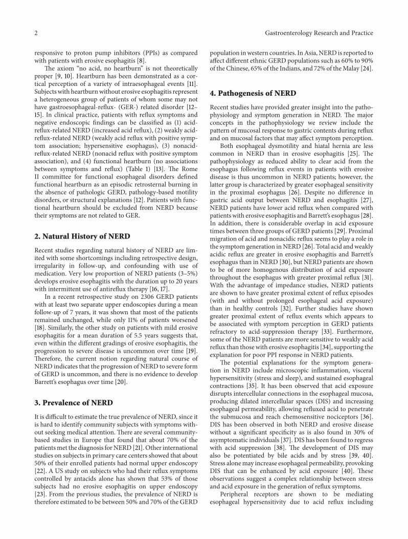
2 Gastroenterology Research and Practice
responsive to proton pump inhibitors (PPIs) as comparedwith patients with erosive esophagitis [8].
The axiom “no acid, no heartburn” is not theoreticallyproper [9, 10]. Heartburn has been demonstrated as a cor-tical perception of a variety of intraesophageal events [11].Subjectswith heartburnwithout erosive esophagitis representa heterogeneous group of patients of whom some may nothave gastroesophageal-reflux- (GER-) related disorder [12–15]. In clinical practice, patients with reflux symptoms andnegative endoscopic findings can be classified as (1) acid-reflux-related NERD (increased acid reflux), (2) weakly acid-reflux-related NERD (weakly acid reflux with positive symp-tom association; hypersensitive esophagus), (3) nonacid-reflux-related NERD (nonacid reflux with positive symptomassociation), and (4) functional heartburn (no associationsbetween symptoms and reflux) (Table 1) [13]. The RomeII committee for functional esophageal disorders definedfunctional heartburn as an episodic retrosternal burning inthe absence of pathologic GERD, pathology-based motilitydisorders, or structural explanations [12]. Patients with func-tional heartburn should be excluded from NERD becausetheir symptoms are not related to GER.
2. Natural History of NERD
Recent studies regarding natural history of NERD are lim-ited with some shortcomings including retrospective design,irregularity in follow-up, and confounding with use ofmedication. Very low proportion of NERD patients (3–5%)develops erosive esophagitis with the duration up to 20 yearswith intermittent use of antireflux therapy [16, 17].
In a recent retrospective study on 2306 GERD patientswith at least two separate upper endoscopies during a meanfollow-up of 7 years, it was shown that most of the patientsremained unchanged, while only 11% of patients worsened[18]. Similarly, the other study on patients with mild erosiveesophagitis for a mean duration of 5.5 years suggests that,even within the different gradings of erosive esophagitis, theprogression to severe disease is uncommon over time [19].Therefore, the current notion regarding natural course ofNERD indicates that the progression of NERD to severe formof GERD is uncommon, and there is no evidence to developBarrett’s esophagus over time [20].
3. Prevalence of NERD
It is difficult to estimate the true prevalence of NERD, since itis hard to identify community subjects with symptoms with-out seeking medical attention. There are several community-based studies in Europe that found that about 70% of thepatientsmet the diagnosis forNERD [21]. Other internationalstudies on subjects in primary care centers showed that about50% of their enrolled patients had normal upper endoscopy[22]. A US study on subjects who had their reflux symptomscontrolled by antacids alone has shown that 53% of thosesubjects had no erosive esophagitis on upper endoscopy[23]. From the previous studies, the prevalence of NERD istherefore estimated to be between 50% and 70% of the GERD
population inwestern countries. InAsia,NERD is reported toaffect different ethnic GERD populations such as 60% to 90%of the Chinese, 65% of the Indians, and 72% of theMalay [24].
4. Pathogenesis of NERD
Recent studies have provided greater insight into the patho-physiology and symptom generation in NERD. The majorconcepts in the pathophysiology we review include thepattern of mucosal response to gastric contents during refluxand on mucosal factors that may affect symptom perception.
Both esophageal dysmotility and hiatal hernia are lesscommon in NERD than in erosive esophagitis [25]. Thepathophysiology as reduced ability to clear acid from theesophagus following reflux events in patients with erosivedisease is thus uncommon in NERD patients; however, thelatter group is characterized by greater esophageal sensitivityin the proximal esophagus [26]. Despite no difference ingastric acid output between NERD and esophagitis [27],NERD patients have lower acid reflux when compared withpatients with erosive esophagitis and Barrett’s esophagus [28].In addition, there is considerable overlap in acid exposuretimes between three groups of GERD patients [29]. Proximalmigration of acid and nonacidic reflux seems to play a role inthe symptomgeneration inNERD [26]. Total acid andweaklyacidic reflux are greater in erosive esophagitis and Barrett’sesophagus than in NERD [30], but NERD patients are shownto be of more homogenous distribution of acid exposurethroughout the esophagus with greater proximal reflux [31].With the advantage of impedance studies, NERD patientsare shown to have greater proximal extent of reflux episodes(with and without prolonged esophageal acid exposure)than in healthy controls [32]. Further studies have showngreater proximal extent of reflux events which appears tobe associated with symptom perception in GERD patientsrefractory to acid-suppression therapy [33]. Furthermore,some of the NERD patients are more sensitive to weakly acidreflux than thosewith erosive esophagitis [34], supporting theexplanation for poor PPI response in NERD patients.
The potential explanations for the symptom genera-tion in NERD include microscopic inflammation, visceralhypersensitivity (stress and sleep), and sustained esophagealcontractions [35]. It has been observed that acid exposuredisrupts intercellular connections in the esophageal mucosa,producing dilated intercellular spaces (DIS) and increasingesophageal permeability, allowing refluxed acid to penetratethe submucosa and reach chemosensitive nociceptors [36].DIS has been observed in both NERD and erosive diseasewithout a significant specificity as is also found in 30% ofasymptomatic individuals [37]. DIS has been found to regresswith acid suppression [38]. The development of DIS mayalso be potentiated by bile acids and by stress [39, 40].Stress alonemay increase esophageal permeability, provokingDIS that can be enhanced by acid exposure [40]. Theseobservations suggest a complex relationship between stressand acid exposure in the generation of reflux symptoms.
Peripheral receptors are shown to be mediatingesophageal hypersensitivity due to acid reflux including
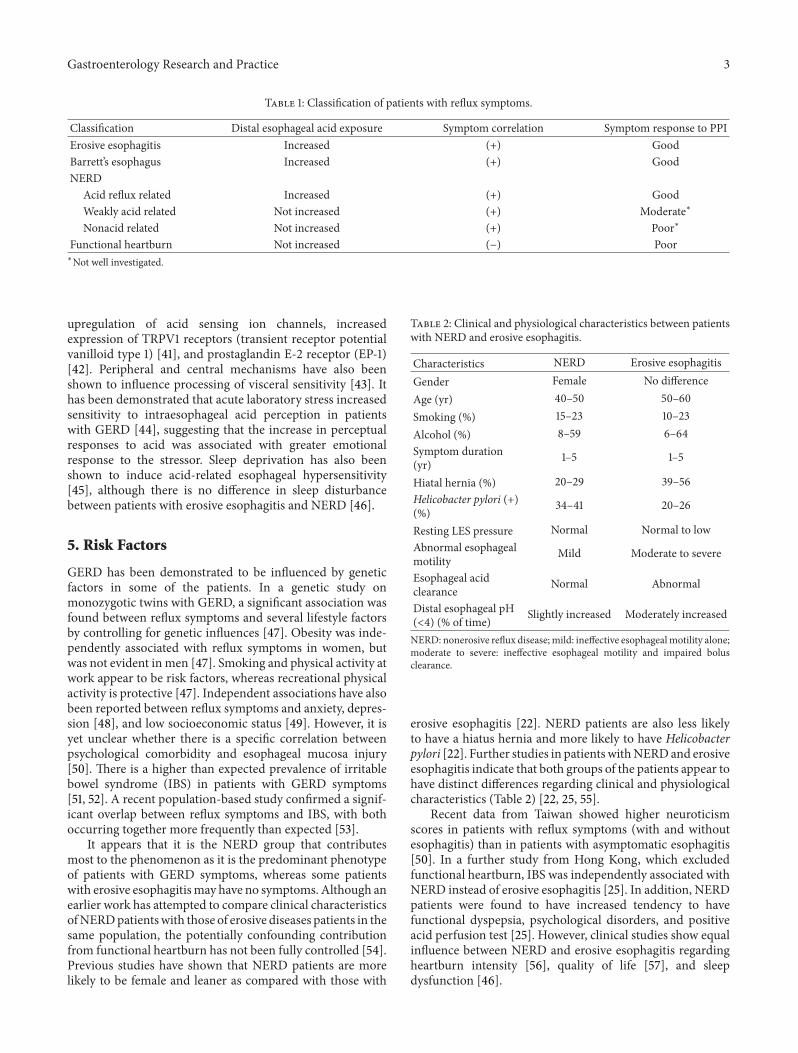
Gastroenterology Research and Practice 3
Table 1: Classification of patients with reflux symptoms.
Classification Distal esophageal acid exposure Symptom correlation Symptom response to PPIErosive esophagitis Increased (+) GoodBarrett’s esophagus Increased (+) GoodNERD
Acid reflux related Increased (+) GoodWeakly acid related Not increased (+) Moderate∗
Nonacid related Not increased (+) Poor∗
Functional heartburn Not increased (−) Poor∗Not well investigated.
upregulation of acid sensing ion channels, increasedexpression of TRPV1 receptors (transient receptor potentialvanilloid type 1) [41], and prostaglandin E-2 receptor (EP-1)[42]. Peripheral and central mechanisms have also beenshown to influence processing of visceral sensitivity [43]. Ithas been demonstrated that acute laboratory stress increasedsensitivity to intraesophageal acid perception in patientswith GERD [44], suggesting that the increase in perceptualresponses to acid was associated with greater emotionalresponse to the stressor. Sleep deprivation has also beenshown to induce acid-related esophageal hypersensitivity[45], although there is no difference in sleep disturbancebetween patients with erosive esophagitis and NERD [46].
5. Risk Factors
GERD has been demonstrated to be influenced by geneticfactors in some of the patients. In a genetic study onmonozygotic twins with GERD, a significant association wasfound between reflux symptoms and several lifestyle factorsby controlling for genetic influences [47]. Obesity was inde-pendently associated with reflux symptoms in women, butwas not evident inmen [47]. Smoking and physical activity atwork appear to be risk factors, whereas recreational physicalactivity is protective [47]. Independent associations have alsobeen reported between reflux symptoms and anxiety, depres-sion [48], and low socioeconomic status [49]. However, it isyet unclear whether there is a specific correlation betweenpsychological comorbidity and esophageal mucosa injury[50]. There is a higher than expected prevalence of irritablebowel syndrome (IBS) in patients with GERD symptoms[51, 52]. A recent population-based study confirmed a signif-icant overlap between reflux symptoms and IBS, with bothoccurring together more frequently than expected [53].
It appears that it is the NERD group that contributesmost to the phenomenon as it is the predominant phenotypeof patients with GERD symptoms, whereas some patientswith erosive esophagitismay have no symptoms. Although anearlier work has attempted to compare clinical characteristicsofNERDpatientswith those of erosive diseases patients in thesame population, the potentially confounding contributionfrom functional heartburn has not been fully controlled [54].Previous studies have shown that NERD patients are morelikely to be female and leaner as compared with those with
Table 2: Clinical and physiological characteristics between patientswith NERD and erosive esophagitis.
Characteristics NERD Erosive esophagitisGender Female No differenceAge (yr) 40–50 50–60Smoking (%) 15–23 10–23Alcohol (%) 8–59 6–64Symptom duration(yr) 1–5 1–5
Hiatal hernia (%) 20–29 39–56Helicobacter pylori (+)(%) 34–41 20–26
Resting LES pressure Normal Normal to lowAbnormal esophagealmotility Mild Moderate to severe
Esophageal acidclearance Normal Abnormal
Distal esophageal pH(<4) (% of time) Slightly increased Moderately increased
NERD: nonerosive reflux disease;mild: ineffective esophagealmotility alone;moderate to severe: ineffective esophageal motility and impaired bolusclearance.
erosive esophagitis [22]. NERD patients are also less likelyto have a hiatus hernia and more likely to have Helicobacterpylori [22]. Further studies in patientswithNERDand erosiveesophagitis indicate that both groups of the patients appear tohave distinct differences regarding clinical and physiologicalcharacteristics (Table 2) [22, 25, 55].
Recent data from Taiwan showed higher neuroticismscores in patients with reflux symptoms (with and withoutesophagitis) than in patients with asymptomatic esophagitis[50]. In a further study from Hong Kong, which excludedfunctional heartburn, IBS was independently associated withNERD instead of erosive esophagitis [25]. In addition, NERDpatients were found to have increased tendency to havefunctional dyspepsia, psychological disorders, and positiveacid perfusion test [25]. However, clinical studies show equalinfluence between NERD and erosive esophagitis regardingheartburn intensity [56], quality of life [57], and sleepdysfunction [46].

4 Gastroenterology Research and Practice
24-hour esophageal impedance and pH monitor
Patients with reflux symptoms and normal endoscopy
Increased acid exposure time63 (42%)
Normal acid exposure time87 (58%)
Positive SAP48 (32%)
Negative SAP39 (26%)
Weakly acid reflux
29 (20%)Nonacid reflux
19 (12%)
NERD acid reflux
NERD nonacid refluxNERD (hypersensitive esophagus)
weakly acid reflux
Functional heartburn
Figure 1: Classification of patients with reflux symptoms and normal endoscopy (SAP: symptom association probability).
6. Diagnosis of True NERD andFunctional Heartburn
6.1. Endoscopic Image. Currently, NERD is differentiatedfrom erosive esophagitis bywhite light endoscopy, andNERDis further differentiated from functional heartburn by usingpH monitoring (±impedance) with symptom reflux associa-tion. Recent technological advances may improve diagnosticsensitivity regarding upper endoscopy. Due to a significantoverlap in the amount of reflux episodes between patientswith NERD and erosive esophagitis [30], it is suggested thatmucosal changes in NERD patients may be too subtle to bedetected by conventional endoscopy. A recent study has con-firmed the clinical utility of magnification endoscopy withnarrow band imaging (NBI) which provides detailed findingsin reflux diseases which are not visible by conventionalendoscopy [58]. This study has shown several subtle changesin the esophageal mucosa which were identified to be highlyassociated with reflux disease. NERD patients appear to haveintrapapillary capillary loops and microerosions identifiedon NBI than controls. The notation is also evident in sub-group analysis when NERD patients and esophagitis patientswere compared with controls. However, despite excellentinterobserver agreement for NBI findings, the drawback ofNBI alone is present as modest intraobserver agreementhas been demonstrated [58]. Further studies of NBI suggestthat combined NBI with conventional findings gives theresolution for improving diagnostic accuracy for NERD byupper endoscopy [59].
6.2. 24-Hour Impedance pH Monitoring. 24-hour esophagealpH monitoring has been criticized for having limited sensi-tivity in diagnosing GERD; however, this technique is stillessential for the diagnosis of NERD. The limitation of con-ventional pH monitoring has been overcome by combining
pH with impedance monitoring [13, 60]. 24-hour impedancepH monitoring enables detection of acidic, weakly acidic,and nonacidic reflux and correlation with symptoms. Thistechnique is able to identify three subsets of NERD (i.e.,patients with an excess of acid, with a hypersensitiveesophagus [to weakly acidic reflux], or with nonacid-reflux-related symptom) and patients with functional heartburn.Savarino et al. investigated the data of combined impedancepH monitoring in 150 patients with reflux symptoms andnegative endoscopy under off-PPI condition (Figure 1). Itwas concluded that adding impedance to pH monitoringimproved the diagnostic sensitivity mainly by identifying apositive symptom association probability with weakly acidor nonacid reflux in patients off PPI therapy [13]. By usingthis advanced technique in a group of patients with refluxsymptoms not taking PPI, it was observed that the value ofadding impedance measurement to standard pH monitoringcould increase the observed positive symptom-reflux eventassociation that might improve the diagnostic sensitivity ofNERD [61]. From the findings previous, although combinedimpedance and pH measurement is necessary to reliablydistinguish NERD patients from patients with functionalheartburn, the test is not commonly used in general practice,and the response to PPI is more realizable than to identifythose with functional heartburn [62]. Furthermore, NERDwith weakly acid reflux is relatively uncommon without thecondition during acid-suppression treatment.
7. Treatment of NERD
7.1. PPIs. PPIs are the most recommended and effectiveagents employed in the treatment of GERD. The advantageof PPIs relieving reflux symptoms is also found in NERDpatients. PPIs are more effective than other acid-suppressingagents such as histamine-2 receptor antagonists (H2RAs).

Gastroenterology Research and Practice 5
It has been demonstrated in NERD patients that the relativerisk for PPIs versus H2RAs was 0.74 (95% CI: 0.53–1.03) forcontrolling heartburn [63].
Initially, patients can be treated by a proton pumpinhibitor (PPI; standard dose, once daily) for 2–4 weeks. Ifinitial treatment fails to elicit adequate symptom control,increasing the PPI dose (standard dose PPI twice daily) isrecommended. In patients with poor response to appropriatePPI treatment, esophageal pH (±impedance) monitoringis indicated to differentiate pathological acid reflux, acid-sensitive (hypersensitive) esophagus, and functional heart-burn. The beneficial effects of PPIs in achieving symptomrelief in NERD have been well documented in several studies.The rates of the relief of symptoms are shown to be 40–60%for omeprazole and rabeprazole 20mg/day and about 30% foromeprazole 10mg/day for 4 weeks [7, 64, 65]. By using thewireless Bravo pH monitoring, normalization of esophagealacid exposure is found in NERD patients within 48 hoursafter starting PPIs [66].
NERD patients have been shown to be less responsive toPPIs as compared with patients with erosive esophagitis byapproximately 20–30% after 4 weeks of the treatment [8].Theoverall PPI symptomatic response rate was 36.7% (95% CI:34.1–39.3) in NERD and 55.5% (95% CI: 51.5–59.5) in erosiveesophagitis, whereas the rate of therapeutic gain was 27.5%in NERD and 48.9% in erosive esophagitis [8]. In NERDpatients, the response rate appears to positively correlate withthe extent of distal esophageal acid exposure with the highersymptom resolution in patients with greater acid exposure[7]. Furthermore, patients with NERD demonstrate similarsymptomatic response to half and full standard dose of PPIas a prior study has shown a similar median time to firstsymptom relief (2 days) and to sustained symptom relief(10–13 days) for pantoprazole (20mg/day) and esomeprazole(20mg/day) [67]. In a subsequent study, administration of alower dose of rabeprazole (5mg/day) is not superior to halfdose rabeprazole (10mg/day) for heartburn relief [68].
Studies have demonstrated that on-demand or inter-mittent PPI therapy is also an effective strategy in NERDtreatment [69]. Due to the fact that most of the NERD isless likely to be progressive [20, 70], treatment for thosepatients can be tailored by the presence of their symptoms.Therefore, on-demand or intermittent therapy is widely usedas alternative PPI treatment forNERDpatients [71, 72], whichalso has the advantage of convenience, stable acid control,cost effectiveness, and reducing the chance of acid rebound.
Dexlansoprazole MR is an R-enantiomer of lansoprazolewith dual delayed-release benefit in prolonging plasma con-centration and pharmacodynamic effects better than thoseof single-release PPIs with its administration allowed at anytime of the day without regard to meals. In patients withNERD, dexlansoprazole MR 30mg daily has been shown tobe more efficacious than placebo in controlling heartburn[73].
7.2. NovelTherapeuticModalities. There are novel therapeuticmodalities developed specifically for NERD patients. Thetargets for novel therapy are thought to be improving the
competence of LES function such as new GABA-B agonists,better acid-suppression therapy, normalizing esophageal sen-sitivity, and augmenting esophageal motility. In patients withfailure to respond to PPI treatment, it has been suggestedthat pain modulators like tricyclics and selective serotoninreuptake inhibitors are an alternative treatment option forcontrolling refractory symptoms such as heartburn and chestpain [74, 75]. However, there is no sufficient evidence tosupport their efficacy in PPI-failure patients. In patients withPPI failure, the use of pain modulators alone or combinedwith PPIs can be a treatment strategy, but further studies needto confirm such approach in PPI-failure patients.
The role of antireflux surgery NERD has not beenwellestablished. In general, NERD patients are less responsive toantireflux surgery [76]. In one earlier study comparing theclinical outcome of antireflux surgery between patients witherosive esophagitis and NERD, it was demonstrated that 91%versus 56% reported heartburn resolution, 24% versus 50%reported dysphagia after surgery, and 94% versus 79% weresatisfied with surgery, respectively [76].
8. Conclusions
The definition of GERD is well established and simplyunderstood, whereas the NERD has been intangibly definedwithmore conditions needed, largely because of the increasedrecognition of functional heartburn due to the evolution ofthe Rome criteria for functional gastrointestinal disorders.NERD is generally accepted as an entity within the broaderdefinition of GERD by excluding functional heartburn.NERDhas been increasingly recognized as themost commoncause of reflux symptoms in community population withimpact on quality of life. Mechanisms of the symptomgeneration in NERD remain complex, and stress may play arole in the symptomgeneration. Treatmentwith PPIs remainsthe choice of the therapy in NERD patients, but may be lesseffective when compared with those with erosive esophagitis.The role of anti-reflux surgery inNERD remains to be furtherinvestigated and defined. PPIs therapy with intermittent oron-demand fashion can be an alternative treatment strategyinmost of the NERD patients due to the relatively low risk forthe progression to erosive esophagitis or Barrett’s esophagus.
References
[1] N. Vakil, S. V. van Zanten, P. Kahrilas et al., “The Montrealdefinition and classification of gastroesophageal reflux disease:a global evidence-based consensus,” American Journal of Gas-troenterology, vol. 101, no. 8, pp. 1900–1943, 2006.
[2] P. Moayyedi and N. J. Talley, “Gastro-oesophageal reflux dis-ease,”The Lancet, vol. 367, no. 9528, pp. 2086–2100, 2006.
[3] K. M. Fock, N. J. Talley, R. Fass et al., “Asia-Pacific consensuson themanagement of gastroesophageal reflux disease: update,”Journal of Gastroenterology and Hepatology, vol. 23, no. 1, pp. 8–22, 2008.
[4] I. Wiklund, “Review of the quality of life and burden of illnessin gastroesophageal reflux disease,” Digestive Diseases, vol. 22,no. 2, pp. 108–114, 2004.

6 Gastroenterology Research and Practice
[5] H. B. El-Serag, “Epidemiology of non-erosive reflux disease,”Digestion, vol. 78, supplement 1, pp. 6–10, 2008.
[6] I. M.Modlin, R. H. Hunt, P.Malfertheiner et al., “Diagnosis andmanagement of non-erosive reflux disease—the vevey NERDconsensus group,” Digestion, vol. 80, no. 2, pp. 74–88, 2009.
[7] T. Lind, T. Havelund, R. Carlsson et al., “Heartburn with-out oesophagitis: efficacy of omeprazole therapy and featuresdetermining therapeutic response,” Scandinavian Journal ofGastroenterology, vol. 32, no. 10, pp. 974–979, 1997.
[8] B. B. Dean, A. D. Gano Jr., K. Knight, J. J. Ofman, and R. Fass,“Effectiveness of proton pump inhibitors in nonerosive refluxdisease,” Clinical Gastroenterology and Hepatology, vol. 2, no. 8,pp. 656–664, 2004.
[9] R. Fass, M. B. Fennerty, and N. Vakil, “Nonerosive refluxdisease—current concepts and dilemmas,” American Journal ofGastroenterology, vol. 96, no. 2, pp. 303–314, 2001.
[10] J. E. Richter and D. O. Castell, “Gastroesophageal reflux. Patho-genesis, diagnosis, and therapy,” Annals of Internal Medicine,vol. 97, no. 1, pp. 93–103, 1982.
[11] R. Fass and G. Tougas, “Functional heartburn: the stimulus, thepain, and the brain,” Gut, vol. 51, no. 6, pp. 885–892, 2002.
[12] The Rome International Working Teams, “Functional heart-burn,” in Rome II: the Functional Gastrointestinal Disorders, D.A. Drossman, E. Corazziari, N. J. Talley et al., Eds., pp. 275–278,Lawrence, Allen Press, 2000.
[13] E. Savarino, P. Zentilin, R. Tutuian et al., “The role of nonacidreflux in NERD: lessons learned from impedance-pH monitor-ing in 150 patients off therapy,” American Journal of Gastroen-terology, vol. 103, no. 11, pp. 2685–2693, 2008.
[14] T. Hershcovici and R. Fass, “Nonerosive Reflux Disease(NERD)—an update,”Neurogastroenterology &Motility, vol. 16,no. 1, pp. 8–21, 2010.
[15] J.W.Winter and R. C. Heading, “The nonerosive reflux disease-gastroesophageal reflux disease controversy,” Current Opinionin Gastroenterology, vol. 24, no. 4, pp. 509–515, 2008.
[16] J. Isolauri, M. Luostarinen, E. Isolauri, P. Reinikainen, M. Vil-jakka, and O. Keyrilainen, “Natural course of gastroesophagealreflux disease: 17-22 year follow-up of 60 patients,” AmericanJournal of Gastroenterology, vol. 92, no. 1, pp. 37–41, 1997.
[17] E. Kuster, E. Ros, V. Toledo-Pimentel et al., “Predictive factorsof the long term outcome in gastro-oesophageal reflux disease:six year follow up of 107 patients,” Gut, vol. 35, no. 1, pp. 8–14,1994.
[18] S. Sontag, T. Schnell, G. Chejfec, A. Metz, and A. Sonnenberg,“The benign natural history of GERD: 20 years with rare seriousprogression,” American Journal of Gastroenterology, vol. 94,1999, abstract 2604.
[19] N. Manabe, M. Yoshihara, A. Sasaki, S. Tanaka, K. Haruma,and K. Chayama, “Clinical characteristics and natural historyof patients with low-grade reflux esophagitis,” Journal of Gas-troenterology and Hepatology, vol. 17, no. 9, pp. 949–954, 2002.
[20] J. Labenz, M. Nocon, T. Lind et al., “Prospective follow-up datafrom the ProGERD study suggest that GERD is not a categorialdisease,” American Journal of Gastroenterology, vol. 101, no. 11,pp. 2457–2462, 2006.
[21] J.-P. Galmiche, P. Barthelemy, and B. Hamelin, “Treating thesymptoms of gastro-oesophageal reflux disease: a double-blindcomparison of omeprazole and cisapride,” Alimentary Pharma-cology andTherapeutics, vol. 11, no. 4, pp. 765–773, 1997.
[22] R. Carlsson, J. Dent, R. Watts et al., “Gastro-oesophageal refluxdisease in primary care: an international study of different
treatment strategies with omeprazole,” European Journal ofGastroenterology and Hepatology, vol. 10, no. 2, pp. 119–124,1998.
[23] M. Robinson, D. Earnest, S. Rodriguez-Stanley et al., “Heart-burn requiring frequent antacid use may indicate significantillness,” Archives of Internal Medicine, vol. 158, no. 21, pp. 2373–2376, 1998.
[24] M. S. Rosaida and K.-L. Goh, “Gastro-oesophageal refluxdisease, reflux oesophagitis and non-erosive reflux disease in amultiracial Asian population: a prospective, endoscopy basedstudy,” European Journal of Gastroenterology and Hepatology,vol. 16, no. 5, pp. 495–501, 2004.
[25] J. C. Y. Wu, C. M. Y. Cheung, V. W. S. Wong, and J. J. Y.Sung, “Distinct clinical characteristics between patients withnonerosive reflux disease and those with reflux esophagitis,”Clinical Gastroenterology and Hepatology, vol. 5, no. 6, pp. 690–695, 2007.
[26] S. Emerenziani, M. Ribolsi, D. Sifrim, K. Blondeau, and M.Cicala, “Regional oesophageal sensitivity to acid and weaklyacidic reflux in patients with non-erosive reflux disease,” Neu-rogastroenterology & Motility, vol. 21, no. 3, pp. 253–258, 2009.
[27] K. Y. Ho and J. Y. Kang, “Reflux esophagitis patients in Sin-gapore have motor and acid exposure abnormalities similarto patients in the Western hemisphere,” American Journal ofGastroenterology, vol. 94, no. 5, pp. 1186–1191, 1999.
[28] S. D.Martinez, I. B.Malagon, H. S. Garewal, H. Cui, and R. Fass,“Non-erosive reflux disease (NERD)—acid reflux and symptompatterns,” Alimentary Pharmacology and Therapeutics, vol. 17,no. 4, pp. 537–545, 2003.
[29] M. Shapiro, C. Green, E.M. Faybush, R. F. Esquivel, and R. Fass,“The extent of oesophageal acid exposure overlap among thedifferent gastro-oesophageal reflux disease groups,” AlimentaryPharmacology andTherapeutics, vol. 23, no. 2, pp. 321–329, 2006.
[30] A. J. Bredenoord, G. J. M. Hemmink, and A. J. P. M. Smout,“Relationship between gastro-oesophageal reflux pattern andseverity of mucosal damage,” Neurogastroenterology & Motility,vol. 21, no. 8, pp. 807–812, 2009.
[31] R. Dickman, J. M. Bautista, W.-M. Wong et al., “Comparisonof esophageal acid exposure distribution along the esophagusamong the different gastroesophageal reflux disease (GERD)groups,” American Journal of Gastroenterology, vol. 101, no. 11,pp. 2463–2469, 2006.
[32] A. J. Bredenoord, B. L. A. M. Weusten, W. L. Curvers, R.Timmer, and A. J. P. M. Smout, “Determinants of perceptionof heartburn and regurgitation,” Gut, vol. 55, no. 3, pp. 313–318,2006.
[33] F. Zerbib, A. Duriez, S. Roman, M. Capdepont, and F. Mion,“Determinants of gastro-oesophageal reflux perception inpatients with persistent symptoms despite proton pumpinhibitors,” Gut, vol. 57, no. 2, pp. 156–160, 2008.
[34] S. Emerenziani, D. Sifrim, F. I. Habib et al., “Presence of gas inthe refluxate enhances reflux perception in non-erosive patientswith physiological acid exposure of the oesophagus,” Gut, vol.57, no. 4, pp. 443–447, 2008.
[35] D. Ang, D. Sifrim, and J. Tack, “Mechanisms of heartburn,”Nature Clinical Practice Gastroenterology and Hepatology, vol.5, no. 7, pp. 383–392, 2008.
[36] H. van Malenstein, R. Farre, and D. Sifrim, “Esophageal dilatedintercellular spaces (DIS) and nonerosive reflux disease,” Amer-ican Journal of Gastroenterology, vol. 103, no. 4, pp. 1021–1028,2008.

Gastroenterology Research and Practice 7
[37] R. Caviglia,M. Ribolsi, N.Maggiano et al., “Dilated intercellularspaces of esophageal epithelium in nonerosive reflux diseasepatients with physiological esophageal acid exposure,” Amer-ican Journal of Gastroenterology, vol. 100, no. 3, pp. 543–548,2005.
[38] C. Calabrese, M. Bortolotti, A. Fabbri et al., “Reversibility ofGERD ultrastructural alterations and relief of symptoms afteromeprazole treatment,” American Journal of Gastroenterology,vol. 100, no. 3, pp. 537–542, 2005.
[39] C. Calabrese, A. Fabbri, M. Bortolotti et al., “Dilated intercel-lular spaces as a marker of oesophageal damage: comparativeresults in gastro-oesophageal reflux disease with or without bilereflux,” Alimentary Pharmacology and Therapeutics, vol. 18, no.5, pp. 525–532, 2003.
[40] R. Farre, R. De Vos, K. Geboes et al., “Critical role of stressin increased oesophageal mucosa permeability and dilatedintercellular spaces,” Gut, vol. 56, no. 9, pp. 1191–1197, 2007.
[41] Y. M. Bhat and K. Bielefeldt, “Capsaicin receptor (TRPV1) andnon-erosive reflux disease,” European Journal of Gastroenterol-ogy and Hepatology, vol. 18, no. 3, pp. 263–270, 2006.
[42] S. Sarkar, A. R. Hobson, A. Hughes et al., “The prostaglandin E2receptor-1 (EP-1)mediates acid-induced visceral pain hypersen-sitivity in humans,” Gastroenterology, vol. 124, no. 1, pp. 18–25,2003.
[43] P. Anand, Q. Aziz, R. Willert, and L. van Oudenhove, “Periph-eral and central mechanisms of visceral sensitization in man,”Neurogastroenterology &Motility, vol. 19, no. 1, supplement, pp.29–46, 2007.
[44] R. Fass, B. D. Naliboff, S. S. Fass et al., “The effect of auditorystress on perception of intraesophageal acid in patients withgastroesophageal reflux disease,” Gastroenterology, vol. 134, no.3, pp. 696–705, 2008.
[45] R. Schey, R. Dickman, S. Parthasarathy et al., “Sleep deprivationis hyperalgesic in patients with gastroesophageal reflux disease,”Gastroenterology, vol. 133, no. 6, pp. 1787–1795, 2007.
[46] C.-H. Yi, C.-T. Hu, and C.-L. Chen, “Sleep dysfunction inpatients with GERD: erosive versus nonerosive reflux disease,”American Journal of theMedical Sciences, vol. 334, no. 3, pp. 168–170, 2007.
[47] Z. Zheng, H. Nordenstedt, N. L. Pedersen, J. Lagergren, and W.Ye, “Lifestyle factors and risk for symptomatic gastroesophagealreflux in monozygotic twins,” Gastroenterology, vol. 132, no. 1,pp. 87–95, 2007.
[48] C. Jansson, H. Nordenstedt, M.-A. Wallander et al., “Severegastro-oesophageal reflux symptoms in relation to anxiety,depression and coping in a population-based study,”AlimentaryPharmacology andTherapeutics, vol. 26, no. 5, pp. 683–691, 2007.
[49] C. Jansson, H. Nordenstedt, S. Johansson et al., “Relationbetween gastroesophageal reflux symptoms and socioeconomicfactors: a population-based study (the HUNT study),” ClinicalGastroenterology and Hepatology, vol. 5, no. 9, pp. 1029–1034,2007.
[50] Y.-C. Lee, H.-P. Wang, H.-M. Chiu et al., “Comparative analysisbetween psychological and endoscopic profiles in patients withgastroesophageal reflux disease: a prospective study based onscreening endoscopy,” Journal of Gastroenterology and Hepatol-ogy, vol. 21, no. 5, pp. 798–804, 2006.
[51] I. Nastaskin, E. Mehdikhani, J. Conklin, S. Park, and M.Pimentel, “Studying the overlap between IBS and GERD: asystematic review of the literature,” Digestive Diseases andSciences, vol. 51, no. 12, pp. 2113–2120, 2006.
[52] M. Pimentel, F. Rossi, E. J. Chow et al., “Increased prevalenceof irritable bowel syndrome in patients with gastroesophagealreflux,” Journal of Clinical Gastroenterology, vol. 34, no. 3, pp.221–224, 2002.
[53] H.-K. Jung, S. Halder, M. McNally et al., “Overlap of gastro-oesophageal reflux disease and irritable bowel syndrome: preva-lence and risk factors in the general population,” AlimentaryPharmacology andTherapeutics, vol. 26, no. 3, pp. 453–461, 2007.
[54] R. Fass, “Erosive esophagitis and nonerosive reflux disease(NERD): comparison of epidemiologic, physiologic, and thera-peutic characteristics,” Journal of Clinical Gastroenterology, vol.41, no. 2, pp. 131–137, 2007.
[55] R. Fass, “Epidemiology and pathophysiology of symptomaticgastroesophageal reflux disease,” American Journal of Gastroen-terology, vol. 98, no. 3, supplement, pp. S2–S7, 2003.
[56] A. J. P. M. Smout, “Endoscopy-negative acid reflux disease,”Alimentary Pharmacology andTherapeutics, vol. 11, supplement2, pp. 81–85, 1997.
[57] T. L. Venables, R. D. Newland, A. C. Patel, J. Hole, C.Wilcock, and M. L. Turbitt, “Omeprazole 10 milligrams oncedaily, omeprazole 20 milligrams once daily, or ranitidine 150milligrams twice daily, evaluated as initial therapy for the reliefof symptoms of gastro-oesophageal reflux disease in generalpractice,” Scandinavian Journal of Gastroenterology, vol. 32, no.10, pp. 965–973, 1997.
[58] P. Sharma, S. Wani, A. Bansal et al., “A feasibility trial of narrowband imaging endoscopy in patients with gastroesophagealreflux disease,” Gastroenterology, vol. 133, no. 2, pp. 454–464,2007.
[59] Y.-C. Lee, J.-T. Lin, H.-M. Chiu et al., “Intraobserver andinterobserver consistency for grading esophagitis with narrow-band imaging,” Gastrointestinal Endoscopy, vol. 66, no. 2, pp.230–236, 2007.
[60] F. Zerbib, S. Roman, A. Ropert et al., “Esophageal pH-impedance monitoring and symptom analysis in GERD: astudy in patients off and on therapy,” American Journal ofGastroenterology, vol. 101, no. 9, pp. 1956–1963, 2006.
[61] A. J. Bredenoord, B. L. A. M. Weusten, R. Timmer, J. M.Conchillo, and A. J. P. M. Smout, “Addition of esophagealimpedance monitoring to pH monitoring increases the yieldof symptom association analysis in patients off PPI therapy,”American Journal of Gastroenterology, vol. 101, no. 3, pp. 453–459, 2006.
[62] A. Gasiorowska and R. Fass, “The proton pump inhibitor (PPI)test in GERD: does it still have a role?” Journal of ClinicalGastroenterology, vol. 42, no. 8, pp. 867–874, 2008.
[63] B. van Pinxteren, M. E. Numans, P. A. Bonis, and J. Lau,“Short-term treatment with proton pump inhibitors, H
2
-receptor antagonists and prokinetics for gastro-oesophagealreflux disease-like symptoms and endoscopy negative refluxdisease,”CochraneDatabase of Systematic Reviews, no. 4, ArticleID CD002095, 2004.
[64] C. M. Bate, S. M. Griffin, P. W. N. Keeling et al., “Reflux symp-tom relief with omeprazole in patients without unequivocalesophagitis,” Alimentary Pharmacology and Therapeutics, vol.10, no. 4, pp. 547–555, 1996.
[65] P. Miner Jr., W. Orr, J. Filippone, L. Jokubaitis, and S. Sloan,“Rabeprazole in nonerosive gastroesophageal reflux disease:a randomized placebo-controlled trial,” American Journal ofGastroenterology, vol. 97, no. 6, pp. 1332–1339, 2002.
[66] C. Calabrese, G. Liguori, V. Gabusi et al., “Ninety-six-hourwireless oesophageal pH monitoring following proton pump
8 Gastroenterology Research and Practice
inhibitor administration in NERD patients,” Alimentary Phar-macology andTherapeutics, vol. 28, no. 2, pp. 250–255, 2008.
[67] H. Monnikes, B. Pfaffenberger, G. Gatz, J. Hein, and K. D.Bardhan, “Novel measurement of rapid treatment success withReQuest: first and sustained symptom relief as outcome param-eters in patients with endoscopy-negative GERD receiving 20mg pantoprazole or 20 mg esomeprazole,”Digestion, vol. 71, no.3, pp. 152–158, 2005.
[68] Y. Kinoshita, K. Ashida, and M. Hongo, “Randomised clinicaltrial: a multicentre, double-blind, placebo-controlled study onthe efficacy and safety of rabeprazole 5 mg or 10 mg oncedaily in patients with non-erosive reflux disease,” AlimentaryPharmacology andTherapeutics, vol. 33, no. 2, pp. 213–224, 2011.
[69] L. B. Gerson, A. S. Robbins, A. Garber, J. Hornberger, and G.Triadafilopoulos, “A cost-effectiveness analysis of prescribingstrategies in the management of gastroesophageal reflux dis-ease,” American Journal of Gastroenterology, vol. 95, no. 2, pp.395–407, 2000.
[70] R. Fass and J. J. Ofman, “Gastroesophageal reflux disease—should we adopt a new conceptual framework?” AmericanJournal of Gastroenterology, vol. 97, no. 8, pp. 1901–1909, 2002.
[71] P. Bytzer and A. L. Blum, “Personal view: rationale and pro-posed algorithms for symptom-based proton pump inhibitortherapy for gastro-oesophageal reflux disease,” AlimentaryPharmacology and Therapeutics, vol. 20, no. 4, pp. 389–398,2004.
[72] P. Juul-Hansen and A. Rydning, “On-demand requirementsof patients with endoscopy-negative gastro-oesophageal refluxdisease: H2-blocker vs. proton pump inhibitor,” AlimentaryPharmacology andTherapeutics, vol. 29, no. 2, pp. 207–212, 2009.
[73] R. Fass, W. D. Chey, S. F. Zakko et al., “Clinical trial: the effectsof the proton pump inhibitor dexlansoprazole MR on daytimeand nighttime heartburn in patients with non-erosive refluxdisease,”Alimentary Pharmacology andTherapeutics, vol. 29, no.12, pp. 1261–1272, 2009.
[74] C. Prakash andR. E. Clouse, “Long-termoutcome from tricyclicantidepressant treatment of functional chest pain,” DigestiveDiseases and Sciences, vol. 44, no. 12, pp. 2373–2379, 1999.
[75] I. Varia, E. Logue, C. O’Connor et al., “Randomized trial ofsertraline in patients with unexplained chest pain of noncardiacorigin,” American Heart Journal, vol. 140, no. 3, pp. 367–372,2000.
[76] P. Fenton, M. L. Terry, K. D. Galloway, C. D. Smith, J. G. Hunter,and J. P.Waring, “Is there a role for laparoscopic fundoplicationin patients with non-erosive reflux disease (NERD),” Gastroen-terology, vol. 118, supplement 2, 2000, abstract A481.

Hindawi Publishing CorporationGastroenterology Research and PracticeVolume 2013, Article ID 481417, 6 pageshttp://dx.doi.org/10.1155/2013/481417
Review ArticleAntireflux Endoluminal Therapies: Past and Present
Kuo Chao Yew1 and Seng-Kee Chuah2
1 Department of Gastroenterology and Hepatology, Tan Tock Seng Hospital, 11 Jalan Tan Tock Seng, Singapore 3084332Division of Hepatogastroenterology, Department of Internal Medicine, Kaohsiung Chang Gung Memorial Hospital,Chang Gung University College of Medicine, 123 Ta-Pei Road, Niao-Sung Hsiang, Kaohsiung 833, Taiwan
Correspondence should be addressed to Seng-Kee Chuah; [email protected]
Received 18 April 2013; Accepted 13 June 2013
Academic Editor: Deng-Chyang Wu
Copyright © 2013 K. C. Yew and S.-K. Chuah. This is an open access article distributed under the Creative Commons AttributionLicense, which permits unrestricted use, distribution, and reproduction in any medium, provided the original work is properlycited.
The basic principle of antireflux procedures employing endoscopic intervention aims to create a mechanical barrier to preventprimary pathophysiology in gastroesophageal reflux disease (GERD). We review, highlight, and discuss the past and presentstatus of endoluminal therapy. Currently, there are 3 commonly employed anti-reflux endoluminal procedures: fundoplication orsuturing techniques (EndoCinch, NDO, and EsophyX), intramural injection or implant techniques (enhancing lower esophagealsphincter (LES) volume and/or strengthening compliance of the LES-Enteryx and Gatekeeper), and radiofrequency ablation ofLES and cardia. EndoCinch plication requires further study and modification of technique before it can be recommended becauseof durability issues. Esophynx, the transoral incisionless fundoplication, may reduce hiatal hernias and increase LES length.Preliminary studies have shown promising reduction in symptoms and medication use but evidence concerning safety and long-term durability is still pending. The safety issue with injection technique is the main concern as evident from the incidences ofimplant withdrawals after reported major adverse events. Future research with cautious monitoring is required before any newimplant material can be recommended for commercial application. Radiofrequency ablation therapy is regaining popularity intreating refractory symptoms despite PPI use due to improved efficacy, durability, and safety after years of refinement of protocol.
1. Introduction
Gastroesophageal reflux disease (GERD) is a disease spec-trum caused by regurgitation of stomach contents causingtroublesome esophageal or extraesophageal symptoms asdefined by Montreal definitions [1]. Either mild heartburnand/or regurgitation for at least 2 days per week or moderateto severe symptoms for at least one day per week quali-fies as significant symptom-based diagnosis [2]. Phenotyp-ical classifications of GERD are nonerosive reflux disease(NERD), erosive esophagitis (EE), and Barrett’s esophagus(BE). Population-based study reported 15%–20% of theWest-ern population experience reflux on a weekly basis which canlead to impoverishment of a country’s economy and qualityof life [3]. Dent et al. reported the prevalence of GERD inSweden (15.5%), Italy (11.8%), China, Japan, Korea (3.4%–8.5%), and Taiwan (9%–24.6%), respectively [4]. Subanalysisshows that EE and hiatus hernia aremore common in Europethan in Asia with the exception of Taiwan which reported
similar EE prevalence as Europe. Over years, the prevalenceofGERD is increasing by approximately 5%annually amongstthe American population and other countries in the Westsuch as Western Europe and Scandinavia [5]. Furthermore,the prevalence of GERD is also increasing in Asian countries[6]. Taiwan, with a population consisting mainly of Chineseancestry, has one of the most published data on GERD. Yehet al. reported 14.5% of prevalence in 1991 [7] only to besuperseded by a prevalence of approximately 25%-26% from2007 to 2011 [8, 9]. The social impact of GERD includes lostwork days and increased public health cost. Medically, thereis a risk of developing esophageal adenocarcinoma (EAC).Barrett’s esophagus will develop in an incidence rate of 0.5%per year and Barrett’s esophagus is a risk for esophagealadenocarcinoma (EAC) or of a 400-fold increased risk ofEAC [10]. GERD increases the risk of EAC by 8.6-fold [11].
GERD and its associated clinical manifestations present adiagnostic challenge. A third of patients with GERD presentwith atypical symptoms or in fact may be asymptomatic.

2 Gastroenterology Research and Practice
Wong et al. reported extraesophageal presentation or atypicalcomplaints such as asthma (4.8%), chronic cough (13%), andlaryngeal disorder (10%) [12]. Some authors have suggesteda trial of proton pump inhibitors (PPI) with a temporalimprovement in symptoms as an indirect diagnostic methodbut the risk is that BE, RE, and even EACmay be missed [13].
PPI have been the most effective treatment for GERDbut discontinuation of medical therapy is likely to lead toclinical relapse. Long-term PPI users and patients who arenoncompliant with daily PPI dosage may run into prob-lems such as refractory GERD, NERD, EE, and BE. Sideeffects of PPI use that are gaining more attention amongstphysicians include an increased incidence of hip fracturein postmenopausal women, pneumonia, enteric infections,and drug-drug interactions with clopidogrel.With long-termPPI use, there is also the issue of compliance and financialhealth costs. Therefore, some patients may be more suitedto other treatment options such as surgery or endoscopicintervention. Antireflux procedures either via surgery orendoscopic intervention aim to create a mechanical barrierto prevent primary pathophysiology in GERD. In this paper,we review, highlight, and discuss the past and present statusof the endoluminal therapies.
2. GERD Pathophysiology
The pathophysiological mechanisms of GERD are transientLES relaxation (TLESR), low LES pressure, andGEJ anatomicdistortions such as hiatal hernia [14]. Dysfunctional esophag-eal motility, impaired barrier function of LES, and gastricemptying in relation to meal intake may all lead to gastriccontent reflux. LES relaxation can be triggered by 3 mainmotor events: deglutitive inhibition during the swallowingprocess, secondary peristalsis from esophageal distention,and cardial distention mediated relaxation of LES. The thirdmechanism is transient TLESR with the sensory trigger pointlocated distal to LES [15]. It is the total relaxation duration andnot the frequency that contributes to this pathophysiology.Classical description involves relaxation of LES, esophagealshortening, and inhibition of crural diaphragm. A disruptionin the musculature plane such as hiatal hernia will blunt theangle of His and impair the flap valve mechanism.TheGERDcondition ismostly accompanied by the formation of a gastricair pocket which can stimulate the acid sensing ion channelprimarily located around the cardia leading to TLESR [15].Often it is bile acid that causes more mucosal damage thangastric secretion.
3. Rationale of AntirefluxEndoluminal Therapy
PPI therapy to decrease acid output cannot provide a phys-ical barrier or restore the LES function [16]. Relapses inesophageal and extraesophageal disease are also indicationsfor more definitive treatment. Chen et al. reported thatbile acid disrupts squamous epithelial barrier function bymodulating TJ proteins, demonstrating the importance ofthe integrity of an antireflux barrier in addition to estab-lished acid suppressive therapy [17]. Some patients may not
tolerate long-term medical therapy or be a candidate forsurgery. Under such circumstances, endoluminal antirefluxinterventions may be a viable option. Antireflux endoscopicintervention aims to create a mechanical barrier to preventthe regurgitation of gastric contents into the esophagus. Anappropriate high pressure zone at LES needs to be producedto aid the closure of diaphragmatic crural fibers and theHis angle of the gastroesophageal junction. An anatomic-physiological flap valve at gastroesophageal junction canbe created by antireflux barrier reconstruction of collarsling musculature at the cardia [18]. The antireflux barrierreconstruction procedure aims to reconstruct the acute angleof His by enveloping the distal esophagus to the proximalstomach mimicking an intragastric valve that will preventregurgitation of food in the presence of intragastric and intra-abdominal pressure [19].
Hill and Kozarek demonstrated that the LES pressuregradient can be increased by suturing a flap valve (valvu-loplasty) which can then be further enhanced with theposterior attachment of gastroesophageal junction [20].Withhiatus hernia, suturing the GEJ to a fixation point intra-abdominally at the preaortic fascia will have a similar effect.The presence of an intragastric mucosal ridge is far moreimportant in determining antireflux effect rather than anincreased LES pressure gradient. The valvular appearance isa good predictor of the reflux status. As there are limitationsin endoscopic manipulation of the esophagus, careful patientselection with little or no hiatal hernia is important todetermine the success of this approach [18]. In general, a largehiatal hernia (especially paraoesophageal) should be referredfor laparoscopic or open surgery. Other contraindicationsfor antireflux endoluminal therapy include patients withrefractory symptoms despite maximum therapy, esophagealstrictures, dysmotility and Barrett’s esophagus, severe liverdisease, portal hypertension, varices, and coagulopathy [18].
Indications for antireflux endoluminal therapy are refrac-tory GERD, PPI intolerance, a desire to stop drug therapywith concerns of long-term side effects, concerns aboutlaparoscopic antireflux surgery side effects such as dysphagia,gas bloat, and finally symptomatic GERD after fundoplica-tion.
4. Antireflux Endoluminal Therapy
Development of the antireflux endoluminal therapy was anattempt at correcting GERD’s pathophysiology by increasingthe LESpressure, reducing the frequency of TLESR, antirefluxbarrier construction, attenuation of esophageal sensationagainst refluxate, and anatomical reconstruction improvingthe angle of His or cardia for flap valve creation [21]. Theavailable antireflux endoluminal therapies can be divided intofundoplication or suturing techniques (EndoCinch, NDO,and EsophyX), intramural injection or implants techniques(Enteryx, Gatekeeper), and radiofrequency ablation of LESand cardia (Stretta system) (Table 1). Procedures such asEndocinch and Stretta RFA are safe outpatient procedures[21, 22].

Gastroenterology Research and Practice 3
Table 1: Anti-reflux endoluminal therapies.
(1) Endoscopic fundoplication or suturing techniques
EndoCinch A landmark procedure approved by FDA in 2000 till today but the durability is poor even after enhancedmodification
NDO PLICATORA full-thickness suturing transmural plicator to address the weakness of EndoCinch with successful creation ofa more effective mechanical barrier but had been retrieved from market due to the company’s poor financialperformance
EsophyxThis transoral incisionless fundoplication (TIF) has the advantage that it can reduce hiatal hernia up to 2 cm,which is often not possible with other anti-reflux endoluminal therapies. Serious complications such asesophageal perforation and postoperative bleeding were reported
(2) Injection/implantation techniques
Gatekeeper refluxrepair system andEnteryx
(1) Creation of an anti-reflux barrier by a bulking effect at LES which included injection of bovine dermalcollagen, Teflon, polymethylmethacrylate microspheres (Plexiglas), and polytetrafluoroethylene (Polytef) withno remarkable benefits(2) They were removed from the market due to unsatisfactory benefit from symptoms control or objectivemeasurement of anti-reflux properties and various degrees of complications
(3) Radiofrequency ablation
Stretta system
(1) It was first introduced in 2000 and utilizes an inflatable balloon-mounted device that introduces 4 electrodesat the LES with RFA energy delivered under controlled temperature to produce a coagulation inflammation,necrosis, and fibrosis. The technology has refined the recommended dosage to avoid serious complications(2) It had gained popularity in recent years and is being used as a first line treatment option for refractoryGERD before surgical salvage
FDA: food and drug administration; TIF: transoral incisionless fundoplication; LES: lower esophageal sphincter; RFA: radiofrequency ablation; GERD:gastroesophageal reflux disease.
4.1. Endoscopic Fundoplication or Suturing Techniques. Endo-scopic fundoplication can be accomplished using the Endo-Cinch (C.R. Bard Inc., Murray Hill, NJ, USA), EsophyX(EndoGastric Solutions, Redwood City, CA, USA), and SRSendoscopic stapling (Medigus Ltd., Tel Aviv, Israel) systems[21]. The procedure may be used to improve LES tone,remodel GEJ, and alter lower esophageal length. This wouldreduce esophageal sensitivity and improve gastroesophagealflap valve grading.
4.1.1. EndoCinch. This landmark procedure was described bySwain and Mills in 1986 and subsequently approved in April2000 by FDA.Till today it remains a popular optionwithwell-studied research and is often compared to surgical fundopli-cation [23].However, intermediate to long-termperformanceof Endocinch is considered poor with endoscopic ultrasounddemonstrating loosening of sutures from lack of full thicknessfundoplication and poor mucosa apposition with sutureseven after enhancedmodification [24, 25].There is nomarkedimprovement in its durability in the sham-controlled study[26]. There was no obvious reversal of esophagitis and noimprovement in pH evaluation [26]. Complications such asmucosal tear and microperforation have been reported butthe number was small. Moreover, the efficacy of EndoCinchwas shown to be inferior to surgical fundoplication [27].
4.1.2. NDO PLICATOR. In 2003, NDO Surgical Company,USA, designed a full-thickness suturing transmural pli-cator to address the weakness of EndoCinch. The pilotstudy conducted by Chuttani et al. proved the safety ofthe procedure in humans [28]. The device created a valveresembling partial fundoplication. Rothstein et al. reported
an extended improvement inQoL and reduction in PPI usageat 5 years when compared to the sham study [29]. The timereduction for pH < 4 after full-thickness plication (FTP)suggested the successful creation of a more effective mechan-ical barrier. Jeansonne IV et al. reported a superiority of NDOFTP against radio frequency ablation (RFA) in an obesecohort and also in patients with major complaints of regur-gitation [30]. Larger hiatus hernia (especially >2 cm) andloose cardia diameter resulted in failure of procedure [13].Technique modification such as application of 2 plications atslight diagonal vector was superior to conventional methods[30]. However, this procedure had similar side effects tofundoplication such as dysphagia, dysphonia, and cough [23].The device was retrieved frommarket in June 2008 due to thecompany’s poor financial performance. However, there areother competitive devices such as Antireflux Device (ARD;Syntheon, Miami, FL, USA), the His-Wiz (Olympus, CenterValley, PA, USA), and the EsophyX (EndoGastric Solutions,Redmond, WA, USA) [25].
4.1.3. EsophyX. EsophynX, the transoral incisionless fundo-plication (TIF), was evaluated in 2006. It utilises suction andtransmural fasteners for the application of an uninterruptedsuture line at the base of LES and opposing gastroesophagealjunction to the fundus, thus creating a neoesophageal valveof 2–6 cm (average 4 cm) and 230∘ in circumference (range160–300∘) and restoring the angle of His, closely resemblingthose of Nissen fundoplication products.There is no repeateddevice intubation requirement [23]. More importantly it canbe used to reduce hiatal hernia up to 2 cm, which is oftennot possible with other antireflux endoluminal therapies [7].Cadiere et al. reported total hiatal hernia reduction and

4 Gastroenterology Research and Practice
beneficial increment of LES length after procedure [31]. How-ever, serious complications such as esophageal perforationand postoperative bleeding were reported [21].
4.2. Injection/Implantation Techniques. There were numer-ous attempts to create an antireflux barrier by a bulk-ing effect at LES which included injection of bovine der-mal collagen, Teflon, polymethylmethacrylate microspheres(Plexiglas), and polytetrafluoroethylene (Polytef) with noremarkable benefits [32]. Devises such as Gatekeeper RefluxRepair System (Medtronic Inc., Minneapolis, MN, USA) andEnteryx (Boston Scientific Corporate, Natick, MA, USA)were removed from the market because of unsatisfactorybenefit from symptoms control or objective measurement ofantireflux properties and various degrees of complications.Cicala et al. reported pharyngeal perforation in a patientresulting inmediastinitis or surrounding organ inflammationafter error in injection techniques at early postmarketingphase in 2005 [33].
To date, the safety issue of injection techniques is stillin the main concern [34]. Therefore, further research andobservation are required before it can be recommended forcommercial application.Themajor obstacle to injection tech-niques is that the implant material must meet the criteria ofproducing minimal inflammation to surrounding organs.
4.3. Radiofrequency Ablation. Radiofrequency Ablation(RFA), Stretta system (Curon Medical, Fremont, CA, USA),was first introduced in 2000 [35]. It utilises an inflatableballoon-mounted device that introduces 4 electrodes at theLESwith RFA energy delivered under controlled temperatureto produce a coagulation inflammation, necrosis, and fibrosis.The RFA energy is emitted circumferentially, extending from2 cm above to 1.5 cm below the gastroesophageal junctionand an additional six sets below the cardia [35–37]. TheRFA energy can induce neurolysis of LES vagal nerve whichresults in reduced frequency of TLESR, improvement ofgastric emptying, and also increasing the gastric yieldpressure level needed to cause reflux episodes [38, 39]. Itcan also reduce esophageal sensitivity, inducing remodelingof the compliance of gastroesophageal junction and henceincreasing the LES resistance [40]. Abdel Aziz et al. reportedthat the total esophageal acid exposure was reduced after-procedure and sustained at 12th month of evaluation [37].
The Stretta system improves GERD symptoms and refluxcontrol, alleviates heartburn, and reduces PPI requirement innearly two-thirds of patients [41]. Reymunde and Santiagoreported a 4-year followup with sustained improvements insymptoms, quality of life, and drug use after the procedure[42]. The efficacy was again demonstrated by Dughera etal. which showed that 72.3% of patients remained PPIfree at month 48 [39]. Most of the antireflux endoluminalprocedures do not alter the esophageal acid exposure and failto demonstrate reversibility of the esophagitis which is theearly manifestation of BA and EAC development. The Strettais able to improve the severity of esophagitis [43].
Over the years, the Stretta technology has refined the rec-ommended dosage to avoid serious complications. The early
Stretta technique made 112 lesions at gastroesophageal junc-tion resulting in some cases of esophageal perforation. Atechnical modification by Aziz group with the total numberof lesions made reduced to 56 per session with a double-doseRFA energy delivered 4 months apart has been proven tobe less harmful than a single vigorous dose with double theefficacy [37]. The adverse events of the Stretta procedure aremostly mild and transient such as transient chest discomfort(26.7%), fever (7.1%), and dysphagia (7.1%) [41]. The para-doxical adverse event of delayed gastric emptying is variablewhich could be due to the effect of double-dose RFA energycausing vagal nerve damage at gastric fundus which resultsin gastroparesis. Other significant complications reported byUnited States Food and Drug Administration are bleeding,mucosal injuries, aspiration, and effusion [44].
As this procedure can be done easily in an outpatientsetting, the Stretta system had gained popularity in recentyears and is being used as a first line treatment option forrefractory GERD before surgical salvage [8]. Patients withsliding hernia more than 2 cm, severe reflux esophagitis (LosAngeles classification grade C/D), erosive esophagitis despiteoptimal PPI therapy, and primary extraesophageal conditionssuch as asthma should be excluded from this procedure [39].
5. Conclusions
Gastroesophageal reflux disease (GERD) is an increasingprevalent clinical condition affecting a significant portionof the population. This increase in incidence may well beassociated with the awareness amongst medical practitionersand more efficient diagnostic techniques as well as otherlifestyle factors. However, with the advent of minimallyinvasive procedures such as the various endoluminal tech-niques, there is now an increased array of managementoptions available in addition to the traditional drug therapyand surgery. Current endoscopic intraluminal proceduresgaining popularity include endoscopic fundoplication andradiofrequency ablation. These and any future minimallyinvasive endoscopic procedures certainly would bewelcomedin addition to themanagement ofGERDandwould hopefullyhelp to further alleviate the suffering ofmanyGERD patients.
Conflict of Interests
All authors declare no commercial association, such as con-sultancies, stock ownership, or other equity interests or pat-ent-licensing arrangements.
References
[1] N. Vakil, S. V. van Zanten, P. Kahrilas et al., “The Montrealdefinition and classification of gastroesophageal reflux disease:a global evidence-based consensus,” American Journal of Gas-troenterology, vol. 101, no. 8, pp. 1900–1943, 2006.
[2] R. M. Lovell and A. C. Ford, “Prevalence of gastro-esophagealreflux-type symptoms in individuals with irritable bowel syn-drome in the community: a meta-analysis,” The AmericanJournal of Gastroenterology, vol. 107, no. 12, pp. 1793–1802, 2012.

Gastroenterology Research and Practice 5
[3] J. Isolauri and P. Laippala, “Prevalence of symptoms suggestiveof gastro-oesophageal reflux disease in an adult population,”Annals of Medicine, vol. 27, no. 1, pp. 67–70, 1995.
[4] J. Dent,H. B. El-Serag,M.-A.Wallander, and S. Johansson, “Epi-demiology of gastro-oesophageal reflux disease: a systematicreview,” Gut, vol. 54, no. 5, pp. 710–717, 2005.
[5] H. El-Serag, “The association between obesity and GERD: areview of the epidemiological evidence,” Digestive Diseases andSciences, vol. 53, no. 9, pp. 2307–2312, 2008.
[6] K. M. Fock, N. J. Talley, R. Fass et al., “Asia-Pacific consensuson themanagement of gastroesophageal reflux disease: update,”Journal of Gastroenterology and Hepatology, vol. 23, no. 1, pp. 8–22, 2008.
[7] C. Yeh, C.-T. Hsu, A.-S. Ho, R. E. Sampliner, and R. Fass, “Ero-sive esophagitis and Barrett’s esophagus in Taiwan. A higherfrequency than expected,” Digestive Diseases and Sciences, vol.42, no. 4, pp. 702–706, 1997.
[8] C.-Y. Chang, Y.-C. Lee, C.-T. Lee et al., “The application ofprague C and M criteria in the diagnosis of barrett’s esophagusin an ethnic chinese population,”American Journal of Gastroen-terology, vol. 104, no. 1, pp. 13–20, 2009.
[9] L.-J. Hung, P.-I. Hsu, C.-Y. Yang, E.-M. Wang, and K.-H. Lai,“Prevalence of gastroesophageal reflux disease in a general pop-ulation in Taiwan,” Journal of Gastroenterology and Hepatology,vol. 26, no. 7, pp. 1164–1168, 2011.
[10] J. Lagergren, “Adenocarcinoma of oesophagus: what exactlyis the size of the problem and who is at risk?” Gut, vol. 54,supplement 1, pp. i1–i5, 2005.
[11] N. Pandeya, P. M. Webb, S. Sadeghi, A. C. Green, and D. C.Whiteman, “Gastro-oesophageal reflux symptoms and the risksof oesophageal cancer: are the effects modified by smoking,NSAIDs or acid suppressants?” Gut, vol. 59, no. 1, pp. 31–38,2010.
[12] W. M. Wong, K. C. Lai, K. F. Lam et al., “Prevalence, clinicalspectrum and health care utilization of gastro-oesophagealreflux disease in a Chinese population: a population-basedstudy,” Alimentary Pharmacology and Therapeutics, vol. 18, no.6, pp. 595–604, 2003.
[13] M. B.Cook,C. P.Wild, andD. Forman, “A systematic review andmeta-analysis of the sex ratio for Barrett’s esophagus, erosivereflux disease, and nonerosive reflux disease,” American Journalof Epidemiology, vol. 162, no. 11, pp. 1050–1061, 2005.
[14] J. J. Liu, J. N. Glickman, X. Li et al., “Smoothmuscle remodelingof the gastroesophageal junction after endoluminal gastroplica-tion,” Gastrointestinal Endoscopy, vol. 65, no. 7, pp. 1023–1027,2007.
[15] T.Hershcovici, H.Mashimo, andR. Fass, “The lower esophagealsphincter,”Neurogastroenterology andMotility, vol. 23, no. 9, pp.819–830, 2011.
[16] A. K. Madan, C. A. Ternovits, and D. S. Tichansky, “Emerg-ing endoluminal therapies for gastroesophageal reflux disease:adverse events,” American Journal of Surgery, vol. 192, no. 1, pp.72–75, 2006.
[17] X. Chen, T. Oshima, T. Tomita et al., “Acidic bile salts mod-ulate the squamous epithelial barrier function by modulatingtight junction proteins,” American Journal of Physiology—Gastrointestinal and Liver Physiology, vol. 301, no. 2, pp. G203–G209, 2011.
[18] K. S. Nason, M. J. Schuchert, B. P. L. Witteman, and B. A.Jobe, “Endoscopic therapies for the treatment of reflux disease,”Seminars in Thoracic and Cardiovascular Surgery, vol. 20, no. 4,pp. 320–325, 2008.
[19] B. A. Jobe, P. J. Kahrilas, A. H. Vernon et al., “Endoscopicappraisal of the gastroesophageal valve after antireflux surgery,”American Journal of Gastroenterology, vol. 99, no. 2, pp. 233–243,2004.
[20] L. D. Hill and R. A. Kozarek, “The gastroesophageal flap valve,”Journal of Clinical Gastroenterology, vol. 28, no. 3, pp. 194–197,1999.
[21] S.-M. Jafri, G. Arora, and G. Triadafilopoulos, “What is left ofthe endoscopic antireflux devices?” Current Opinion in Gas-troenterology, vol. 25, no. 4, pp. 352–357, 2009.
[22] M. Parker and C. D. Smith, “Comparing the effectiveness ofendoscopic full-thickness plication and endoscopic radiofre-quency treatments for patients with GERD,” Expert Review ofGastroenterology and Hepatology, vol. 4, no. 4, pp. 387–390,2010.
[23] M. P. Schwartz and A. J. P. M. Smout, “Review article: theendoscopic treatment of gastro-oesophageal reflux disease,”Alimentary Pharmacology andTherapeutics, vol. 26, supplement2, pp. 1–6, 2007.
[24] I. Schiefke, A. Zabel-Langhennig, S. Neumann, J. Feisthammel,J. Moessner, and K. Caca, “Long term failure of endoscopicgastroplication (EndoCinch),” Gut, vol. 54, no. 6, pp. 752–758,2005.
[25] P. Mosler, A. M. A. Aziz, K. Hieston, C. Filipi, and G. Lehman,“Evaluation of supplemental cautery during endoluminal gas-troplication for the treatment of gastroesophageal reflux dis-ease,” Surgical Endoscopy and Other Interventional Techniques,vol. 22, no. 10, pp. 2158–2163, 2008.
[26] M. P. Schwartz, H. Wellink, H. G. Gooszen, J. M. Conchillo, M.Samsom, and A. J. P. M. Smout, “Endoscopic gastroplicationfor the treatment of gastro-oesophageal reflux disease: a ran-domised, sham-controlled trial,” Gut, vol. 56, no. 1, pp. 20–28,2007.
[27] Z. Mahmood, P. J. Byrne, B. P. McMahon et al., “Comparison oftransesophageal endoscopic plication (TEP) with laparoscopicnissen fundoplication (LNF) in the treatment of uncomplicatedreflux disease,” American Journal of Gastroenterology, vol. 101,no. 3, pp. 431–436, 2006.
[28] R. Chuttani, R. Sud, G. Sachdev et al., “A novel endoscopic full-thickness plicator for the treatment of GERD: a pilot study,”Gastrointestinal Endoscopy, vol. 58, no. 5, pp. 770–776, 2003.
[29] D. Pleskow, R. Rothstein, R. Kozarek et al., “Endoscopic full-thickness plication for the treatment of GERD: five-year long-term multicenter results,” Surgical Endoscopy and Other Inter-ventional Techniques, vol. 22, no. 2, pp. 326–332, 2008.
[30] L. O. Jeansonne IV, B. C. White, V. Nguyen et al., “Endolumi-nal full-thickness plication and radiofrequency treatments forGERD: an outcomes comparison,” Archives of Surgery, vol. 144,no. 1, pp. 19–24, 2009.
[31] G.-B. Cadiere, M. Buset, V. Muls et al., “Antireflux transoralincisionless fundoplication using EsophyX: 12-month results ofa prospective multicenter study,” World Journal of Surgery, vol.32, no. 8, pp. 1676–1688, 2008.
[32] C. Feretis, P. Benakis, C. Dimopoulos et al., “Endoscopicimplantation of Plexiglas (PMMA) microspheres for the treat-ment of GERD,” Gastrointestinal Endoscopy, vol. 53, no. 4, pp.423–426, 2001.
[33] M. Cicala, A. Gabbrielli, S. Emerenziani et al., “Effect of endo-scopic augmentation of the lower oesophageal sphincter (Gate-keeper reflux repair system) on intraoesophageal dynamiccharacteristics of acid reflux,” Gut, vol. 54, no. 2, pp. 183–186,2005.

6 Gastroenterology Research and Practice
[34] R. F. Wong, T. V. Davis, and K. A. Peterson, “Complicationsinvolving the mediastinum after injection of Enteryx forGERD,” Gastrointestinal Endoscopy, vol. 61, no. 6, pp. 753–756,2005.
[35] G. Triadafilopoulos, “Clinical experience with the Stretta proce-dure,” Gastrointestinal Endoscopy Clinics of North America, vol.13, no. 1, pp. 147–155, 2003.
[36] J. K. DiBaise, R. E. Brand, and E. M. M. Quigley, “Endoluminaldelivery of radiofrequency energy to the gastroesophageal junc-tion in uncomplicatedGERD: efficacy and potentialmechanismof action,” American Journal of Gastroenterology, vol. 97, no. 4,pp. 833–842, 2002.
[37] A. M. Abdel Aziz, H. R. El-Khayat, A. Sadek et al., “Aprospective randomized trial of sham, single-dose Stretta,and double-dose Stretta for the treatment of gastroesophagealreflux disease,” Surgical Endoscopy and Other InterventionalTechniques, vol. 24, no. 4, pp. 818–825, 2010.
[38] M.D. Noar and E. Noar, “Gastroparesis associated with gastroe-sophageal reflux disease and corresponding reflux symptomsmay be corrected by radiofrequency ablation of the cardiaand esophagogastric junction,” Surgical Endoscopy and OtherInterventional Techniques, vol. 22, no. 11, pp. 2440–2444, 2008.
[39] L. Dughera, M. Navino, P. Cassolino et al., “Long-term resultsof radiofrequency energy delivery for the treatment of GERD:results of a prospective 48-month study,” Diagnostic andThera-peutic Endoscopy, vol. 2011, Article ID 507157, 2011.
[40] J. Arts, A. D. vanOlmen, G.Haens et al., “Radiofrequency deliv-ery at the gastroesophageal junction in GERD improves acidexposure and symptoms and decreases esophageal sensitivityto acid infusion,” Gastroenterology, vol. 124, no. 1, pp. A1–A19,2003.
[41] H.-F. Liu, J.-G. Zhang, J. Li, X.-G. Chen, and W.-A. Wang,“Improvement of clinical parameters in patients with gastroe-sophageal reflux disease after radiofrequency energy delivery,”World Journal of Gastroenterology, vol. 17, no. 39, pp. 4429–4433,2011.
[42] A. Reymunde and N. Santiago, “Long-term results of radiofre-quency energy delivery for the treatment of GERD: sustainedimprovements in symptoms, quality of life, and drug use at 4-year follow-up,” Gastrointestinal Endoscopy, vol. 65, no. 3, pp.361–366, 2007.
[43] W. C. E. Tam, M. N. Schoeman, Q. Zhang et al., “Delivery ofradiofrequency energy to the lower oesophageal sphincter andgastric cardia inhibits transient lower oesophageal sphincterrelaxations and gastro-oesophageal reflux in patientswith refluxdisease,” Gut, vol. 52, no. 4, pp. 479–485, 2003.
[44] R. W. Yeh and G. Triadafilopoulos, “Endoscopic antirefluxtherapy: the Stretta procedure,”Thoracic Surgery Clinics, vol. 15,no. 3, pp. 395–403, 2005.

Hindawi Publishing CorporationGastroenterology Research and PracticeVolume 2013, Article ID 983653, 12 pageshttp://dx.doi.org/10.1155/2013/983653
Review ArticleCurrent Pharmacological Management of GastroesophagealReflux Disease
Yao-Kuang Wang,1 Wen-Hung Hsu,1,2 Sophie S. W. Wang,1,3
Chien-Yu Lu,1,4 Fu-Chen Kuo,5 Yu-Chung Su,4,6 Sheau-Fang Yang,7
Chiao-Yun Chen,3,8 Deng-Chyang Wu,1,2,3,4 and Chao-Hung Kuo1,3,4
1 Division of Gastroenterology, Department of Internal Medicine, Kaohsiung Medical University Hospital, Kaohsiung, Taiwan2Division of Internal Medicine, Kaohsiung Municipal Hsiao-Kang Hospital, Kaohsiung Medical University, Kaohsiung, Taiwan3 Cancer Center, Kaohsiung Medical University Hospital, Kaohsiung, Taiwan4Department of Medicine, Faculty of Medicine, College of Medicine, Kaohsiung Medical University, Kaohsiung, Taiwan5Department of Health Management, I-Shou University, E-Da Hospital, Kaohsiung, Taiwan6Department of Internal Medicine, Kaohsiung Municipal Ta-Tung Hospital, Kaohsiung, Taiwan7Department of Pathology, Kaohsiung Medical University Hospital, Kaohsiung, Taiwan8Department of Medical Imaging, Kaohsiung Medical University Hospital, Kaohsiung, Taiwan
Correspondence should be addressed to Chao-Hung Kuo; [email protected]
Received 7 May 2013; Accepted 3 June 2013
Academic Editor: Ping-I Hsu
Copyright © 2013 Yao-Kuang Wang et al. This is an open access article distributed under the Creative Commons AttributionLicense, which permits unrestricted use, distribution, and reproduction in any medium, provided the original work is properlycited.
Gastroesophageal reflux disease (GERD), a common disorder with troublesome symptoms caused by reflux of gastric contentsinto the esophagus, has adverse impact on quality of life. A variety of medications have been used in GERD treatment, and acidsuppression therapy is the mainstay of treatment for GERD. Although proton pump inhibitor is the most potent acid suppressantand provides good efficacy in esophagitis healing and symptom relief, about one-third of patients with GERD still have persistentsymptoms with poor response to standard dose PPI. Antacids, alginate, histamine type-2 receptor antagonists, and prokineticagents are usually used as add-on therapy to PPI in clinical practice. Development of novel therapeutic agents has focused on theunderlying mechanisms of GERD, such as transient lower esophageal sphincter relaxation, motility disorder, mucosal protection,and esophageal hypersensitivity. Newer formulations of PPI with faster and longer duration of action and potassium-competitiveacid blocker, a newer acid suppressant, have also been investigated in clinical trials. In this review, we summarize the current anddeveloping therapeutic agents for GERD treatment.
1. Introduction
Gastroesophageal reflux disease (GERD) is a common gas-trointestinal disorder in the general population, and its prev-alence is increasing worldwide [1]. According to theMontrealdefinition, GERD is diagnosed when the reflux of stomachcontents causes troublesome symptoms and/or complica-tions [2], and it is the most common outpatient gastrointesti-nal disease diagnosed inUSA [3]. Reflux from stomach causessymptoms like heartburn and regurgitation, which are thecardinal symptoms of GERD, and other symptoms, suchas chest pain, asthma, hoarseness, and sleep disturbance,
are also considered as atypical or extraesophageal symptomsof GERD [4]. Troublesome symptoms of GERD have adverseimpact on health-related quality of life (HRQL) [5], and pa-tients withmore frequent ormore severe symptoms have low-er HRQL, work productivity, and sleep quality [5, 6]. Chronicreflux is also an important risk factor of esophageal adenocar-cinoma [7].
There are many factors contributing to GERD, includingtransient lower esophageal sphincter relaxation (TLESR), re-duced LES pressure, impaired esophageal mucosal defense,poor esophageal clearance, visceral hypersensitivity, hiatalhernia, and delayed gastric emptying, and TLESRs is the

2 Gastroenterology Research and Practice
predominant mechanism of reflux formation [8]. Obesity isan independent risk factor for development of GERD andis also associated with its complications, including erosiveesophagitis, Barrett’s esophagus, and esophageal adenocarci-noma [9, 10]. Acid pocket is a short zone of unbuffered highlyacidic gastric juice after meal. Discovery of acid pocket hasbeen helpful in understanding postprandial acid reflux andhas an influence on management strategies [11, 12]. Both ero-sive esophagitis and nonerosive reflux disease (NERD) are in-cluded in GERD, and the difference between them is whethermucosal damage is detected by endoscopy or not. Patientswith NERD have increased sensitivity to weakly acidic ornonacid reflux and abnormal peripheral and central sensiti-zations resulting in symptoms in these patients [13].
Acid suppression is themainstay of therapy forGERDandproton pump inhibitors (PPIs) are the most potent drug inthis regard.Although the use of PPIs is the treatment of choicefor GERD, still approximately one-third of patients withGERD fail to response symptomatically to a standard dosePPI, either partially or completely [14, 15]. Refractory GERD,defined as reflux symptoms either completely or incompletelyresponsive to PPI therapy, has become an important issue inclinical practice. Treatment options, such as histamine type-2receptor antagonist (H2RA), TLESR reducers, prokineticagents, and alginates, could be considered as an add-on toPPI therapy for symptomatic patients after taking PPI. Newerdrug and other therapeutic strategies targeting mechanismof GERD, other than acid suppression, are also being devel-oped for patients with incomplete response to PPI. In thisreview,we summarize the current and developing therapeuticoptions for GERD treatment:
AntacidsAlginateSucralfateAcid suppressants
Histamine type-2 receptor antagonistProton pump inhibitorPotassium-competitive acid blocker
TLESR reducers
GABAB receptor agonistmGluR5 antagonist
Prokinetic agents
MetoclopramideDomperidoneTegaserodMosaprideItoprideRikkunshito
Pain modulators
Tricyclic antidepressantsTrazodoneSelective serotonin reuptake inhibitorsSerotonin-norepinephrine reuptake inhibitorTheophylline.
2. Therapy Focused on Antacids and Alginate
2.1. Antacids. BeforeH2RAdevelopment, antacidswerewide-ly used as initial treatment for patient with reflux symptoms.Antacids are compounds containing different combinations,such as calcium carbonate, sodium bicarbonate, aluminum,and magnesium hydroxide. They provide rapid but short-term symptom relief by buffering gastric acid. Antacids area convenient over-the-counter treatment for GERD, but onlyone-quarter of patients have symptom relief after antacid use.Nevertheless, these drugs have no efficacy in healing erosiveesophagitis [16].
2.2. Alginate. Alginate is anionic polysaccharide occurringnaturally in brown algae and has a unique property differentfrom traditional antacids. Alginate and bicarbonate, usuallycontained in alginate-based formulations, interact with gas-tric acid to form a foamy gel, and this foamy gel, like a raftfloating on the surface of gastric contents, creates a relativepH-neutral barrier [17]. Alginate-antacid formulations canreduce postprandial symptoms by neutralizing the acidity ofgastric contents and, more importantly, by forming a gel-like barrier to displace the “acid pocket” from the esopha-gogastric junction and protect the esophageal mucosa [18].Like antacids, alginate-based formulations demonstrate animmediate onset of effect within 1 hour of administration,faster than PPI and H2RA [19]. Furthermore, alginate-basedformulations have longer duration [17] and higher efficacythan traditional antacids in relieving reflux symptoms, evenin NERD patients [20]. The mechanism of symptom reliefin NERD patients treated with alginate is possibly related toprotection of esophageal mucosal integrity [21]. The otherpotential role of alginate in GERD patients is reducing thedamaging of nonacid reflux, like pepsin and bile acids [22]. Arandomized double-blind double-dummy trial in moderateGERD patients showed that an alginate-based formulation,Gaviscon (4 × 10mL/day), was noninferior to omeprazole(20mg/day) in achieving a 24 h heartburn-free period [23].Although alginate has less benefit in healing erosive esophagi-tis [24], it could be considered as an alternative or add-ontherapy for symptom relief in GERD patients refractory toPPI [25].
3. Therapy Focused on Mucosal Protection
3.1. Sucralfate. Sucralfate, a complex salt of sucrose sulfateand aluminum hydroxide, contributes to mucosal protectionby several different actions. It provides a physical barrier toblock diffusion of acid, pepsin, and bile acids across esophag-eal mucosa and attenuate the erosive injury of acid and alkali.The potential benefits of sucralfate includemucosa repair andulcer healing [26]. Sucralfate shows its efficacy in improvingreflux symptoms in patients with reflux esophagitis andNERD patients [27, 28]. Like antacids and alginate, sucralfatehas a limited role in healing of erosive esophagitis and isusually considered as add-on therapy for GERD treatment.For its low maternal adverse events and no teratogenicity,sucralfate is a safe drug for pregnant woman with refluxsymptoms [29].

Gastroenterology Research and Practice 3
4. Therapy Focused on Acid Suppression
4.1. Histamine Type-2 Receptor Antagonist (H2RA). Beforedevelopment of PPIs, H2RAs were the first acid-suppressiveagents and have better efficacy than antacids in healing oferosive esophagitis and alleviating reflux symptoms. H2RAreduces gastric acid output as well as gastric acid volumeby competitive inhibition of histamine at H2 receptors andreducing pepsin secretion. However, patients with severe ero-sive esophagitis have poorer therapeutic response to H2RA,and most patients with GERD have only improved, but noteliminated, reflux symptoms after H2RA use. H2RAs alsohave their limitations in treating erosive esophagitis, such astheir relatively short duration of action (comparedwith PPIs),development of tolerance, and incomplete inhibition of acidsecretion in response to ameal [30]. Inmeta-analysis, H2RAsare less effective than PPIs in healing of erosive esophagitisand reliving heartburn [31, 32].
Although H2RAs are not as effective as PPI in acid sup-pression, the potential effect ofH2RAs on the nighttimehista-mine-driven surge in gastric acid secretion makes H2RAs anadd-on therapy for patients with nighttime symptoms on PPItreatment such as nocturnal acid breakthrough (NAB). NABis defined as a gastric pH < 4 for a period greater than 1 hourovernight in patients on twice-daily PPI therapy and occursinmore than 70% of patients on PPI therapy [33]. Addition ofa nighttime H2RA to twice-daily PPI can reduce the per-centage of NAB and lead to an improvement of nighttimereflux symptoms and sustained efficacy in short-term andlong-term use [34, 35]. There are no significant differencesbetween different H2RA agents in suppressing gastric acid,and different H2RAs are considered to have equivalent ef-ficacy. At present, H2RAs are still popular over-the-countermedicines and widely used for controlling GERD symptomsbecause of their rapid onset of action [36].
4.2. Proton Pump Inhibitor (PPI). PPI blocks the gastric H+/K+-adenosine triphosphatase (ATPase) via covalent bindingto cysteine residues of the proton pump to inhibit gastric acidsecretion and is the most potent type of acid suppressantsnowadays. Inhibition ofH+/K+-ATPase ismore effective thanantagonism of H2R in suppressing gastric acid secretionbecauseH+/K+-ATPase is the final step of acid secretion. Sev-eral trials and reviews have shown that PPIs aremore effectivein healing of erosive esophagitis and symptomatic relief thanH2RAs [31, 37–39]. Eighty-three percent of patients withGERD symptoms and 78% of patients with erosive esophagi-tis have response to PPI treatment [40]. Many studies haveevaluated the efficacy or superiority between different PPIs(esomeprazole, lansoprazole, pantoprazole, and rabeprazole)and, the results were inconsistent [41, 42].
Although PPI is the most successful acid suppressant inthe treatment of GERD, unsatisfactory results still exist dur-ing PPI therapy. Fifty-nine percent of GERD patients withlong-term PPI therapy still have persistent reflux symptoms[43]. About one-third of patients fail to adequately responseto PPI therapy, and different groups of GERD, like erosiveesophagitis, NERD, and Barrett’s esophagus, have differentresponse rates to PPI. NERD patients demonstrate the lowest
response rate to PPI, and PPI symptomatic response rate inNERD patients is only about 50–60% [43]. The definition ofPPI failure is controversial, and refractory GERD is a termused to describe incomplete esophageal healing and/or un-satisfactory symptomatic response after a full course of PPItreatment. The mechanisms of failure of PPI therapy arecomplicated and multifactorial [44, 45]:
Non-reflux-related causes
Esophageal motility disorder, like achalasia,sclerodermaOther esophagitis, like eosinophilic, pill, infec-tionFunctional heartburn or functional chest pain
Reflux-related causes
ComplianceRapid PPI metabolism (CYP2C19 polymor-phisms)Nocturnal acid breakthroughGastric acid hypersecretory states, like Zolling-er-Ellison syndromeAnatomic abnormality, like large hiatal herniaDelayed gastric emptyingWeakly acidic refluxDuodenogastroesophageal (bile) refluxImpairment of esophageal mucosal integrityEsophageal hypersensitivityPsychological comorbidity, like depression, anx-iety, life stressConcomitant functional bowel disorder.
Traditional PPIs (omeprazole, lansoprazole, pantopra-zole, rabeprazole, and esomeprazole) have relatively slowonset of action and provide insufficient 24-hour suppressionof gastric acid under a once-daily dosage regime. Novel PPIshave been designed to improve the PPI efficacy with theadvantage of rapid onset of action, extended-released profile,and longer half-life.
Tenatoprazole is a novel PPI characterized by an imida-zopyridine ring in place of the benzimidazole moiety foundin other proton pump inhibitors. Tenatoprazole has longerplasma half-life in comparison with other PPIs, providing aprolonged duration of acid inhibition and a shorter nocturnalacid breakthrough [46, 47]. Even though the plasma half-life of tenatoprazole is about seven times longer than thatof other PPIs, tenatoprazole is considered a good alternativePPI for patients with ineffective once-daily PPI therapy [48].However, the real efficacy of tenatoprazole on patients withGERDneeds further investigation becausemost clinical trialshave been performed in healthy volunteers. On the otherhand, dexlansoprazole MR is a modified release formulationof dexlansoprazole and has a unique dual delayed-releaseformulation, which results in a dual-peak time-concentrationprofile as opposed to the single peak seen with conventional

4 Gastroenterology Research and Practice
PPIs.The dual delayed-release technology,made by two typesof granules containing Dexlansoprazole MR capsule, pro-vides two distinct drug-release periods in the small intestine,which extends plasma drug concentrations and prolongsthe therapeutic time [49]. In previous reviews, dexlansopra-zole MR has shown its greater effect in healing of erosiveesophagitis, maintenance of esophagitis healing, and relief ofsymptoms in NERD patients as compared with traditionaldelay-released (DR) PPI [50, 51]. However, the therapeuticpotential of dexlansoprazoleMR in refractoryGERDpatientsneeds further evaluation. The other potential benefits ofdexlansoprazole MR used in GERD patients include greaterdosing flexibility without regard to meals, effective control ofnocturnal heartburn and GERD-related sleep disturbances,and less drug-drug interaction with clopidogrel as comparedwith omeprazole or esomeprazole [52–54]. A single-blind,multicenter study which enrolled patients taking twice-dailyPPI for heartburn control evaluated the efficacy of once-dailydexlansoprazole MR 30mg as a step-down therapy for twice-daily PPI. This trial demonstrated that heartburn remainedwell controlled in 88% of patients after step-down to once-daily dexlansoprazoleMR 30mg. However, this study did notcompare the efficacy between once-daily dexlansoprazoleMRand once-daily traditional PPI as step-down therapy in thispatient group [55].
Traditional PPIs are DR PPI because they are acid-labile and need enteric coating to prevent degradation in thestomach, resulting in relatively slow onset of pharmacologicalaction. Traditional PPIs require several doses to achieve ade-quate acid suppression but fail to achieve adequate 24-houracid suppression, allowing nocturnal acid breakthrough.Unlike DR PPI, immediate-release (IR) omeprazole is a for-mulation of nonenteric-coated omeprazole combined withsodium bicarbonate, which protects omeprazole from degra-dation by gastric acid, and is characterized by more rapidonset of antisecretory action compared with DR PPIs. Basedon administration time, IR omeprazole provides profoundcontrol of postprandial and nocturnal intragastric acidity.The faster action of IR omeprazole is not influenced byconcomitant antacid or food, which attenuates the efficacy oftraditional DR PPI on acid suppression [56]. A randomizedstudy conducted in GERD patients with nocturnal symptomsshowed that bedtime dosing of IR omeprazole provided sig-nificant faster control of nighttime gastric pH and decreasednocturnal acid breakthrough compared with esomeprazoleand lansoprazole. IR omeprazole also provided better noc-turnal gastric acid control than lansoprazole and compara-ble efficacy with esomeprazole, suggesting that immediate-release omeprazolemay be useful in treating nighttime heart-burn [57]. IR omeprazole also provides adequate controlof daytime gastric acidity compared with traditional PPIs.Howden et al. evaluated 24-hour intragastric acidity inGERDpatients treated with once-daily IR omeprazole and foundthatmorning dosing of IR omeprazole achieved better controlof 24-hour intragastric acidity than lansoprazole and panto-prazole [58]. Buffered esomeprazole is another IR formula-tion and is an oral preparation consisting of an inner coreof nonenteric-coated esomeprazole. Buffered esomeprazoleachieved significantly faster control of intragastric acidity
and higher 24-hour median intragastric pH compared withpantoprazole in healthy volunteers [59]. The advantages ofbuffered esomeprazole use in GERD patients need furtherevaluation.
Extended-release (ER) rabeprazole is designed to provideinitial acid suppression similar to DR PPI and maintain theplasma exposure of PPI over a longer period, achieving suf-ficient duration of acid suppression over a 24-hour period.Each ER rabeprazole formulation contains a single rabe-prazole enteric-coated DR tablet and multiple rabeprazolepulsatile-release tablets, with prolonged pharmacodynamicseffect performed by releasing rabeprazole in the intestine andcolon separately. A study conducted in healthy volunteersshowed that once-daily ER rabeprazole demonstrated a sig-nificantly longer gastric acid suppression (mean percentageof time with gastric pH > 4) over a 24-hour period com-paredwith esomeprazole 40mg and standardDR rabeprazole20mg, and formulations containing 50mg ER rabeprazoleshowed the best pharmacodynamics profile compared withother dosages [60]. ER rabeprazole 50mg once daily is aseffective as esomeprazole 40mg once daily in healingmoder-ate-to-severe erosive esophagitis and heartburn resolution ina combined analysis of two studies, and the subgroup analysissuggests a better healing rate of severe esophagitis in an ERrabeprazole group [61].
VECAM is a combination of a PPI and succinic acid (anacid pump activator that has the same acid-stimulating activi-ty as pentagastrin) and has a meal-independent antisecretoryeffect. Coadministration of succinic acid with PPI resulted inaugmented PPI effects in animal models. A recent study thatevaluated efficacy of once-daily VECAM and omeprazole inhealthy volunteers showed that VECAM was significantlybetter in maintaining intragastric pH > 4 during the night-time than omeprazole 20mg, which may provide a therapeu-tic gain in nocturnal symptom control [62].
Long-termuse of PPI asmaintenance treatment raises theconcern of long-term safety of PPI use. Several studies suggestthat PPI use may be associated with osteoporotic fractures,enteric infections, community-acquired pneumonia, benignfundic gland polyps, malabsorption of calcium, magnesium,vitamin B12, and iron and decreasing efficacy of clopidogrel.However, most of these results came from observation inepidemiologic case-control studies, and many confoundersmay contribute to these associations. To date, the evidence ofserious side effects from long-term PPI use is poor, and abso-lute risk of complications attributed to PPIs is low [63, 64].
4.3. Potassium-Competitive Acid Blocker (P-CAB). Potassi-um-competitive acid blockers (P-CABs) are another class ofacid suppressants developed in the last few years and inhibitproton pumps via a different mechanism than PPIs. By com-peting with binding of the potassium-binding site of protonpump, P-CABs reversibly inhibit gastric H+/K+-ATPase anddo not require acidactivation, which means that they aremealtimeindependent in contrast to PPIs. P-CAB is absorbedvery quickly and provides rapid and profound acid sup-pression by achieving peak plasma concentration rapidly.Several P-CABs such as revaprazan (YH1885), soraprazan,

Gastroenterology Research and Practice 5
and AZD0865 have been evaluated in animal model andhealthy volunteers, and these results have suggested that thisgroup of acid suppressive drugs has a much faster onset ofaction and may provide greater acid suppression than con-ventional PPIs [65–67]. However, initial clinical trials withAZD0865 did not show better results than conventionalPPI in GERD treatment. In treatment of erosive esophagi-tis, AZD0865 once daily only provided similar efficacy toesomeprazole 40mg once daily in healing and controllingsymptoms of erosive esophagitis [68]. In another clinical trialof AZD0865 and esomeprazole for the treatment of patientswith NERD, AZD0865 also failed to demonstrate betterheartburn control than esomeprazole in patients with NERD[69]. Liver toxicity was also observed in several P-CABsduring early stages of drug development.
TAK-438 is a new type of P-CAB developed recently andhas a slower dissociation rate from proton pumps than otherP-CABs by higher pKa. In animal studies, TAK-438 showeda more potent and longer-lasting antisecretory effect thanlansoprazole and other P-CABs [70–72].
5. Therapy Focused on TLESR
TLESRs are defined as periods of spontaneous, simultane-ous relaxation of the lower esophageal sphincter and cruraldiaphragm. Reflux of gastric content during TLESRs causesreflux symptoms, and TLESRs are the main mechanism of alltypes of gastroesophageal reflux, including acid and nonacidreflux episodes [73]. TLESRs are primarily triggered by gas-tric distension through a vagovagal reflex initiated by acti-vation of mechanoreceptors in the cardiac of stomach [74].Several pharmacologic agents, including nitric oxide syn-thase inhibitors, cannabinoid agonists (CB1 receptor ago-nists), cholecystokinin receptor 1 (CCK1) antagonists, 𝛾-ami-nobutyric acid type B (GABAB) receptor agonists, and met-abotropic glutamate receptor 5 (mGluR5) antagonists, havebeen developed as TLESR reducers. However, some of thesecompounds did not provide clinically relevant effect anddemonstrated undesirable pharmacologic side effects in clin-ical trials. At present, only GABAB receptor agonists andmGluR5 antagonists have reached the stage of clinical use andare the most promising agents of TLESR reduction [75].
5.1. GABA𝐵Receptor Agonists. GABAB receptors are located
at many sites within the central and peripheral nervous sys-tems. GABA, as a major inhibitory neurotransmitter withinthe central nervous system, controls TLESRS by GABABreceptors expressed in LES-projecting neurons of the vagalnerve and the subnucleus centralis of the nucleus tractussolitarius. Other than effect from central nuclei, peripheralGABAB receptors also have inhibitory effect on gastric vagalmechanoreceptors and gastric distention-related TLESRs[76].
Baclofen, usually used in the management of spasticity, isa prototypical GABAB agonist and has effects in the controlof TLESRs, initially noted in animal and healthy humanstudies [77, 78]. In patients with GERD, baclofen significantlydecreases the number of reflux events and reflux symptoms
by reducing the incidence of TLESRs [79–81]. The effect ofbaclofen is also seen in patients with hiatal hernia [79]. Inaddition to control of acid reflux, baclofen also has inhibitoryeffect on nonacid and duodenal reflux as well as associatedsymptoms, suggesting a potential role of baclofen as add-ontreatment in the management of refractory GERD [82, 83].In recent studies, baclofen is also effective in attenuating ex-traesophageal symptoms of GERD. A study of patients withnighttime heartburn showed that baclofen reduced the num-ber of reflux events during sleep and significantly improvedsleep quality [84]. In a case series study enrolling three pa-tients with refractory chronic cough due to GERD and beingnonrsponsive to PPI, baclofen 20mg three times a day wasgiven to substitute for domperidone and the cough was re-solved after a 2–4-week course of baclofen in all patients [85].Although baclofen is a promising agent of GABAB agonistsin themanagement of GERD, the routine usage of baclofen inclinical practice is limited because of poor tolerability due tocentral nervous system-related side effects, such as weakness,drowsiness, confusion, dizziness, headache, and trembling.In an attempt to overcome these limitations, other GABABagonists, such as arbaclofen placarbil or lesogaberan havebeen developed to improve tolerability.
Arbaclofen placarbil is an actively transported prodrugof the active R-isomer of baclofen and is efficiently absorbedthroughout the intestine and colon, which allows it to be de-veloped in a sustained release formulation. Arbaclofen pla-carbil has lower dosing frequency and more stable plasmaconcentration compared with baclofen to improve the safetyprofile [86]. A study to evaluate arbaclofen placarbil as mon-otherapy in 44 patients with GERD demonstrated that arba-clofen placarbil 60mg once daily significantly decreased thenumber of reflux episodes and number of reflux-associatedheartburn events over a period of 12 hours compared withplacebo. Arbaclofen placarbil also provides a favorable tolera-bility and safety profile in this study [87].However, arbaclofenplacarbil was not superior to placebo in relieving heartburn ina subsequent randomized, double-blind, placebocontrolledtrial of 156 patients with GERD [88]. Recently, no fur-ther studies with arbaclofen placarbil in GERD have beenreported, and further development of this agent seems to bestopped.
Lesogaberan, a GABAB agonist that does not cross theblood-brain barrier and mainly acts on peripheral GABABreceptors, is designed to overcome the side effects of baclofen.In healthy volunteers, lesogaberan significantly reduces thenumber of TLESRs by 36% and acid reflux episodes byapproximately 44% and increases LES pressure by 39% com-pared with placebo [89]. These effects are also found in pa-tients with reflux symptoms despite PPI treatment and leso-gaberan being well tolerated [90]. Based on successful resultsmentioned above, lesogaberan was evaluated as an add-onto PPI therapy in patients with persistent GERD symptomsdespite receiving PPI therapy in the following two double-blinded, placebo-controlled, randomized studies. In a phaseIIa study with a total of 244 randomised patients, 232 adultpatients (114 lesogaberan- and 118 placebo-treated) receivedeither lesogaberan (65mg twice daily) or placebo in additionto PPI therapy for a period of 4 weeks and were analyzed for

6 Gastroenterology Research and Practice
efficacy. Treatment with lesogaberan, comparedwith placebo,resulted in increasing proportion of responders from 8%to 16% and increasing proportion of symptom-free daysfrom 23% to 37% in heartburn and from 25% to 38% inregurgitation [91]. A recent dose-finding phase IIb studywas conducted in 661 patients with partial response to PPItherapy, and persistent GERD symptoms demonstrated thatlesogaberan at a dose 240mg twice daily in addition to PPIwas found to achieve a statistically significant response com-pared with placebo (26.2% versus 17.9%, 𝑃 < 0.1). Themajor side effect noted in this study was reversible elevatedalanine transaminase levels (1.1%) [92]. The aforementionedstudies demonstrate a relatively modest therapeutic effect oflesogaberan, yet this is insufficient for lesogaberan to be con-sidered as a treatment option for refractory GERD. Furtherdevelopment of this compound was terminated.
5.2. mGluR5 Antagonists. Glutamate is the primary neuro-transmitter involved in signalling from visceral and somaticprimary afferents to the central nervous system. Peripherallylocated mGluR5 receptors have been associated with controlof TLESRs, noted by animal studies initially, and mGluR5antagonists are considered as potential therapy for patientwith GERD [93].
ADX10059 is a potent selective negative allosteric mod-ulator of the mGluR5 and is the most extensively studiedagent of mGluR5 antagonists. In the first proof-of-conceptstudy, two groups of 12 patients with GERD demonstratedADX10059 250mg three times daily significantly reducedesophageal acid exposure and symptomatic reflux episodesand were welltolerated [94]. A modified release (MR) formu-lation of ADX10059 had been tested in healthy volunteers,andADX10059MR 125mg twice daily significantly decreasedpostprandial weakly acidic reflux episodes and esophagealacid exposure [95]. In a larger randomized clinical trial in-volving 103 patients withGERD,ADX10059 120mg twice dai-ly as monotherapy for 2 weeks significantly increased GORDsymptom-free days and heartburn-free days, reduced antaciduse, and improved total symptom score comparedwith place-bo. ADX10059waswell tolerated and common adverse eventsin this study were mild-to-moderate dizziness and vertigo[96]. Despite good safety and tolerability in these short-termtrials, further development of ADX10059 has been haltedbecause of high incidence of adverse hepatic effects in a largemulticenter trial of ADX10059 in migraine patients.
AZD2066 is a novel elective, noncompetitive antagonistof mGluR5 and has been studied in healthy volunteers. In arandomized crossover study, AZD2066 significantly reducedTLESRs and reflux episodes in healthy volunteers and hadacceptable safety and tolerability profile [97]. The efficacy ofAZD2066 in the management of GERD needs further inves-tigation.
6. Therapy Focused onGastroesophageal Motility
Function of gastroesophageal motility is an important factorinfluencing the pathophysiology of GERD, and disorderedgastroesophageal motility includes reduced LES pressure,
ineffective esophageal motility, and delayed gastric emptying[98]. Prokinetic agents are a heterogenous class of com-pounds acting on different receptors, including 5-hydroxy-tryptamine
4(5-HT
4) receptor agonists, dopamine
2(D2) re-
ceptor antagonists, and motilin and ghrelin receptor ago-nists, and these compounds are proposed to improve GERDsymptoms by enhancing esophageal motility and gastricemptying. However, prokinetic agents are usually not highlyselective and provide off-target effects, which lead to con-troversial therapeutic benefits and undesirable side effects.Metoclopramide (D
2antagonist), domperidone (dopamine
antagonist), cisapride (5-HT4agonist) and tegaserod (5-HT
4
agonist) were usually used in patients with GERD in thepast, but routine use of these agents was not suggested byguidelines because of limited benefits and high side-effectprofile [40]. Erythromycin and ABT-229 are motilin receptoragonists, which are proposed to accelerate gastric emptyingand increase LES pressure, and are still not routinely usedas prokinetics in GERD because of several limitations [99].Prokinetic agents are usually used in combination with acidsuppression agents as an adjunctive, rather than as soletreatment of GERD.
6.1. Mosapride and Itopride. Mosapride, a prokinetic withselective 5-HT
4receptor agonist and weak 5-HT
3receptor
antagonist actions, is effective in reducing acid reflux in theesophagus by improving esophageal motility and gastricemptying. Furthermore, mosapride is well tolerated and noserious adverse events are reported [100]. Mosapride is lesseffective than PPI as monotherapy in the management ofGERD and is usually used as an adjunct to PPI therapy.Coadministration of mosapride has favorable influence onpharmacokinetics of PPI by accelerating the absorption ofPPI and increasing maximum plasma concentration and thearea under the time-plasma concentration curve and combi-nation therapy withmosapride and PPI increases intragastricpH more rapidly than using PPI alone [101, 102]. However,mosapride as add-on therapy to PPI in patients with ero-sive esophagitis fails to provide better symptom relief thanplacebo, and additional benefits of mosapride are only pos-sibly seen in patients with severe symptoms [103]. A double-blind, placebocontrolled study with mosapride in NERDpatients demonstrated that addition of mosapride to PPI wasnot more effective than placebo in improving reflux symp-toms [104]. In another study investigating efficacy of mo-sapride as add-on therapy to omeprazole in PPI-resistantNERDpatients, improving reflux symptoms and gastric emp-tying was found in patients with delayed gastric emptying[105]. A recent small study showed that the addition of mosa-pride to esomeprazole improved esophageal peristaltic func-tion in patients with GERD, but treatment response was notdifferent between mosapride and placebo groups. Moreover,in the same study, better response seemed to be found inpatientswith dyspepsia than in thosewithout dyspepsia [106].Mosapride may provide additional benefit as add-on therapyin some special groups like those withmotility disorder, rath-er than the general population.
Itopride, a D2antagonist with anticholinesterase activity,
accelerates gastric emptying through both antidopaminergic

Gastroenterology Research and Practice 7
and antiacetylcholinesterase actions. It is usually used inthe treatment of patients with functional dyspepsia and hasgood efficacy in postprandial fullness and early satiety. Apilot study conducted in 26 patients with GERD symptomsshowed that itopride 100mg three times a day improvedGERD symptoms and decreased esophageal acid exposure,and no serious adverse events were noted [107]. However,recent mechanistic studies demonstrated that itopride hadno significant influence on gastric emptying, esophagealperistaltic function, and LES pressure. Therapeutic benefit ofitopride may come from influence on brain-gut correlation,visceral hypersensitivity, gastric accommodation, distension-induced adaptation, and TLESRs [108, 109]. Itopride has alsobeen used in patients with laryngopharyngeal reflux as anadd-on therapy to PPI for extraesophageal symptoms, butitopride did not provide better efficacy than placebo, onlyaccelerated improvement rate [110, 111].
6.2. Rikkunshito (TJ-43). Rikkunshito, a traditional Japanesemedicine, is composed of eight crude herbs and is widelyused in Japan for patients with various gastrointestinal symp-toms such as anorexia, nausea, and vomiting. Rikkunshitoameliorates the effects of nitric oxide-mediated gastric func-tion to improve gastric emptying; besides, it also increasesghrelin levels, a potent stimulant for gastric emptying andgastrointestinal motility [112]. Rikkunshito reduced distalesophageal acid exposure by improving esophageal acidclearance in a small study conducted in children with GERD[113]. In healthy volunteers, standard doseRikkunshito has nosignificant influence on postprandial acid or nonacid refluxevents and does not accelerate esophageal clearance time[114]. In a study with Rikkunshito as combination therapywith rabeprazole (10mg/day) in patients with refractoryGERD showing resistant symptoms after a 4-week courseof rabeprazole, combination therapy had similar efficacy ofsymptom relief compared with double-dose rabeprazole. Inthis study, subgroup analysis demonstrated that combinationtherapy was more effective than double-dose PPI in malepatients with NERD [115]. Furthermore, Rikkunshito hasstrong binding capacity of bile salts and adsorption of bilesalt, giving it a potential role in themanagement of refractoryGERD related to duodenogastroesophageal reflux, whichdeserves further evaluation [116].
7. Therapy Focused onVisceral Hypersensitivity
Visceral hypersensitivity has been suggested to be an im-portant mechanism of refractory GERD in patients withNERD and functional heartburn. The pathophysiology ofesophageal hypersensitivity is complex, and visceral hyper-sensitivity resulting from upregulation of nociceptive path-ways by peripheral and central sensitization and psychoneuroimmune interactions is proposed. Heightened percep-tion threshold and response function for stimulus withinphysiology range, like weakly acidic, nonacidic, or bile reflux,cause chest pain, heartburn, or reflux symptoms in thesepatients [117, 118]. Furthermore, psychological comorbid-ity also influences GERD symptom burden and treatment
response to PPI [119]. Tricyclic antidepressants, trazodone,and selective serotonin reuptake inhibitors have been usedas pain modulators to improve esophageal pain in patientswith noncardiac chest pain [120]. Serotonin-norepinephrinereuptake inhibitor and theophylline also improve esophagealhypersensitivity in patients with functional chest pain [121,122]. Although these pain modulators are used in low non-mood-altering doses, side effects are relatively common.At present, these visceral analgesics provide a therapeuticalternative for PPI failure patients as add-on therapy or mon-otherapy [120].
Transient receptor potential vanilloid 1 (TRPV1) is a pol-ymodal receptor, sensitive to noxious heat, change in pH (aci-dosis and alkalosis), endovanilloids, and numerous pungentplant products such as capsaicin, piperine, and eugenol, and itcan be both upregulated and sensitized during inflammationand injury via peripheral and central nervous pathways.Studies have demonstrated that TRPV1 is a critical channelfor mediating thermal hyperalgesia from noxious heat stimu-lation in mice, and these results have generated great interestin developing TRPV1 antagonists as pain modulators [123].AZD1386 is a new TRPV1 antagonist and currently underinvestigation for esophageal pain in humans. In healthy men,AZD1386 reduces the threshold of esophageal pain percep-tion in response to heat, but not to acid, mechanical, or elec-trical stimulation, as compared with placebo. A rise in bodytemperature and feeling cold reported by volunteers wereobserved in an AZD1386 group in this study [124]. Anotherstudy with AZD1386 in NERD patients with insufficientresponse to PPI demonstrated that AZD1386 did not signifi-cantly change pain threshold for heat,mechanical or electricalstimulation [125].
8. Pharmacological Options forRefractory GERD
The mechanisms of refractory GERD are complicated, andclarification of the possible causes of PPI failure is importantto deal with these patients. Compliance to therapy shouldbe checked first by physician, and the presence of functionalgastrointestinal disorders, psychological distress, functionalheartburn, or other esophagitis not related to reflux shouldalso be carefully evaluated in these patients.
With some proven benefits, switching to another PPI ordoubling the PPI dose has become the most common ther-apeutic strategy for patients who failed PPI once-daily treat-ment in clinical practice. When prescribing high-dose PPI,the dose is given twice daily before breakfast and dinner tohave better control of intragastric pH [45, 126]. Although newformulations of PPIs can provide more immediate, potent,or consistent acid suppression, the real efficacy of newerPPIs for refractory GERD is still limited. Alginate and H2RAprovide additional benefit on symptom relief in patientswith persistent symptoms despite PPI therapy and can beconsidered as add-on therapy for refractory GERD [25, 35].Under the concern of tolerance, H2RA is suggested to betaken on demand or intermittently. Baclofen is the mostpromising agent of TLESR reducer, but routine use in patientswith refractory GERD is not favored because of neurological

8 Gastroenterology Research and Practice
side effects. Mosapride may provide additional benefit asadd-on therapy in patients with severe symptoms or gastroe-sophagealmotility disorder [103, 105]. Rikkunshito is a potentprokinetic and can be used as add-on therapy to PPI [115].Thevalue of pain modulators in the management of refractoryGERD needs further evaluation.
9. Conclusion
To date, PPIs are still the most effective therapeutic tool andshould be suggested as mainstay of treatment in patientswith GERD. If symptoms continue despite adequate PPI use,the poor compliance or inadequate dosing time should beexcluded before diagnosing refractory GERD in patients withpoor response to PPI. The causes of refractory GERD arecomplex, and symptoms fromweakly acidic or nonacid refluxsuggest that acid suppression cannot be the only solutionfor all patients with GERD. New PPI formulations and newacid suppressants, P-CABs, have not shown clinical superi-ority to current PPIs. Nevertheless, newer PPI formulationswith longer duration of action provide additional benefit inpatients with poor compliance or nocturnal symptoms. Inaddition to PPI, TLESR reducers have been considered asthe most promising strategies in the management of GERD.However, the therapeutic gain of TLESR reducers observedin patients with GERD was relatively small. Prokinetics havepotential role as add-on therapy to PPIs and may provideadditional benefit in special groups. Pain modulators thatattenuate esophageal hypersensitivity are in the early phaseof development, and the efficacy as well as tolerability needsfurther investigation. Overall, the target population for thesenew therapeutic agents remains to be defined by futurestudies. Despite the well-established benefits of current PPIsin the management of GERD, unmet needs are still presentand require further pharmacologic development to provideviable options for better GERD treatment.
Acknowledgments
The authors are thankful for the supports from Excel-lence for Cancer Research Center Grant, DOH102-TD-C-111-002, Department of Health, Executive Yuan, Taiwan, andKaohsiungMedicalUniversityHospital (KMUH101-1R02 andKMUH101-1R01).
References
[1] H. B. El-Serag, “Time trends of gastroesophageal reflux disease:a systematic review,” Clinical Gastroenterology and Hepatology,vol. 5, no. 1, pp. 17–26, 2007.
[2] N. Vakil, S. V. Van Zanten, P. Kahrilas et al., “The Montrealdefinition and classification of gastroesophageal reflux disease:a global evidence-based consensus,” American Journal of Gas-troenterology, vol. 101, no. 8, pp. 1900–1943, 2006.
[3] A. F. Peery, E. S. Dellon, J. Lund et al., “Burden of gastrointesti-nal disease in the United States: 2012 update,” Gastroenterology,vol. 143, no. 5, pp. 1179–1187.
[4] I. M. Modlin and S. F. Moss, “Symptom evaluation in gastroe-sophageal reflux disease,” Journal of Clinical Gastroenterology,vol. 42, no. 5, pp. 558–563, 2008.
[5] P. Wahlqvist, M. Karlsson, D. Johnson, J. Carlsson, S. C. Bolge,and M. A. Wallander, “Relationship between symptom load ofgastro-oesophageal reflux disease and health-related quality oflife, work productivity, resource utilization and concomitantdiseases: survey of a US cohort,” Alimentary Pharmacology andTherapeutics, vol. 27, no. 10, pp. 960–970, 2008.
[6] J. Tack, A. Becher, C. Mulligan, and D. A. Johnson, “Systematicreview: the burden of disruptive gastro-oesophageal reflux dis-ease on health-related quality of life,” Alimentary PharmacologyandTherapeutics, vol. 35, no. 11, pp. 1257–1266, 2012.
[7] J. Lagergren, R. Bergstrom, A. Lindgren, and O. Nyren, “Symp-tomatic gastroesophageal reflux as a risk factor for esophagealadenocarcinoma,” New England Journal of Medicine, vol. 340,no. 11, pp. 825–831, 1999.
[8] G. E. E. Boeckxstaens, “Review article: the pathophysiology ofgastro-oesophageal reflux disease,” Alimentary PharmacologyandTherapeutics, vol. 26, no. 2, pp. 149–160, 2007.
[9] H. B. El-Serag, D. Y. Graham, J. A. Satia, and L. Rabeneck,“Obesity is an independent risk factor for GERD symptoms anderosive esophagitis,” American Journal of Gastroenterology, vol.100, no. 6, pp. 1243–1250, 2005.
[10] A. M. Ryan, M. Duong, L. Healy et al., “Obesity, metabolic syn-drome and esophageal adenocarcinoma: epidemiology, etiologyand new targets,” Cancer Epidemiology, vol. 35, no. 4, pp. 309–319, 2011.
[11] K. E. L. McColl, A. Clarke, and J. Seenan, “Acid pocket, hiatushernia and acid reflux,” Gut, vol. 59, no. 4, pp. 430–431, 2010.
[12] G. E. Boeckxstaens, “Alterations confined to the gastro-oeso-phageal junction: the relationship between low LOSP, TLOSRs,hiatus hernia and acid pocket,” Best Practice and Research:Clinical Gastroenterology, vol. 24, no. 6, pp. 821–829, 2010.
[13] Y. C. Chua and Q. Aziz, “Perception of gastro-oesophagealreflux,” Best Practice and Research: Clinical Gastroenterology,vol. 24, no. 6, pp. 883–891, 2010.
[14] R. Carlsson, J. Dent, R. Watts et al., “Gastro-oesophageal refluxdisease in primary care: an international study of different treat-ment strategies with omeprazole. International GORD StudyGroup,” European Journal of Gastroenterology and Hepatology,vol. 10, no. 2, pp. 119–124, 1998.
[15] J. M. Inadomi, L. McIntyre, L. Bernard, and A. M. Fendrick,“Step-down from multiple- to single-dose proton pump in-hibitors (PPIs): a prospective study of patients with heartburnor acid regurgitation completely relieved with PPIs,” AmericanJournal of Gastroenterology, vol. 98, no. 9, pp. 1940–1944, 2003.
[16] J. Behar, D. G. Sheahan, P. Biancani, H. M. Spiro, and E. H.Storer, “Medical and surgical management of reflux esophagitis.A 38 month report on a prospective clinical trial,”New EnglandJournal of Medicine, vol. 293, no. 6, pp. 263–268, 1975.
[17] K. G. Mandel, B. P. Daggy, D. A. Brodie, and H. I. Jacoby,“Review article: alginate-raft formulations in the treatmentof heartburn and acid reflux,” Alimentary Pharmacology andTherapeutics, vol. 14, no. 6, pp. 669–690, 2000.
[18] M. A. Kwiatek, S. Roman, A. Fareeduddin, J. E. Pandolfino,and P. J. Kahrilas, “An alginate-antacid formulation (GavisconDouble Action Liquid) can eliminate or displace the postpran-dial ’acid pocket’ in symptomatic GERD patients,” AlimentaryPharmacology andTherapeutics, vol. 34, no. 1, pp. 59–66, 2011.
[19] P. W. Dettmar, J. Sykes, S. L. Little, and J. Bryan, “Rapid on-set of effect of sodium alginate on gastro-oesophageal refluxcompared with ranitidine and omeprazole, and relationshipbetween symptoms and reflux episodes,” International Journalof Clinical Practice, vol. 60, no. 3, pp. 275–283, 2006.

Gastroenterology Research and Practice 9
[20] I. R. Lai, M. S. Wu, and J. T. Lin, “Prospective, randomized,and active controlled study of the efficacy of alginic acid andantacid in the treatment of patients with endoscopy-negativereflux disease,”World Journal of Gastroenterology, vol. 12, no. 5,pp. 747–754, 2006.
[21] P. Woodland, C. Lee, Y. Duraysami, R. Farre, P. Dettmar, andD. Sifrim, “Assessment and protection of esophageal mucosalintegrity in patients with heartburn without esophagitis,”Amer-ican Journal of Gastroenterology, vol. 108, no. 4, pp. 535–543,2013.
[22] V. Strugala, J. Avis, I. G. Jolliffe, L. M. Johnstone, and P. W.Dettmar, “The role of an alginate suspension on pepsin andbile acids—key aggressors in the gastric refluxate. Does thishave implications for the treatment of gastro-oesophageal refluxdisease?” Journal of Pharmacy and Pharmacology, vol. 61, no. 8,pp. 1021–1028, 2009.
[23] D. Pouchain, M. A. Bigard, F. Liard, M. Childs, A. Decaudin,and D. McVey, “Gaviscon versus omeprazole in symptomatictreatment of moderate gastroesophageal reflux. A direct com-parative randomised trial,” BMC Gastroenterology, vol. 12,article 18, 2012.
[24] H. L. Smart and M. Atkinson, “Comparison of a dimethicone/antacid (Asilone gel) with an alginate/antacid (Gaviscon liquid)in the management of reflux oesophagitis,” Journal of the RoyalSociety of Medicine, vol. 83, no. 9, pp. 554–556, 1990.
[25] N. Manabe, K. Haruma, M. Ito et al., “Efficacy of adding sodi-um alginate to omeprazole in patients with nonerosive refluxdisease: a randomized clinical trial,” Diseases of the Esophagus,vol. 25, no. 5, pp. 373–380, 2012.
[26] D. M. McCarthy, “Drug therapy: sucralfate,” New England Jour-nal of Medicine, vol. 325, no. 14, pp. 1017–1025, 1991.
[27] B. Simon and P. Mueller, “Comparison of the effect of sucralfateand ranitidine in reflux esophagitis,” American Journal of Med-icine, vol. 83, no. 3, pp. 43–47, 1987.
[28] B. Simon, G. P. Ravelli, and H. Goffin, “Sucralfate gel versusplacebo in patients with non-erosive gastro-oesophageal refluxdisease,”Alimentary Pharmacology andTherapeutics, vol. 10, no.3, pp. 441–446, 1996.
[29] R. A. R. Ali and L. J. Egan, “Gastroesophageal reflux disease inpregnancy,” Best Practice and Research: Clinical Gastroenterol-ogy, vol. 21, no. 5, pp. 793–806, 2007.
[30] D. G. Colin-Jones, “The role and limitations of H2-receptorantagonists in the treatment of gastro-oesophageal reflux dis-ease,” Alimentary Pharmacology and Therapeutics, vol. 9, sup-plement 1, pp. 9–14, 1995.
[31] M. Khan, J. Santana, C. Donnellan, C. Preston, and P.Moayyedi,“Medical treatments in the short term management of refluxoesophagitis,” Cochrane Database of Systematic Reviews, no. 2,Article ID CD003244, 2007.
[32] B. van Pinxteren, K. E. Sigterman, P. Bonis, J. Lau, and M. E.Numans, “Short-term treatment with proton pump inhibitors,H2-receptor antagonists andprokinetics for gastro-oesophagealreflux disease-like symptoms and endoscopy negative refluxdisease,” Cochrane Database of Systematic Reviews, no. 11,Article ID CD002095, 2010.
[33] R. Tutuian, P. O. Katz, and D. O. Castell, “Nocturnal acidbreakthrough: pH, drugs and bugs,” European Journal of Gas-troenterology and Hepatology, vol. 16, no. 5, pp. 441–443, 2004.
[34] I. Mainie, R. Tutuian, and D. O. Castell, “Addition of a H2receptor antagonist to PPI improves acid control and decreasesnocturnal acid breakthrough,” Journal of Clinical Gastroenterol-ogy, vol. 42, no. 6, pp. 676–679, 2008.
[35] A. Rackoff, A. Agrawal, A. Hila, I. Mainie, R. Tutuian, and D.O. Castell, “Histamine-2 receptor antagonists at night improvegastroesophageal reflux disease symptoms for patients on pro-ton pump inhibitor therapy,” Diseases of the Esophagus, vol. 18,no. 6, pp. 370–373, 2005.
[36] T. Tran, A. M. Lowry, and H. B. El-Serag, “Meta-analysis: theefficacy of over-the-counter gastro-oesophageal reflux diseasetherapies,” Alimentary Pharmacology and Therapeutics, vol. 25,no. 2, pp. 143–153, 2007.
[37] N. Chiba, C. J. DeGara, J.M.Wilkinson, andR.H.Hunt, “Speedof healing and symptom relief in grade II to IV gastroesophagealreflux disease: a meta-analysis,”Gastroenterology, vol. 112, no. 6,pp. 1798–1810, 1997.
[38] Y. Habu, K. Maeda, T. Kusuda et al., ““Proton-pump inhibitor-first” strategy versus “step-up” strategy for the acute treatment ofreflux esophagitis: a cost-effectiveness analysis in Japan,” Journalof Gastroenterology, vol. 40, no. 11, pp. 1029–1035, 2005.
[39] W. H. Wang, J. Q. Huang, G. F. Zheng et al., “Head-to-head comparison of H2-receptor antagonists and proton pumpinhibitors in the treatment of erosive esophagitis: a meta-analysis,” World Journal of Gastroenterology, vol. 11, no. 26, pp.4067–4077, 2005.
[40] K. R. DeVault and D. O. Castell, “Updated guidelines for thediagnosis and treatment of gastroesophageal reflux disease,”American Journal of Gastroenterology, vol. 100, no. 1, pp. 190–200, 2005.
[41] S. Ip, M. Chung, D. Moorthy et al., “Comparative effectivenessof management strategies for gastroesophageal reflux disease:update [internet],” 2011, http://www.ncbi.nlm.nih.gov/pubmed/22091471.
[42] “Gastroesophageal reflux disease: drug therapy,” Revista daAssociacao Medica Brasileira, vol. 57, no. 6, pp. 617–628, 2011.
[43] A. S. Raghunath, A. P. S. Hungin, J. Mason, and W. Jackson,“Symptoms in patients on long-term proton pump inhibitors:prevalence and predictors,”Alimentary Pharmacology andTher-apeutics, vol. 29, no. 4, pp. 431–439, 2009.
[44] R. Fass, “Proton-pump inhibitor therapy in patientswith gastro-oesophageal reflux disease: putative mechanisms of failure,”Drugs, vol. 67, no. 11, pp. 1521–1530, 2007.
[45] D. Sifrim and F. Zerbib, “Diagnosis andmanagement of patientswith reflux symptoms refractory to proton pump inhibitors,”Gut, vol. 61, no. 9, pp. 1340–1354, 2012.
[46] J. P. Galmiche, S. Bruley Des Varannes, P. Ducrotte et al., “Ten-atoprazole, a novel proton pump inhibitor with a prolongedplasma half-life: effects on intragastric pH and comparisonwithesomeprazole in healthy volunteers,” Alimentary PharmacologyandTherapeutics, vol. 19, no. 6, pp. 655–662, 2004.
[47] R.H.Hunt, D. Armstrong, C. James et al., “Effect on intragastricpH of a PPI with a prolonged plasma half-life: comparisonbetween tenatoprazole and esomeprazole on the duration ofacid suppression in healthy male volunteers,” American Journalof Gastroenterology, vol. 100, no. 9, pp. 1949–1956, 2005.
[48] R. H. Hunt, D. Armstrong, M. Yaghoobi, and C. James, “Thepharmacodynamics and pharmacokinetics of S-tenatoprazole-Na 30 mg, 60 mg and 90 mg versus esomeprazole 40 mg inhealthymale subjects,”Alimentary Pharmacology andTherapeu-tics, vol. 31, no. 6, pp. 648–657, 2010.
[49] D. C. Metz, M. Vakily, T. Dixit, and D. Mulford, “Review article:dual delayed release formulation of dexlansoprazole MR, anovel approach to overcome the limitations of conventionalsingle release proton pump inhibitor therapy,”Alimentary Phar-macology andTherapeutics, vol. 29, no. 9, pp. 928–937, 2009.

10 Gastroenterology Research and Practice
[50] C. R. Emerson and N. Marzella, “Dexlansoprazole: a protonpump inhibitor with a dual delayed-release system,” ClinicalTherapeutics, vol. 32, no. 9, pp. 1578–1596, 2010.
[51] T. Hershcovici, L. K. Jha, and R. Fass, “Dexlansoprazole MR—areview,” Annals of Medicine, vol. 43, no. 5, pp. 366–374, 2011.
[52] R. D. Lee, M. Vakily, D. Mulford, J. Wu, and S. N. Atkinson,“Clinical trial: the effect and timing of food on the pharmacoki-netics and pharmacodynamics of dexlansoprazole MR, a novelDualDelayedRelease formulation of a proton pump inhibitor—evidence for dosing flexibility,” Alimentary Pharmacology andTherapeutics, vol. 29, no. 8, pp. 824–833, 2009.
[53] R. Fass, D. A. Johnson, W. C. Orr et al., “The effect of dexlanso-prazole MR on nocturnal heartburn and GERD-related sleepdisturbances in patients with symptomatic GERD,” AmericanJournal of Gastroenterology, vol. 106, no. 3, pp. 421–431, 2011.
[54] A. L. Frelinger, R. D. Lee, D. J. Mulford et al., “A random-ized, 2-period, crossover design study to assess the effects ofdexlansoprazole, lansoprazole, esomeprazole, and omeprazoleon the steady-state pharmacokinetics and pharmacodynamicsof clopidogrel in healthy volunteers,” Journal of the AmericanCollege of Cardiology, vol. 59, no. 14, pp. 1304–1311, 2012.
[55] R. Fass, J. Inadomi, C. Han, R. Mody, J. O’Neil, andM. C. Perez,“Maintenance of heartburn relief after step-down from twice-daily proton pump inhibitor to once-daily dexlansoprazolemodified release,”Clinical Gastroenterology andHepatology, vol.10, no. 3, pp. 247–253, 2012.
[56] C. W. Howden, “Review article: immediate-release proton-pump inhibitor therapy—potential advantages,” AlimentaryPharmacology andTherapeutics, vol. 22, no. 3, pp. 25–30, 2005.
[57] P. O. Katz, F. K. Koch, E. D. Ballard et al., “Comparisonof the effects of immediate-release omeprazole oral suspen-sion, delayed-release lansoprazole capsules and delayed-releaseesomeprazole capsules on nocturnal gastric acidity after bed-time dosing in patients with night-time GERD symptoms,” Ali-mentary Pharmacology andTherapeutics, vol. 25, no. 2, pp. 197–205, 2007.
[58] C. W. Howden, E. D. Ballard, F. K. Koch, T. C. Gautille, and R.G. Bagin, “Control of 24-hour intragastric acidity withmorningdosing of immediate-release and delayed-release proton pumpinhibitors in patients with GERD,” Journal of Clinical Gastroen-terology, vol. 43, no. 4, pp. 323–326, 2009.
[59] R. Banerjee, D. N. Reddy, N. M. Guda et al., “Oral bufferedesomeprazole is superior to i.v. pantoprazole for rapid riseof intragastric pH: a wireless pH metry analysis,” Journal ofGastroenterology and Hepatology, vol. 25, no. 1, pp. 43–47, 2010.
[60] G. Morelli, H. Chen, G. Rossiter, B. Rege, and Y. Lu, “An open-label, parallel, multiple-dose study comparing the pharmacoki-netics and gastric acid suppression of rabeprazole extended-release with esomeprazole 40 mg and rabeprazole delayed-release 20 mg in healthy volunteers,” Alimentary PharmacologyandTherapeutics, vol. 33, no. 7, pp. 845–854, 2011.
[61] L. Laine, P. O. Katz, D. A. Johnson et al., “Randomised clinicaltrial: a novel rabeprazole extended release 50 mg formulationversus esomeprazole 40 mg in healing of moderate-to-severeerosive oesophagitis—the results of two double-blind studies,”Alimentary Pharmacology and Therapeutics, vol. 33, no. 2, pp.203–212, 2011.
[62] Y. Chowers, T. Atarot, V. S. Pratha, and R. Fass, “The effectof once daily omeprazole and succinic acid (VECAM) vs oncedaily omeprazole on 24-h intragastric pH,” Neurogastroenterol-ogy and Motility, vol. 24, no. 5, pp. 426–431, 2012.
[63] J. J. Heidelbaugh, D. C. Metz, and Y. X. Yang, “Proton pumpinhibitors: are they overutilised in clinical practice and do theypose significant risk?” International Journal of Clinical Practice,vol. 66, no. 6, pp. 582–591, 2012.
[64] N. Vakil, “Prescribing proton pump inhibitors: is it time topause and rethink?” Drugs, vol. 72, no. 4, pp. 437–445, 2012.
[65] K. S. Yu, K. S. Bae, J. H. Shon et al., “Pharmacokinetic andpharmacodynamic evaluation of a novel proton pump inhibitor,YH1885, in healthy volunteers,” Journal of Clinical Pharmacolo-gy, vol. 44, no. 1, pp. 73–82, 2004.
[66] W. A. Simon, M. Herrmann, T. Klein et al., “Soraprazan: settingnew standards in inhibition of gastric acid secretion,” Journal ofPharmacology and ExperimentalTherapeutics, vol. 321, no. 3, pp.866–874, 2007.
[67] K. Gedda, C. Briving, K. Svensson, I. Maxvall, and K. Ander-sson, “Mechanism of action of AZD0865, a K+-competitiveinhibitor of gastricH+,K+-ATPase,”Biochemical Pharmacology,vol. 73, no. 2, pp. 198–205, 2007.
[68] P. J. Kahrilas, J. Dent, K. Lauritsen et al., “A Randomized, com-parative study of three doses of AZD0865 and esomeprazolefor healing of reflux esophagitis,” Clinical Gastroenterology andHepatology, vol. 5, no. 12, pp. 1385–1391, 2007.
[69] J. Dent, P. J. Kahrilas, J. Hatlebakk et al., “A randomized,comparative trial of a potassium-competitive acid blocker(AZD0865) and esomeprazole for the treatment of patients withnonerosive reflux disease,” American Journal of Gastroenterol-ogy, vol. 103, no. 1, pp. 20–26, 2008.
[70] J. Matsukawa, Y. Hori, H. Nishida, M. Kajino, and N. Inatomi,“A comparative study on the modes of action of TAK-438, anovel potassium-competitive acid blocker, and lansoprazole inprimary cultured rabbit gastric glands,”Biochemical Pharmacol-ogy, vol. 81, no. 9, pp. 1145–1151, 2011.
[71] Y. Hori, J. Matsukawa, T. Takeuchi, H. Nishida, M. Kajino, andN. Inatomi, “A study comparing the antisecretory effect of TAK-438, a novel potassium-competitive acid blocker, with lanso-prazole in animals,” Journal of Pharmacology and ExperimentalTherapeutics, vol. 337, no. 3, pp. 797–804, 2011.
[72] Y. Arikawa, H. Nishida, O. Kurasawa et al., “Discovery ofa novel pyrrole derivative 1-[5-(2-fluorophenyl)-1-(pyridin-3-ylsulfonyl)-1H-pyrrol-3-yl]-N-methylmethanamin e fumarate(TAK-438) as a potassium-competitive acid blocker (P-CAB),”Journal of Medicinal Chemistry, vol. 55, no. 9, pp. 4446–4456,2012.
[73] D. Sifrim, R. Holloway, J. Silny, J. Tack, A. Lerut, and J. Janssens,“Composition of the postprandial refluxate in patients withgastroesophageal reflux disease,”American Journal of Gastroen-terology, vol. 96, no. 3, pp. 647–655, 2001.
[74] T.Hershcovici, H.Mashimo, andR. Fass, “The lower esophagealsphincter,”Neurogastroenterology andMotility, vol. 23, no. 9, pp.819–830, 2011.
[75] F. Zerbib, “Medical treatment of GORD. Emerging therapeutictargets and concepts,” Best Practice and Research: Clinical Gas-troenterology, vol. 24, no. 6, pp. 937–946, 2010.
[76] C. M. McDermott, T. P. Abrahams, E. Partosoedarso et al., “Siteof action of GABAB receptor for vagal motor control of thelower esophageal sphincter in ferrets and rats,” Gastroenterol-ogy, vol. 120, no. 7, pp. 1749–1762, 2001.
[77] A. Lehmann,M.Antonsson,M. Bremner-Danielsen,M. Flardh,L. Hansson-Branden, and L. Karrberg, “Activation of theGABA(B) receptor inhibits transient lower esophageal sphinc-ter relaxations in dogs,”Gastroenterology, vol. 117, no. 5, pp. 1147–1154, 1999.

Gastroenterology Research and Practice 11
[78] I. Lidums, A. Lehmann, H. Checklin, J. Dent, and R. H.Holloway, “Control of transient lower esophageal sphincter re-laxations and reflux by theGABA(B) agonist baclofen in normalsubjects,” Gastroenterology, vol. 118, no. 1, pp. 7–13, 2000.
[79] L. Cange, E. Johnsson, H. Rydholm et al., “Baclofen-mediatedgastro-oesophageal acid reflux control in patients with estab-lished reflux disease,” Alimentary Pharmacology and Therapeu-tics, vol. 16, no. 5, pp. 869–873, 2002.
[80] Q. Zhang, A. Lehmann, R. Rigda, J. Dent, and R. H. Holloway,“Control of transient lower oesophageal sphincter relaxationsand reflux by the GABAB agonist baclofen in patients withgastro-oesophageal reflux disease,”Gut, vol. 50, no. 1, pp. 19–24,2002.
[81] M. A. Van Herwaarden, M. Samsom, H. Rydholm, and A. J. P.M. Smout, “The effect of baclofen on gastro-oesophageal reflux,lower oesophageal sphincter function and reflux symptomsin patients with reflux disease,” Alimentary Pharmacology andTherapeutics, vol. 16, no. 9, pp. 1655–1662, 2002.
[82] M. F. Vela, R. Tutuian, P. O. Katz, and D. O. Castell, “Baclofendecreases acid and non-acid post-prandial gastro-oesophagealrefluxmeasured by combinedmultichannel intraluminal impe-dance and pH,”Alimentary Pharmacology andTherapeutics, vol.17, no. 2, pp. 243–251, 2003.
[83] G. H. Koek, D. Sifrim, T. Lerut, J. Janssens, and J. Tack, “Effectof the GABAB agonist baclofen in patients with symptoms andduodeno-gastro-oesophageal reflux refractory to proton pumpinhibitors,” Gut, vol. 52, no. 10, pp. 1397–1402, 2003.
[84] W. C. Orr, S. Goodrich, S. Wright, K. Shepherd, andM.Mellow,“The effect of baclofen on nocturnal gastroesophageal refluxand measures of sleep quality: a randomized, cross-over trial,”Neurogastroenterology and Motility, vol. 24, no. 6, pp. 553–559,2012.
[85] X. Xu, Q. Chen, S. Liang, H. Lu, and Z. Qiu, “Successful reso-lution of refractory chronic cough induced by gastroesophagealreflux with treatment of baclofen,” Cough, vol. 8, no. 1, article 8,2012.
[86] R. Lal, J. Sukbuntherng, E. H. L. Tai et al., “Arbaclofenplacarbil, a novel R-baclofen prodrug: improved absorption,distribution, metabolism, and elimination properties comparedwith R-baclofen,” Journal of Pharmacology and ExperimentalTherapeutics, vol. 330, no. 3, pp. 911–921, 2009.
[87] L. B. Gerson, F. J. Huff, A. Hila et al., “Arbaclofen placarbildecreases postprandial reflux in patients with gastroesophagealreflux disease,” American Journal of Gastroenterology, vol. 105,no. 6, pp. 1266–1275, 2010.
[88] N. B. Vakil, F. J. Huff, A. Bian, D. S. Jones, and D. Stamler,“Arbaclofen placarbil in GERD: a randomized, double-blind,placebo-controlled study,” American Journal of Gastroenterol-ogy, vol. 106, no. 8, pp. 1427–1438, 2011.
[89] G. E. Boeckxstaens, H. Rydholm, A. Lei, J. Adler, and M.Ruth, “Effect of lesogaberan, a novel GABAB-receptor agonist,on transient lower oesophageal sphincter relaxations in malesubjects,” Alimentary Pharmacology and Therapeutics, vol. 31,no. 11, pp. 1208–1217, 2010.
[90] G. E. Boeckxstaens, H. Beaumont, V. Mertens et al., “Effects oflesogaberan on reflux and lower esophageal sphincter functionin patients with gastroesophageal reflux disease,”Gastroenterol-ogy, vol. 139, no. 2, pp. 409–417, 2010.
[91] G. E. Boeckxstaens, H. Beaumont, J. G. Hatlebakk et al., “Anovel reflux inhibitor lesogaberan (AZD3355) as add-on treat-ment in patients with GORD with persistent reflux symptoms
despite proton pump inhibitor therapy: a randomised placebo-controlled trial,” Gut, vol. 60, no. 9, pp. 1182–1188, 2011.
[92] N. J. Shaheen, H. Denison, K. Bjorck, M. Karlsson, and D. G.Silberg, “Efficacy and safety of lesogaberan in gastro-oesophag-eal reflux disease: a randomised controlled trial,” Gut, 2012.
[93] C. L. Frisby, J. P. Mattsson, J. M. Jensen, A. Lehmann, J. Dent,and L. A. Blackshaw, “Inhibition of transient lower esophagealsphincter relaxation and gastroesophageal reflux by metabo-tropic glutamate receptor ligands,” Gastroenterology, vol. 129,no. 3, pp. 995–1004, 2005.
[94] C. Keywood, M. Wakefield, and J. Tack, “A proof-of-conceptstudy evaluating the effect of ADX10059, a metabotropic gluta-mate receptor-5 negative allosteric modulator, on acid exposureand symptoms in gastro-oesophageal reflux disease,” Gut, vol.58, no. 9, pp. 1192–1199, 2009.
[95] F. Zerbib, C. Keywood, and G. Strabach, “Efficacy, tolerabilityand pharmacokinetics of a modified release formulation ofADX10059, a negative allosteric modulator of metabotropicglutamate receptor 5: an esophageal pH-impedance study inhealthy subjects,” Neurogastroenterology and Motility, vol. 22,no. 8, pp. 859–865, 2010.
[96] F. Zerbib, S. BruleyDes Varannes, S. Roman et al., “Randomisedclinical trial: effects of monotherapy with ADX10059, amGluR5inhibitor, on symptoms and reflux events in patients withgastro-oesophageal reflux disease,” Alimentary PharmacologyandTherapeutics, vol. 33, no. 8, pp. 911–921, 2011.
[97] W. O. Rohof, A. Lei, D. P. Hirsch et al., “The effects of a novelmetabotropic glutamate receptor 5 antagonist (AZD2066) ontransient lower oesophageal sphincter relaxations and refluxepisodes in healthy volunteers,” Alimentary Pharmacology andTherapeutics, vol. 35, no. 10, pp. 1231–1242, 2012.
[98] D. Ang, K. Blondeau, D. Sifrim, and J. Tack, “The spectrumof motor function abnormalities in gastroesophageal refluxdisease and barrett’s esophagus,” Digestion, vol. 79, no. 3, pp.158–168, 2009.
[99] D. Armstrong and D. Sifrim, “New pharmacologic approachesin gastroesophageal reflux disease,” Gastroenterology Clinics ofNorth America, vol. 39, no. 3, pp. 393–418, 2010.
[100] M. P. Curran and D.M. Robinson, “Mosapride: in gastrointesti-nal disorders,” Drugs, vol. 68, no. 7, pp. 981–991, 2008.
[101] K. Arai, Y. Takeuchi, H.Watanabe, A. Tsukurimichi, N. Uchida,and M. Imawari, “Prokinetics influence the pharmacokineticsof rabeprazole,” Digestion, vol. 78, no. 2-3, pp. 67–71, 2008.
[102] H. Iida, M. Inamori, T. Fujii et al., “Early effect of oral adminis-tration of omeprazole with mosapride as compared with thoseof omeprazole alone on the intragastric pH,” BMC Gastroenter-ology, vol. 12, article 25, 2012.
[103] Y. C. Hsu, T. H. Yang,W. L. Hsu et al., “Mosapride as an adjunctto lansoprazole for symptom relief of reflux oesophagitis,”British Journal of Clinical Pharmacology, vol. 70, no. 2, pp. 171–179, 2010.
[104] H. Miwa, K. Inoue, K. Ashida et al., “Randomised clinical trial:efficacy of the addition of a prokinetic, mosapride citrate, toomeprazole in the treatment of patients with non-erosive refluxdisease—a double-blind, placebo-controlled study,” AlimentaryPharmacology andTherapeutics, vol. 33, no. 3, pp. 323–332, 2011.
[105] S. Futagami, K. Iwakiri, T. Shindo et al., “The prokinetic effect ofmosapride citrate combined with omeprazole therapy improvesclinical symptoms and gastric emptying in PPI-resistant NERDpatients with delayed gastric emptying,” Journal of Gastroen-terology, vol. 45, no. 4, pp. 413–421, 2010.

12 Gastroenterology Research and Practice
[106] Y. K. Cho, M. G. Choi, E. Y. Park et al., “Effect of mosapridecombined with esomeprazole improves esophageal peristalticfunction in patients with gastroesophageal reflux disease: astudy using high resolution manometry,”Digestive Diseases andSciences, vol. 58, no. 4, pp. 1035–1041, 2013.
[107] Y. S. Kim, T. H. Kim, C. S. Choi et al., “Effect of itopride, a newprokinetic, in patients with mild GERD: a pilot study,” WorldJournal of Gastroenterology, vol. 11, no. 27, pp. 4210–4214, 2005.
[108] T. Nonaka, T. Kessoku, Y. Ogawa et al., “Does postprandialitopride intake affect the rate of gastric emptying?: a crossoverstudy using the continuous real time13C breath test (BreathIDsystem),”Hepato-Gastroenterology, vol. 58, no. 105, pp. 224–228,2011.
[109] P. Joshi, S. Bhoir, A. M. Bhagwat, K. Vishwanath, and R. K. Jad-hav, “Identification of forced degradation products of itoprideby LC-PDA and LC-MS,” Indian Journal of PharmaceuticalSciences, vol. 73, no. 3, pp. 287–291, 2011.
[110] W. F. Ezzat, S. A. Fawaz, H. Fathey, and A. El Demerdash,“Virtue of adding prokinetics to proton pump inhibitors inthe treatment of laryngopharyngeal reflux disease: prospectivestudy,” Journal of Otolaryngology, vol. 40, no. 4, pp. 350–356,2011.
[111] B. J. Chun and D. S. Lee, “The effect of itopride combined withlansoprazole in patients with laryngopharyngeal reflux disease,”EuropeanArchives of Oto-Rhino-Laryngology, vol. 270, no. 4, pp.1385–1390, 2013.
[112] H. Suzuki, J. M. Inadomi, and T. Hibi, “Japanese herbal med-icine in functional gastrointestinal disorders,” Neurogastroen-terology and Motility, vol. 21, no. 7, pp. 688–696, 2009.
[113] H. Kawahara, A. Kubota, T. Hasegawa et al., “Effects of rikkun-shito on the clinical symptoms and esophageal acid exposure inchildren with symptomatic gastroesophageal reflux,” PediatricSurgery International, vol. 23, no. 10, pp. 1001–1005, 2007.
[114] T.Morita, K. Furuta, K.Adachi et al., “Effects of rikkunshito (TJ-43) on esophageal motor function and gastroesophageal reflux,”Journal of Neurogastroenterology Motility, vol. 18, no. 2, pp. 181–186, 2012.
[115] K. Tominaga, R. Iwakiri, K. Fujimoto et al., “Rikkunshito im-proves symptoms in PPI-refractory GERD patients: a prospec-tive, randomized, multicenter trial in Japan,” Journal of Gastro-enterology, vol. 47, no. 3, pp. 284–292, 2012.
[116] Y. Araki, K. I. Mukaisho, Y. Fujiyama, T. Hattori, and H.Sugihara, “The herbal medicine rikkunshito exhibits strong anddifferential adsorption properties for bile salts,” Experimentaland Therapeutic Medicine, vol. 3, no. 4, pp. 645–649, 2012.
[117] R. Fass, M. Shapiro, R. Dekel, and J. Sewell, “Systematic review:proton-pump inhibitor failure in gastro-oesophageal reflux‘dis-ease—where next?” Alimentary Pharmacology and Therapeu-tics, vol. 22, no. 2, pp. 79–94, 2005.
[118] C. H. Knowles and Q. Aziz, “Visceral hypersensitivity innonerosive reflux disease,”Gut, vol. 57, no. 5, pp. 674–683, 2008.
[119] B. Nojkov, J. H. Rubenstein, S. A. Adlis et al., “The influenceof co-morbid IBS and psychological distress on outcomes andquality of life following PPI therapy in patients with gastro-oesophageal reflux disease,” Alimentary Pharmacology andTherapeutics, vol. 27, no. 6, pp. 473–482, 2008.
[120] R. Fass and D. Sifrim, “Management of heartburn not respond-ing to proton pump inhibitors,” Gut, vol. 58, no. 2, pp. 295–309,2009.
[121] H. Lee, J. H. Kim, B. H. Min et al., “Efficacy of venlafaxinefor symptomatic relief in young adult patients with functional
chest pain: a randomized, double-blind, placebo-controlled,crossover trial,” American Journal of Gastroenterology, vol. 105,no. 7, pp. 1504–1512, 2010.
[122] S. S. C. Rao, R. S. Mudipalli, J. M. Remes-Troche, C. L.Utech, and B. Zimmerman, “Theophylline improves esophagealchest pain—a randomized, placebo-controlled study,”AmericanJournal of Gastroenterology, vol. 102, no. 5, pp. 930–938, 2007.
[123] J. D. Brederson, P. R. Kym, and A. Szallasi, “Targeting TRPchannels for pain relief,” European Journal of Pharmacology,2013.
[124] A. L. Krarup, L. Ny, M. Astrand et al., “Randomised clinicaltrial: the efficacy of a transient receptor potential vanilloid 1antagonist AZD1386 in human oesophageal pain,” AlimentaryPharmacology and Therapeutics, vol. 33, no. 10, pp. 1113–1122,2011.
[125] A. L. Krarup, L. Ny, J. Gunnarsson et al., “Randomized clinicaltrial: inhibition of the TRPV1 system in patients with nonero-sive gastroesophageal reflux disease and a partial response toPPI treatment is not associated with analgesia to esophagealexperimental pain,” Scandinavian Journal of Gastroenterology,vol. 48, no. 3, pp. 274–284, 2013.
[126] R. Fass, “Therapeutic options for refractory gastroesophagealreflux disease,” Journal of Gastroenterology and Hepatology, vol.27, supplement 3, pp. 3–7, 2012.

Hindawi Publishing CorporationGastroenterology Research and PracticeVolume 2013, Article ID 714564, 12 pageshttp://dx.doi.org/10.1155/2013/714564
Review ArticlePharmacological Therapy of Gastroesophageal Reflux inPreterm Infants
Luigi Corvaglia,1,2 Caterina Monari,1 Silvia Martini,1
Arianna Aceti,1 and Giacomo Faldella1,2
1 Neonatology and Neonatal Intensive Care Unit, S. Orsola-Malpighi Hospital, via Massarenti 11, 40138 Bologna, Italy2 Department of Medical and Surgical Sciences (DIMEC), University of Bologna, 40126 Bologna, Italy
Correspondence should be addressed to Luigi Corvaglia; [email protected]
Received 2 May 2013; Revised 22 May 2013; Accepted 12 June 2013
Academic Editor: Khean Lee Goh
Copyright © 2013 Luigi Corvaglia et al.This is an open access article distributed under the Creative Commons Attribution License,which permits unrestricted use, distribution, and reproduction in any medium, provided the original work is properly cited.
Although gastroesophageal reflux (GER) is a very common phenomenon among preterm infants, its therapeutic management isstill an issue of debate among neonatologists. A step-wise approach should be advisable, firstly promoting nonpharmacologicalinterventions and limiting drugs to selected infants unresponsive to the conservative measures or who are suffering from severeGERwith clinical complications. Despite of this, a concerning pharmacological overtreatment has been increasingly reported.Mostof the antireflux drugs, however, have not been specifically assessed in preterm infants; moreover, serious adverse effects have beennoticed in association to their administration. This review mainly aims to draw the state of the art regarding the pharmacologicalmanagement of GER in preterm infants, analyzing the best piecies of evidence currently available on the most prescribed anti-reflux drugs. Although further trials are required, sodium alginate-based formulations might be considered promising; however,data regarding their safety are still limited. Few piecies of evidence on the efficacy of histamine-2 receptor blockers and protonpump inhibitors in preterm infants with GER are currently available. Nevertheless, a significantly increased risk of necrotizingenterocolitis and infections has been largely reported in association with their use, thereby leading to an unfavorable risk-benefitratio.The efficacy ofmetoclopramide inGER’s improvement still needs to be clarified.Other prokinetic agents, such as domperidoneand erythromycin, have been reported to be ineffective, whereas cisapride has beenwithdrawn due to its remarkable cardiac adverseeffects.
1. IntroductionGastroesophageal reflux (GER) is very frequent in preterminfants. The incidence in those babies born before 34 weeksof gestation approximately amounts to 22% [1]. In thepreterm population GER should not be usually considereda pathological phenomenon, as it might be promoted by anumber of physiological factors. Among these, are includedthe supine posture, which enhances the migration of liquidgastric content through the looser gastroesophageal junction,the immature esophageal motility, which leads to a poorclearance of refluxate, and, eventually, the relatively abundantmilk intakes [2].
The linkage between GER, apneas [3] and chronic lungdisease is still controversial [4, 5]. In few cases, however, GERmay be associated to clinical complications as, for instance,feeding problems, failure to thrive, esophagitis, and lung aspi-ration [6], thereby lengthening the hospital stay [7].
The therapeutic management of GER is still debated. Astep-wise approach,which firstly promotes nonpharmacolog-ical interventions such as body positioning, modification offeeding modalities, or milk thickening, is currently consid-ered an advisable strategy to manage GER in preterm infants[3, 6], limiting drug administration to those infants whodo not benefit from conservative measures or with clinicalcomplications of GER [8].
In the last decades, a widespread use of empirical antire-flux medications in preterm infants, both during hospitalrecovery and after discharge, has been reported [9]. Mostof these drugs, however, have not been specifically studiedin these patients; moreover, antireflux medications havebeen noticed to cause serious adverse effects. For instance,inhibitors of acid gastric secretion as histamine-2 receptorblockers and proton pump inhibitors (PPIs) have beenrecently associatedwith an increased incidence of necrotizing

2 Gastroenterology Research and Practice
enterocolitis (NEC) [10, 11] and infections [12], whereas alinkage between cisapride administration and QTc prolonga-tion was previously established [13, 14]. Therefore, a carefulbalance between risk and benefits for each drug should becarried out before starting a pharmacological therapy.
We aimed to provide a complete overview on the pharma-cological management of GER in preterm infants, analyzingthe evidences currently available conceiving the most pre-scribed antireflux drugs: surface protective agents as alginate-based formulations, histamine-2 receptor blockers, protonpump inhibitors, and prokinetics.
2. Gastroesophageal Reflux: Pathogenesis
Gastroesophageal reflux is very common in early child-hood, being particularly frequent among preterm infants [3].Indeed, several promoting factors may contribute to triggerGER in this specific population [15]. Preterm infants charac-teristically show a short and narrow esophagus, subsequentlyresulting in a slight displacement of lower esophageal sphinc-ter (LES) above the diaphragm [16]. As Henry previouslydisclosed [17], gastrointestinal motor innervation graduallydevelops as postmenstrual age (PMA) increases. Hence, anonperistaltic esophageal motility is frequently observed inpreterm infants, therefore resulting in a subsequent ineffec-tive clearance of the refluxate from the esophageal lumen [18].Additionally, esophageal and upper esophageal sphincter(UES)motor responses to an abrupt intraluminal stimulation(i.e., due to the refluxate of gastric content) have been shownto be incomplete before 33-week PMA [19].
Neonates are usually lying in the supine position, whichmay additionally lead to GER worsening as well as therelatively abundant milk intakes that elicit LES relaxationthrough the enhancement of gastric distension [2].
It has been previously demonstrated that the occurrenceof transient LES relaxations (TLESRs) represents the mainGER’s pathogenic mechanism in preterm infants, beinglinked to the 92–94% of the overall GER episodes detected inthis population [2].Unexpectedly, no differencewas observedin the frequency of TLESRs between healthy infants andthose affected by gastroesophageal reflux disease (GERD);however, the latter were disclosed to have a significantlyhigher proportion of TLESRs associated with acid GER [2].
3. Gastroesophageal Reflux:Clinical Presentation
In early childhood, the occurrence of GER may vary withina wide range of clinical manifestations, being vomiting andregurgitations the most frequent nonpathological symptoms.Generally, healthy babies who are experiencing frequentregurgitations in the absence of clinical complications arecommonly referred as “happy spitters” [20].
Other common but less specific symptoms are repre-sented by irritability, sleep disturbances, feeding refusal, orunexplained crying [8], especially if associated with backarching [21]. In fewer, severe cases, GERD may be combinedwith the presence of spastic torticollis and dystonic bodymovements, outlining the so-called Sandifer syndrome [22].
Sometimes frequent regurgitations or vomiting may becomplicated by failure to thrive, despite an adequate caloricintake; thus, diagnosis other than GER, as, for instance, cow’smilk protein allergy (CMPA), should be carefully ruled out[3, 8, 23].
Furthermore, due to the higher risk of gastric content’saspiration, the occurrence of GER in the neonatal populationmay contribute to the development of wheezing or pneumo-nia [24], whereas its linkage with the chronic lung disease isstill controversial [4, 5, 25].
With regard to the preterm population, the linkageexisting between GER, apneas, and cardiorespiratory eventsrepresents an actual issue of debate. On one hand, Di Fioreet al. recently reported that the rate of cardiorespiratoryevents (CEs), defined as episodes of apnea, bradycardia, anddesaturations, following GER in healthy preterm infants, isirrelevant when compared to the overall number of eventsrecorded during a 12-hour plethysmographic and pH-MIImonitoring [26]. Similarly, no temporal relationship hasbeen previously observed neither between the occurrenceof cardiorespiratory events and acid refluxes detected by apH-probe [27], nor between apneas and GERs recorded bymultiple intraluminal impedance (MII) monitoring [28].
Conversely, we have previously perceived an increasedrate of apneas occurring within the 30 seconds following aGER episode [29]. Moreover, as we have subsequently shown[30], the number of apneas is significantly higher after non-acid GER episodes, which prevail in the early postprandialperiod [31], confirming Wenzl’s previous findings [32]. Inaccordance with these results, neither thickened formulas[33] nor the administration of sodiumalginate [34]was foundto improve the rate of apneas in symptomatic preterm infants.Eventually, a significant temporal association between car-diorespiratory events and GER, particularly remarkableamong obstructive apneas and MII-GER, has been recentlyreported by Nunez et al. [35] in a small cohort of bothterm and preterm infants. Even so, the analysis of piecies ofcurrent evidence conceiving the relationship between apneasand GER in preterm infants is partially affected by the smallsample sizes and the relevant methodological differences,thereby leaving this issue unsolved.
4. Diagnostic Procedures
The presence of GER, generally suspected on the basis ofsuggestive clinical symptoms, might be confirmed and char-acterized by specific diagnostic investigations. EsophagealpH-metry is generally accepted as a standard technique fordiagnosing GERD [6], enabling the detection of acid GERepisodes, defined by the decrease of intraesophageal pHvalues below 4, and other parameters as, for instance, refluxindex and symptom index. However, a relevant limitation ofthis technique is its capability of detecting only acid refluxes.Thereby, as the acidity of gastric juice is age-dependent [36]and milk feeds are reported to buffer gastric content’s pH[31, 37], pH-metry might result to be flawed in the pretermpopulation.
Multiple intraluminal impedance (MII) monitoring anal-yses the variations of esophageal electrical impedance

Gastroenterology Research and Practice 3
throughmultiple intraluminal electrodes [38]. Due to its spe-cific ability to detect nonacid reflux events, MII monitoringis considered a sensitive diagnostic tool, particularly usefulduring the postprandial period or in other conditions inwhich the gastric content is mainly nonacidic [39].
A combined MII and pH monitoring allows to assessacid, weakly acid and alkaline reflux, proximal extent, andnature of the reflux episodes being gas, liquid, or mixed[31, 40, 41], thereby achieving a relevant diagnostic abil-ity. Combined pH-MII has been recognized by the NorthAmerican Society for Pediatric Gastroenterology, Hepatol-ogy, and Nutrition (NASPGHAN) and the European Societyfor Pediatric Gastroenterology, Hepatology, and Nutrition(ESPGHAN) to be superior to pH monitoring alone forthe evaluation of the temporal relation between symptomsand gastroesophageal reflux, particularly if nonacidic, andfor the assessment of pharmacological antireflux therapy’seffectiveness [23, 41]. Hence, combined pH-MII monitoringis progressively emerging as the best diagnostic choice forGER’s detection in preterm infants.
Nonetheless, even if these diagnostic techniques arehighly accurate in detecting reflux events, on the other handthe presence of a probe through LES could potentially con-tribute to trigger GER episodes [42]. Therefore, therapeuticdecisions should be guided by the presence of clinical man-ifestations and not just on the basis of instrumental GERdetection.
A reflux questionnaire aimed to guide pediatricians’ deci-sions regardingGER’s diagnosis and treatmentwas developedin 1993 by Orenstein et al. [43]. The need of simpler and lessinvasive tests for diagnosingGERD in the pretermpopulationhas recently led Birch andNewell [6] to design a similar refluxscoring system based on clinical observation, adapting theOrenstein’s questionnaire for hospitalized preterm infants.As the authors noticed, however, this questionnaire couldnot supplant the need for standard diagnostic investigations;moreover, it needs to be largely validated before beingrecommended as a diagnostic tool.
5. Conservative Management
A step-wise therapeutic approach is advisable in the manage-ment of GER in preterm infants. Conservative managementof GER should be considered the first-line treatment insymptomatic babies who are experiencing frequent vomitingand effortless regurgitations without significant clinical com-plications.
On the basis of current evidences, body positioningcan be considered a well-established and safe treatment inpreterm babies symptomatic for noncomplicated GER, bothacid and nonacid [6]. A reduction of GER has been observedin left lateral and prone positions [44–46], whereas rightlateral and supine positions were reported to worsen GER[47, 48]. However, due to the risk of sudden infant deathsyndrome (SIDS) associated to prone position [49], thismeasure should be restricted to hospitalized infants.
Furthermore, supplemental benefits can be attained bydietary changes as, for instance, the reduction of feeding flowrate [50] or the use of an extensively hydrolyzed formula
[51]. Feed thickening has been found to be almost ineffectivein the preterm population [52, 53]. Besides, the concernof a possible association between milk thickening and thedevelopment of necrotizing enterocolitis has been raised [54,55]. Eventually, it should be noticed that a worsening inacid GER’s features has been reported after HM fortification[56], while evidencess regarding the effect of nonnutritivesucking [57] and intragastric tubes [42, 49] are still limitedand controversial.
6. Pharmacological Therapy
The provision of drugs in preterm infants with GER shouldbe taken into account when conservative measures do notprovide effective results on GER symptoms, or it might beconsidered at first instance in those symptomatic infantswho are suffering from severe GER clinical complications,as failure to thrive, weight loss despite an adequate caloricintake, hematemesis, aspiration pneumonia, and Sandifersyndrome. We provide a comprehensive analysis of thecurrently available evidences, regarding the main antirefluxmedications administered in the neonatal population, withparticular reference to preterm infants.
6.1. Alginate-Based Formulations. Alginate-based formula-tions, acting as a physical protection of the gastric mucosa,are commonly employed to treat GERD, both in adultand pediatric populations. In the presence of gastric acid,sodiumalginate precipitates to forma low-density but viscousgel, while sodium bicarbonate, usually contained in theseformulations, is converted to carbon dioxide. The latter isentrapped within the gel, forming a foam which floats onthe surface of gastric content, preferentially moving into theesophagus instead of acidic gastric contents during GERepisodes [58].
With regard to the pediatric population, the first placebo-controlled study, disclosing the effect of sodium alginateon vomiting and regurgitation in symptomatic infants andchildren, dates back to 1987 [59]. This finding has beensubsequently confirmed in an open-label trial [60] testinga sodium alginate liquid formulation at daily doses of 1-2mL/Kg.Moreover, these comforting data have been eventu-ally proved by Miller [61], who studied a new aluminum-freeformula of sodium alginate in infants with recurrent GER,compared to a placebo group.
On the contrary, Del Buono et al. [62] did not notice anydifference in acid GER indexes between an alginate formulaand placebo, except for the lower esophageal peaks reachedby the refluxate. This opposite result might be explained bythe use of a powder formulation, which did not containbicarbonate, thus mainly exerting a thickening action ratherthan a buffering one.
Alginate-based formulations are reported to be the mostcommonly prescribed antireflux medications in preterminfants symptomatic for GER [1]. Despite of this, the evi-dences currently available on the efficacy and safety of sodiumalginate in this specific population are still limited.
In a previous study [63] we have evaluated the effec-tiveness of a formulation containing sodium alginate and

4 Gastroenterology Research and Practice
sodium bicarbonate (Gaviscon Reckitt Benckiser Health-care), administered 4 times a day at a dosage of 0.25mL/kg,to improve many GER’s features in preterm infants. Sodiumalginate decreased the number of acid GER episodes andtotal acid esophageal exposure, detected by pH-monitoring.Moreover, it also reduced the number of refluxes reachingproximal esophagus, whereas it had no influence on nonacidrefluxes, detected by MII.
The two substances contained in the formulation seem towork together as thickening and buffering factors, exertinga complementary effect in lowering acid GER’s indexes. Theefficacy of sodium alginate is particularly relevant in decreas-ing acid GER, which is known to be the most importantdeterminant of GERD [2]. Additionally, due to the bicar-bonate buffering effect, GER’s pH may probably rise up.
Depending on its physical and chemical characteristics,GER may be classified into acid and nonacid. While thelatter occurs in the early postprandial period, when thegastric fullness promotes the passage of gastric contentinto the proximal esophagus, the former occurs in the latepostprandial period,when the stomach is partially empty, andit is suggested to represent the main trigger for reflux-relatedapneas [64]. The remarkable improvement in acid refluxessuggests that this preparation remains inside the stomach forquite a long period after feeding, also because of the longertime of gastric emptying of preterm infants.
As for safety, drugs containing sodium alginate have beenlinked to bezoar formation [65] and to adverse events duealuminum’s toxicity [66, 67]. Furthermore, the content ofsodium within this medication is quite high for preterminfants, thereby potentially leading to hypernatremia.
The results of our study are in agreement with thosedisclosed byAtasay et al. [68], who have evaluated the efficacyof a formulation containing sodium alginate and potassiumbicarbonate, administered 4 times a day at a dose of 1mL/kgin a cohort of 41 preterm infants with GERD. Eighty-threepercent of the patients with pathologic GER responded to thetherapy, showing a significant reduction of acid GER param-eters and improving clinical features such as vomiting andweight gain. Moreover, the occurrence of possible side effectsas abdominal distension, constipation, diarrhea, thickeningof the stool, and anal fissure was also analyzed; none of thesemanifestations was noticed, except for stool thickening inthree infants.
These encouraging butmerely preliminary data should bedeeply investigated in larger trials, in order to have a completeand faithful scenario particularly regarding the safety profileof sodium alginate in preterm infants.
As van den Anker [69] suggests in a recent comment onthe study by Atasay et al. [68], this background is urgentlyneeded before recommending the routine use of alginate-based formulations in this specific population.
6.2. Histamine-2 Receptor Blockers. Histamine-2 (H2) block-
ers are a group of drugs which compete with histamine for theselective linkage to the H
2receptor, placed in the gastric wall.
This bond leads to a lowered secretion of the hydrochloricacid by the parietal cells in the stomach and, thus, to anincreased intragastric pH [70].
Several reports support the effectiveness of H2-blockers
in children and infants affected byGERDand esophagitis [71–73].
Ranitidine is the main H2-blocker used in Neonatal
Intensive Care Units (NICUs). Like many other medications,it has not been approved by the Food and Drug Adminis-tration (FDA) for the use in the preterm population, beingtherefore prescribed in an off-label manner because of theperceived safety and potential benefits [9]. Ranitidine isfrequently administered in a wide range of situations. Itis usually employed either as prophylaxis and therapy inpreterm infants with stress-induced gastric bleeding [74]or, mostly, in infants with GERD, despite the lack of high-level evidences supporting its efficacy. Ranitidine may bealso administered in association with steroids, in order tominimize the risk of gastritis [70]. Nevertheless, the efficacyof H2-blockers in the preterm population is still an issue of
debate [9].A research performed in critically ill term and preterm
infants, aiming to establish the required optimal dose forthese two different populations, proved that ranitidine atthe dose of 0.5mg/kg/twice daily effectively keeps gastricpH over 4 in preterm infants, whereas the optimal dose forterm infants amounts to 1.5mg/kg, three times a day [74].After the first month of life, oral doses range between 2and 5mg/kg twice daily, whereas the intravenous dosage isreported to be 2–4mg/kg/day, divided in 2 daily doses [75].However, the chronic use of ranitidine is discouraged, dueto the frequent development of tachyphylaxis within 6 weeksfrom the beginning of the therapy, which leads to a decline ofits efficacy [8, 75].
With regard to the safety profile of H2-blockers, numer-
ous trials have investigated their short run effects on preterminfants [10–12], disclosing no encouraging results.
As a matter of fact, gastric juice, which is mainly com-posed by HCl and pepsin, is one of the most importantnonimmune protection systems [76], which directly reducesintragastric bacterial proliferation and indirectly modulatesthe composition of the intestinal microflora [77]. HCl hasa powerful bactericidal effect on the exogenous bacteriaintroduced into the stomach: at pH < 3, gastric juice is ableto kill bacteria within 15 minutes [78]. According to thisfinding, a higher growth of pathogens in the gastroenterictract has been associated to intragastric pH levels >4 in acohort of preterm infants [79]. With regard to the effectsof H2-blockers on gut’s bacterial colonization, a lowered
fecal microbial diversity and a shift toward a Proteobacteriapattern have recently been disclosed by Gupta et al. [80],therefore potentially predisposing to NEC development.
The association of gastric acidity inhibitors, such as H2-
blockers, with a higher incidence of necrotizing enterocolitisand infections in very-low-birth-weight (VLBW) preterminfants represents the most daunting ensue in the currentliterature.
Guillet et al. [10] performed a retrospective case-controlstudy onVLBW infants to investigate the association betweenthe incidence of NEC and the use of H
2-blockers, as raniti-
dine, famotidine, and cimetidine. A significant linkage hasbeen proven, with an overall incidence of NEC of 7.1%. In

Gastroenterology Research and Practice 5
particular, the administration of these drugs started at amean of 18.9 ± 15.5 days before NEC development. Thesedata have been recently confirmed by Terrin et al. [12],who have acquired information about VLBW infants fromfour different Italian NICUs. The patients were clusteredinto two different groups: infants treated with ranitidine asprophylaxis or treatment for stress-induced peptic diseaseor suspected GERD, and infants not exposed to this drug,as control cohort. According to their results, NEC wasmore frequent in infants treated with ranitidine (rate 9.8%)compared to thosewho did not receive it (rate 1.6%), althoughthe risk of NEC was not associated neither with the dosenor with the duration of treatment. Moreover, the authorsdocumented a higher rate of infections (overall infections,sepsis, pneumonia, and urinary tract infections) and fataloutcome in the treated VLBW infants.
The latest evidence on the linkage between H2-blockers
and NEC has been provided by Bilali et al. [81] in a case-control trial: the authors documented a higher incidenceof NEC in preterm infants treated with ranitidine whencompared to the control group (17.2% versus 4.3%, resp.).
Moreover, the provision ofH2-blockers has been reported
to strike down several leukocyte’s functions, thus leading toan insufficient control of the production of inflammatorycytokines in the intestinal tract [82, 83].Therefore, the factorsmentioned above contribute importantly to increase therisks of infections. According to these findings, Canani etal. [84] demonstrated a more frequent onset of infectionsin children aged 4–36 month, symptomatic for GERD,and treated with GA inhibitors. In particular, a significanthigher rate of acute gastroenteritis and community-acquiredpneumonia was observed. These findings were probably dueto hypochlorhydria, induced by an 8-week treatment withranitidine, at a daily dose of 10mg/kg, or omeprazole, at adosage of 1mg/kg/day.
Stoll et al. [85] demonstrated an increased rate of bac-teremia, late onset sepsis, and meningitis in VLBW treatedwith both H
2-blockers and postnatal steroids, in order to
prevent the risk of gastrointestinal bleeding.A previous analysis of the risk factors for the development
of bloodstream infections in a cohort of both term andpreterm newborn admitted to NICU registered a highlysignificant associationwithH
2-blockers’ administration [86].
H2-blockers are probably overused in most of the NICUs
to treat many clinical conditions, without any evidence ofbenefits, and mostly burdened by an adverse risk-benefitsratio.
6.3. Proton Pump Inhibitors. PPIs act as long-termblockers ofthe gastric proton pump,which catalyzes the final phase of theacid secretory process, hindering both basal and stimulatedacid secretion by the parietal cells.
Data collected by MII in preterm and term infants withGERD showed that PPIs increase the esophagus baselinelevels of impedance, which is known to be related to theesophageal mucosal integrity [87], suggesting an ameliorativeeffect.
The prescription of PPIs as therapeutic agents for thetreatment of GERD in the pediatric population has largely
increased over the last 10 years, in particular after thetherapeutic failure of H
2-blockers [88]. Currently available
PPIs, however, are not approved for being prescribed belowone year of life, with the exception of esomeprazole, which hasrecently gained the indication for the short-term treatment oferosive esophagitis in infants from 1 to 12 months of age.
Data on the safety and efficacy of PPIs in the pretermpop-ulation are few and controversial. The effectiveness of omep-razole on preterm infants with GERD has been investigatedby Omari et al. [89]. This drug, administered at a dailydose of 0.7mg/kg, yielded a significant decrease of acidGER frequency and of the overall degree of esophageal acidexposure, which fell even below the currently defined normallevels. However, despite this clear pharmacodynamic effect,omeprazole appeared clinically ineffective to relieve GERsymptoms, confirming the previous finding of a double-blindplacebo-controlled trial, performed on infants aged 3 to 12months [90].
Similarly, Orenstein et al. [91] assessed the efficacy of lan-soprazole versus placebo on a large cohort of both term andpreterm symptomatic infants, showing no significant advan-tage over placebo in the reduction of symptoms attributedto GERD (i.e., crying, regurgitation, refuse of feeding, backarching, wheezing, and coughing). Besides, a trend towardsincreasing serious adverse effects was reported in the lanso-prazole group, regarding, in particular, lower respiratory tractinfections. However, as the enrolled infants did not undergoa pH-MII evaluation, the authors hypothesized a causal roleof predominant nonacid reflux events, for which PPIs areineffective, on GER symptoms.
On the contrary, a recent study by Omari et al. [92], onthe effectiveness of esomeprazole in preterm infants, demon-strated a significant decrease in the number of GERD-relatedsymptoms, a remarkable reduction of the overall esophagealacid exposure and, as previously found [93], a lowerednumber of acid bolus reflux episodes whereas, as expected,nonacid GER features were not influenced. However, theseresults were not controlled for placebo effects; therefore, theyshould be confirmed in further placebo-controlled trials.
With regard to pantoprazole, a daily dose of 1.2mg/kghas been recently reported to improve the frequency of acidGER as well as its mean clearance time in both term andpreterm infants. Nonetheless, adverse effects were perceivedin more than half of the cohort, being anemia, hypoxia, andconstipation themost frequently observed [94, 95]. However,as preterm infants were not analyzed separately from terminfants, the specific role of pantoprazole on this specificpopulation cannot be currently ascertained.
PPIs are known to decrease gastricmucosal viscosity [96],to reduce gastrointestinal motility, and to delay gastric emp-tying [97], potentially enhancing the growth of pathogenicbacteria and leading to a disruption of gut microbiota [98].Moreover, PPIs have been shown to inhibit neutrophils’chemotactic migration [99], to constrain their phagocyticactivity [100], and to decrease the adherence of these cells tothe endothelium [101], consequently leading to an increasedrisk of bacterial infections. According to the issues describedso far, a higher incidence of intragastric bacterial infec-tions [102] and community-acquired pneumonia has been

6 Gastroenterology Research and Practice
reported in association with PPIs’ therapy [103]. As men-tioned above, children with gastric acid suppression, inducedboth by PPIs and H
2-blockers, showed a higher incidence of
community-acquired pneumonia and gastroenteritis [84].As asserted in a recent systematic review, a higher inci-
dence of NEC has been reported in pretermVLBW infants inassociation with the suppression of gastric acidity, inducedboth by H
2-Blockers and PPIs [11]. The state of gastric
hypochlorhydria, induced by acid suppression, may allowbacterial survival, enhancing gut colonization and potentiallyleading to bacterial overgrowth, which is known to play animportant role in the pathogenesis of NEC [80]. Additionally,it should be considered that gastric juice becomes more acidas gestational and postnatal age increases [36]. Therefore theadministration of gastric acidity inhibitors in preterm infants,who already have a lower gastric acidity, will make themmoresusceptible to bacterial overgrowth, potentially enhancing therisk of NEC development.
So far, it is not possible to fit these evidences specificallyfor PPIs, as data currently available on the occurrence ofNEC and infections are jointly concerning both PPIs andH
2-
blockers.Hence, further systematic and controlled assessments
should be carried out to clarify the clinical efficacy of PPIs onGERD’s symptoms and their safety in the pretermpopulation.On the basis of the present evidences, pharmacologicaltherapy with PPIs seems to result in an adverse benefit-risk balance; therefore, it is not routinely recommended inpreterm infants with symptomatic GERD.
6.4. Prokinetic Agents. Promotility agents (cisapride, meto-clopramide, erythromycin, and domperidone) belong to afamily of drugswhich have beenwidely employed in pediatricpractices, in order to reduce the symptoms of GER [104].
In particular, these drugs seem to improve gastric empty-ing, to reduce emesis, and to enhance LES tone, thus allowingto treat clinical features of GER [105].
6.4.1. Cisapride. Cisapride is the most largely investigatedprokinetic drug, being used as a treatment of GER in adults,children, and neonates.
Cisapride is able to enhance the release of acetylcholinefrom the mesenteric plexus [13], therefore decreasing GER.However, this medication seems to be an important antago-nist of the rapid component of the delayed rectifier currentof potassium in cardiac cells, thus acting as a III classantiarrhythmic medicament [13, 105].
The clinical efficacy of cisapride in reducing GER inpreterm infants has been demonstrated by Ariagno et al.[106]. The authors found a significant reduction in refluxindexes and in the number of GER episodes lasting morethan 5 minutes, whereas the therapy was ineffective on thetotal number of refluxes/24 hours and on the duration of thelongest episode.
On the contrary, McClure et al. [107] raised concernson the efficacy of cisapride in preterm infants, as it wasobserved to cause a delay in gastric emptying, which led toan amplification of refluxes and their symptoms. Therefore,
the authors did not recommend its use in this particularpopulation.
As the metabolism of cisapride occurs through the cyto-chrome P 450 (CYP 450) system, which is not fully developedin preterm infants, the simultaneous provision of otherdrugs inhibiting the CYP 450, such as azole antifungals andmacrolides, may further reduce cisapride clearance, increas-ing its serum levels and, therefore, resulting in amajor toxicity[13, 106].
Due to its cardiac effects, the relationship existingbetween the administration of cisapride in preterm infantsand the prolongation of QTc interval has been deeply inves-tigated.
A prolongation of QTc interval in infants and childrenreceiving cisapride has been previously reported by severalauthors [108, 109]. Semama et al. [110] confirmed a significantincrease in theQTc interval in a cohort of term infants treatedwith cisapride at the dose of 0.2mg/kg 4 times a day; inparticular, the prolongation of the interval resulted to be dosedependent, probably due to the immaturity of liver enzymeswhich leads to an accumulation of cisapride.
With regard to the preterm population, as Dubin et al.have demonstrated, 48% of the infants treated with cisapridedeveloped anomalies of repolarization; QTc values weresignificantly longer, especially in babies with gestational agelower than 32 weeks [13].
In a previous study [14], we have examined the possibleexistence of a relationship between fetal growth and QT pro-longation, in a cohort of preterm infants receiving cisapridecompared to a control group. In relation to the fetal growthpattern, the infants enrolled were classified as adequate-for-gestational-age (AGA) or small-for-gestational-age (SGA).Both baseline QTc and in-treatment QTc were significantlyhigher in the SGA group when compared to the values ofAGA infants. Therefore, according to these results, intrauter-ine growth retardation might represent a risk factor forcisapride-inducted QT interval’s prolongation in preterminfants.
Hence, due to the possible cardiac toxicity of cisaprideand the increased risk of potentially lethal cardiac arrhyth-mias or sudden death, cisapride has been gradually with-drawn [111], and it is no longer an approved therapy for GER.
However, if an isoform of this medicament, which has nocardiac side effects, becomes available, more detailed studiesshould be initiated, in order to investigate the real effects ofcisapride on GER and its clinical features.
6.4.2. Domperidone. Domperidone is a peripheral dopamineD2-receptor antagonist, commonly provided to treat regurgi-
tation and vomiting. As a matter of fact, it is able to enhancemotility and gastric emptying and to reduce postprandialreflux time [112].
To date, there are few evidences of its efficacy in infantsand children with GERD [113, 114], and none in preterminfants [6]. In their review dated 2005, Pritchard et al. [112]demonstrated no convincing efficacy of domperidone inthe treatment of GER or GERD in young children, mainlybecause of several limitations, such as the small numberof trials or the high methodological heterogeneity in the
Gastroenterology Research and Practice 7
studies analyzed. In fact, domperidone does not seem to bemore effective in improving symptoms of GER comparedto placebo [113]. Recently, Scott [114] confirmed the abovementioned findings, showing little convincing evidence forthe efficacy of domperidone in infants with GER. A recentstudy by Cresi et al. [115] aimed to assess the effectiveness ofdomperidone on both term and preterm infants symptomaticfor GER. The authors showed a paradoxical increase in thenumber of GER episodes as well as a reduction of theirduration, whereas no effects were found in height and pHof refluxes. As hypothesized by the authors, domperidonemay amplify the motor incoordination of neonatal gastroe-sophageal tract. Therefore, the efficacy of this drug in themanagement of neonatal GER still appears controversial.
Despite no side effects have been reported in all thefour trials, domperidone might provoke serious neurologicsymptoms, such as extrapyramidal symptoms, oculogyriccrises, and long-term hyperprolactinemia [112].The pediatricpopulation is particularly susceptible to these problems, dueto an immaturity of the nervous system and blood-brainbarrier.
Moreover, domperidone, such as cisapride, is metabo-lized by the cytochrome P450; the immaturity of this system,or the simultaneous provision of drugs, which may inhibitits functionality, may lead to higher concentrations of thismedicament, consequently enhancing its toxicity.
6.4.3. Erythromycin. Erythromycin, a common used macro-lide antibiotic, acts as a strong nonpeptide motilin receptoragonist that contributes to enhance gastric emptying andinduces phase III activity of the interdigestive migratorymotor complex (MMC), propagating from the stomachto the ileum [116]. Erythromycin increases the release ofendogenous motilin and stimulates cholinergic nerves of thegastrointestinal tract, thus resulting in a major release ofcalcium and in the contraction of muscles of the gut [117].
Oral erythromycin has been proposed as a rescuemedica-ment for feeding intolerance [118]. Specifically, three differentoral doses have been investigated: a high dose (12.5mg/kgadministered 4 times a days for an overall period of 14 days)[116], an intermediate dose (10mg/kg administered 4 times aday for 2 days followed by 4mg/kg 4 times a day for the next 5days) [119], and finally a low dose (6–15mg/kg/die) [118, 120].Although an improvement of gastrointestinal dysmotility, aswell as a reduction of days gained to establish an adequateenteral nutrition, has been reported in these trials, the actionof erythromycin in promoting enteral feeding appears to bedose as well as age-dependent. In fact, a decrease of theeffectiveness of this medication has been observed in themore preterm infants (<32weeks of gestational age), probablydue to gut immaturity [117].
Recently, a large randomized controlled trial demon-strated in a preterm cohort a significant improvement onparenteral-nutrition associated cholestasis [121]. This findingmay be justified by the quicker attainment of full enteralfeeding, at the intermediate-dose of erythromycin (5mg/kg4 times/day for 14 days), therefore resulting in a shorterduration of parental nutrition [121, 122]. Regarding theerythromycin’s effectiveness on GER, one of the mentioned
trials [116], performed in a small number of preterm infants,reported no significant improvement inGER indexes after thelow-dose provision.
Possible adverse effects have been observed in relationto erythromycin’s administration. Among them, an increasedrisk of infantile hypertrophic pyloric stenosis has been repo-rted, especially in association to an early use, that is, duringthe first 2 weeks of life [123]. Moreover, cardiac arrhythmiashave been related to erythromycin’s intravenous administra-tion [124].
6.4.4. Metoclopramide. Metoclopramide is a dopamine ago-nist, which improves the responses of the upper gastrointesti-nal tract to acetylcholine [125]. Moreover, metoclopramidehas been previously shown to enhance LES tone [126].
Therefore, thanks to its promotility properties, metoclo-pramide has been widely used as treatment of GERD ininfants and children, despite the lack of rigorous evidencesapproving its usage [127].
Because of its widespread employment and an increasingnumber of concerns about its toxicity in infants, Hibbs andLorch [127] carried out a systematic review regarding theprovision of metoclopramide for GERD in infants aged 0 to23 months. Twelve studies, testing metoclopramide at dosesranged between 0.1 and 1mg/kg, were evaluated. Converselyto a Cochrane review published in 2004 [128], which affirmedthe effectiveness of metoclopramide in reducing both clinicalsymptoms and reflux indexes in infants with GERD, theconflicting results of the studies and the lack of a validdemonstration of the metoclopramide‘s efficacy or toxicitydid not allow the authors to assess a risk-benefit profile ofmetoclopramide in infants affected by GERD. However, onlyfew studies evaluated in this review had been performed inthe preterm population.
Another trial performed in preterm infants regardingmetoclopramide’s effectiveness failed to demonstrate theimprovement of bradycardia clinically attributed to GER[129].
Eventually, metoclopramide’s administration might beassociated to adverse effects [130]; particularly, irritability wasthe most frequent side effect, followed by dystonic reactions,drowsiness, oculogyric crisis, emesis, and, eventually, apnea.
Therefore, the current literature is insufficient to eithersupport or contrast the employment of metoclopramide inthe usual GERD’s treatment.
7. Conclusions
Although GER is a very common condition among preterminfants, its therapeutic management in this peculiar popula-tion still remains controversial.
A step-wise therapeutic approach, primarily based onnonpharmacological strategies, should be advisable in themanagement of preterm infants affected by noncomplicatedclinical GER, especially in the so-called “happy spitters” [8].When conservative measures do not provide effective results,or in the presence of clinical complications, the provision ofa pharmacological therapy should be considered.

8 Gastroenterology Research and Practice
Although the empirical prescription of antireflux drugsin preterm infants affected by GERD is widespread [9], theoverall available evidences regarding the efficacy and thesafety of antireflux drugs in the preterm population are quitelimited. As a matter of fact, most of these medications havenot been neither assessed nor approved for being used inpreterm infants. Additionally, serious side effects have beenreported in association to their provision.
On the basis of preliminary results, alginate-based formu-lations might be considered a promising treatment of GERD,both buffering the gastric content and physically hamperingthe refluxate. However, further trials are advisable in order toconfirm these findings and, in particular, to test out the safetyof these medications before recommending their routine use.With regard to inhibitors of gastric acidity, as H
2-blockers
or PPIs, evidences conceiving their effectiveness in preterminfants with GERD are limited. Furthermore, a significantlyincreased risk of NEC and infections has been noticed,therefore leading to an unfavorable risk-benefit ratio. Dueto conflicting evidences, the efficacy of metoclopramide inGERD’s improvement is still controversial. Other prokineticagents, such as domperidone and erythromycin, have beenreported to be ineffective, whereas cisapride, largely used totreat GERD in the preterm population up to a decade ago, hasbeen withdrawn due to its remarkable cardiac adverse effects.
Hence, to avoid a harmful overtreatment in the pretermpopulation, pharmacological therapy should be limited toselected infants suffering from GER complications or afterthe failure of the conservative management. Finally, thetherapeutic choice among the several antireflux medicationscurrently available should represent the result of a careful andtargeted risk-benefit balance.
References
[1] A. S. Dhillon and A. K. Ewer, “Diagnosis and management ofgastro-oesophageal reflux in preterm infants in neonatal inten-sive care units,” Acta Paediatrica, vol. 93, no. 1, pp. 88–93, 2004.
[2] T. I. Omari, C. P. Barnett, M. A. Benninga et al., “Mechanismsof gastro-oesophageal reflux in preterm and term infants withreflux disease,” Gut, vol. 51, no. 4, pp. 475–479, 2002.
[3] C. F. Poets, “Gastroesophageal reflux: a critical review of itsrole in preterm infants,” Pediatrics, vol. 113, no. 2, pp. e128–e132,2004.
[4] S. Farhath, Z. He, T. Nakhla et al., “Pepsin, a marker of gastriccontents, is increased in tracheal aspirates from preterm infantswho develop bronchopulmonary dysplasia,” Pediatrics, vol. 121,no. 2, pp. e253–e259, 2008.
[5] E. Akinola, T. S. Rosenkrantz, M. Pappagallo, K. McKay, and N.Hussain, “Gastroesophageal reflux in infants <32 weeks gesta-tional age at birth: lack of relationship to chronic lung disease,”The American Journal of Perinatology, vol. 21, no. 2, pp. 57–62,2004.
[6] J. L. Birch and S. J. Newell, “Gastrooesophageal reflux diseasein preterm infants: current management and diagnostic dilem-mas,” Archives of Disease in Childhood, vol. 94, no. 5, pp. F379–F383, 2009.
[7] G. Frakaloss, G. Burke, and M. R. Sanders, “Impact of gas-troesophageal reflux on growth and hospital stay in premature
infants,” Journal of Pediatric Gastroenterology andNutrition, vol.26, no. 2, pp. 146–150, 1998.
[8] J. R. Lightdale, D. A. Gremse, L. A. Heitlinger et al., “Gastroe-sophageal reflux: management guidance for the pediatrician,”Pediatrics, vol. 131, no. 5, pp. 1684–1695, 2013.
[9] W. F. Malcolm, M. Gantz, R. J. Martin, R. F. Goldstein, R. N.Goldberg, and C. M. Cotten, “Use of medications for gastroe-sophageal reflux at discharge among extremely low birth weightinfants,” Pediatrics, vol. 121, no. 1, pp. 22–27, 2008.
[10] R. Guillet, B. J. Stoll, C. M. Cotten et al., “Association of H2-blocker therapy and higher incidence of necrotizing enterocoli-tis in very low birth weight infants,” Pediatrics, vol. 117, no. 2, pp.e137–e142, 2006.
[11] K. More, G. Athalve-Jape, S. Rao, and S. Patole, “Associationof inhibitors of gastricacid secretion and higher incidence ofNEC in preterm VLBW infants,” The American Journal ofPerinatology. In press.
[12] G. Terrin, A. Passariello, M. De Curtis et al., “Ranitidine isassociated with infections, necrotizing enterocolitis, and fataloutcome in newborns,” Pediatrics, vol. 129, no. 1, pp. e40–e45,2012.
[13] A. Dubin,M. Kikkert,M.Mirmiran, and R. Ariagno, “Cisaprideassociated with QTc prolongation in very low birth weightpreterm infants,” Pediatrics, vol. 107, no. 6, pp. 1313–1316, 2001.
[14] L. Corvaglia, G. Faldella, R. Rotatori, M. Lanari, M. G. Capretti,and G. P. Salvioli, “Intrauterine growth retardation is a risk fac-tor for Cisapride-induced QT prolongation in preterm infants,”Cardiovascular Drugs and Therapy, vol. 18, no. 5, pp. 371–375,2004.
[15] C. F. Poets and P. E. Brockmann, “Myth: gastroesophageal refluxis a pathological entity in the preterm infant,” Seminars in Fetaland Neonatal Medicine, vol. 16, no. 5, pp. 259–263, 2011.
[16] P. Schurr andC.K. Findlater, “Neonatalmythbusters: evaluatingthe evidence for and against pharmacological and non phar-macological management of gastroesophageal reflux,”NeonatalNetwork, vol. 31, no. 4, pp. 229–241, 2012.
[17] S. M. Henry, “Discerning differences: gastroesophageal refluxand gastroesophageal reflux disease in infants,” Advances inNeonatal Care, vol. 4, no. 4, pp. 235–247, 2004.
[18] T. I. Omari, K.Miki, R. Fraser et al., “Esophageal body and loweresophageal sphincter function in healthy premature infants,”Gastroenterology, vol. 109, no. 6, pp. 1757–1764, 1995.
[19] S. R. Jadcherla, H. Q. Duong, R. G. Hoffmann, and R. Shaker,“Esophageal body and upper esophageal sphincter motorresponses to esophageal provocation during maturation inpreterm newborns,” Journal of Pediatrics, vol. 143, no. 1, pp. 31–38, 2003.
[20] P. Brown, “Medical management of gastroesophageal reflux,”Current Opinion in Pediatrics, vol. 12, no. 3, pp. 247–250, 2000.
[21] Y. Vandenplas, H. Badriul, M. Verghote, B. Hauser, and L.Kaufman, “Oesophageal pHmonitoring and reflux oesophagitisin irritable infants,” European Journal of Pediatrics, vol. 163, no.6, pp. 300–304, 2004.
[22] N. Lehwald, M. Krausch, C. Franke, B. Assmann, R. Adam, andW. T. Knoefel, “Sandifer syndrome: a multidisciplinary diag-nostic and therapeutic challenge,” European Journal of PediatricSurgery, vol. 17, no. 3, pp. 203–206, 2007.
[23] Y. Vandenplas, C. D. Rudolph, C. Di Lorenzo et al., “Pediatricgastroesophageal reflux clinical practice guidelines: joint rec-ommendations of the North American Society for PediatricGastroenterology, Hepatology, and Nutrition (NASPGHAN)

Gastroenterology Research and Practice 9
and the European Society for Pediatric Gastroenterology,Hepatology, and Nutrition (ESPGHAN),” Journal of PediatricGastroenterology andNutrition, vol. 49, no. 4, pp. 498–547, 2009.
[24] C. D. Rudolph, L. J. Mazur, G. S. Liptak et al., “Guidelinesfor evaluation and treatment of gastroesophageal reflux ininfants and children: recommendations of the North AmericaSociety for Pediatric Gastroenterology and Nutrition,” Journalof Pediatric Gastroenterology and Nutrition, vol. 32, supplement2, pp. S1–S31, 2001.
[25] S. Farhart, Z. He, J. Saslow et al., “Detection of pepsin inmouth swab: correlation with clinical gastroesophageal refluxin preterm infants,” Journal of Maternal-Fetal and NeonatalMedicine, vol. 26, no. 8, pp. 819–824, 2013.
[26] J. Di Fiore, M. Arko, B. Herynk, R. Martin, and A. M. Hibbs,“Characterization of cardiorespiratory events following gas-troesophageal reflux in preterm infants,” Journal of Perinatology,vol. 30, no. 10, pp. 683–687, 2010.
[27] J. M. Di Fiore, M. Arko, M. Whitehouse, A. Kimball, and R. J.Martin, “Apnea is not prolonged by acid gastroesophageal refluxin preterm infants,” Pediatrics, vol. 116, no. 5, pp. 1059–1063,2005.
[28] C. S. Peter, N. Sprodowski, B. Bohnhorst, J. Silny, and C. F.Poets, “Gastroesophageal reflux and apnea of prematurity: notemporal relationship,” Pediatrics, vol. 109, no. 1, pp. 8–11, 2002.
[29] L. Corvaglia, D. Zama, S. Gualdi, M. Ferlini, A. Aceti, and G.Faldella, “Gastro-oesophageal reflux increases the number ofapnoeas in very preterm infants,” Archives of Disease in Child-hood, vol. 94, no. 3, pp. F188–F192, 2009.
[30] L. Corvaglia, D. Zama, M. Spizzichino et al., “The frequencyof apneas in very preterm infants is increased after non-acidgastro-esophageal reflux,” Neurogastroenterology and Motility,vol. 23, no. 4, pp. e303–e152, 2011.
[31] L. Corvaglia, E. Mariani, A. Aceti, M. G. Capretti, G. Ancora,and G. Faldella, “Combined oesophageal impedance-pH mon-itoring in preterm newborn: comparison of two options forlayout analysis,” Neurogastroenterology and Motility, vol. 21, no.10, pp. 1027–e81, 2009.
[32] T. G. Wenzl, S. Schenke, T. Peschgens, J. Silny, G. Heimann,and H. Skopnik, “Association of apnea and nonacid gastroe-sophageal reflux in infants: Investigations with the intraluminalimpedance technique,” Pediatric Pulmonology, vol. 31, no. 2, pp.144–149, 2001.
[33] L. Corvaglia, M. Spizzichino, A. Aceti et al., “A thickenedformula does not reduce apneas related to gastroesophagealreflux in preterm infants,” Neonatology, vol. 103, no. 2, pp. 98–102, 2013.
[34] L. Corvaglia, M. Spizzichino, D. Zama et al., “Sodium Algi-nate (Gaviscon) does not reduce apnoeas related to gastro-oesophageal reflux in preterm infants,” Early Human Develop-ment, vol. 87, no. 12, pp. 775–778, 2011.
[35] J. Nunez, E. Cristofalo, B. McGinley, R. Katz, D. R. Glen, and E.Gauda, “Temporal association of polysomnographic cardiores-piratory events with GER detected by MII-pH probe in thepremature infant at term,” Journal of Pediatric Gastroenterologyand Nutrition, vol. 52, no. 5, pp. 523–531, 2011.
[36] E. J. Kelly, S. J. Newell, K. G. Brownlee, J. N. Primrose, and P. R. F.Dear, “Gastric acid secretion in preterm infants,” Early HumanDevelopment, vol. 35, no. 3, pp. 215–220, 1993.
[37] D. J. Mitchell, B. G. McClure, and T. R. J. Tubman, “Simul-taneous monitoring of gastric and oesophageal pH revealslimitations of conventional oesophageal pHmonitoring inmilk
fed infants,” Archives of Disease in Childhood, vol. 84, no. 3, pp.273–276, 2001.
[38] T. I. Omari and G. P. Davidson, “Multipoint measurement ofintragastric pH in healthy preterm infants,” Archives of Diseasein Childhood, vol. 88, no. 6, pp. F517–F520, 2003.
[39] H. M. Mousa, R. Rosen, F. W. Woodley et al., “Esophagealimpedance monitoring for gastroesophageal reflux,” Journal ofPediatric Gastroenterology and Nutrition, vol. 52, no. 2, pp. 129–139, 2011.
[40] T. G. Wenzl, C. Moroder, M. Trachterna et al., “Esophageal pHmonitoring and impedance measurement: a comparison of twodiagnostic tests for gastroesophageal reflux,” Journal of PediatricGastroenterology and Nutrition, vol. 34, no. 5, pp. 519–523, 2002.
[41] R. J. van der Pol, M. J. Smits, L. Venmans, N. Boluyt, M. A.Benninga, and M. M. Tabbers, “Diagnostic accuracy of tests inpediatric gastroesophageal reflux disease,” Journal of Pediatrics,vol. 162, no. 5, pp. 983–987, 2013.
[42] C. S. Peter, C. Wiechers, B. Bohnhorst, J. Silny, and C. F. Poets,“Influence of nasogastric tubes on gastroesophageal reflux inpreterm infants: a multiple intraluminal impedance study,”Journal of Pediatrics, vol. 141, no. 2, pp. 277–279, 2002.
[43] S. R. Orenstein, J. F. Cohn, T. M. Shalaby, and R. Kartan,“Reliability and validity of an infant Gastroesophageal RefluxQuestionnaire,” Clinical Pediatrics, vol. 32, no. 8, pp. 472–484,1993.
[44] A. K. Ewer, M. E. James, and J. M. Tobin, “Prone and leftlateral positioning reduce gastro-oesophageal reflux in preterminfants,”Archives of Disease in Childhood, vol. 81, no. 3, pp. F201–F205, 1999.
[45] T. I. Omari, N. Rommel, E. Staunton et al., “Paradoxical impactof body positioning on gastroesophageal reflux and gastricemptying in the premature neonate,” Journal of Pediatrics, vol.145, no. 2, pp. 194–200, 2004.
[46] L. Corvaglia, R. Rotatori, M. Ferlini, A. Aceti, G. Ancora, andG. Faldella, “The effect of body positioning on gastroesophagealreflux in premature infants: evaluation by combined impedanceand pHmonitoring,” Journal of Pediatrics, vol. 151, no. 6, pp. 591–596, 2007.
[47] M. P. van Wijk, M. A. Benninga, J. Dent et al., “Effect of BodyPosition Changes on Postprandial Gastroesophageal Reflux andGastric Emptying in theHealthy PrematureNeonate,” Journal ofPediatrics, vol. 151, no. 6, pp. 585–590, 2007.
[48] M. P. vanWijk, M. A. Benninga, G. P. Davidson, R. Haslam, andT. I. Omari, “Small volumes of feed can trigger transient loweresophageal sphincter relaxation and gastroesophageal reflux inthe right lateral position in infants,” Journal of Pediatrics, vol.156, no. 5, pp. 744–748, 2010.
[49] R. Y. Moon, R. A. Darnall, M. H. Goodstein et al., “SIDS andother sleep-related infant deaths: expansion of recommenda-tions for a safe infant sleeping environment,” Pediatrics, vol. 128,no. 5, pp. 1030–1039, 2011.
[50] S. R. Jadcherla, C. Y. Chan, R. Moore, M. Malkar, C. J.Timan, and C. J. Valentine, “Impact of feeding strategies on thefrequency and clearance of acid and nonacid gastroesophagealreflux events in dysphagic neonates,” Journal of Parenteral andEnteral Nutrition, vol. 36, no. 4, pp. 449–455, 2012.
[51] L. Corvaglia, E. Mariani, A. Aceti, S. Galletti, and G. Faldella,“Extensively hydrolyzed protein formula reduces acid gastro-esophageal reflux in symptomatic preterm infants,” EarlyHuman Development, vol. 89, no. 7, pp. 453–455, 2013.
[52] L. Corvaglia, M. Ferlini, R. Rotatori et al., “Starch thickeningof human milk is ineffective in reducing the gastroesophageal

10 Gastroenterology Research and Practice
reflux in preterm infants: a crossover study using intraluminalimpedance,” Journal of Pediatrics, vol. 148, no. 2, pp. 265–268,2006.
[53] L. Corvaglia, A. Aceti, E. Mariani et al., “Lack of efficacy of astarch-thickened preterm formula on gastro-oesophageal refluxin preterm infants: a pilot study,” Journal of Maternal-Fetal andNeonatal Medicine, vol. 25, no. 12, pp. 2735–2738, 2012.
[54] P. Clarke andM. J. Robinson, “Thickeningmilk feedsmay causenecrotising enterocolitis,” Archives of Disease in Childhood, vol.89, article F280, no. 3, 2004.
[55] J. Beal, B. Silverman, J. Bellant, T. E. Young, and K. Klontz,“Late Onset Necrotizing Enterocolitis in Infants following Useof a Xanthan Gum-Containing Thickening Agent,” Journal ofPediatrics, vol. 161, no. 2, pp. 354–356, 2012.
[56] A. Aceti, L. Corvaglia, V. Paoletti et al., “Protein content andfortification of human milk influence gastroesophageal refluxin preterm infants,” Journal of Pediatric Gastroenterology andNutrition, vol. 49, no. 5, pp. 613–618, 2009.
[57] C.-X. Zhao, X.-H. Yue, H. Lu, and X.-D. Xue, “Effects ofnonnutritive sucking on gastric emptying and gastroesophagealreflux in premature infants,” Zhonghua er Ke Za Zhi, vol. 42, no.10, pp. 772–776, 2004.
[58] P. Zentilin, P. Dulbecco, E. Savarino et al., “An evaluation of theantireflux properties of sodium alginate by means of combinedmultichannel intraluminal impedance and pH-metry,” Alimen-tary Pharmacology and Therapeutics, vol. 21, no. 1, pp. 29–34,2005.
[59] J. P. Buts, C. Barudi, and J. B. Otte, “Double-blind controlledstudy on the efficacy of sodium alginate (Gaviscon) in reducinggastroesophageal reflux assessed by 24 h continuous pH mon-itoring in infants and children,” European Journal of Pediatrics,vol. 146, no. 2, pp. 156–158, 1987.
[60] B. Le Luyer, J. F. Mougenot, L. Mashako et al., “Multicenter clin-ical study of sodium alginate for the treatment of regurgitationsin infants,” Annales de Pediatrie, vol. 39, no. 10, pp. 635–640,1992.
[61] S. Miller, “Comparison of the efficacy and safety of a newaluminium-free paediatric alginate preparation and placeboin infants with recurrent gastro-oesophageal reflux,” CurrentMedical Research and Opinion, vol. 15, no. 3, pp. 160–168, 1999.
[62] R. Del Buono, T. G. Wenzl, G. Ball, S. Keady, and M.Thomson,“Effect of Gaviscon Infant on gastro-oesophageal reflux ininfants assessed by combined intraluminal impedance/pH,”Archives of Disease in Childhood, vol. 90, no. 5, pp. 460–463,2005.
[63] L. Corvaglia, A. Aceti, E. Mariani, M. De Giorgi, M. G. Capretti,and G. Faldella, “The efficacy of sodium alginate (Gaviscon) forthe treatment of gastro-oesophageal reflux in preterm infants,”Alimentary Pharmacology and Therapeutics, vol. 33, no. 4, pp.466–470, 2011.
[64] L. Corvaglia, D. Zama, M. Spizzichino et al., “The frequencyof apneas in very preterm infants is increased after non-acidgastro-esophageal reflux,” Neurogastroenterology and Motility,vol. 23, no. 4, pp. 303–e152, 2011.
[65] A. L. Sorbie, D. N. Symon, and E. J. Stockdale, “Gavisconbezoars,” Archives of Disease in Childhood, vol. 120, pp. 1599–1606, 1984.
[66] V. M. Tsou, R. M. Young, M. H. Hart, and J. A. Vanderhoof,“Elevated plasma aluminum levels in normal infants receivingantacids containing aluminum,” Pediatrics, vol. 87, no. 2, pp.148–151, 1991.
[67] W. J. Klish, S. S. Baker, C. A. Flores et al., “Aluminum toxicity ininfants and children,” Pediatrics, vol. 97, no. 3, pp. 413–416, 1996.
[68] B. Atasay, O. Erdeve, S. Arsan, and T. Turmen, “Effect of sodiumalginate on acid gastroesophageal reflux disease in preterminfants: a pilot study,” Journal of Clinical Pharmacology, vol. 50,no. 11, pp. 1267–1272, 2010.
[69] J. N. van den Anker, “Sodium alginate in preterm infants: game,set, andmatch?” Journal of Clinical Pharmacology, vol. 52, no. 3,p. 446, 2012.
[70] E. J. Kelly, S. L. Chatfield, K. G. Brownlee et al., “The effect ofintravenous ranitidine on the intragastric pH of preterm infantsreceiving dexamethasone,”Archives of Disease in Childhood, vol.69, no. 1, pp. 37–39, 1993.
[71] S. Cucchiara, R. Minella, C. Iervolino et al., “Omeprazole andhigh dose ranitidine in the treatment of refractory reflux oeso-phagitis,” Archives of Disease in Childhood, vol. 69, no. 6, pp.655–659, 1993.
[72] S. R. Orenstein, D. A. Gremse, C. D. Pantaleon, D. F. Kling,and K. S. Rotenberg, “Nizatidine for the treatment of pediatricgastroesophageal reflux symptoms: an open-label, multiple-dose, randomized, multicenter clinical trial in 210 children,”Clinical Therapeutics, vol. 27, no. 4, pp. 472–483, 2005.
[73] S. R. Orenstein, T. M. Shalaby, S. N. Devandry et al., “Famo-tidine for infant gastro-oesophageal reflux: a multi-centre,randomized, placebo-controlled, withdrawal trial,” AlimentaryPharmacology and Therapeutics, vol. 17, no. 9, pp. 1097–1107,2003.
[74] A.-L. Kuusela, “Long term gastric pHmonitoring for determin-ing optimal dose of ranitidine for critically ill preterm and termneonates,” Archives of Disease in Childhood, vol. 78, no. 2, pp.F151–F153, 1998.
[75] S. J. Czinn and S. Blanchard, “Gastroesophageal reflux diseasein neonates and infants,” Pediatric Drugs, vol. 15, pp. 19–27, 2013.
[76] T. C. Martinsen, K. Bergh, and H. L. Waldum, “Gastric juice: abarrier against infectious diseases,” Basic and Clinical Pharma-cology and Toxicology, vol. 96, no. 2, pp. 94–102, 2005.
[77] F. Guarner and J.-R. Malagelada, “Gut flora in health anddisease,”The Lancet, vol. 361, no. 9356, pp. 512–519, 2003.
[78] R. A. Giannella, S. A. Broitman, andN. Zamcheck, “Gastric acidbarrier to ingested microorganisms in man: studies in vivo andin vitro,” Gut, vol. 13, no. 4, pp. 251–256, 1972.
[79] V. Carrion and E. A. Egan, “Prevention of neonatal necrotizingenterocolitis,” Journal of Pediatric Gastroenterology and Nutri-tion, vol. 11, no. 3, pp. 317–323, 1990.
[80] R. W. Gupta, L. Tran, J. Norori et al., “Histamine-2 receptorblockers alter the fecalmicrobiota in premature infants,” Journalof Pediatric Gastroenterology and Nutrition, vol. 56, no. 4, pp.397–400, 2013.
[81] A. Bilali, P. Galanis, C. Bartsocas, L. Sparos, and E. Velonakis,“H2-blocker therapy and incidence of necrotizing enterocolitisin preterm infants: a case-control study,” Pediatrics and Neona-tology, vol. 54, pp. 141–142, 2013.
[82] A. K. Moharana, S. K. Bhattacharya, P. K. Mediratta, and K.K. Sharma, “Possible role of histamine receptors in the centralregulation of immune responses,” Indian Journal of Physiologyand Pharmacology, vol. 44, no. 2, pp. 153–160, 2000.
[83] T.C. T.M. van der PouwKraan,A. Snijders, L. C.M. Boeije et al.,“Histamine inhibits the production of interleukin-12 throughinteraction with H2 receptors,” Journal of Clinical Investigation,vol. 102, no. 10, pp. 1866–1873, 1998.

Gastroenterology Research and Practice 11
[84] R. B. Canani, P. Cirillo, P. Roggero et al., “Therapy with gastricacidity inhibitors increases the risk of acute gastroenteritis andcommunity-acquired pneumonia in children,” Pediatrics, vol.117, no. 5, pp. e817–e820, 2006.
[85] B. J. Stoll, M. Temprosa, J. E. Tyson et al., “Dexamethasone ther-apy increases infection in very low birth weight infants,” Pedi-atrics, vol. 104, no. 5, p. e63, 1999.
[86] C. M. Beck-Sague, P. Azimi, S. N. Fonseca et al., “Bloodstreaminfections in neonatal intensive care unit patients: results of amulticenter study,” Pediatric Infectious Disease Journal, vol. 13,no. 12, pp. 1110–1116, 1994.
[87] C. M. Loots, R. Wijnakker, M. P. van Wijk, G. Davidson, M. A.Benninga, and T. I. Omari, “Esophageal impedance baselinesin infants before and after placebo and proton pump inhibitortherapy,” Neurogastroenterology and Motility, vol. 24, no. 8, pp.758–162, 2012.
[88] J. J. Barron, H. Tan, J. Spalding, A. W. Bakst, and J. Singer,“Proton pump inhibitor utilization patterns in infants,” Journalof Pediatric Gastroenterology and Nutrition, vol. 45, no. 4, pp.421–427, 2007.
[89] T. I. Omari, R. R. Haslam, P. Lundborg, and G. P. Davidson,“Effect of omeprazole on acid gastroesophageal reflux andgastric acidity in preterm infants with pathological acid reflux,”Journal of Pediatric Gastroenterology and Nutrition, vol. 44, no.1, pp. 41–44, 2007.
[90] D. J. Moore, B. Siang-Kuo Tao, D. R. Lines, C. Hirte, M. L.Heddle, and G. P. Davidson, “Double-blind placebo-controlledtrial of omeprazole in irritable infants with gastroesophagealreflux,” Journal of Pediatrics, vol. 143, no. 2, pp. 219–223, 2003.
[91] S. R. Orenstein, E. Hassall, W. Furmaga-Jablonska, S. Atkin-son, and M. Raanan, “Multicenter, double-blind, randomized,placebo-controlled trial assessing the efficacy and safety ofproton pump inhibitor lansoprazole in infants with symptomsof gastroesophageal reflux disease,” Journal of Pediatrics, vol.154, no. 4, pp. 514–520, 2009.
[92] T. Omari, P. Lundborg, M. Sandstrom et al., “Pharmacody-namics and systemic exposure of esomeprazole in preterminfants and termneonateswith gastroesophageal reflux disease,”Journal of Pediatrics, vol. 155, no. 2, pp. 222–228, 2009.
[93] T. Omari, G. Davidson, P. Bondarov, E. Naucler, C. Nilsson, andP. Lundborg, “Pharmacokinetics and acid-suppressive effectsof esomeprazole in infants 1-24 months old with symptoms ofgastroesophageal reflux disease,” Journal of Pediatric Gastroen-terology and Nutrition, vol. 45, no. 5, pp. 530–537, 2007.
[94] R. M. Ward, B. Tammara, S. E. Sullivan et al., “Single-dose,multiple-dose, and population pharmacokinetics of pantopra-zole in neonates and preterm infants with a clinical diagnosis ofgastroesophageal reflux disease (GERD),” European Journal ofClinical Pharmacology, vol. 66, no. 6, pp. 555–561, 2010.
[95] J. Kierkus, W. Furmaga-Jablonska, J. E. Sullivan et al., “Phar-macodynamics and safety of pantoprazole in neonates, preterminfants, and infants aged 1 through 11 months with a clinicaldiagnosis of gastroesophageal reflux disease,”Digestive Diseasesand Sciences, vol. 56, no. 2, pp. 425–434, 2011.
[96] A. F. Goddard and R. C. Spiller, “The effect of omeprazoleon gastric juice viscosity, pH and bacterial counts,” AlimentaryPharmacology andTherapeutics, vol. 10, no. 1, pp. 105–109, 1996.
[97] M. Sanaka, T. Yamamoto, and Y. Kuyama, “Effects of protonpump inhibitors on gastric emptying: a systematic review,”Digestive Diseases and Sciences, vol. 55, no. 9, pp. 2431–2440,2010.
[98] S. J. Lewis, S. Franco, G. Young, and S. J. D. O’Keefe, “Alteredbowel function and duodenal bacterial overgrowth in patientstreated with omeprazole,” Alimentary Pharmacology and Ther-apeutics, vol. 10, no. 4, pp. 557–561, 1996.
[99] J. H.Wandall, “Effects of omeprazole on neutrophil chemotaxis,superoxide production, degranulation, and translocation ofcytochrome b-245,” Gut, vol. 33, no. 5, pp. 617–621, 1992.
[100] G. Agastaya, B. C. West, and J. M. Callahan, “Omeprazoleinhibits phagocytosis and acidification of phagolysosomes ofnormal human neutrophils in vitro,” Immunopharmacology andImmunotoxicology, vol. 22, no. 2, pp. 357–372, 2000.
[101] N. Yoshida, T. Yoshikawa, Y. Tanaka et al., “A new mecha-nism for anti-inflammatory actions of proton pump inhibitors:inhibitory effects on neutrophil-endothelial cell interactions,”Alimentary Pharmacology andTherapeutics, vol. 14, supplement1, pp. 74–81, 2000.
[102] K. Wang, H.-J. Lin, C.-L. Perng et al., “The effect of H2-receptor antagonist and proton pump inhibitor on microbialproliferation in the stomach,” Hepato-Gastroenterology, vol. 51,no. 59, pp. 1540–1543, 2004.
[103] R. J. Laheij, M. C. Sturkenboom, R. J. Hassing, J. Dieleman,B. H. Stricker, and J. B. Jansen, “Risk of community-acquiredpneumonia and use of gastric acid-suppressive drugs,” JAMA,vol. 292, no. 16, pp. 1955–1960, 2004.
[104] V. Khoshoo, D. Edell, and R. Clarke, “Effect of cisapride on theQT interval in infants with gastroesophageal reflux,” Pediatrics,vol. 105, article E24, no. 2, 2000.
[105] B. Drolet, M. Khalifa, P. Daleau, B. A. Hamelin, and J. Turgeon,“Block of the rapid component of the delayed rectifier potas-sium current by the prokinetic agent cisapride underlies drug-related lengthening of the QT interval,” Circulation, vol. 97, no.2, pp. 204–210, 1998.
[106] R. L. Ariagno, M. A. Kikkert, M. Mirmiran, C. Conrad, andR. B. Baldwin, “Cisapride decreases gastroesophageal reflux inpreterm infants,” Pediatrics, vol. 107, article E58, no. 4, 2001.
[107] R. J. McClure, J. H. Kristensen, and A. Grauaug, “Randomisedcontrolled trial of cisapride in preterm infants,” Archives ofDisease in Childhood, vol. 80, no. 3, pp. F174–F177, 1999.
[108] A. Khongphatthanayothin, J. Lane, D. Thomas, L. Yen, D.Chang, and B. Bubolz, “Effects of cisapride on QT interval inchildren,” Journal of Pediatrics, vol. 133, no. 1, pp. 51–56, 1998.
[109] S. Bernardini, D. S. Semama, F. Huet, C. Sgro, and J. B. Gouyon,“Effects of cisapride on QTC interval in neonates,” Archives ofDisease in Childhood, vol. 77, no. 3, pp. F241–F243, 1997.
[110] D. S. Semama, S. Bernardini, S. Louf, S. Laurent-Atthalin, andJ.-B. Gouyon, “Effects of cisapride on QTc interval in termneonates,” Archives of Disease in Childhood, vol. 84, no. 1, pp.F44–F46, 2001.
[111] S. MacLennan Augood C, L. Cash-Gibson, S. Logan, and R.E. Gilbert, “Cisapride treatment for gastro-esophageal reflux inchildren,” Cochrane Database of Systematic Reviews, vol. 14, no.4, Article ID CD002300, 2010.
[112] D. S. Pritchard, N. Baber, and T. Stephenson, “Should domperi-done be used for the treatment of gastro-oesophageal reflux inchildren? Systematic review of randomized controlled trials inchildren aged 1 month to 11 years old,” British Journal of ClinicalPharmacology, vol. 59, no. 6, pp. 725–729, 2005.
[113] A. Carroccio, G. Iacono,G.Montalto, F. Cavataio,M. Soresi, andA. Notarbartolo, “Domperidone plus magnesium hydroxideand aluminum hydroxide: a valid therapy in children withgastroesophageal reflux. A double-blind randomized study

12 Gastroenterology Research and Practice
versus placebo,” Scandinavian Journal of Gastroenterology, vol.29, no. 4, pp. 300–304, 1994.
[114] B. Scott, “Question 2. How effective is domperidone at reducingsymptoms of gastro-oesophageal reflux in infants?” Archives ofDisease in Childhood, vol. 97, no. 8, pp. 752–755, 2012.
[115] F. Cresi, C. Marinaccio, M. C. Russo, R. Miniero, and L. Silve-stro, “Short-term effect of domperidone on gastroesophagealreflux in newborns assessed by combined intraluminal imped-ance and pH monitoring,” Journal of Perinatology, vol. 28, no.11, pp. 766–770, 2008.
[116] S. C.-Y. Ng, J. M. Gomez, V. S. Rajadurai, S.-M. Saw, and S.-H. Quak, “Establishing enteral feeding in preterm infants withfeeding intolerance: a randomized controlled study of low-dose erythromycin,” Journal of Pediatric Gastroenterology andNutrition, vol. 37, no. 5, pp. 554–558, 2003.
[117] C.-M. Chen, “Erythromycin for the treatment of feeding intol-erance in preterm infants,” Pediatrics and Neonatology, vol. 53,no. 1, pp. 2–3, 2012.
[118] P. C. Ng, K. W. So, K. S. C. Fung et al., “Randomised controlledstudy of oral erythromycin for treatment of gastrointestinal dys-motility in preterm infants,” Archives of Disease in Childhood,vol. 84, no. 3, pp. F177–F182, 2001.
[119] P. Nuntnarumit, P. Kiatchoosakun,W. Tantiprapa, and S. Boon-kasidecha, “Efficacy of oral erythromycin for treatment offeeding intolerance in preterm infants,” Journal of Pediatrics,vol. 148, no. 5, pp. 600–605, 2006.
[120] A. A. ElHennawy, J. W. Sparks, D. Armentrout, V. Huseby, andC. L. Berseth, “Erythromycin fails to improve feeding outcomein feeding-intolerant preterm infants,” Journal of PediatricGastroenterology and Nutrition, vol. 37, no. 3, pp. 281–286, 2003.
[121] P. C. Ng, C. H. Lee, S. P. S. Wong et al., “High-dose oral ery-thromycin decreased the incidence of parenteral nutrition-associated cholestasis in preterm infants,”Gastroenterology, vol.132, no. 5, pp. 1726–1739, 2007.
[122] Y.-Y. Ng, P.-H. Su, J.-Y. Chen et al., “Efficacy of intermediate-dose oral erythromycin on very low birth weight infants withfeeding intolerance,” Pediatrics and Neonatology, vol. 53, no. 1,pp. 34–40, 2012.
[123] B. E. Mahon, M. B. Rosenman, and M. B. Kleiman, “Maternaland infant use of erythromycin and other macrolide antibioticsas risk factors for infantile hypertrophic pyloric stenosis,”Journal of Pediatrics, vol. 139, no. 3, pp. 380–384, 2001.
[124] J. B. Gouyon, A. Benoit, P. Betremieux et al., “Cardiac toxicityof intravenous erythromycin lactobionate in preterm infants,”Pediatric Infectious Disease Journal, vol. 13, no. 9, pp. 840–841,1994.
[125] M. F. Chicella, L. A. Batres, M. S. Heesters, and J. E. Dice,“Prokinetic drug therapy in children: a review of currentoptions,”Annals of Pharmacotherapy, vol. 39, no. 4, pp. 706–711,2005.
[126] R.W.McCallum,M.M.Kline,N.Curry, andR.A. L. Sturdevant,“Comparative effects of metoclopramide and bethanechol onlower esophageal sphincter pressure in reflux patients,” Gas-troenterology, vol. 68, no. 5, pp. 1114–1118, 1975.
[127] A.M.Hibbs and S.A. Lorch, “Metoclopramide for the treatmentof gastroesophageal reflux disease in infants: a systematicreview,” Pediatrics, vol. 118, no. 2, pp. 746–752, 2006.
[128] W. R. Craig, A. Hanlon-Dearman, C. Sinclair, S. Taback, andM.Moffatt, “Metoclopramide, thickened feedings, and positioningfor gastro-oesophageal reflux in children under two years,”Cochrane Database of Dystematic Reviews, no. 4, Article IDCD003502, 2004.
[129] E. Wheatley and K. A. Kennedy, “Cross-over trial of treatmentfor bradycardia attributed to gastroesophageal reflux in preterminfants,” Journal of Pediatrics, vol. 155, no. 4, pp. 516–521, 2009.
[130] H.M.Machida, D. A. Forbes, D. G. Gall, and R. B. Scott, “Meto-clopramide in gastroesophageal reflux of infancy,” Journal ofPediatrics, vol. 112, no. 3, pp. 483–487, 1988.

Hindawi Publishing CorporationGastroenterology Research and PracticeVolume 2013, Article ID 863527, 8 pageshttp://dx.doi.org/10.1155/2013/863527
Review ArticleSurgical Management of Pediatric GastroesophagealReflux Disease
Hope T. Jackson1 and Timothy D. Kane2,3
1 Department of Surgery, The George Washington University School of Medicine & Health Sciences, Washington, DC, USA2Division of Pediatric Surgery, Department of Surgery, Sheikh Zayed Institute for Pediatric Surgical Innovation,Children’s National Medical Center, Washington, DC, USA
3 Surgical Residency Training Program, Children’s National Medical Center, Division of Pediatric Surgery,111 Michigan Avenue, NWWashington, DC 20010-2970, USA
Correspondence should be addressed to Timothy D. Kane; [email protected]
Received 21 December 2012; Revised 11 March 2013; Accepted 27 March 2013
Academic Editor: Deng-Chyang Wu
Copyright © 2013 H. T. Jackson and T. D. Kane.This is an open access article distributed under the Creative Commons AttributionLicense, which permits unrestricted use, distribution, and reproduction in any medium, provided the original work is properlycited.
Gastroesophageal reflux (GER) is common in the pediatric population. Most cases represent physiologic GER and as the loweresophageal sphincter (LES)matures and a solid diet is introduced,many of these patients (>65%) experience spontaneous resolutionof symptoms by two years of age. Those who continue to have symptoms and develop complications such as failure to thrive,secondary respiratory disease, and others are classified as having gastroesophageal reflux disease (GERD).Goals ofGERD treatmentinclude the resolution of symptoms and prevention of complications. Treatment options to achieve these goals include dietary orbehavioral modifications, pharmacologic intervention, and surgical therapy. This paper will review the clinical presentation ofGERD and discuss options for surgical management and outcomes in these patients.
1. Introduction
Gastroesophageal reflux (GER) is a common and oftenbenign occurrence in the pediatric population that refersto the regurgitation of gastric contents into the esophagus.The majority of these patients (>65%) will experience spon-taneous resolution of their symptoms by two years of age[1–3]. Those who continue to have symptoms and developcomplications such as failure to thrive, secondary respi-ratory disease, laryngospasm, esophagitis, and esophagealstrictures are classified as having gastroesophageal refluxdisease (GERD).The overall goals for the treatment of GERDare to relieve symptoms, maintain remission of symptoms,and manage or prevent complications. Treatment optionsto achieve these goals include dietary or behavioral modi-fications, pharmacologic intervention, and surgical therapy.Increased understanding of GERD pathophysiology has ledto improved diagnostic techniques, pharmacologic agents,and invariable approaches to surgical management [4]. Thispaper will review the classification of physiologic and patho-logic GER and clinical presentation and diagnosis of GERD
as well as discuss options for surgical management andoutcomes in these patients.
2. Classification
2.1. Physiologic GER and Pathologic GER/GERD. Up to 60%of healthy infants 0–6 months of age experience occasionalrefluxing of gastric contents into the esophagus.This percent-age declines to 5% at one year of age [5]. The mechanism ofreflux is believed to be due to an immature lower esophagealsphincter (LES) and a predominately liquid diet and it isconsidered physiologic. As the LES matures and solids areintroduced into the diet, reflux resolves, typically by 12months of age [6]. Those children who do not experienceresolution of their reflux may go on to develop GERD whichdescribes the complications that can result from persistentGER (i.e., secondary respiratory disease, apnea, acute-lifethreatening events (ALTE), and esophageal stricture). Thepathophysiology of GERD is believed to have anatomi-cal (short esophagus, stricture, and hiatal hernia) and/or

2 Gastroenterology Research and Practice
Table 1: Common symptoms of infant and adolescent patientspresenting with GERD [8].
Infants Older children andadolescents
Regurgitation and vomiting Hoarseness
Feeding difficulties andfeeding refusal
Chronic cough
Failure to thriveEpigastric pain andirritability
Apnea or apparentlife-threatening event(ALTE)
Dysphagia
Sandifer syndrome orspasmodic torsionaldystonia [8] (arching of theback and neck (Sandiferposturing) and abdominalwall contractions)
Bronchospasm and asthma
functional components (pharmacologic agents, poor dietaryhabits, and abnormal gastric motility) [7].
3. Clinical Presentation/Diagnosis
The clinical symptoms of reflux that lead to GERD mayvary according to the age of the child. Infants commonlypresent with regurgitation, vomiting, and irritability whilethe older child or adolescent may more commonly presentwith dysphagia, epigastric/substernal pain, and heartburn[17]; see Table 1. Management of GERD in both adults andchildren is based on disease severity, the degree of symptoms,and presence or absence of complications of GER determinedby diagnostic evaluation [4]. Given these variables, it is nosurprise that GERD is rarely diagnosed solely on the basisof one diagnostic test, but usually a combination of studies.WhileGERDcan be diagnosed by typical history and physicalexamination findings as a basis for a trial of therapy, typicalsymptoms are not always present and do not always predictwhich patients will respond to treatment. See Table 2 for themost commonly used diagnostic tests in the evaluation ofGERD.
4. Management
The management of children with GERD initially beginswith nonoperative measures that often result in resolution ofsymptoms.Thegoals ofmedicalmanagement include lifestylemodifications, acid-suppressivemedications designed to altergastric pH, and prokinetic agents that seek to improve thetransit of gastric contents. Lifestyle modifications consist offormula changes and thickened feeds in infants and reducedcaffeine intake and weight reduction in adolescents. Ado-lescents also make up a portion of the smoking population.While pediatricians should counsel against smoking andadvocate for cessation regardless of the presence of GERD, it
Table 2: Most commonly used diagnostic tests in the evaluation ofGERD.
Esophageal 24 hr pH monitoring∗
EsophagramUpper gastrointestinal seriesGastric emptying studyEsophagogastroduodenoscopy (EGD)Esophageal impedanceEsophageal manometry∗Gold standard.
Table 3: Common indications for antireflux procedures.
Failed or refractive medical managementSevere pulmonary disease associated with GERD∗
GERD in neurologically impaired childrenEsophageal stricture, recurrent esophagitis∗, Barrett’s esophagusFeeding disorders associated with reflux∗
Failure to thrive∗∗Most common indications for antireflux surgery in the US [10].
is evenmore important in those patients experiencing GERDsymptoms.
Surgical management of GERD typically becomes neces-sary in presence of GER complications and/or failed medicaltherapy. It is considered for the patient with esophagitis,stricture, pulmonary symptoms such as asthma and recurrentpneumonia, and finally those with failure or inadequateresponse to medical therapy associated with neurologichandicaps [18, 19]. SeeTable 3 for a list of common indicationsfor surgical management. Antireflux procedures are usuallyperformed to eradicate the reflux of gastric contents into theesophagus which should control GERD related symptoms,prevent complications, and permit adequate caloric intaketo achieve growth [20]. This is achieved by reestablishingthe gastroesophageal barrier through creation of a partial orcomplete valve mechanism at the gastroesophageal junction(fundoplication) [7]. Over the years, laparoscopic antirefluxprocedures (first reported in children in 1993) have replacedthe open approach to become the primary surgical approachfor the treatment of GERD [11, 21].
4.1. Fundoplication. Fundoplication provides definitive treat-ment for GERD and is highly effective inmost circumstances.The fundus of the stomach can be wrapped around the distalesophagus either 360 degrees (i.e., Nissen fundoplication)or to lesser degrees (i.e., Thal or Toupet fundoplication).Initially described in 1954 by Rudolph Nissen, the Nissenfundoplication has evolved to become the standard operationfor the surgical treatment of GERD in children and adults[17]. Nissen described the procedure as a 360 degree gastricfundoplication around the distal esophagus for a distanceof 4-5 centimeters. This provided solid control of reflux butwas associated with numerous side effects that encouragedmodifications to the procedure.These changes included using

Gastroenterology Research and Practice 3
Table 4: Essential steps to the laparoscopic Nissen. See Figures 1–3for images of these steps.
Gastroesophageal junction (GEJ) mobilization with identificationof main vagi trunksHiatal dissection and creation of retroesophageal windowDivision of short gastric vessels/gastrosplenic ligamentCrural approximationCreation of a 360∘ wrap with a bougie in place
Figure 1: Laparoscopic Nissen fundoplication technique.Esophageal mobilization with creation of a retroesophagealwindow and crural approximation (sutures).
only the gastric fundus to surround the esophagus andlimiting the length of the fundoplication to 1 to 2 centimeters[22]. The essential steps to performing both an open andlaparoscopic fundoplication can be found in Table 4. Figures1, 2, and 3 also highlight key steps of the laparoscopicfundoplication.
4.2. Partial Fundoplication. Partial fundoplication proce-dures involve wrapping the distal esophagus to a lesser degreethan required in the Nissen procedure (e.g., 270 degrees).Partial wraps are often performed in those patients withesophageal motility disorders to prevent dysphagia that mayresult from a complete fundoplication. The most commonlyperformed partial fundoplications are Toupet (posterior)and Thal (anterior). The steps of the Toupet procedure aresimilar to the Nissen; however, once the fundus is mobilizedposteriorly around the esophagus, the edges of the fundusare sutured to the right and left sides of the distal esophaguswhich ensures that the wrap only partially encircles theesophagus (posteriorly). AThal is performed by approximat-ing the hiatus posterior to the esophagus and then the fundusis sutured anteriorly with fixation to the esophagus and thediaphragm (anteriorly).
4.3. Nissen versus Partial Fundoplication. Currently, there arefour large retrospective studies in the literature that compare
FundusEsophagus
Figure 2: Laparoscopic Nissen fundoplication technique. Creationof the fundoplication “wrap” by passing the fundus of the stomachbehind the esophagus (through the retroesophageal window). Thisis called the “shoe shine maneuver.”
FundusEsophagus
Figure 3: Laparoscopic Nissen fundoplication technique. Com-pleted 360∘ fundoplication.
the different laparoscopic fundoplication techniques in chil-dren. In 2001, Esposito et al. [23] showed that laparoscopicfundoplication was feasible even in pediatric patients lessthan one year of age. In 2006 Esposito et al. then observed nostatistical significance in outcome between laparoscopic Nis-sen, Toupet, and Thal procedures in neurologically normalchildren in the hands of experienced pediatric surgeons [24](Table 5). Similarly, Chung and Georgeson [12] and Steyaertet al. [13] reported that Nissen and Toupet procedures werecomparable with regard to reflux control. Among the fourstudies, the reoperation rate ranged between 2.1% and 11.1%,with the highest incidence reported by Esposito et al. [23]in 36 infants [12, 13, 23, 24]. Kubiak et al. [15] published thefirst prospective randomized trial seeking to compare thelong-term outcomes and control of symptoms after Nissenand Thal fundoplications in children. In this study, theNissen fundoplication had a significantly lower recurrencerate than the Thal (5.9% versus 15.9%) in patients with

4 Gastroenterology Research and Practice
Table 5: Outcomes in antireflux surgery. Ranges based on retrospective reviews by Mattioli et al. [11], Chung and Georgeson [12], Steyaert etal. [13], and Subramaniam andDickson [14] and randomized prospective study byKubiak et al. [15].Those categories with only one percentagevalue represent the only study that individually looked at a particular outcome category for either Nissen, Toupet, or Thal.
Dysphagia Postoperative complications Recurrence rates Repeat surgical interventionNissen 4% to 24% 4% to 22% 3% to 46% 2% to 14%Toupet 2% 3% to 8% 1% to 25% ∼2%–11%Thal 2% to 22% 3% 6%–20% 10%–14%
underlying neurological disorders. There was no significantdifference between the fundoplications in normal children. Interms of control of symptoms, the incidence of postoperativedysphagia was similar in both groups, but significantly morepatients in the Nissen group required intervention for severedysphagia (11.8% versus 2.4%). In those patients who hada recurrence of moderate symptoms, there was no signifi-cant difference in the need to restart antireflux medicationbetween both groups.
4.4. Learning Curves for Laparoscopic Fundoplication. Aswith any surgical technique, a period of learning is expectedto master approach, technique, and avoid complications.With respect to antireflux surgery (complete and partialfundoplication), the laparoscopic approach requires the needfor intracorporeal suturing and specific dissection and mobi-lization techniques that can be challenging to even the mostexperienced surgeon.
There are both adult and pediatric studies that address thelearning curves associated with laparoscopic surgery.Watsonet al. [25] reported an institutional learning curve of 50procedures and individual learning curves of 20 operationsfrom an initial experience of 280 laparoscopic antirefluxprocedures in adults. They also noted that the adverse effectsof the learning curve could be avoided if new surgeonsperformed their initial cases under the direct supervision ofan experienced surgeon.
In children, Meehan and Georgeson [26] looked atthe learning curve in their first 160 cases of laparoscopicfundoplications and suggested a learning curve in terms ofconversion to open and operative times between 20 and 25cases. In his series of 220 procedures, Rothenberg [27] alsoreported an estimated learning curve for laparoscopic Nissenfundoplication to be between 20 and 50 cases.
As this learning period is to be expected, the pres-ence/consultation of a senior surgeon during this periodmay mitigate longer operative times and increased risk ofsurgical complications. It is also important to note that thesurgeon’s learning curve extends as technique improves andmore complicated patients are referred for operation [28].
4.5. Fundoplication Plus Gastrostomy. Though a large num-ber of patients who require a fundoplication also receive agastrostomy, children with intact swallowing or those whowere not dependent on gastrostomy or tube feeding beforeantireflux surgery are candidates for fundoplication alone [7].In those children with a preexisting gastrostomy, the tube caninterfere with dissection of the hiatus or performance of the
wrap and create too much tension on the fundus to performan adequate fundoplication. Therefore, if leaving the oldgastrostomy tube in place will compromise the performanceof the fundoplication, the authors prefer to take down the oldsite, close with suture repair, and replace it at the end of theprocedure.
4.6. Fundoplication Plus Pyloroplasty. Delayed gastric emp-tying is associated with a significant number of patientswith GERD and has also been reported in the postoper-ative period [7]. This has brought into question whethera pyloroplasty should be performed at the time of fundo-plication. The outcomes of children who have undergoneNissen fundoplication with pyloroplasty are similar to thosewho have been treated without pyloroplasty in terms ofrecurrence of symptoms, reoperation, and readmission [29].However, short-term postoperative complications have beenreported to be higher when pyloroplasty was added to theantireflux procedure [29]. Lastly, improved gastric emptyingafter fundoplication as documented by preoperative andpostoperative gastric emptying scans in both adults andchildren has led to the common practice for surgeons toperform fundoplication without pyloroplasty [30, 31].
4.7. Gastrostomy. The challenges that result from failed fun-doplication have led to the implementation of alternative sur-gical management strategies for GERD [17]. Many childrenwho require gastrostomy placement often have coexistentGER [32].This is particularly true in neurologically impairedchildren. In the past, those requiring a gastrostomy tubewould also receive an antireflux procedure at the timeof tube placement. Neurologically impaired children havebeen shown to have a poorer prognosis following antirefluxsurgery compared to neurologically normal children [33].Consequently, several studies sought to challenge the notionthat an antireflux surgery should still be performed irre-spective of GER symptom resolution with gastrostomy tubeplacement. A retrospective analysis by Wilson et al. [32] in2006 reported that symptoms of GERD were alleviated in68% of children with gastrostomy alone. Fourteen percentof those who had persistent GER symptoms respondedwith the addition of antireflux medications and only 7%of the included patients eventually required an antirefluxprocedure. While the mechanism for symptom improvementis unclear, this study does suggest that it may be a viablesurgical alternative, particularly in neurologically impairedchildren that may have other coexisting medical conditions

Gastroenterology Research and Practice 5
that increase preoperative surgical risk.This issue is still con-troversial, however, as a retrospective analysis by Srivastava etal. in 2009 revealed that reflux-related hospital admissions forneurologically impaired childrenwhounderwentNissen fun-doplication were reduced compared to hospital admissionsbefore-fundoplication [34].
4.8. Gastrojejunal Feeding. Of all the surgical managementprocedures, the least invasive is the placement of a nasoje-junal or gastrojejunal feeding tube. This allows the stomachto be bypassed, preventing food contents from entering theesophagus, and often results in symptom improvement. Thistechnique is limited, however, as a long-term managementstrategy. Patient comfort, tube dislodgement, inability tobolus feed, the need for lifelong antireflux medications, andrarely enteroenteric intussusceptions are often cited as disad-vantages to thismanagement option. Some literature suggeststhat this option is best reserved for those neurologicallyimpaired children with increased operative risk [35].
4.9. Total Esophagogastric Dissociation. Originally describedby Bianchi in 1997, total esophagogastric dissociationemerged as a surgical option for those who have repeatedlyfailed attempts at fundoplication or have severe neurologicimpairment [36]. This procedure permanently eliminatesGERD by transecting the esophagus from the stomachand creating an esophagojejunal (EJ) anastomosis. Thebiliopancreatic limb is then anastomosed to the jejunal loopapproximately 30 cm distal to the EJ anastomosis in order todrain the gastric contents.This procedure was recently shownto be feasible laparoscopically in children [37]. In addition,gastric feedings may still be utilized via a gastrostomy tubein the remnant stomach without the risk of reflux.
4.10. Endoscopic Approaches. During the past few years, anumber of endoscopic procedures aimed at improvement ofthe barrier function of the lower esophageal sphincter (LES)have emerged. In general, these endoscopic techniques usetwo different approaches to reduce reflux and improve thegastroesophageal barrier function. In one approach the GEjunction can be tightened by the endoscopic creation of pli-cations and in another radiofrequency energy is delivered tothe lower esophagus and cardia to obtain collagen remodelingand augment LES pressure.
Endoluminal gastroplication involves the endoscopic cre-ation ofmultiple folds or plicae in the stomach below the LES.In 2004,Thomson et al. [38] reported their initial experienceperforming this procedure in children. In 2008, they reportedtheir medium-term outcome which showed 88% of patientssymptom free with no need for antireflux medications at1 year, 56% at 3 years and a rate of symptom recurrencerequiring reoperation of 25% at 3 years [39]. There are nodata regarding the long-term outcomes of gastroplication inchildren.
The next endoscopic procedure that has been describedin children is the Stretta procedure. In this procedure,radiofrequency energy is delivered in multiple levels aroundthe GE junction (approximately 2-3 cm). The intent is to
create a high pressure zone that reduces reflux throughscarring of the lower esophagus.This scarring not only createsa high pressure zone but it also causes a decrease in thenumber of transient LES relaxations due to disruption ofadjacent vagal afferent fibers [40]. Studies in adults showquestionable improvement in GERD symptoms, patient sat-isfaction, quality of life, and need for medication sustainedover 4 years of followup, and the use of Stretta in childrenis based on type III evidence [9, 17, 41]. At this point basedon the limited data and lack of long-term outcomes, boththe Stretta and endoluminal gastroplication techniques areincluded for historical perspective and context. They cannotbe recommended as surgical options for the treatment ofGERD in children.
5. Surgical Complications
Antireflux surgery complications can be divided into shortand long-term events. Short-termwill describe intraoperativeand initial postoperative period complications. Long-termcomplications will refer to those complications developingseveral months to years after the initial procedure.
5.1. Intraoperative Complications. Bleeding, esophageal andgastric perforation (all repaired laparoscopically), vagusnerve injury, bowel injury, and pneumothorax have all beenreported as intraoperative complications of laparoscopicantireflux surgery. The reported rate of these complicationsis between 0.5% and 11% [42–44].
5.2. Postoperative Complications. The challenge of any antire-flux procedure is to reestablish the gastroesophageal barrierand eradicate symptoms of refluxwithout inducing dysphagiaand hyperflatulence, symptoms that often characterize wrapsthat have been too tightly placed. Complications of surgery inthe initial postoperative period are uncommon but includedysphagia and gas bloat. Dysphagia rates are reported torange from <1% to 23% [15, 42]. For dysphagia, the childis kept on liquid and semisolid foods until the dysphagiaresolves which usually occurs by 3 weeks following the opera-tion. Asmentioned earlier in this paper, while dysphagia rateshave been reported to be similar across all fundoplicationtypes [24], the Nissen fundoplication has been shown to havea higher rate of severe dysphagia that required interventionthan those patients who received aThal fundoplication [15].
5.3. Long-Term Complications. Failed laparoscopic fundopli-cation defined as abnormal pH studies with symptoms hasbeen shown to occur in 2% of neurologically normal and inup to 12% of neurologically impaired children [45]. Recur-rence or persistence of reflux symptoms (i.e., heartburn andregurgitation) and postoperative persistent dysphagia are themost common indicators for failure of Nissen fundoplication.See Table 6 for common causes of fundoplication failure andFigure 4 for radiographic imaging which shows a slippedfundoplication with intrathoracic herniation.
When patient symptoms persist, a “redo” fundoplication,whether open or laparoscopic, has been shown to be a safe
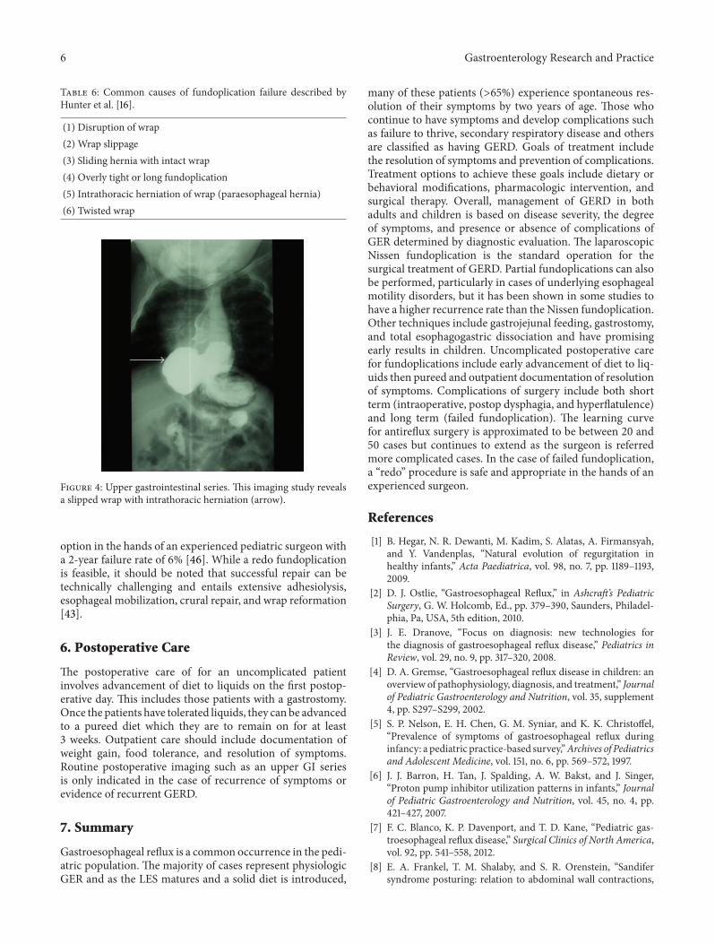
6 Gastroenterology Research and Practice
Table 6: Common causes of fundoplication failure described byHunter et al. [16].
(1) Disruption of wrap(2)Wrap slippage(3) Sliding hernia with intact wrap(4) Overly tight or long fundoplication(5) Intrathoracic herniation of wrap (paraesophageal hernia)(6) Twisted wrap
Figure 4: Upper gastrointestinal series. This imaging study revealsa slipped wrap with intrathoracic herniation (arrow).
option in the hands of an experienced pediatric surgeon witha 2-year failure rate of 6% [46]. While a redo fundoplicationis feasible, it should be noted that successful repair can betechnically challenging and entails extensive adhesiolysis,esophageal mobilization, crural repair, and wrap reformation[43].
6. Postoperative Care
The postoperative care of for an uncomplicated patientinvolves advancement of diet to liquids on the first postop-erative day. This includes those patients with a gastrostomy.Once the patients have tolerated liquids, they can be advancedto a pureed diet which they are to remain on for at least3 weeks. Outpatient care should include documentation ofweight gain, food tolerance, and resolution of symptoms.Routine postoperative imaging such as an upper GI seriesis only indicated in the case of recurrence of symptoms orevidence of recurrent GERD.
7. Summary
Gastroesophageal reflux is a common occurrence in the pedi-atric population. The majority of cases represent physiologicGER and as the LES matures and a solid diet is introduced,
many of these patients (>65%) experience spontaneous res-olution of their symptoms by two years of age. Those whocontinue to have symptoms and develop complications suchas failure to thrive, secondary respiratory disease and othersare classified as having GERD. Goals of treatment includethe resolution of symptoms and prevention of complications.Treatment options to achieve these goals include dietary orbehavioral modifications, pharmacologic intervention, andsurgical therapy. Overall, management of GERD in bothadults and children is based on disease severity, the degreeof symptoms, and presence or absence of complications ofGER determined by diagnostic evaluation. The laparoscopicNissen fundoplication is the standard operation for thesurgical treatment of GERD. Partial fundoplications can alsobe performed, particularly in cases of underlying esophagealmotility disorders, but it has been shown in some studies tohave a higher recurrence rate than the Nissen fundoplication.Other techniques include gastrojejunal feeding, gastrostomy,and total esophagogastric dissociation and have promisingearly results in children. Uncomplicated postoperative carefor fundoplications include early advancement of diet to liq-uids then pureed and outpatient documentation of resolutionof symptoms. Complications of surgery include both shortterm (intraoperative, postop dysphagia, and hyperflatulence)and long term (failed fundoplication). The learning curvefor antireflux surgery is approximated to be between 20 and50 cases but continues to extend as the surgeon is referredmore complicated cases. In the case of failed fundoplication,a “redo” procedure is safe and appropriate in the hands of anexperienced surgeon.
References
[1] B. Hegar, N. R. Dewanti, M. Kadim, S. Alatas, A. Firmansyah,and Y. Vandenplas, “Natural evolution of regurgitation inhealthy infants,” Acta Paediatrica, vol. 98, no. 7, pp. 1189–1193,2009.
[2] D. J. Ostlie, “Gastroesophageal Reflux,” in Ashcraft’s PediatricSurgery, G. W. Holcomb, Ed., pp. 379–390, Saunders, Philadel-phia, Pa, USA, 5th edition, 2010.
[3] J. E. Dranove, “Focus on diagnosis: new technologies forthe diagnosis of gastroesophageal reflux disease,” Pediatrics inReview, vol. 29, no. 9, pp. 317–320, 2008.
[4] D. A. Gremse, “Gastroesophageal reflux disease in children: anoverview of pathophysiology, diagnosis, and treatment,” Journalof Pediatric Gastroenterology and Nutrition, vol. 35, supplement4, pp. S297–S299, 2002.
[5] S. P. Nelson, E. H. Chen, G. M. Syniar, and K. K. Christoffel,“Prevalence of symptoms of gastroesophageal reflux duringinfancy: a pediatric practice-based survey,”Archives of Pediatricsand Adolescent Medicine, vol. 151, no. 6, pp. 569–572, 1997.
[6] J. J. Barron, H. Tan, J. Spalding, A. W. Bakst, and J. Singer,“Proton pump inhibitor utilization patterns in infants,” Journalof Pediatric Gastroenterology and Nutrition, vol. 45, no. 4, pp.421–427, 2007.
[7] F. C. Blanco, K. P. Davenport, and T. D. Kane, “Pediatric gas-troesophageal reflux disease,” Surgical Clinics of North America,vol. 92, pp. 541–558, 2012.
[8] E. A. Frankel, T. M. Shalaby, and S. R. Orenstein, “Sandifersyndrome posturing: relation to abdominal wall contractions,

Gastroenterology Research and Practice 7
gastroesophageal reflux, and fundoplication,”Digestive Diseasesand Sciences, vol. 51, no. 4, pp. 635–640, 2006.
[9] A. Reymunde and N. Santiago, “Long-term results of radiofre-quency energy delivery for the treatment of GERD: sustainedimprovements in symptoms, quality of life, and drug use at 4-year follow-up,” Gastrointestinal Endoscopy, vol. 65, no. 3, pp.361–366, 2007.
[10] R. M. Sydorak and C. T. Albanese, “Laparoscopic antirefluxprocedures in children: evaluating the evidence,” Seminars inLaparoscopic Surgery, vol. 9, no. 3, pp. 133–138, 2002.
[11] G. Mattioli, C. Esposito, M. Lima et al., “Italian multicentersurvey on laparoscopic treatment of gastro-esophageal refluxdisease in children,” Surgical Endoscopy and Other Interven-tional Techniques, vol. 16, no. 12, pp. 1666–1668, 2002.
[12] D. H. Chung and K. E. Georgeson, “Fundoplication andgastrostomy,” Seminars in Pediatric Surgery, vol. 7, no. 4, pp. 213–219, 1998.
[13] H. Steyaert, M. Al Mohaidly, M. A. Lembo, L. Carfagna, S.Tursini, and J. S. Valla, “Long-term outcome of laparoscopicNissen andToupet fundoplication in normal and neurologicallyimpaired children,” Surgical Endoscopy andOther InterventionalTechniques, vol. 17, no. 4, pp. 543–546, 2003.
[14] R. Subramaniam and A. P. Dickson, “Long-term outcome ofBoix-Ochoa and Nissen fundoplication in normal and neuro-logically impaired children,” Journal of Pediatric Surgery, vol. 35,no. 8, pp. 1214–1216, 2000.
[15] R. Kubiak, J. Andrews, andH.W.Grant, “Long-termoutcome oflaparoscopic nissen fundoplication comparedwith laparoscopicthal fundoplication in children: a prospective, randomizedstudy,” Annals of Surgery, vol. 253, no. 1, pp. 44–49, 2011.
[16] J. G. Hunter, C. D. Smith, G. D. Branum et al., “Laparoscopicfundoplication failures: patterns of failure and response tofundoplication revision,” Annals of Surgery, vol. 230, no. 4, pp.595–606, 1999.
[17] “IPEG guidelines for the surgical treatment of pediatric gastroe-sophageal reflux disease (GERD),” Journal of Laparoendoscopic& Advanced Surgical Techniques A, vol. 19, supplement 1, 2009.
[18] H. Kawahara, H. Okuyama, A. Kubota et al., “Can laparoscopicantireflux surgery improve the quality of life in children withneurologic and neuromuscular handicaps?” Journal of PediatricSurgery, vol. 39, no. 12, pp. 1761–1764, 2004.
[19] G. Mattioli, O. Sacco, V. Gentilino et al., “Outcome of laparo-scopic Nissen-Rossetti fundoplication in children with gas-troesophageal reflux disease and supraesophageal symptoms,”Surgical Endoscopy and Other Interventional Techniques, vol. 18,no. 3, pp. 463–465, 2004.
[20] D. M. Diaz, T. E. Gibbons, K. Heiss, M. L. Wulkan, R. R.Ricketts, and B. D. Gold, “Antireflux surgery outcomes in pedi-atric gastroesophageal reflux disease,” The American Journal ofGastroenterology, vol. 100, no. 8, pp. 1844–1852, 2005.
[21] E. W. Fonkalsrud, K. W. Ashcraft, A. G. Coran et al., “Surgicaltreatment of gastroesophageal reflux in children: a combinedhospital study of 7467 patients,” Pediatrics, vol. 101, no. 3 I, pp.419–422, 1998.
[22] B. A. Jobe and J. H. Peters, “Esophagus and diaphragmatichernia,” in Schwartz’s Principles of Surgery, F. C. Brunicardi, T.R. Billiar, D. L. Dunn, J. G. Hunter, J. B. Matthews, and R. E.Pollock, Eds., McGraw-Hill, New York, NY, USA, 9th edition,2010.
[23] C. Esposito, P. Montupet, and O. Reinberg, “Laparoscopicsurgery for gastroesophageal reflux disease during the first year
of life,” Journal of Pediatric Surgery, vol. 36, no. 5, pp. 715–717,2001.
[24] C. Esposito, P. Montupet, D. van Der Zee et al., “Long-termoutcome of laparoscopic Nissen, Toupet, and Thal antirefluxprocedures for neurologically normal children with gastroe-sophageal reflux disease,” Surgical Endoscopy and Other Inter-ventional Techniques, vol. 20, no. 6, pp. 855–858, 2006.
[25] D. I.Watson, R. J. Baigrie, andG. G. Jamieson, “A learning curvefor laparoscopic fundoplication: definable, avoidable, or a wasteof time?” Annals of Surgery, vol. 224, no. 2, pp. 198–203, 1996.
[26] J. J. Meehan and K. E. Georgeson, “The learning curve asso-ciated with laparoscopic antireflux surgery in infants andchildren,” Journal of Pediatric Surgery, vol. 32, no. 3, pp. 426–429, 1997.
[27] S. S. Rothenberg, “Experience with 220 consecutive laparo-scopic nissen fundoplications in infants and children,” Journalof Pediatric Surgery, vol. 33, no. 2, pp. 274–278, 1998.
[28] J. Gill,M. I. Booth, J. Stratford, andT. C. B.Dehn, “The extendedlearning curve for laparoscopic fundoplication: a cohort analy-sis of 400 consecutive cases,” Journal of Gastrointestinal Surgery,vol. 11, no. 4, pp. 487–492, 2007.
[29] R. T. Maxson, S. Harp, R. J. Jackson, S. D. Smith, and C. W.Wagner, “Delayed gastric emptying in neurologically impairedchildren with gastroesophageal reflux: the role of pyloroplasty,”Journal of Pediatric Surgery, vol. 29, no. 6, pp. 726–729, 1994.
[30] M. Pacilli, A. Pierro, K. J. Lindley, J. I. Curry, and S. Eaton,“Gastric emptying is accelerated following laparoscopic Nissenfundoplication,” European Journal of Pediatric Surgery, vol. 18,no. 6, pp. 395–397, 2008.
[31] J. E. Bais, M. Samsom, E. A. J. Boudesteijn, P. P. van Rijk, L.M. A. Akkermans, and H. G. Gooszen, “Impact of delayedgastric emptying on the outcome of antireflux surgery,” Annalsof Surgery, vol. 234, no. 2, pp. 139–146, 2001.
[32] G. J. P.Wilson,D. C. van der Zee, andN.M.A. Bax, “Endoscopicgastrostomy placement in the child with gastroesophagealreflux: is concomitant antireflux surgery indicated?” Journal ofPediatric Surgery, vol. 41, no. 8, pp. 1441–1445, 2006.
[33] R. H. Pearl, D. K. Robie, S. H. Ein et al., “Complications ofgastroesophageal antireflux surgery in neurologically impairedversus neurologically normal children,” Journal of PediatricSurgery, vol. 25, no. 11, pp. 1169–1173, 1990.
[34] R. Srivastava, J. G. Berry, M. Hall et al., “Reflux related hospitaladmissions after fundoplication in children with neurologicalimpairment: retrospective cohort study,” British Medical Jour-nal, vol. 339, Article ID b4411, 2009.
[35] P. W. Wales, I. R. Diamond, S. Dutta et al., “Fundoplicationand gastrostomy versus image-guided gastrojejunal tube forenteral feeding in neurologically impaired children with gas-troesophageal reflux,” Journal of Pediatric Surgery, vol. 37, no.3, pp. 407–412, 2002.
[36] A. Bianchi, “Total esophagogastric dissociation: an alternativeapproach,” Journal of Pediatric Surgery, vol. 32, no. 9, pp. 1291–1294, 1997.
[37] J. Boubnova, G. Hery, F. Ughetto, A. Charpentier, J. M. Guys,and P. de Lagausie, “Laparoscopic total esophagogastric disso-ciation,” Journal of Pediatric Surgery, vol. 44, no. 10, pp. e1–e3,2009.
[38] M. Thomson, A. Fritscher-Ravens, S. Hall, N. Afzal, P. Ash-wood, and C. P. Swain, “Endoluminal gastroplication in chil-dren with significant gastro-oesophageal reflux disease,” Gut,vol. 53, no. 12, pp. 1745–1750, 2004.

8 Gastroenterology Research and Practice
[39] M. Thomson, B. Antao, S. Hall et al., “Medium-term outcomeof endoluminal gastroplication with the EndoCinch device inchildren,” Journal of Pediatric Gastroenterology and Nutrition,vol. 46, no. 2, pp. 172–177, 2008.
[40] W. C. E. Tam, M. N. Schoeman, Q. Zhang et al., “Delivery ofradiofrequency energy to the lower oesophageal sphincter andgastric cardia inhibits transient lower oesophageal sphincterrelaxations and gastro-oesophageal reflux in patientswith refluxdisease,” Gut, vol. 52, no. 4, pp. 479–485, 2003.
[41] M. D. Noar and S. Lotfi-Emran, “Sustained improvement insymptoms of GERD and antisecretory drug use: 4-year follow-up of the Stretta procedure,”Gastrointestinal Endoscopy, vol. 65,no. 3, pp. 367–372, 2007.
[42] S. S. Rothenberg, “The first decade’s experience with laparo-scopic Nissen fundoplication in infants and children,” Journalof Pediatric Surgery, vol. 40, no. 1, pp. 142–147, 2005.
[43] T. D. Kane, M. F. Brown, and M. K. Chen, “Position paper onlaparoscopic antireflux operations in infants and children forgastroesophageal reflux disease,” Journal of Pediatric Surgery,vol. 44, no. 5, pp. 1034–1040, 2009.
[44] T. E. Lobe, “The current role of laparoscopic surgery for gas-troesophageal reflux disease in infants and children,” SurgicalEndoscopy and Other Interventional Techniques, vol. 21, no. 2,pp. 167–174, 2007.
[45] C. Capito, M. D. Leclair, H. Piloquet, V. Plattner, Y. Heloury,and G. Podevin, “Long-term outcome of laparoscopic Nissen-Rossetti fundoplication for neurologically impaired and normalchildren,” Surgical Endoscopy and Other Interventional Tech-niques, vol. 22, no. 4, pp. 875–880, 2008.
[46] S. S. Rothenberg, “Laparoscopic redo Nissen fundoplication ininfants and children,” Surgical Endoscopy and Other Interven-tional Techniques, vol. 20, no. 10, pp. 1518–1520, 2006.
Hindawi Publishing CorporationGastroenterology Research and PracticeVolume 2013, Article ID 181604, 4 pageshttp://dx.doi.org/10.1155/2013/181604
Clinical StudyDuodenal Tube Feeding: An Alternative Approach forEffectively Promoting Weight Gain in Children withGastroesophageal Reflux and Congenital Heart Disease
Seiko Kuwata,1 Yoichi Iwamoto,1 Hirotaka Ishido,1
Mio Taketadu,1 Masanori Tamura,1 and Hideaki Senzaki1,2
1 Department of Pediatrics and Pediatric Cardiology, Saitama Medical University, Saitama, Japan2Department of Pediatric Cardiology, Saitama Medical Center, Saitama Medical University, Staff Office Building 101,1985 Kamoda, Kawagoe, Saitama 350-1981, Japan
Correspondence should be addressed to Hideaki Senzaki; [email protected]
Received 21 December 2012; Accepted 24 March 2013
Academic Editor: Ping-I Hsu
Copyright © 2013 Seiko Kuwata et al. This is an open access article distributed under the Creative Commons Attribution License,which permits unrestricted use, distribution, and reproduction in any medium, provided the original work is properly cited.
We tested whether duodenal tube feeding effectively improves the clinical symptoms and body weight gain in children withcongenital heart disease (CHD) and gastroesophageal reflux (GER). In the retrospective analysis of 17 consecutive children withCHD who were treated with duodenal tube feeding for symptomatic GER, we found that clinical symptoms of persistent emesis orrespiratory wheezing after feeding disappeared with duodenal tube feeding in all patients. Duodenal tube feeding facilitated a stablenutritional supply, resulting in marked improvement of weight gain from 6 to 21 g/day (𝑃 < .0001). In a patient with trisomy 21 andpersistent pulmonary hypertension after the closure of a ventricular septal defect, duodenal tube feeding ameliorated pulmonaryhypertension, as evidenced by the improvement of the pressure gradient of tricuspid regurgitation from 77 to 41mmHg. In 14 of the17 patients, the duodenal tube was successfully removed, with the spontaneous improvement of GER (median duration of duodenaltube feeding: 7 months). In conclusion, duodenal tube feeding improves the weight gain of infants with GER who need treatmentfor CHD-associated heart failure. It also allows for the improvement of pulmonary hypertension.
1. Introduction
Body weight gain is important for the successful treatmentfor infants with heart failure associated with congenital heartdisease (CHD). Gastroesophageal reflux (GER) is known tobe relatively common in this condition and is occasionallyan important cause of growth failure in affected patients[1]. It can also cause aspiration pneumonia and pulmonaryarterial hypertension, thus potentially complicating the clin-ical course of heart failure [2]. Medical therapy with gastricacidity inhibitors, including histamine-2 receptor antagonistsand proton pump inhibitors, is the first line of treatment;however, it is not always effective [3–6]. In such cases,antireflux surgical procedures are selected [7–10]. Anothertreatment optionmay be the administration of duodenal tubefeeding, which is less invasive than surgical procedures andthusmay be beneficial for this particular group of patients for
whom invasive interventions with general anesthesia carry arisk for worsening heart failure. However, little informationis available about the efficacy of duodenal tube feeding forinfants with GER and heart failure associated with CHD.In this study, we reviewed our experience of duodenal tubefeeding performed in 17 children with CHD-associated heartfailure, focusing on its efficacy in terms of body weight gain.We also evaluated its effect on GER-induced pulmonaryhypertension.
2. Methods
Seventeen consecutive infants and childrenwith preoperative(𝑛 = 3) and postoperative (𝑛 = 14) CHD and heartfailure who were treated with duodenal tube feeding wereanalyzed. These patients had episodes of frequent vomiting

2 Gastroenterology Research and Practice
Table 1: Patients’ Characteristics.
Birth weight, g 2607 ± 321Age at the time of diagnosis, months 4.7 ± 0.5Body weight at the time of GER diagnosis 3667 ± 420Types of congenital heart defects (𝑛)
Tetralogy of Fallot 4 (1)Single ventricle 2 (1)Atrioventricular septal defect 2 (1)Coarctation of the aorta 1Coarctation of the aorta with ventricular septaldefect 1
Transposition of the great arteries 1Double outlet right ventricle 2Ventricular septal defect 1The Ebstein anomaly 1Patent ductus arteriosus 1
Other abnormalities (𝑛)Trisomy 21 8 (2)Trisomy 18 122q1- 2Asplenia syndrome 2 (1)
Numbers in parentheses indicate postoperative patients.
and/or wheezing after oral or tube feeding and therefore weresuspected of having GER. They underwent gastrography,which showed a reflux of contrast medium from the stomachto the esophagus beyond the halfway point between theseorgans. After gastrography, a weighted duodenal tube (5 Fr)was inserted under fluoroscopic guidance using a guidewirewithin a tube to facilitate manipulation of the tube thatwas then advanced beyond the descending portion of theduodenum. The appropriate position of the tube was finallyconfirmed by injecting a small amount of contrast mediumthrough the tube, which showed the jejunum directly. A gas-tric tube is routinely placed for medication but not for gastricacid drainage. Because our patients had no gastrointestinaltract obstruction, gastric tube drainage was not performed inorder to avoid potential electrolyte disturbance. Medicationfor reducing acid levels was continued only for severe GERpatients who showed a reflux of contrast medium up to thepharynx.
We compared the body weight gain averaged for 14 to 21days before and after duodenal tube feeding in each patient.In 1 patient (trisomy 21) who showed persistent pulmonaryhypertension after the closure of a ventricular septal defect,changes in the severity of pulmonary hypertension wereassessed by measuring the Doppler flow velocity of tricuspidregurgitation (TR).
3. Results
Table 1 summarizes the characteristics of the studied patients.Of note, 13 patients had underlying conditions of chro-mosomal abnormalities (𝑛 = 10) or anomaly syndromes
Pre Post
5040302010
0−10
−20
−30
(g/d
ay)
Figure 1: Changes in body weight gain per day before and afterduodenal tube feeding.
(𝑛 = 3). The patients’ age at the time of the initiation ofduodenal tube feeding ranged from 0 to 16 months, witha median of 2 months. No adverse events occurred duringthe insertion of the duodenal tube. In all patients, clinicalsymptoms of persistent emesis or respiratory wheezing afterfeeding disappeared after duodenal tube feeding. Duodenaltube feeding facilitated a stable nutritional supply, resultingin marked improvement of weight gain from 6 to 21 g/day(𝑃 < .0001, Figure 1). In the patient with trisomy 21 andpersistent pulmonary hypertension after the closure of aventricular septal defect, duodenal tube feeding amelioratedthe pulmonary hypertension, as evidenced by the improvedpressure gradient of TR from 77 to 41mmHg.
In 14 of the 17 patients, the duodenal tubewas successfullyremoved, with the spontaneous improvement of GER. Themedian duration of duodenal tube feeding was 7 months,ranging from 4 to 10 months. One patient who had singleventricular physiology complicated with asplenia syndromeunderwent laparoscopic fundoplication after the initial car-diac surgery (the Blalock-Taussig shunt). In this particularpatient, the procedure of fundoplication was difficult becauseof the unusual anatomical relation between the heart andthe stomach, and the patient developed hypotension andcyanosis during the procedure. Another patient died of severeheart failure after cardiac surgery for the Ebstein anomaly.The remaining patient had trisomy 18 and continued duo-denal tube feeding, considering the risk for both generalanesthesia and antireflux surgery.
Tube-related complications associated with duodenaltube feeding included accidental removal, obstruction, ordamage; in most of the patients, these complications neces-sitated the replacement of the tube before the scheduled 3-month replacement. In addition, enterocolitis due to mul-tiresistant Staphylococcus aureus (MRSA) was observed in 1patient during the study period.
4. Discussion
To the best of our knowledge, our study is the first to demon-strate that duodenal tube feeding is effective in promotingweight gain in infants and children with CHD and GER.
In general, pharmacologic therapy with gastric acid-ity inhibitors, including histamine-2 receptor antagonists

Gastroenterology Research and Practice 3
and proton pump inhibitors, together with maintaining anupright posture during feeding and the administration ofthickened feedings, is the mainstay of GER treatment [3–6]. However, such medical therapy often fails to resolve thesymptoms of GER in children [4–6]. In a particular group ofinfants with CHD, Weesner and Rosenthal [11] also reportedvery low success rates of medical therapy in resolving theinfants’ respiratory symptoms. In addition, the use of gas-tric acidity inhibitors was reported to be associated withan increased risk of acute gastroenteritis and community-acquired pneumonia in a multicenter, prospective study ofchildren with GER [6].
In contrast, antireflux surgery for GER (i.e., the Nissenfundoplication) has been generally shown to result in asubstantial improvement of reflux and the alleviation ofits consequences in children without CHD [7–10]. Severalstudies have also reported that laparoscopic procedures canbe safely performed even in infants with CHD, with carefulmonitoring of arterial carbon dioxide levels during insuffla-tion [12, 13]. In addition, a recent study by Cribbs et al. [2]showed that the surgical repair of GER in their population ofinfants and children with severe CHD was safely performedand effectively promoted weight gain. However, they alsoreported 1 death among 112 procedures and 3 potentiallylethal complications in the early postoperative period. Thesedata evidence that pediatric cardiac anesthesia providers areessential for the safe performance of antireflux proceduresin this population, with postoperative care administered ina dedicated cardiac intensive care unit, as suggested by theauthors. Our patient with asplenia syndromewho underwentlaparoscopic fundoplication indeed experienced unexpectedhemodynamic instability during the procedure even underthe care of our pediatric anesthesiologists.
Compared with surgical procedures, insertion of a duo-denal tube does not require general anesthesia or postop-erative intensive care, which would be a major advantageof this approach. With the low procedural risk, duodenaltube feeding consistently amelioratedGER-related symptomsand resulted in a dramatic improvement of weight gain.In addition to being less invasive, duodenal tube feedingmay have another merit in that it can be terminated uponthe subsequent resolution of GER. There has been no clearinformation about whether GER associated with CHD canimprove over time. In this sense, our study clearly demon-strated that most of our studied population outgrew reflux,and the duodenal tube was successfully removed within 10months after the initiation. Because the timing of the follow-up gastrography to check for GER status was arbitrary in ourpatients, the periods of duodenal tube feeding could havebeen even shorter than the actual duration of 10 months. Inthe least, duodenal tube feeding can avoid antireflux surgeryin symptomatic GER patients with CHD. Duodenal tubefeeding may also be useful as a bridge to fundoplicationparticularly in preoperative CHD patients, as in our aspleniapatient who underwent fundoplication after the temporaladministration of duodenal tube feeding. Fundoplicationmay be more safely performed under stable hemodynamicconditions established after cardiac surgery.
Our study also demonstrated the improvement of pul-monary hypertension by duodenal tube feeding in a trisomy21 patient after a corrective surgery for CHD. This is consis-tent with our previous report on a Down syndrome infantwithout structural cardiac anomaly [14]. Down syndromeis known to be associated with an increased prevalenceof GER. The syndrome is also known to pose a high riskfor postoperative persistent pulmonary hypertension. Thepresent case, together with our previous one, highlights theimportance of a high index of suspicion for GER as a curablecause of pulmonary artery hypertension in this syndrome.
Finally, tube dislodgement,migration, diarrhea, and ente-rocolitis are known to occur occasionally with duodenal tubefeeding. In fact, we experienced tube-related complicationssuch as accidental removal, obstruction, and damage thatnecessitated unscheduled tube replacement in most patients.Education and training of both family and medical staff areessential for minimizing these events. In addition to thesetube-related complications, 1 patient in our study experiencedMRSA enterocolitis during duodenal tube feeding. Althougha cause-effect relationship between duodenal tube feedingand the occurrence of MRSA enterocolitis was not clear,intestinal tract infection is a known potential importantcomplication associated with duodenal tube feeding that mayoccur due to reduced protective effects of gastric acid. Thisfact also should be kept in mind during the application ofduodenal tube feeding.
5. Conclusions
Duodenal tube feeding is a less invasive method for promot-ingweight gain in symptomatic childrenwithGER associatedwith CHD. It can be terminated upon the spontaneousresolution of GER with the patient’s growth. Thus, duodenaltube feeding should be considered a useful alternative tosurgery in CHD patients with GER.
References
[1] M. Glassman, D. George, and B. Grill, “Gastroesophageal refluxin children: clinical manifestations, diagnosis, and therapy,”Gastroenterology Clinics of North America, vol. 24, no. 1, pp. 71–98, 1995.
[2] R. K. Cribbs, K. F. Heiss, M. L. Clabby, and M. L. Wulkan,“Gastric fundoplication is effective in promoting weight gainin children with severe congenital heart defects,” Journal ofPediatric Surgery, vol. 43, no. 2, pp. 283–289, 2008.
[3] D. G. Johnson and S. G. Jolley, “Gastroesophageal reflux ininfants and children. Recognition and treatment,” SurgicalClinics of North America, vol. 61, no. 5, pp. 1101–1115, 1981.
[4] S. R. Orenstein, E. Hassall, W. Furmaga-Jablonska, S. Atkin-son, and M. Raanan, “Multicenter, double-blind, randomized,placebo-controlled trial assessing the efficacy and safety ofproton pump inhibitor lansoprazole in infants with symptomsof gastroesophageal reflux disease,” Journal of Pediatrics, vol.154, no. 4, pp. 514.e4–520.e4, 2009.
[5] R. J. van der Pol, M. J. Smits, M. P. van Wijk, T. I. Omari, M.M. Tabbers, and M. A. Benninga, “Efficacy of proton-pumpinhibitors in children with gastroesophageal reflux disease: asystematic review,” Pediatrics, vol. 127, pp. 925–935, 2011.

4 Gastroenterology Research and Practice
[6] R. B. Canani, P. Cirillo, P. Roggero et al., “Therapy with gastricacidity inhibitors increases the risk of acute gastroenteritis andcommunity-acquired pneumonia in children,” Pediatrics, vol.117, no. 5, pp. e817–e820, 2006.
[7] E. W. Fonkalsrud, K. W. Ashcraft, A. G. Coran et al., “Surgicaltreatment of gastroesophageal reflux in children: a combinedhospital study of 7467 patients,” Pediatrics, vol. 101, no. 3, part 1,pp. 419–422, 1998.
[8] S. G. Jolley, J. J. Herbst, and D. G. Johnson, “Surgery in childrenwith gastroesophageal reflux and respiratory symptoms,” Jour-nal of Pediatrics, vol. 96, no. 2, pp. 194–198, 1980.
[9] S. S. Rothenberg, “Experience with 220 consecutive laparo-scopic nissen fundoplications in infants and children,” Journalof Pediatric Surgery, vol. 33, no. 2, pp. 274–278, 1998.
[10] E. W. Fonkalsrud, J. Bustorff-Silva, C. A. Perez, R. Quintero,L. Martin, and J. B. Atkinson, “Antireflux surgery in childrenunder 3 months of age,” Journal of Pediatric Surgery, vol. 34, no.4, pp. 527–531, 1999.
[11] K. M. Weesner and A. Rosenthal, “Gastroesophageal reflux inassociation with congenital heart disease,” Clinical Pediatrics,vol. 22, no. 6, pp. 424–426, 1983.
[12] M. L. Wulkan and S. A. Vasudevan, “Is end-tidal CO2
an accu-rate measure of arterial CO
2
during laparoscopic procedures inchildren and neonates with cyanotic congenital heart disease?”Journal of Pediatric Surgery, vol. 36, no. 8, pp. 1234–1236, 2001.
[13] E. R. Mariano, M. G. Boltz, C. T. Albanese, C. T. Abrajano, andC. Ramamoorthy, “Anesthetic management of infants with pal-liated hypoplastic left heart syndrome undergoing laparoscopicnissen fundoplication,”Anesthesia and Analgesia, vol. 100, no. 6,pp. 1631–1633, 2005.
[14] M. Seki, T. Kato, S. Masutani, T. Matsunaga, and H. Senzaki,“Pulmonary arterial hypertension associated with gastroe-sophageal reflux in a 2-month-old boy with Down syndrome,”Circulation Journal, vol. 73, no. 12, pp. 2352–2354, 2009.

Hindawi Publishing CorporationGastroenterology Research and PracticeVolume 2013, Article ID 504816, 8 pageshttp://dx.doi.org/10.1155/2013/504816
Research ArticleChanges in Ghrelin-Related Factors in Gastroesophageal RefluxDisease in Rats
Miwa Nahata,1 Yayoi Saegusa,1 Yumi Harada,1 Naoko Tsuchiya,1
Tomohisa Hattori,1 and Hiroshi Takeda2,3
1 Tsumura & Co., Tsumura Research Laboratories, 3586 Yoshiwara, Ami-machi, Inashiki-gun, Ibaraki 300-1192, Japan2Department of Gastroenterology and Hematology, Hokkaido University Graduate School of Medicine, N15 W7 Kita-ku,Sapporo, Hokkaido 060-8638, Japan
3Department of Pathophysiology andTherapeutics, Division of Pharmasciences, Faculty of Pharmaceutical Sciences,Hokkaido University, N12 W6 Kita-ku, Sapporo, Hokkaido 060-0812, Japan
Correspondence should be addressed to Tomohisa Hattori; hattori [email protected]
Received 21 December 2012; Revised 5 March 2013; Accepted 9 March 2013
Academic Editor: Ping-I Hsu
Copyright © 2013 Miwa Nahata et al. This is an open access article distributed under the Creative Commons Attribution License,which permits unrestricted use, distribution, and reproduction in any medium, provided the original work is properly cited.
To examine gastrointestinal hormone profiles and functional changes in gastroesophageal reflux disease (GERD), blood levelsof the orexigenic hormone ghrelin were measured in rats with experimentally induced GERD. During the experiment, plasmaacyl ghrelin levels in GERD rats were higher than those in sham-operated rats, although food intake was reduced in GERD rats.Although plasma levels of the appetite-suppressing hormone leptin were significantly decreased in GERD rats, no changes wereobserved in cholecystokinin levels. Repeated administration of rat ghrelin to GERD rats had no effect on the reduction in bodyweight or food intake.Therefore, these results suggest that aberrantly increased secretion of peripheral ghrelin and decreased ghrelinresponsiveness may occur in GERD rats. Neuropeptide Y and agouti-related peptide mRNA expression in the hypothalamus ofGERD rats was significantly increased, whereas proopiomelanocortin mRNA expression was significantly decreased comparedto that in sham-operated rats. However, melanin-concentrating hormone (MCH) and prepro-orexin mRNA expression in thehypothalamus of GERD rats was similar to that in sham-operated rats. These results suggest that although GERD rats have higherplasma ghrelin levels, ghrelin signaling in GERD rats may be suppressed due to reduced MCH and/or orexin synthesis in thehypothalamus.
1. Introduction
Gastroesophageal reflux disease (GERD) is caused whengastric acid flows back into the esophagus, resulting in ero-sion of the esophageal mucosal epithelium. Gastric secretioninhibitors, such as proton pump inhibitors, can alleviateGERD symptoms [1]. Moreover, gastric acid reflux into theesophagus characteristically increases after eating [2], andalthough GERD patients sometimes complain of nauseaand a loss of appetite, the acid reflux into the esophagusthat causes GERD may instead be partially due to theamount and content of food.There are various peripheral andcentral appetite-related hormones involved in the control ofappetite and satiation; ghrelin is secreted by the stomach andstimulates appetite and gastrointestinal motility [3], whereas
cholecystokinin (CCK) and leptin, which are secreted inresponse to food intake, suppress appetite [4, 5]. Ghrelin issecreted by X/A-like cells found in the gastric mucosa, and itbinds to the growth hormone secretagogue receptor (GHS-R) present at the end of the vagus nerve to stimulate feedingbehavior by suppressing the satiety stimulus transmitted byCCK [6, 7]. In contrast, leptin is secreted by fat cells andacts directly on the hypothalamus by crossing the blood-brainbarrier, thereby suppressing food intake desire caused byghrelin [5]. Until date, changes in appetite-related hormonesin GERD patients have not been sufficiently characterized.We hypothesized that the progression of GERDmay bemedi-ated by the abnormal function of appetite-related hormones.As a first step in elucidating the involvement of appetite-related hormones in GERD, we examined the profiles of

2 Gastroenterology Research and Practice
peripheral appetite-related peptides, with a focus on changesin ghrelin levels and ghrelin responsiveness.
2. Materials and Methods
2.1. Animals. Eight-week-old male Wistar rats (CLEA Japan,Tokyo, Japan) were used during the experiment. Duringtesting, 4-5 animals were housed in a single cage andwere allowed free access to food and water. Animal roomswere illuminated between 07:00 and 19:00, and temperatureand humidity were maintained at constant levels. All testswere performed between 09:00 and 18:00, according to theguidelines of the Experimental Animal Ethics Committee ofTsumura.
2.2. Chemicals. Rat ghrelin was obtained from the PeptideInstitute (Osaka, Japan) and was dissolved in 0.9% sterilizedphysiological saline (Otsuka Pharmaceutical, Tokyo, Japan).
2.3. Preparation of GERDRats. GERDwas surgically inducedby Omura’s method [8]. Rats deprived of food for 24 h wereanesthetized with ether. The abdomen was opened using a2 cm upper-median abdominal incision. The stomach andduodenum were exteriorized, and the boundary between theforestomach and the glandular stomach was sutured with 1-0 silk thread (Natsume Seisakusho, Tokyo, Japan). A precut2mm wide 18-Fr Nelaton catheter (Terumo, Tokyo, Japan)was used to cover the area proximal to the pylorus on theduodenal side, and a 5–0 nylon thread (Natsume Seisakusho,Tokyo, Japan)was used to suture and fix it to the surface of thepyloric serous membrane. The stomach and duodenum wereplaced back into the abdominal cavity, whichwas then closed.Sham-operated rats were first laparotomized to expose theirstomach and duodenum for about 1min, after which theirabdominal cavities were closed. After surgery, rats were fastedfor an additional 24 h (resulting in a total of 48 h). GERD-induced animals that were noted to have developed organadhesions or abscesses or extreme weight loss or weaknesswere excluded from the experiment. To avoid a dramaticreduction in the sample, more GERD-induced animals werecreated than sham-operated rats.
2.4. Measurement of Body Weight and Food Intake. The ratswere housed individually after GERD induction. Daily bodyweight was measured from the day of surgery, and dailyfood intake, calculated as the difference between preprandialand postprandial weight of the food, was measured from 2days after surgery (day 2). On day 9, rats were deprived offood for 24 h and then sacrificed to performhistopathologicalassessment of the esophagus.
2.5. Histopathological Assessment. After 24 h of fasting (day10), rats were exsanguinated via the abdominal vena cavaunder ether anesthesia, and the esophagus was excised.Histopathological assessment was performed as previouslydescribed [9].The esophagus was opened with a longitudinalincision and immobilized on a rubber plate with insect pins.The entire esophagus was photographed, and each image
was imported into an image analysis software (WinROOF;Mitani Corporation, Tokyo, Japan). Sites showing esophagealmucosal erosion were identified and their total area wasmeasured.
2.6. Effect of Exogenous Ghrelin in GERD Rats. DuringGERD-inducing surgery, rats were anesthetized by intraperi-toneal injection of pentobarbital sodium (Kyoritsu Seiyaku,Tokyo, Japan), and a catheter filled with heparin in phys-iological saline was fixed to the jugular vein. The catheterwas passed subcutaneously and pulled out from the back,whichwas then coveredwith ametal spring to prevent it frombeing bitten. After surgery, rats were housed individually andfasted for an additional 24 h. From the next day, rat ghrelin(3 nmol/rat) was administered to the rats once daily throughthe jugular vein for 6 days. The control group of GERDand sham-operated rats were administered saline. Daily bodyweight was measured from the day of surgery and daily foodintake from day 2 after surgery.
2.7. Determination of Plasma Ghrelin and Appetite-RelatedHormones. Blood was collected from the abdominal venacava under ether anesthesia on days 3, 7, and 10 after surgery.Plasma samples were obtained as previously reported [10].In brief, blood was collected in a tube containing EDTA-2Na (Dojindo Laboratories, Kumamoto, Japan) and aprotinin(Wako Pure Chemical Industries, Osaka, Japan). Blood sam-ples were immediately centrifuged at 4∘C, and the super-natant was acidified with 1mol/L HCl (1/10 volume). Plasmawas stored at−80∘Cuntil measurement. Plasma ghrelin levelswere measured using the Active Ghrelin ELISA Kit andDesacyl Ghrelin ELISA Kit (Mitsubishi Chemical Medience,Tokyo, Japan). Plasma CCK levels were measured with theCCK EIA Kit (Phoenix Pharmaceuticals, Burlingame, CA,USA) using nonacidified plasma samples obtained in thesame manner. Plasma leptin levels were measured usingthe Bio-Plex suspension array system (BioRad Laboratories,Hercules, CA, USA) with the Bio-Plex Pro Rat Diabetes assaypanel (BioRad Laboratories).
2.8. RNA Extraction, Reverse Transcription, and Real-TimePolymerase Chain Reaction. After collection of blood sam-ples, the stomach and hypothalamus were immediatelyexcised on days 3, 7, and 10 and stored at −80∘C untilmeasurement. The tissue was homogenized and total RNAwas extracted using the RNeasyUniversal Tissue Kit (Qiagen,Valencia, CA, USA). Total RNA from each sample wasdiluted to 100 ng/𝜇L, allowed to react for 5min at 70∘C,and immediately cooled on ice. An aliquot of 1 𝜇g of totalRNA was reverse transcribed using the TaqMan ReverseTranscription Reagents (Applied Biosystems, Foster City, CA,USA) according to the manufacturer’s protocol.
Quantitative polymerase chain reaction (PCR) was per-formed with the PRISM 7900HT Sequence Detection System(Applied Biosystems) using the TaqMan Universal PCRMaster Mix (Applied Biosystems). To compensate for thedifferences in the amount of total RNA added to eachreaction, mRNA expression was normalized to 𝛽-actin as an

Gastroenterology Research and Practice 3
Sham
GERD
(a)
150
200
250
300
350
0 2 4 6 8 10Time after GERD induction (day)
Body
wei
ght (
g)
Sham (𝑛 = 8)GERD (𝑛 = 8)
∗∗∗∗∗∗∗∗∗
∗∗∗
∗∗∗ ∗∗∗
(b)
0
10
20
30
0 2 4 6 8 10Time after GERD induction (day)
Dai
ly fo
od in
take
(g)
Sham (𝑛 = 8)GERD (𝑛 = 8)
∗∗∗∗∗∗
∗∗∗
∗∗∗
∗∗∗ ∗∗∗
(c)
Figure 1: Comparison of esophageal mucosa, body weight, and food intake between sham-operated and GERD rats. (a) Esophagus on day 10after GERD induction showed obvious erosion. Bar, 10mm. (b) Body weight and (c) daily food intake. ∗∗∗𝑃 < 0.001 versus sham-operatedrats.
endogenous control as expressed by the Δ threshold cycle(Δ𝐶𝑡) value:
Δ𝐶𝑡= 2(−|𝐴 − 𝐵|)
, (1)
where 𝐴 is the number of cycles that reached the 𝛽-actingene threshold and 𝐵 is the number of cycles that reachedthe target gene threshold. The set of oligonucleotide primersand fluorescent probes used in TaqMan quantitative PCRwas provided by Applied Biosystems: cytoplasmic 𝛽-actin,Rn00667869 m1; prepro-ghrelin, Rn00572319 m1; GHS-R, Rn00821417 m1; membrane bound O-acyltransferasedomain containing 4 (ghrelin O-acyltransferase; GOAT),Rn02079102 s1; neuropeptide Y (NPY): Rn00561681 m1;agouti-related protein (AgRP): Rn01431703 g1; proopiome-lanocortin (POMC): Rn00595020 m1; promelanin-concen-trating hormone (MCH): Rn00561766 g1; and hypocretin(prepro-orexin), Rn00565995 m1.
2.9. Statistical Analysis. Statistical significance was examinedusing Student’s t-test and𝑃 < 0.05was considered statisticallysignificant. Data were expressed as the mean ± SEM of eachgroup.
3. Results
3.1. General Condition and Histology in GERD Rats. Mucosalerosion was clearly observed in the esophagus of GERDrats on day 10 (Figure 1(a)). The number of erosion sites inGERD rats was 2.5 ± 0.4 with a total area of 39.7 ± 9.8mm2.Moreover, the body weight and food intake in these ratshad significantly decreased compared with those in sham-operated rats (Figures 1(b) and 1(c)).
3.2. Changes in Plasma Ghrelin Levels in GERD Rats. Plasmaacyl and desacyl ghrelin levels significantly increased fromday 3 to day 10 (Figures 2(a) and 2(b); desacyl ghrelin levelson day 10: sham-operated, 520.4 ± 94.0 versus GERD, 832.9 ±92.7 fmol/mL; 𝑃 = 0.06).
3.3. Plasma Leptin and CCK Levels in GERD Rats. Plasmaleptin levels significantly decreased on day 10 (Table 1). Therewere no significant differences in plasma CCK levels betweenGERD and sham-operated rats.
3.4. Effect of Ghrelin Administration on BodyWeight and FoodIntake. Body weight of GERD rats significantly decreased

4 Gastroenterology Research and Practice
0
100
200
300
Day 3 Day 7 Day 10
Plas
ma a
cyl g
hreli
n (fm
ol/m
L)
Sham (𝑛 = 4–8)
∗
∗∗
∗∗
GERD (𝑛 = 7-8)
(a)
0
300
600
900
1200
Day 3 Day 7 Day 10
Plas
ma d
esac
yl g
hreli
n (fm
ol/m
L)
Sham (𝑛 = 4–8)
∗∗
∗∗
GERD (𝑛 = 7-8)
(b)
Figure 2: Plasma ghrelin levels in sham-operated and GERD rats on day 3, 7, and 10 after GERD induction. (a) Plasma acyl ghrelin and (b)desacyl ghrelin levels. ∗, ∗∗𝑃 < 0.05, 0.01 versus sham-operated rats on each day.
100
150
200
250
300
Body
wei
ght (
g)
0 1 2 3 4 5 6
Time after GERD induction (day)
Sham + saline (𝑛 = 6)GERD + saline (𝑛 = 10)GERD + ghrelin (𝑛 = 7)
∗∗∗
(a)
2 3 4 5 6
0
5
10
15
20
25 D
aily
food
inta
ke (g
)
Time after GERD induction (day)
Sham + saline (𝑛 = 6)GERD + saline (𝑛 = 10)GERD + ghrelin (𝑛 = 7)
∗∗
∗∗∗∗∗
∗∗∗
(b)
Figure 3: Effect of repeated administration of ghrelin to GERD rats. (a) Body weight and (b) daily food intake in sham-operated and GERDrats. Exogenous ghrelin (3 nmol/rat/day) was intravenously administered to rats once daily. Sham-operated rats and control group of GERDrats were administered saline. There were no significant differences in body weight or food intake between the saline-administered andghrelin-administered groups in GERD rats. ∗, ∗∗, and ∗∗∗𝑃 < 0.05, 0.01, and 0.001 versus sham-operated rats on each day.
Table 1: Plasma leptin and cholecystokinin (CCK) levels in sham-operated and GERD rats on day 10 after GERD induction.
Sham (𝑛 = 4 or 8) GERD (𝑛 = 8)(pg/mL) (pg/mL)
Leptin 714.6 ± 49.3 358.1 ± 78.5∗
CCK 133.8 ± 8.2 112.3 ± 6.8
∗P < 0.05 versus sham-operated rats.
compared with that of sham-operated rats (Figure 3(a)). Therepeated administration of ghrelin toGERD rats had no effect
on body weight reduction. Furthermore, there were no differ-ences in daily food intake between GERD rats administeredsaline and those administered ghrelin (Figure 3(b)).
3.5. Changes in Gastric or Hypothalamic mRNA Expres-sion in GERD Rats. There were no major differences inprepro-ghrelin andGHS-RmRNA expression in the stomachthroughout the experiment between sham-operated andGERD rats (Figures 4(a) and 4(b)). In contrast, GOATmRNAexpression in GERD rats significantly decreased from day7 to day 10 (Figure 4(c)). NPY mRNA expression in the

Gastroenterology Research and Practice 5
0
0.5
1
2
Day 3 Day 7 Day 10
Relat
ive e
xpre
ssio
n
1.5
Prepro-ghrelin
Sham (𝑛 = 8)GERD (𝑛 = 6-7)
(a)
0
0.5
1
1.5
2
2.5
3
Day 3 Day 7 Day 10
Relat
ive e
xpre
ssio
n
GHS-R
Sham (𝑛 = 8)GERD (𝑛 = 6-7)
(b)
0
0.5
1.0
1.5
Day 3 Day 7 Day 10
Relat
ive e
xpre
ssio
n
GOAT
Sham (𝑛 = 8)
∗
∗∗∗
GERD (𝑛 = 6-7)
(c)
Figure 4: Gastric mRNA expression in sham-operated and GERD rats on days 3, 7, and 10 after GERD induction. (a) Prepro-ghrelin, (b)growth hormone secretagogue receptor (GHS-R), and (c) ghrelin O-acyltransferase (GOAT) mRNA expression. ∗, ∗∗∗𝑃 < 0.05, 0.001 versussham-operated rats on each day.
hypothalamus of GERD rats significantly increased on day 10(Figure 5(a)), whereas AgRP mRNA expression significantlyincreased from day 3 (Figure 5(b)). In contrast, a significantdecrease in POMCmRNA expression was observed inGERDrats from day 7 (Figure 5(c)). In addition, MCH (Figure 5(d))and prepro-orexin (Figure 5(e)) mRNA expression in GERDrats remained unchanged compared with that in sham-operated rats.
4. Discussion
Despite the rapid increase in plasma acyl and desacyl ghrelinlevels in 24-h fasted GERD rats, their food intake and bodyweight decreased. Repeated administration of acyl ghrelin didnot suppress the reduction in food intake and body weight.Hypothalamic NPY/AgRP neuronal activity, but not MCHor orexin neurons, significantly increased. Peripheral ghrelin
signals in GERD rats were sent to the arcuate nucleus in thehypothalamus, but MCH and orexin neurons in the lateralhypothalamic area (LH) might have failed to be activated,leading to inhibition of food intake.
Ghrelin is an orexigenic hormone producedmainly in thestomach [7]. Ghrelin increases food intake and suppressesenergy expenditure [3, 11]. In our previous study, we usedGERD rats to demonstrate lack of responsiveness throughan acute bolus administration of ghrelin (3 nmol/rat) [12]. Incomparison with sham-operated rats, the growth hormonesecretory effect in GERD rats intravenously administeredacyl ghrelin decreased, and acute administration of acylghrelin did not suppress the decrease in food intake, gastricemptying, or gastricmotility. Repeated administration of acylghrelin (3 nmol/rat/day) to GERD rats had no effect on foodintake or body weight throughout the experiment in thisstudy. We previously demonstrated that acute intravenous
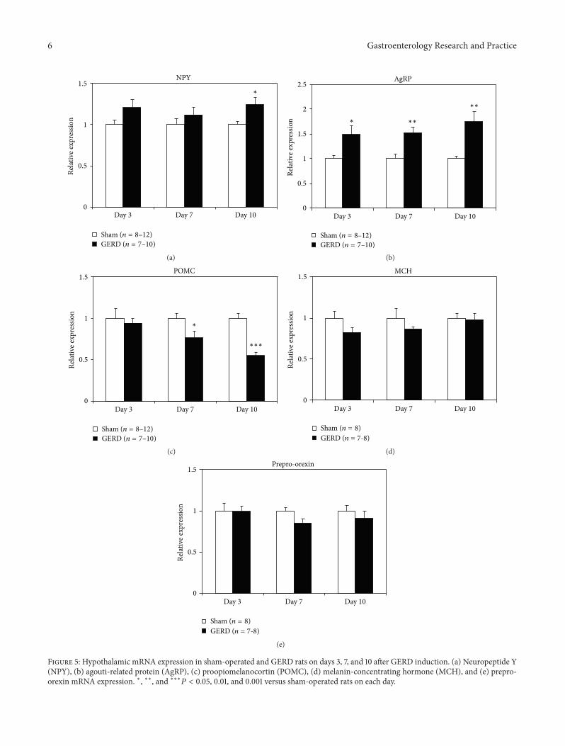
6 Gastroenterology Research and Practice
0
0.5
1
1.5
Day 3 Day 7 Day 10
Relat
ive e
xpre
ssio
nNPY
Sham (𝑛 = 8–12)GERD (𝑛 = 7–10)
∗
(a)
0
0.5
1
1.5
2
2.5
Day 3 Day 7 Day 10
Relat
ive e
xpre
ssio
n
AgRP
Sham (𝑛 = 8–12)GERD (𝑛 = 7–10)
∗ ∗∗
∗∗
(b)
0
0.5
1
1.5
Day 3 Day 7 Day 10
Relat
ive e
xpre
ssio
n
POMC
Sham (𝑛 = 8–12)GERD (𝑛 = 7–10)
∗
∗∗∗
(c)
0
0.5
1
1.5
Day 3 Day 7 Day 10
Relat
ive e
xpre
ssio
n
MCH
Sham (𝑛 = 8)GERD (𝑛 = 7-8)
(d)
0
0.5
1
1.5
Day 3 Day 7 Day 10
Rela
tive e
xpre
ssio
n
Prepro-orexin
Sham (𝑛 = 8)GERD (𝑛 = 7-8)
(e)
Figure 5: Hypothalamic mRNA expression in sham-operated and GERD rats on days 3, 7, and 10 after GERD induction. (a) Neuropeptide Y(NPY), (b) agouti-related protein (AgRP), (c) proopiomelanocortin (POMC), (d) melanin-concentrating hormone (MCH), and (e) prepro-orexin mRNA expression. ∗, ∗∗, and ∗∗∗𝑃 < 0.05, 0.01, and 0.001 versus sham-operated rats on each day.

Gastroenterology Research and Practice 7
administration of acyl ghrelin at a dose of 3 nmol/rat to sham-operated rats increased food intake significantly [12]. In nor-mal rats, administration of ghrelin at a dose of 1.5 nmol/ratalso increased food intake significantly [13]. Food intakedid not increase with repeated administration of ghrelin;therefore, GERD rats may require a higher dose of exogenousghrelin.
The mechanism whereby peripheral blood ghrelin levelsare increased in GERD rats is not well understood. Therewere no differences in relation to prepro-ghrelin or GHS-RmRNA expression in the stomach between sham-operatedand GERD rats. These results were not caused by an increasein ghrelin synthesis in the stomach and do not necessarilypromote the synthesis of receptors that would increase signalresponsiveness. Moreover, expression of the gene encodingGOAT, which is an enzyme that adds an octanoyl group toproghrelin, was significantly decreased.This result is in accor-dance with the findings that GOATmRNAmay be negativelyregulated during long-time fasting [14]. This may be due tonegative feedback as a result of abundant acyl ghrelin presentin the peripheral blood. Therefore, ghrelin secretion raw thestomach may contribute to the high peripheral blood ghrelinlevels.
GHS-R is a G-protein-coupled receptor, and these recep-tors typically undergo depolarization after ligand binding[15]. Sustained high plasma ghrelin levels in GERD ratsmay cause systemic GHS-R depolarization. However, inthis study we found that hypothalamic NPY/AgRP mRNAexpression significantly increased from day 3 in GERD ratscompared with that in sham-operated rats. Ghrelin bindsto GHS-R at the end of the vagus nerve in the stomach,stimulates NPY/AgRP neurons present in the hypothalamicarcuate nucleus, and increases NPY/AgRPmRNA expression[3, 13]. Because NPY/AgRP mRNA expression is increasedin GERD rats, ghrelin signaling may be maximal. Leptininhibits NPY/AgRP expression, stimulates POMC neurons,and produces POMC mRNA [16, 17]. Since plasma leptinwas only examined on day 10 after surgery, we can onlysuggest that increased ghrelin levels were inversely corre-lated with plasma leptin levels. However, in addition tothe increased plasma ghrelin levels, decreased plasma leptinlevels might have contributed to increased NPY/AgRP andsignificantly reduced POMC mRNA expression in GERDrats.
In this study, prepro-orexin andMCHmRNA expressionwas not altered in GERD rats. The orexigenic signal, via theactivation of the NPY/AgRP neurons, is transmitted to theLH, leading to the activation of orexin or MCH neurons [18–21]. Orexin and MCH are primarily synthesized in the LHwhen these neurons are activated. MCH-1R antagonism ordepletion of the peptide results in hypophagia, and MCH-1R-deficient mice are lean [22–24]. It is well known that areward system, including appetite or learning, is mediatedby activation of these neurons via the dopamine neurons[25, 26] in the ventral tegmental area or nucleus accumbens.We speculated that orexin or MCH neuron activation mightbe suppressed in GERD rats, leading to the inhibition of foodintake. However, a detailed mechanism of action and theidentity of the factors that cause suppression of these neurons
is still unknown. Further study is needed to clarify why orexinor MCH neurons are unresponsive.
Currently, there exists no GERD model that suitablyreflects human GERD. The model presented in the currentexperiment describes an extensive operation involving liga-tion of the forestomach and fixing of a ring to the pyloricregion, making it differ greatly from human GERD. Further-more, although these are preliminary results, administrationof an effective dose of PPI [27] did not affect initial bodyweight, food intake, or ghrelin concentration in GERD rats.It is possible to conclude that overexposure of the esophagusto stomach acid is not involved in the increased secretionof ghrelin or abnormal food intake-related factor expression.Moreover, because oral administration of Cisapride clearlyimproved gastric emptying in the present model [12], itappears that treatment with a pyloric region ring and ligationdoes not cause an irreversible reduction in gastric motility.However, the possibility that extensive surgery affects foodintake-related parameters could not be excluded.The validityof the GERDmodel used in this study needs to be sufficientlyverified in the future. In addition, the effect of physicalimpairment and stomach acid exposure in surgery on foodintake-related parameters and hormone levels needs to becarefully examined.
5. Conclusion
In comparison to normal rats, GERD rats characteristicallyhave increased peripheral acyl ghrelin levels, decreased leptinlevels, and might have impaired ghrelin signal transmission.However, it remains necessary to verify the validity of themodel used and to further examine details regarding PPIadministration.
Conflict of Interests
There is no conflict of interests with any financial organiza-tion regarding the material discussed in the paper.
Acknowledgments
H. Takeda received grant support from Tsumura. M. Nahata,Y. Saegusa, Y. Harada, N. Tsuchiya, and T. Hattori areemployed by Tsumura.
References
[1] R. Fass, “Therapeutic options for refractory gastroesophagealreflux disease,” Journal of Gastroenterology and Hepatology, vol.27, supplement 3, pp. 3–7, 2012.
[2] L. F. Johnson and T. R. Demeester, “Twenty four hour pHmonitoring of the distal esophagus. A quantitative measure ofgastroesophageal reflux,” American Journal of Gastroenterology,vol. 62, no. 4, pp. 325–332, 1974.
[3] A. Asakawa, A. Inui, T. Kaga et al., “Ghrelin is an appetite-stimulatory signal from stomach with structural resemblanceto motilin,” Gastroenterology, vol. 120, no. 2, pp. 337–345, 2001.
[4] G. J. Dockray, “Cholecystokinin,” Current Opinion in Endocri-nology, Diabetes and Obesity, vol. 19, no. 1, pp. 8–12, 2012.

8 Gastroenterology Research and Practice
[5] J. G. Mercer, N. Hoggard, L. M. Williams, C. B. Lawrence, L.T. Hannah, and P. Trayhurn, “Localization of leptin receptormRNA and the long form splice variant (Ob-Rb) in mousehypothalamus and adjacent brain regions by in situ hybridiza-tion,” FEBS Letters, vol. 387, no. 2-3, pp. 113–116, 1996.
[6] Y. Date, K. Toshinai, S. Koda et al., “Peripheral interac-tion of ghrelin with cholecystokinin on feeding regulation,”Endocrinology, vol. 146, no. 8, pp. 3518–3525, 2005.
[7] M. Kojima, H. Hosoda, Y. Date, M. Nakazato, H. Matsuo, andK. Kangawa, “Ghrelin is a growth-hormone-releasing acylatedpeptide from stomach,”Nature, vol. 402, no. 6762, pp. 656–660,1999.
[8] N. Omura, H. Kashiwagi, G. Chen, Y. Suzuki, F. Yano, and T.Aoki, “Establishment of surgically induced chronic acid refluxesophagitis in rats,” Scandinavian Journal of Gastroenterology,vol. 34, no. 10, pp. 948–953, 1999.
[9] H. Miwa, J. Koseki, T. Oshima et al., “Rikkunshito, a tradi-tional Japanese medicine, may relieve abdominal symptoms inrats with experimental esophagitis by improving the barrierfunction of epithelial cells in esophageal mucosa,” Journal ofGastroenterology, vol. 45, no. 5, pp. 478–487, 2010.
[10] H. Takeda, C. Sadakane, T. Hattori et al., “Rikkunshito, anherbal medicine, suppresses cisplatin-induced anorexia in ratsVia 5-HT2 receptor antagonism,” Gastroenterology, vol. 134, no.7, pp. 2004–2013, 2008.
[11] M. Nakazato, N. Murakami, Y. Date et al., “A role for ghrelin inthe central regulation of feeding,” Nature, vol. 409, no. 6817, pp.194–198, 2001.
[12] M. Nahata, S. Muto, N. Oridate et al., “Impaired ghrelin signal-ing is associated with gastrointestinal dysmotility in rats withgastroesophageal reflux disease,”American Journal of PhysiologyGastrointestinal and Liver Physiology, vol. 303, no. 1, pp. G42–G53, 2012.
[13] Y. Date, N. Murakami, K. Toshinai et al., “The role of the gastricafferent vagal nerve in Ghrelin-induced feeding and growthhormone secretion in rats,” Gastroenterology, vol. 123, no. 4, pp.1120–1128, 2002.
[14] H. Kirchner, J. A. Gutierrez, P. J. Solenberg et al., “GOAT linksdietary lipids with the endocrine control of energy balance,”Nature Medicine, vol. 15, no. 7, pp. 741–745, 2009.
[15] J. P. Camina, “Cell biology of the ghrelin receptor,” Journal ofNeuroendocrinology, vol. 18, no. 1, pp. 65–76, 2006.
[16] M. W. Schwartz, R. J. Seeley, L. A. Campfield, P. Burn, andD. G. Baskin, “Identification of targets of leptin action in rathypothalamus,” Journal of Clinical Investigation, vol. 98, no. 5,pp. 1101–1106, 1996.
[17] T.M.Mizuno, S. P. Kleopoulos, H. T. Bergen, J. L. Roberts, C. A.Priest, and C. V. Mobbs, “Hypothalamic pro-opiomelanocortinmRNA is reduced by fasting in ob/ob and db/db mice, but isstimulated by leptin,” Diabetes, vol. 47, no. 2, pp. 294–297, 1998.
[18] T. Sakurai, A. Amemiya, M. Ishii et al., “Orexins and orexinreceptors: a family of hypothalamic neuropeptides and Gprotein-coupled receptors that regulate feeding behavior,” Cell,vol. 92, no. 4, pp. 573–585, 1998.
[19] C. F. Elias, C. B. Saper, E. Maratos-Flier et al., “Chemicallydefined projections linking the mediobasal hypothalamus andthe lateral hypothalamic area,” Journal of Comparative Neurol-ogy, vol. 402, no. 4, pp. 442–459, 1998.
[20] D. Qu, D. S. Ludwig, S. Gammeltoft et al., “A role for melanin-concentrating hormone in the central regulation of feedingbehaviour,” Nature, vol. 380, no. 6571, pp. 243–247, 1996.
[21] K. Toshinai, Y. Date, N.Murakami et al., “Ghrelin-induced foodintake is mediated via the orexin pathway,” Endocrinology, vol.144, no. 4, pp. 1506–1512, 2003.
[22] M. Shimada, N. A. Tritos, B. B. Lowell, J. S. Flier, and E.Maratos-Flier, “Mice lacking melanin-concentrating hormoneare hypophagic and lean,” Nature, vol. 396, no. 6712, pp. 670–679, 1998.
[23] S.Mashiko, A. Ishihara, A. Gomori et al., “Antiobesity effect of amelanin-concentrating hormone 1 receptor antagonist in diet-induced obese mice,” Endocrinology, vol. 146, no. 7, pp. 3080–3086, 2005.
[24] D. J. Marsh, D. T. Weingarth, D. E. Novi et al., “Melanin-concentrating hormone 1 receptor-deficient mice are lean,hyperactive, and hyperphagic and have altered metabolism,”Proceedings of the National Academy of Sciences of the UnitedStates of America, vol. 99, no. 5, pp. 3240–3245, 2002.
[25] G. C.Harris,M.Wimmer, andG.Aston-Jones, “A role for lateralhypothalamic orexin neurons in reward seeking,” Nature, vol.437, no. 7058, pp. 556–559, 2005.
[26] A. Sherwood, M. Wosiski-Kuhn, T. Nguyen et al., “The role ofmelanin-concentrating hormone in conditioned reward learn-ing,” European Journal of Neuroscience, vol. 36, no. 8, pp. 3126–3133, 2012.
[27] H. Miwa, T. Oshima, J. Sakurai et al., “Experimental oesophagi-tis in the rat is associated with decreased voluntary movement,”Neurogastroenterology and Motility, vol. 21, no. 3, pp. 296–303,2009.