“diabetic retinopathy; from glycation to clinical aspects” · “diabetic retinopathy; from...
TRANSCRIPT

“Diabetic retinopathy;
from glycation to clinical aspects”
by
Dag Sigurd Fosmark
Centre of Endocrinology, Aker University Hospital,
Department of Ophthalmology, Ullevål University Hospital,
Diabetes Research Centre of Aker & Ullevål University Hospitals
Oslo, Norway
2007

© Dag Sigurd Fosmark, 2008 Series of dissertations submitted to the Faculty of Medicine, University of Oslo No. 671 ISBN 978-82-8072-897-5 All rights reserved. No part of this publication may be reproduced or transmitted, in any form or by any means, without permission. Cover: Inger Sandved Anfinsen. Printed in Norway: AiT e-dit AS, Oslo, 2008. Produced in co-operation with Unipub AS. The thesis is produced by Unipub AS merely in connection with the thesis defence. Kindly direct all inquiries regarding the thesis to the copyright holder or the unit which grants the doctorate. Unipub AS is owned by The University Foundation for Student Life (SiO)


List of errata
� Page 5 within 4.3: The title of Paper III; ”Serum levels of the Advanced Glycation End product hydroimidazolone is associated with retinopathy in type 1 diabetes patients” should read: ”Serum levels of the Advanced Glycation End product hydroimidazolone are associated with retinopathy in type 1 diabetes patients”.
� Pages 2 and 15: “5.5” should read “5.4”
� Page 7 within 5.2: "Organs mainly affected are the eyes, kidneys and nerves (15); diabetic retinopathy." should read: "Organs mainly affected are the eyes, kidneys and nerves (15) with diabetic retinopathy, nephropathy and neuropathy, respectively."
� Page 28: "(Blood tests were collected after overnight fasting." Omit initial parenthesis.

1
To my patients

2
Contents � 1. Preface � 2. Acknowledgements � 3. Abbreviations � 4. List of papers � 5. Introduction � 5.1 Diabetes
� 5.2 Hyperglycemia and vascular complications of diabetes
� 5.3 Diabetic retinopathy; background � Pathogenesis � Risk � Clinical aspects � Classification � Biochemical mechanisms � ROS and Oxidative stress
� 5.5 Advanced Glycation Endproducts (AGEs): Background, pathologic relevance and detection
� AGEs; definition and examples � Methylglyoxal derived AGEs � AGEs and disease � Circulating AGEs; AGE/RAGE interaction � Inhibition of AGE � AGEs, VEGF and the eye � AGEs; different methods for detection
� 6. Aims of research
� 7. Subjects and methods
� 7.1 Subjects
� 7.2 Methods
� 8. Summary of main results
� 9. Discussion
� 9.1 Methods � Clinical and Photographic methods of retinopathy diagnosis � Immunoassay method � 9.2 Results
� 10. Concluding remarks
� 11. References
Papers I - IV

3
1. Preface “There is only one thing about which I am certain, and that is that there is very little about which one can be certain.” W. Somerset Maugham 2. Acknowledgements This piece of work was carried out during intervals between 2000 and 2007 at the Diabetes Research Centre of Aker and Ullevål University Hospitals, Oslo, with the facilities of Aker University Hospital and the Hormone Laboratory, Oslo. Financial support has been granted by the Norwegian Foundation for Health and Rehabilitation after recommendations from the Norwegian Diabetes Association; the Research Forum of Ullevål University Hospital (FUS) and the Research Forum of Aker University Hospital (FAS); the Diabetes Research Centre of Aker and Ullevål University Hospitals, the Regional Health Board (Helse Øst), my own pocket and the Remembrance Foundation of Inger Holm. First and foremost, I would like to thank the core of the Diabetes Research Centre, in this context meaning my supervisor, Professor Kristian F. Hanssen for his support and stimulation through his immense knowledge and optimism, and secretary Beth Thyrdal for reiterating positive feed-back. I am also grateful to the former professor at the Ophthalmological Department at Ullevål University Hospital, Dr. Olaf Brinchmann-Hansen, who triggered me into getting seriously involved in diabetes research. I would like to thank my Swedish co-authors, Professor Elisabet Agardh and Professor Carl-David Agardh for having faith in me as a novice; for their professional attitude and sharing of knowledge. I owe greatful thoughts to professor Kåre Birkeland for including me as scientist and to Professor Egil Haug of the Hormone Laboratory for providing me with excellent facilities and the following co-workers; Peter Torjesen, Jens Petter Berg, Terje Lund and Aase-Brith Jensen, whose contributions have been inspiring for my clinical mind. Furthermore, at the Hormone laboratory; Turi Arnesen Siegwarth for initially letting me work on AGE immunoassays and Bård Rasmussen for PC support, the remaining “white coats” for including me as member of staff. Thanks of appreciation go to Bente K. Kilhovd and Tore J. Berg for letting me continue working with their assays and for fruitful discussions. I want to thank the first-author Torild Skrivarhaug in particular, and the other colleagues and co-authors at the Pediatric Department of Ullevål University Hospital, Geir Joner and Hans-Jacob Bangstad for inclusive discussions. In addition to contribution from Ragnhei�ur Bragadóttir and Ingar Stene-Johansen and their surgical assistance, I owe thanks to the friends and colleagues at the Ophthalmological Department of Ullevål University Hospital; to Sonja Lerdal for her enthusiasm, and former and current head of the Ophthalmological Department, Kristin Eidal and Ketil O. Eriksen for not forgetting about me during my time as a “sub-clinician”. I thank Lars Christian Stene at the Norwegian Institute of Public Health for inspiring discussions and Leiv Sandvik at the Centre for Clinical Research at Ullevål University Hospital for his positive attitude and original thoughts; often - through extracurricular agendas - contributing beyond what is expected. Thank you, librarians at Ullevål and Aker University hospitals! Thank you, friends and colleagues at the Institute of Aviation Medicine for support, high spirit and for keeping me grounded (!). For teaching endurance; Thank you, friends at the rowing club for contagious mental strength and laughter, and for having given me fantastic experiences of achieving common goals backwards. Thanks to my stand-by American friends: Hy & Rena and the Songy family. Thanks to my father Terje, my mother Annemone and my brother Per for their mere being. A humble “Thank you all” to my family Kristin, Sigurd, Jon and Eivind for continuously letting me feel important on other arenas. To the ones not mentioned; thank you for the open doors. ________________________________________________________________________

4
3. Abbreviations �2�1 �2�1 trans-membrane cell-linker ADRB3 �3 adrenergic receptor gene AGE(s) advanced glycation end product(s) ALR aldose reductase ALT-711 alagebrium chloride AMD age-related macular degeneration BRB blood-retinal barrier BSA bovine serum albumin CEC carboxyethylcysteine CEL carboxyethyllysine CI confidence interval CMC carboxymethylcysteine CML carboxymethyllysine CSME clinically significant macular edema DAG diacylglycerol DCCT diabetes control and complications study DELFIA dissociation enhanced lanthanide fluoro-immunoassay DNA deoxyribonucleic acid DNR diabetes and no retinopathy DOLD 3-Deoxyglucosone-derived lysine dimer DR diabetic retinopathy ELISA enzyme-linked immunosorbent assay ETDRS early treatment of diabetic retinopathy study GOLD glyoxal imidazolium crosslink HbA1c hemoglobin A1c HFE HLA-H antigen HPLC high performance liquid chromatography iBRB inner blood-retinal barrier ICAM-1 intercellular adhesion molecule KHL keyhole limpet hemocyanin LC-MS liquid chromatography mass spectrometry LMW low–molecular weight ME macular edema MG methylglyoxal MG-BSA methylglyoxal-modified bovine serum albumin MG-H1 methylglyoxal hydroimidazolone isomer 1 MOLD methylglyoxal imidazolium crosslink MS mass spectrometry MSMS tandem mass spectrometry NADPH reduced form of Nicotinamide adenine dinucleotide phosphate NO nitric oxide NPDR non-proliferative diabetic retinopathy

5
NVD neovascularization of the optic disc NVE neovascularization elsewhere (than of the optic disc) oBRB outer blood-retinal barrier OR odds ratio PAI-1 plasminogen activator inhibitor-1 PDR proliferative diabetic retinopathy PKC protein kinase C PTB N-phenacylthiazolium bromide RAGE receptor for AGE ROS reactive oxygen species RPE retinal pigment epithelium RR relative risk SD standard deviation sRAGE endogenous secretory RAGE TGF-�1 transforming growth factor- �1 UDP-GlcNAc N-acetyl glucosamine UKPDS United Kingdom Prospective Diabetes Study VCAM-1 endothelial vascular cell adhesion molecule 1 VEGF vascular endothelial growth factor
________________________________________________________________________
4. List of papers
1. Increased serum levels of the specific advanced glycation end product methylglyoxal-derived hydroimidazolone are associated with retinopathy in patients with type 2 diabetes mellitus. (Metabolism. 2006; 55 (2):232-6.)
2. Increased vitreous levels of hydroimidazolone in type 2 diabetes patients are associated with retinopathy. (Acta Ophthalmol Scand. 2007; 85(6):618-22.)
3. Serum levels of the Advanced Glycation End product hydroimidazolone is associated with retinopathy in type 1 diabetes patients. (Submitted)
4. Low cumulative incidence of proliferative retinopathy in childhood-onset type 1 diabetes in Norway. (Diabetologia. 2006; 49(10):2281-90.)

6
5. Introduction:
5.1 Diabetes
Diabetes mellitus is a heterogeneous group of diseases characterized by high glucose levels in
the blood and urine. The earliest known mentioning of the word “diabetes” dates back to a roll
of papyrus from the 3rd Egyptian dynasty and the physician Hesy-Ra. “Diabetes” means
“going through”, referring to the polyuria which is one of the symptoms in diabetes. The Latin
word “mellitus” means “honey-sweet”, referring to the sweet taste of the urine from a diabetic
subject (1).
The two main forms are type 1- and type 2-diabetes mellitus.
The signs and symptoms of diabetes mellitus are caused by lack of insulin or inability to
respond to insulin, or both. Type 1-diabetes is characterized by autoimmune destruction of the
pancreatic beta cells, causing insulin deficiency. Type 2-diabetes is due to a relative insulin
deficit, its pathogenic mechanisms combining resistance to insulin with a relative defect of
insulin secretion. There are huge variations in incidence rates of diabetes mellitus type 1-
between different geographical areas and ethnical groups, the Scandinavian countries ranging
among the highest (2). The prevalence of both type 1- and type 2-diabetes seems to increase
worldwide, Norway included (3). Studies linking WHO’s global database with demographic
estimates of the UN calculate a drastic increase in the prevalence of diabetes mellitus on a
world basis within the next 20 years, estimating the number of adults with diabetes in the
world rising (from 135 million in 1995) to 300 million in the year 2025 (3). In Norway today,
90,000 – 120,000 people have been diagnosed with diabetes, but it is estimated that there are
almost as many undiagnosed cases (4). How to face this epidemic challenge of diabetes is an
up-to-date political issue (5). Additional challenges as diabetes increases not only include how
to deal with the disease itself, but how to deal with its vascular complications; especially since
preventive measures produce better individual health and far less health expenses (6) (7). As
of yet, there are no Norwegian figures available, but the cost of treatment of diabetes and its
complications in Sweden amounts to 8% of its direct healthcare expenses (8). Type 2-diabetes
among persons at risk of developing the disease can be primarily prevented or postponed
through life-style intervening measures (9).
5.2 Hyperglycemia and vascular complications of diabetes
Diabetes leads to disease in both large and small vessels (macro- and microvascular
complications).

7
Macrovascular complications are due to atherosclerosis and can occur in large blood vessels
in any part of the body, affecting the brain, heart, and peripheral arteries (e.g of the lower
extremities) giving rise to stroke, cardiac heart disease and peripheral arterial disease. The risk
for - and burden of - macrovascular mortality and morbidity are increasing (10) (11), in
particular when microvascular disease already exists (12) (13) (14).
Microvascular complications are due to progressive pathology of the small arterioles. Organs
mainly affected are the eyes, kidneys and nerves (15); diabetic retinopathy. Diabetic
retinopathy is the most important cause of permanent visual impairment and/or secondary
blindness in the working-age population in the Western world (16) (17). The findings from
the small, but early Oslo and Stockholm studies were confirmed in the DCCT, stating that
decreasing blood glucose levels by intensive insulin treatment could prevent or delay the
development of diabetic microvascular complications (18) (19) (20) (21). This was later also
confirmed in type 2-diabetes in the UKPDS (22). However, with time the vast majority of all
patients with diabetes develops background retinopathy, as many as 40–50% observed
progressing to proliferative retinopathy within 25 years of diabetes onset (23). Over the last
decades, proliferative retinopathy is declining in many centres (24) (25) (26).
Susceptibility to diabetic microvascular complications reveals a great interindividual variation
(27). Not all patients with diabetes develop complications, but multiple vascular
complications occur in almost 20 % of patients (28). It has also been speculated whether there
may be a chronological order in which microvascular complications develop (29) (30). In
longitudinal studies, diabetic patients with retinopathy at baseline are at increased risk of
developing diabetes-associated peripheral neuropathy (31) and coronary heart disease (32).
In type 1-diabetes, almost all patients develop signs of retinopathy in the first 20 years.
In type 2-diabetes, up to a third of patients have retinopathy at the time of diagnosis (33),
increasing to two-thirds within 20 years. However; as the incidence and prevalence of both
type 1- and type 2-diabetes are increasing, on a population level complications are expected to
increase. It is thus important for the prevention of diabetic retinopathy and its progression that
organized measures are implemented. However, in order to identify treatable retinopathy and
apply further prophylactic measures, joint medical efforts are warranted. Inevitable and silent
progression of diabetic retinopathy may otherwise be overlooked until the patients experience
vision loss. In general, current recommendations apply for retinopathy screening to be
undertaken annually.
8
5.3 Diabetic retinopathy; background.
Diabetic retinopathy was first recognized after the invention of the direct ophthalmoscope in
the middle of the 19th century (34). In 1856, Eduard Jäger observed and described diabetic
macular changes (35), his findings and interpretations meeting skepticism from the
establishment of ophthalmology. Histopathological proof came in 1872 with Nettleship’s
publication (36). The proliferative changes of diabetic retinopathy, including vitreous
hemorrhages and retinal detachment due to traction, were described in 1876 by Wilhelm
Mantz (37). With the discovery of insulin in 1921 by Banting and Best (38) the treatment of
diabetes was revolutionized. The expected “final cure” for diabetes appeared too optimistic,
though, before the growing awareness of the association between diabetes and its
complications. In insulin treated survivors, the prevalence of diabetic complications increased
dramatically, too, further challenging the patients, clinicians and researchers. Dr. Elliot P.
Joslin (already in 1931, only 10 years after the discovery of insulin) concisely described his
clinical observations: “With the advent of insulin, we moved from the era of diabetic coma to
the era of diabetic complications” (39). Ballantyne described in 1943 diabetic retinopathy as a
unique vasculopathy rather than caused by hypertension and atherosclerosis (40). Finally, in
1954, the work of Knud Lundbæk created acceptance for the concept of diabetic
microvasculopathy as a diabetic entity (41).
In the following second half of the 20th century, experimental treatment modalities evolved
with the changing needs of the diabetic patient. Meyer-Schwickerath pioneered
photocoagulation. His principles of using light on retinal conditions (initially reflected
sunlight, and later light from a xenon arc) were applied and improved by several others
(Morón-Salas, Wetzig, Amalric, Okun, Wessing et al) (42). Laser treatment eventually turned
out the most successful means of photocoagulation (43) (44), still used world-wide. Several
surgical approaches for treating diabetic retinopathy were also developed, even experimental
procedures such as pituitary ablation (45). In 1968, The Airlie House Symposium gathered
devoted ophthalmologists and diabetologists to define the future course for research and
development in the field of diabetic retinopathy. Systematic approaches to the growing
challenges of diabetic retinopathy continue to produce valuable insight. Basic epidemiological
studies on both type 1 and type 2 diabetes have shown beneficial effects of glycemic control
on onset and progression of diabetes retinopathy (21) (22).

9
Pathogenesis
Diabetic retinopathy is caused by deranged systemic and local (retinal) metabolism,
potentially capable of destroying the architecture and specialized function of this vascularized
neural tissue of the eye. Alterations in and between the functional and supportive cells of the
retina occur before ophthalmoscopical signs of retinopathy become clinically evident. As
vascular and neural cell defects seem to depend on one another, one might question at what
point diabetic retinopathy actually starts. Electroretinographically observed alterations of
retinal nerve cell function may occur quite early in the course of retinopathy (46), and
neuroretinal dysfunction in general (measured by ERG, dark adaptation, contrast sensitivity,
colour vision) has in fact been shown even before vascular lesions are seen on the fluorescein
angiogram (47). In addition, symptoms such as vision changes can be transitory or occur late,
hence an insufficient parameter for both severity grading and visual prognosis.
Apoptosis of the contractile pericytes that embrace the microvessels denotes a primal
anatomical change, hereby facilitating later microaneurysm development (48) (See Fig. 1).
Fig. 1 Early capillary targets (effects) in the development of diabetic retinopathy The endothelium The pericytes (aneurysm formation) (apoptosis)
Schematic diagram of pathogenesis of diabetic retinopathy (From Kohner, EM et al 1995, Diabetes)

10
In the earliest, preclinical stages of diabetic retinopathy, the retinal blood flow increases.
Leukocytes and monocytes later adhere to the vessel walls, and localized hypoxia results from
capillary closure (49) (50) (51). Further autoregulatory and hemodynamic dysfunction cause
the retinal veins to dilate, eventually appearing bead-like. Cellular and intercellular barrier
derangement may cause intraretinal hemorrhages and leakage of plasma and precipitation of
its lipid and protein constituents (48). Of all tissues, retina has the highest rate of oxygen
consumption (52). When poor perfusion (revealed by fluorescein angiography) and
microinfarctions of the retinal neurons occur (denoted by soft exudates), ischemia may
include larger retinal areas. Further progression may trigger neovascularization, initiated by
sprouting of new (but leaky!) retinal vessels due to stimulation by growth factors. These
factors may leak from the systemic circulation or be of intraocular origin. VEGF is considered
the most important, whereas examples of others are insulinlike growth factor 1, hepatocyte
growth factor and basic fibroblast growth factor (53) (54) (55). At this stage, the clinical
condition of PDR is reached, and panretinal laser treatment will decrease the stimuli for
further worsening.
Macular edema is caused by increased vascular permeability in the macular area. Generalized
macular edema is probably due to a widespread blood-retinal barrier (BRB) breakdown,
secondary to glucose-induced microvascular damage. Anatomically, there are actually two
blood-retinal barriers that may break due to ocular disease: The outer BRB consists of the
retinal pigment epithelium, regulating trans-epithelial diffusion by the intercellular tight
junctions, further separating the choroidal circulation from the retina. The inner BRB consists
of tight junctions of the endothelial cells of retinal vessels and provides a selective mechanism
for the retina to regulate its environment during varying metabolic demands. It is most fragile
in diabetic retinopathy; “retinal disease of diabetes mellitus”.
Diabetic retinitis, the old descriptive term of proposed inflammatory changes of the retinae in
diabetic patients, may in part be justified etymologically: Compared with patients with less
severe retinopathy, patients with severe NPDR or worse have elevated serum levels of
chemokines, supporting the hypothesis that inflammation is involved in the pathogenesis of
retinopathy (56).
Risk
The longer the duration of diabetes, the greater the risk of diabetic retinopathy (57) (58). The
chronic hyperglycemia or total glycemic load associated with diabetes over time is thought to
be the primary cause of diabetic retinopathy (DR) (See Fig 2.) (21).

11
One of the most important treatable risk factor besides hyperglycemia is hypertension; both in
patients with diabetes type 1 (59) (60) and type 2 (61).
Indirectly, underlying genetic predispositions for the background disease (diabetes mellitus)
may be of etiological importance for its complications, too. Especially in diabetic
nephropathy, direct genetic associations have been revealed (62), already suggested by its
much lower incidence over time compared to retinopathy. Among the most intriguing
candidate genes in diabetic retinopathy involve the expression of VEGF (vascular endothelial
growth factor), ALR (rate limiting enzyme of the polyol pathway), RAGE (receptor for
AGE), ICAM-1 (intercellular adhesion molecule), ADRB3 (�3 adrenergic receptor gene),
HFE (or HLA-H antigen), and �2�1 integrin (specific trans-membrane cell-linker)(63).
Evidence suggests that familial factors strongly influence the susceptibility to complications,
especially regarding nephropathy (64) (65) (66). Recent studies report of female
preponderance for diabetic complications (67) and genome-wide linkage analysis linking
chromosome 1p to diabetic retinopathy (68).
12
An association between nephropathy and retinopathy has clinically been shown, and a
previous study (29) demonstrated that diabetic nephropathy is strongly associated with
diabetic retinopathy. Other recent studies have confirmed the presence of diabetic retinopathy
itself revealing patients at risk of diabetic nephropathy (69); in type 1 diabetics maybe even
more so (70). Squandrito and Cucinotta (1991) reported that the severity of diabetic
nephropathy in type 1- and type 2-diabetes increases the prevalence of diabetic retinopathy
(71). However, both complications have hyperglycemia in common, making it difficult to
dissect the relationship.
Unfavourable lipid profiles have been associated with the development and progression of
diabetic retinopathy (72), and a new study suggests that lowering lipids by fenofibrate reduces
the need for laser treatment (73).
Clinical aspects
Preserving vision is, however, the most important treatment goal for both patient and
ophthalmologist, being far simpler and more successful a task in the early stages of
retinopathy development, and – even better; before its evolvement, when preventive measures
may be applied more efficiently. Additional helpful management strategies as DR progresses
to macular edema (ME/CSME) and/or PDR include retinal laser therapy, albeit often at the
expense of functioning retina (74). With retinal traction and/or non-resorbing vitreous
hemorrhage, vitreoretinal surgery may become necessary.
Classification
Diabetic retinopathy is classified according to stage and treatment indications, also revealing
its potential vision threat (75). The terms no retinopathy (DNR), non-proliferative retinopathy
(NPDR) and proliferative retinopathy (PDR) of diabetes reflect the chronological
development of this complication left untreated/suboptimally treated; with or without macular
edema (ME) or clinically significant macular edema (CSME). As mentioned, these latter
entities may coexist with DR. Although uncommon, they may be solely present.
Biochemical mechanisms and ROS
A definite biochemical explanation of diabetic retinopathy has not been established, but
interaction of various pathways is obvious. Four main candidate mechanisms for the
deleterious effect of hyperglycemia are the following (76) (See Fig. 3):

13
I Polyol/Aldose reductase pathway
II Hexosamine pathway
III Protein kinase C pathway with activation of vascular endothelial growth factor
IV Advanced Glycation End products pathway (77).
I Increased glucose flux through the polyol or aldose reductase pathway will lead to sorbitol;
a reaction catalyzed by the enzyme aldose reductase. The depletion of cellular cofactor
NADPH may decrease NO production in endothelial cells, altering the redox balance of the
cell. NADPH is also an essential cofactor for regeneration of the important intracellular
antioxidant, reduced glutathione, hereby increasing the susceptibility to intracellular oxidative
stress. Increased intracellular sorbitol may reduce levels of myo-inositol, which in animal
studies is associated with the development of neuropathy, nephropathy and retinopathy (78).
II Increased hexosamine pathway activity: Some of the fructose-6-phosphate (Fructose-6-P)
from glycolysis is diverted via N-acetyl glucosamine (UDP-GlcNAc), changing gene
expression of transcription factors by modification of serine and threonine residues.
14
(79) (e.g. increased modification of transcription factor Sp1 increases expression of TGF-�1
and PAI-1, promoting blood vessel pathology.)
III Increased protein kinase C pathway activity: Hyperglycemia increases intracellular
content of glyceraldehyde-3-phosphate, which stimulates synthesis of diacylglycerol (DAG);
further activating protein kinase C (PKC). Activation of the �-isoforms of PKC in the vessels
of nerves, kidneys and retinae of diabetic animals may produce vascular damage including
increased permeability, altered blood flow, NO dysregulation and leukocyte adhesion.
IV Increased production of AGE and its precursors appear to cause cellular dysfunction via
three routes (see also Fig 5):
1. By modification of intracellular protein; regulatory proteins of gene transcription among
the most important.
2. By modification of intercellular signaling between cell and matrix after diffusion of AGE
precursors out of the cell.
3. By modification of extracellular proteins of the bloodstream (e.g. albumin), enabling
AGE-receptors interaction and activation. This in turn causes vascular pathology from
production of inflammatory cytokines and growth factors.
ROS and oxidative stress
Reactive oxygen species (ROS) are highly reactive molecules in biological systems,
potentially harmful when insufficiently removed. Oxidative stress is the result of a relative
imbalance of ROS and antioxidative status and may interfere with pathways I – IV.
Generation of ROS occurs when oxygen is converted to the free radical O2�¯ (superoxide),
which then is dismutated to H2O2 by the enzyme superoxide dismutase. H2O2 may be
enzymatically converted to H2O by catalase or glutathione peroxidase, or to HO� by reaction
with copper or iron (80).
ROS have various sources, including normal oxidative phosphorylation in the mitochondria;
and various effects, including cell membrane dysfunction. If DNA is altered, the expression of
a range of signaling or enzymatic proteins may also be altered, via modified transcription
factors. Oxidative stress is central in diabetic complications, and there is most probably an
interaction between ROS and AGEs (81) (76). AGEs – directly or indirectly – stimulate ROS
production (82) and studies have shown a decreased formation of AGEs due to antioxidant
effect (83). In diabetes, the formation of ROS/AGE may be a self-perpetuating cycle (84).
15
Potential sources of ROS in the Maillard reaction are many; auto-oxidation of glucose; Schiff
bases; Amadori adducts and AGEs (85). (See Fig. 3 and Fig. 4)
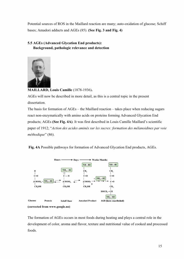
5.5 AGEs (Advanced Glycation End products): Background, pathologic relevance and detection
MAILLARD, Louis Camille (1878-1936).
AGEs will now be described in more detail, as this is a central topic in the present
dissertation.
The basis for formation of AGEs – the Maillard reaction – takes place when reducing sugars
react non-enzymatically with amino acids on proteins forming Advanced Glycation End
products; AGEs (See Fig. 4A). It was first described in Louis Camille Maillard’s scientific
paper of 1912; “Action des acides aminés sur les sucres: formation des mélanoidines par voie
méthodique” (86).
Fig. 4A Possible pathways for formation of Advanced Glycation End products, AGEs.
(corrected from www.google.no)
The formation of AGEs occurs in most foods during heating and plays a central role in the
development of color, aroma and flavor, texture and nutritional value of cooked and processed
foods.

16
However, these processes were later also revealed in biological systems.
HbA1c, which reflects mean level of blood glucose, is an Amadori product (see Fig. 4A) and
the most important predictor for development and progression of vascular complications in
diabetes (21) (19).
AGEs; definition and examples
The term “AGEs” refers to posttranslationally glycated modifications on end-standing
aminogroups on proteins, lipoproteins, lipids and nucleic acids that non-enzymatically have
undergone irreversible dehydration and condensation processes via various reactive
intermediates. There may be several modifications per molecule and several different proteins
may be modified. The modification itself is often referred to as an “adduct”. (see Fig. 4A).
There are several alternative routes into forming AGEs, and the predominant substrate
fuelling the glycation is glucose. However, carbohydrates and sugars other than glucose, such
as glyceraldehyde, fructose and ribose also glycate to form AGEs.
Early glycation products may later form advanced glycation end products, like glyoxal from
auto-oxidative glycation. Glyoxal may, through further steps of oxidation (glycoxidation)
form N�-(carboxymethyl)lysine (CML), whereas methylglyoxal (MG) may form
hydroimidazolones and e.g. argpyrimidine. MG levels are increased in diabetes, and
hydroimidazolone is one of the most important AGEs (87). CML and other AGEs may also
form without carbohydrates from lipid peroxidation, phospholipids and the nucleotides of
DNA. When oxidation accompanies glycation, examples of additional glycoxidation products
to CML are CEL and pentosidine, the latter with intrinsic fluorescent properties (80). Tissue
and plasma fluorescence may be used as indirect markers of accumulation of AGEs. In
addition to CML and pentosidine, glyoxal-lysine dimer (GOLD) is considered marker of
glycoxidation (88).
Examples of AGEs formed through nonoxidative processes are pyrraline, MOLD and DOLD.
A list of biologically important AGEs are found in Table 1 (89).

17
Methylglyoxal derived AGEs
In addition to sugars, many different aldehydes and ketones can form AGEs in vivo.
Methylglyoxal (MG) is an �-oxoaldehyde capable of provoking oxidative stress, and is
present at higher concentrations in diabetes (90). It generates from spontaneous
decomposition of triose-phosphate intermediates in aerobic glycolysis and oxidative
degradation of both carbohydrates (pentoses and ascorbate) and lipids (arachidonate) (91). It
is also a substrate of the glyoxalase system (92), which detoxifies it to D-lactate.
By irreversible glycation, methylglyoxal forms AGEs both intracellularly and extracellularly.
It may modify a range of different proteins in different compartments, from collagen in tissue
to proteins of the circulation. The type of AGE modification formed is determined by the free
aminogroup to which it binds and modifies.
There are three main aminoacids that react non-enzymatically with methylglyoxal forming
physiological AGEs; cysteine, lysine and arginine.
Examples of cysteine modifications are CMC and CEC, which are increased in plasma of
diabetic nephropathy (93).
N(epsilon)-(carboxyethyl)lysine (CEL) and the imidazolium crosslink, methylglyoxal-lysine
dimer (MOLD) are examples of AGEs formed from MG and lysine. Lysine modifications
may also crosslink with eachother via lysine bridging forming imidozolysine.
When MG reacts with arginine, argpyrimidine may form, which is a fluorescent AGE.
However, the main AGEs formed when MG reacts with arginine in proteins are isomers of
hydroimidazolone called methylglyoxal hydroimidazolones, of which MG-H1 (N-�-acetyl-N-

18
�-(5-hydro-5-methyl)-4-imidazolone is the one quantitatively dominating in vivo, often
referred to as hydroimidazolone only, and of special focus of this thesis
(See Fig. 4B).
AGEs and disease
The formation of AGEs is observed in the human body at all ages and increases as a process
of normal aging, contributing to cross-linking of extracellular, long-lived proteins, and may
lead to browning and fluorescence, and – where these reactions are accelerated – to
development of diabetic complications and inflammatory processes linked to
neurodegenerative diseases (94), hypertension, rheumatoid arthritis and atherosclerosis (95).
There are several reasons for AGEs believed to be of pathogenic importance in diabetic
vascular complications (see Table 2):
19
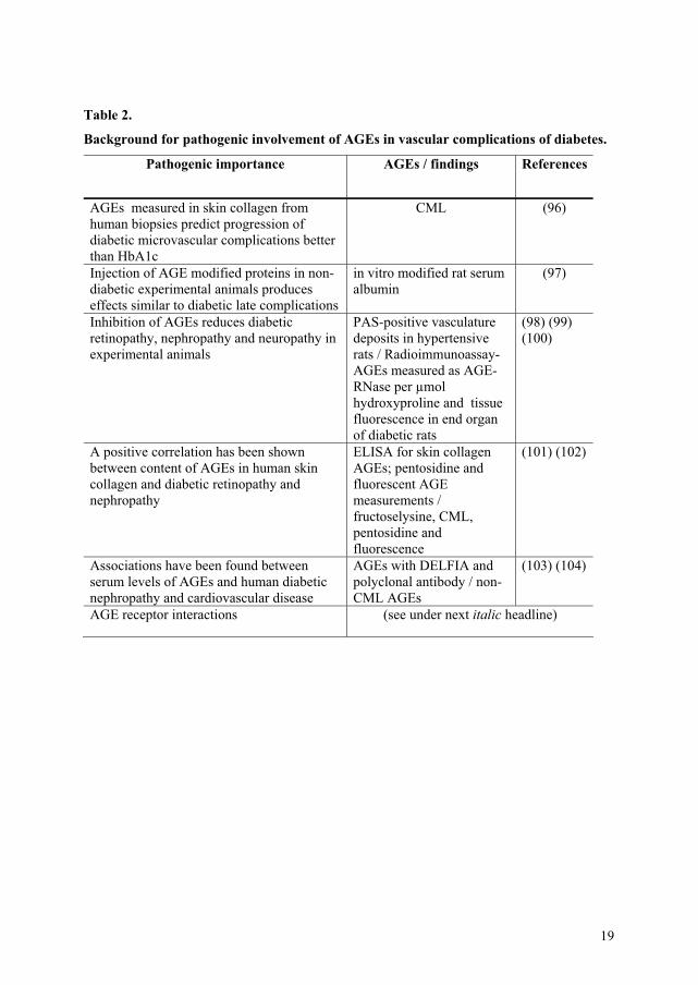
Table 2.
Background for pathogenic involvement of AGEs in vascular complications of diabetes.
Pathogenic importance AGEs / findings References
AGEs measured in skin collagen from human biopsies predict progression of diabetic microvascular complications better than HbA1c
CML (96)
Injection of AGE modified proteins in non-diabetic experimental animals produces effects similar to diabetic late complications
in vitro modified rat serum albumin
(97)
Inhibition of AGEs reduces diabetic retinopathy, nephropathy and neuropathy in experimental animals
PAS-positive vasculature deposits in hypertensive rats / Radioimmunoassay-AGEs measured as AGE-RNase per μmol hydroxyproline and tissue fluorescence in end organ of diabetic rats
(98) (99) (100)
A positive correlation has been shown between content of AGEs in human skin collagen and diabetic retinopathy and nephropathy
ELISA for skin collagen AGEs; pentosidine and fluorescent AGE measurements / fructoselysine, CML, pentosidine and fluorescence
(101) (102)
Associations have been found between serum levels of AGEs and human diabetic nephropathy and cardiovascular disease
AGEs with DELFIA and polyclonal antibody / non-CML AGEs
(103) (104)
AGE receptor interactions (see under next italic headline)
20
Figure 5 depicts the three routes as mentioned in 6.3 IV 1-3, through which AGEs may cause
cellular dysfunction:
Circulating AGEs; AGE/RAGE interaction
The liver and kidney are both involved in the catabolism and excretion of AGEs from the
circulation (105) (106). Renal tissue is among the targets of AGEs that cause cellular damage
and reduce kidney function. Hence, a reduced clearance of AGEs from the circulation may
further increase both cause and effect of damage from glycation (107). Clinical studies have
reported increased serum levels of CML in subjects with type 1- (108) and type 2-diabetes
(90) and elevated levels of CML are associated with microvascular complications of diabetes
such as retinopathy (101). In addition, patients with type 2-diabetes have increased circulating
levels of the AGE hydroimidazolone (109) and its precursor MG (90). Positive associations
between serum levels of hydroimidazolone and retinopathy have been shown in clinical
studies of type 1-diabetes (110). The effects of circulating AGEs may be caused by interaction
with receptors or without receptors involved (See Fig. 5). There are several AGE receptors, of
which the multiligand receptor RAGE probably is among the most important, participating in
chronic inflammatory and immune responses (111). Proposed endogenous ligands for RAGE
other than certain AGEs (like CML), are S100/calgranulins (a family of closely related
calcium-binding polypeptides that accumulate extracellularly at sites of chronic
inflammation); amphoterin (or protein HMGB1, released by cells undergoing necrosis) and

21
amyloid (amyloid -� peptide accumulating in Alzheimer’s disease; amyloid A accumulating
in systemic amyloidosis).
A truncated, soluble form of RAGE also exists; sRAGE (endogenous secretory
RAGE), which is capable of binding extracellular ligands without cell contact. sRAGE in
excess can competitively bind ligands meant for RAGE, thus preventing cellular signalling
mediated via this receptor. The balance between levels and actions of RAGE and sRAGE may
be central in AGE-mediated pathology (80). AGE/RAGE interaction activates a cascade of
signal transductions, of which the PKC pathway is one. Generation of reactive oxygen species
(ROS) may follow, triggering NF-�B. This in turn elicits release of proinflammatory
cytokines, expression of adhesion molecules and growth factors (e.g. VCAM-1, TGF-B1,
VEGF), all implicated in the pathogenesis of diabetic complications, like diabetic retinopathy
(111) (80).
Whether hydroimidazolones interact with RAGE is uncertain. However, methylglyoxal
modification of arginine residues by may be particularly damaging because arginine residues
occur at a high frequency in substrate and ligand recognition sites in enzymes and receptors
(112).
AGEs, VEGF and the eye
Retinal pericytes have a low regenerative capacity. Loss of these supportive cells is an early
event in the course of diabetic retinopathy (48). As AGEs in vitro are toxic to bovine retinal
pericytes and cells of the microvasculature (113) (114), they may play an important role in
pericyte loss. Further, capillary non-perfusion and closure gradually increase hypoxia and
stimulation of growth factors (Fig. 1). VEGF is produced in the eye by retinal pigment
epithelium (RPE) cells and is up-regulated by hypoxia. It is considered to be involved in the
progression of diabetic retinopathy (115) (116) as it stimulates vascular permeability and new
vessel growth. It has been shown that AGEs induce VEGF expression in retinal cell culture
and animals. There are four major biologically active human isoforms. VEGF165
predominates in the human eye, appearing to be responsible for pathological ocular
neovascularization (117). If AGE modified protein is injected in non-diabetic rats, VEGF in
the eye is up-regulated (118), producing dysfunction of the inner BRB (119). Utilizing “anti-
VEGF” as a therapeutic concept has expanded over the last few years: VEGF antibodies are
used to reduce neovascularization and edema of not only cancer but also ophthalmologic
entities such as AMD and ME of DR (120) (121).

22
Interestingly, AGEs are associated with degenerative changes of other tissues of the
eye as well:
- Accumulation of AGEs (CML) is found on the corneal basement membrane suggesting a
causative role in corneal epithelial disorders in diabetes (122).
- Methylglyoxal hydroimidazolones are quantitatively major AGEs of the lens proteins in
humans. These lens protein modifications may stimulate further glycation, oxidation, and
protein aggregation to form cataract (123).
- Accumulation of AGEs (pyrraline) is found at the optics disc; in the cribriform plate and
around vessels of the optic nerve, possibly contributing to the development of neuropathy of
the optic disc and nerve in diabetes (124).
- Elevated levels of AGEs (non-CML, hydroimidazolone) have recently been described in the
human vitreous of diabetic patients compared to non-diabetic controls (125) (126).
AGEs; different methods for detection
1) Immunoassays are widely used in the field of AGE research. The immunoassay used in the
present study was developed by Kilhovd et al (109) based on work by Berg et al with minor
modifications (110) in our determination of N�-acetyl-N�-(5-hydro-5-methyl)-4-imidazolone
(MG-H1) which is a methylglyoxal (MG) -modified arginine compound. The antibodies of
the DELFIA assay (dissociation enhanced lanthanide fluoro-immunoassay) used are marked
with Europium chelate for fluorimetric visualization. An advantage of the DELFIA system
compared to the enzyme linked immunoassay (ELISA) is its ability to diminish interfering
background fluorescence when applying a delayed fluorescent visualization technique. With
either immunoassay the quantification is relative, depending on the quality of the standard
quantification.
2) Separation methods coupled to MS (mass spectrometry): Due to the complexity of
biological samples, chromatographic techniques (e.g. HPLC; high pressure liquid
chromatography) have been used for initial separation of molecules into relatively
homogenous groups. This may be used in combination with later, spectrometric methods,
such as LC-MS (liquid chromatography mass spectrometry). MS may be performed in a
coupled fashion called MS/MS, or tandem mass spectrometry. These are all methods for a
more absolute quantification with a relatively high sensitivity.

23
With mass spectrometry (MS), the constituents of chemical samples are measured as the
mass-to-charge ratio of ions: The sample is ionized; ions of different masses are separated,
and finally; their relative abundance is measured by ion flux intensity. This is the golden
standard for AGE measurements at the present time. However, initial preparation of the AGE
sample is still necessary before LC-MSMS (127), usually by enzymatic digestion or acid
hydrolysis. This implies that the intact modification of the protein is not measured, but rather
peptide fragments of the modified proteins.
3) Autofluorescence may be measured using an autofluorescence meter applied to the skin
(128). This method is not AGE specific, as compounds other than AGEs also may fluoresce.
It measures both glycation and oxidation adduct fluorophores, but the phenomenon of
fluorescence in tissue and plasma can be used as a marker for the presence of AGEs. Over
time, tissue fluorescence increases in diabetes (83), (129), (130) as observed within the
kidney, the retina, the skin and other sites of diabetic microvascular pathology. Some studies
have suggested that fluorescent AGEs may be better associated with microvascular
complications than with non-fluorescent compounds such as CML (131). It is thought that
incomplete degradation of AGE-modified proteins from the diet or endogenous sources
produce so called low–molecular weight (LMW) AGEs. A simple and indirect way of
measuring tissue florescence is by measuring fluorescence in the LMW fraction of serum
(108). The findings of Januszewski et al support the association between LMW AGEs and
end-organ damage in diabetes (132).
6. Aims of research
� To measure and stepwise compare serum hydroimidazolone within a group of diabetic
patients with different degree of retinopathy
� To measure and compare in patients with diabetes and in age matched controls:
- hydroimidazolone in vitreous of patients with diabetes and in age matched
controls
- hydroimidazolone and VEGF both in serum and in vitreous fluid
� To compare retinopathy from fundus photographs in a follow-up study of patients with type 1
diabetes
� To apply a new, clinical method of retinopathy classification on fundus photographs

24
7. Subjects and methods
7.1 Subjects
Paper 1
From a Scandinavian outpatient clinic we recruited 227 patients with type 2 diabetes mellitus
– 124 men and 103 women – with retinopathy ranging from none to proliferative. At the time
of diagnosis, 221 patients were older than 30 years and 6 patients were younger than 30 years,
but were not in need of insulin treatment. 86 had no retinopathy; 89 had NPDR and 52 had
PDR. The retinopathy group (NPDR + PDR) was cross-sectionally compared to the non-
retinopathy group (DNR). The two groups differed significantly on several parameters; hence
logistic regression analysis was applied for comparison regarding retinopathy and serum
levels of hydroimidazolone. Patients with plasma creatinine values > 200 mg/dl were
excluded as this level of reduced kidney function may be associated with increased serum
hydroimidazolone values.
Paper 2
Vitreous from 23 consecutive patients with type 2-diabetes and median known diabetes
duration of 12 years were included and compared to 32 non-diabetic and age-matched control
subjects who also underwent vitrectomy. The median age of both groups was 67 years.
Vitrectomy within the last 6 months before hospitalization and vitreous of reddish colour were
criteria for exclusion; as were plasma creatinine values > 200 mg/dl. Serum and vitreous
parameters were cross-sectionally compared with regard to hydroimidazolone and
retinopathy, in particular.
Paper 3
We randomly selected 61 patients with diabetes mellitus type 1 from a Scandinavian
outpatient clinic for comparison of degree of retinopathy and serum levels of
hydroimidazolone. DNR patients had a mean duration of diabetes of 14 years, which was
significantly lower than the mean duration of 20 years in the DR group. The ratio PDR/NPDR
was 36/11. Quartiles of serum hydroimidazolone were compared with occurrence of
retinopathy.

25
Paper 4
In 1989, of the reachable 1868 incident type 1 diabetes cases in Norway from the period
between 1973 and 1982, 600 were randomly selected for participation in the study. The
baseline examination for diabetic complications of the 368 enrolled took place in 1989/-90.
355 were eligible for follow-up examination in 2002/-03.
All new-onset cases of type 1 diabetes occurring in Norwegian children below 15 years of age
within the decade 1973/-82 were retrospectively registered by the Norwegian Childhood
Registry between 1985 and 1986. The analyses at follow-up included 294 subjects with
proper retinal photographs. The participants belonged to different hospital catchment areas,
and the main investigator visited 24 different hospitals all over Norway. The examination
included a medical history, blood pressure measurements, collection of overnight timed urine,
random venous blood samples and fundus photography. Patient characteristics at baseline,
divided into participants and non-participants at follow-up, were compared.
7.2 Methods
MG modification of BSA was prepared by incubating BSA in sodium phosphate buffer (100
mM, pH 7.4 and 37C) with 1 mM MG for 4 days, before dialyzed against ammonium
bicarbonate buffer (30 mM, pH 7.9 and 4C) and lyophilized to dryness. Preparations of
lyophilized MG-BSA were stored in liquid nitrogen. As determined by Thornalley’s group
(133) using HPLC amino acid analysis, this low-modified MG-BSA contains 23% modified
arginine residues per molecule of serum albumin.
Hydroimidazolone immunoassay
We have previously developed specific solid-phase, time-resolved competitive immunoassays
(DELFIA Wallac, Turku, Finland) for determining AGEs in serum (103); further developed
by Kilhovd et al (109). The primary antiserum of the assay is a monoclonal anti-
hydroimidazolone (IG7) antiserum. (It was obtained by injecting mice with keyhole limpet
haemocyanin (KHL) and modified by incubating with 70 mmol/l of MG for 6 hours at 37C.
Epitope specificity of the anti-hydroimidazolone antibody was evaluated using both dot blots
and an indirect competitive ELISA. The IG7 antibody reacted specifically with N�-acetyl-N�-
(5-hydro-5-methyl)-4-imidazolone, showed 1% cross-reaction against its oxidized form
methylimidazolone, and to some extent recognized the analogue glyoxal derived arginine
hydroimidazolone compound. It did not react with N�-acetyl-argpyrimidine, bis(N�-

26
acetyl)lys-4-methyl-imidazolium chloride or carboxyethyllysine.) The IG7 antibody relatively
specifically recognized MG-induced modifications of arginine residues, and did not cross-
react with the imidazolones produced by the reaction of 3-deoxyglucosone with arginine
(134), (135).
Essentially as described by Kilhovd et al (109), levels of hydroimidazolone immunoreactivity
were determined with this monoclonal anti-MG-modified arginine antibody, except that we
coated the microtiter wells in 0.05 mol/L Tris buffer (pH 7.8) and set up our assay in
duplicates: Microtiter strips of 12 wells each were coated with 0.1 ml of MG-modified BSA
(25 g/ml) diluted in 0.05 mol/L Tris buffer (pH 7.8). They were then covered, and incubated
overnight while shaking at room temperature. The strips were further stored at 4C before
washed. The wells were washed 6 times in DELFIA washing buffer before use. Duplicates of
100 l MG-modified BSA standard or serum (diluted 1:4) were added to each well along with
50 l of anti-hydroimidazolone (IG7) antiserum diluted 1:5000 in DELFIA assay buffer*. 7
standard solutions of 0, 2.5, 5, 10, 20, 40 and 100 g/ml MG modified BSA were used per
assay. The strips were incubated while shaking in room temperature for two hours, and then
washed six times in washing buffer. 100l/well of Europium-labeled anti-mouse-IgG-
antibodies were then added in a final concentration of 0.1 g/ml in DELFIA assay buffer.
(The indicator antibody for the DELFIA was marked with Europium chelate for fluorimetric
visualization). All strips were incubated while shaking for one hour in room temperature,
before subsequently washed 6 times and incubated for 5 minutes while shaking with DELFIA
Enhancement solution prior to measurement of the Europium-ion chelate specific
fluorescence in a 1232 DELFIA Fluorometer.
This assay was applied in paper 1, 2 and 3. The arbitrary hydroimidazolone unit (U) was
defined as the competitive activity of 1 g of MG-modified BSA standard, expressed as U/mg
protein.
*When determining vitreous levels of hydroimidazolone, the same assay was used. Vitreous
samples were set up in non-diluted triplicates.
VEGF assay
We determined serum and vitreous levels of the angiogenic isoforms of natural human
VEGF165. The Quantikine ® VEGF immunoassay (R & D Systems, Inc., Minneapolis, MN,
USA) was used. Optical density was determined at 450 nm using a Rosys Anthos HT2 micro

27
plate absorption spectrophotometer (Anthos Labtec Instruments, Salzburg, Austria). A
correction wavelength of 620 nm was used.
Vitreous hemoglobin
Harboe’s method together with updated validations for measuring haemoglobin (Hb) in
micromolar concentrations were used (136). We measured absorbance with an Ultrospec®
3300 pro spectrophotometer (Amersham Biosciences, Cambridge, UK; acquired by General
Electric Company, Fair- field, CT, USA) at wavelengths of 380 nm, 415 nm and 450 nm. The
following equation then expresses in g �l the concentration of hemoglobin (137): CHb = 1.67 x
A415 – 0.83 x A380 - 0.83 x A450.
Vitreous albumin
Albumin was measured in duplicates of 50 l vitreous samples using the Tina-quant
immunoturbidimetric albumin assay (F. Hoffmann-La- Roche Ltd, Basel, Switzerland).
Asessment of Retinopathy
Paper I & Paper III: After pupillary dilation, stereo photographs from 7 standard fields were
taken of each eye, using a 30 fundus camera (Topcon TRC-50, Tokyo, Japan). Grading was
performed in a masked fashion. The patients were characterized according to the ETDRS
level of retinopathy in the worse affected eye by a centrally located, highly skilled team. If
former treatment with panretinal photocoagulation had been given, retinopathy was
automatically classified as PDR; that is, ETDRS level 61.
We then grouped these results in three categories: DNR, NPDR and PDR for comparison of
retinopathy with other parameters.
Paper II: The patients were clinically diagnosed during their period of hospitalization:
Diagnoses were pre- and peroperatively confirmed by the eye surgeon through mydriatic
pupils, using indirect ophthalmoscope and operating microscope, respectively.
Paper IV: In paper 4, the photographic procedure of taking colour retinal photographs of
each eye using Kodachrome 64 ISO 35-mm colour slide film (Kodak, Rochester, NY, USA)
with a non-mydriatic 45 retinal camera (45NM-CR; Canon, Tokyo, Japan) was applied by
the same investigator (T. Skrivarhaug) after instilling one drop of cyclopentolate 1% and
epinephrine 10% in the cul-de-sac of each eye to obtain dilated pupils. The standard
procedure of centering the photographs midway between the fovea and the temporal edge of

28
the optic disc was executed in the exact same way at follow-up and baseline. For back-up
safety in case of reduced quality affecting the readability of one or more pictures, double sets
of pictures were taken of each eye. Without knowledge of the subjects’ identity, the pictures
were graded centrally for retinopathy (by D. Fosmark), status of the worse eye deciding level
of retinopathy. Between the extremes PDR and DNR; NPDR was further sub-classified with a
new, simplified clinical method (Table 1) based on evidence from the ETDRS and the
WESDR (Diabetic Retinopathy Disease Severity Scale (75)). Macular edema was not
detectable using our method. Patients with fibrous proliferations, vitreous hemorrhage or scars
from panretinal photocoagulation were assigned to the PDR category.
HbA1c was analyzed with high-performance liquid chromatography (VARIAN II
Hemoglobin A1c program, BioRad, Hercules, CA) (reference range, 4.0%-5.3%).
(Blood tests were collected after overnight fasting.
Three overnight timed urine samples were collected at home.
Creatinine in urine was analyzed using a kinetic method (Beckman Synchron LX20, Brea,
CA).
Urinary albumin was measured using nephelometry (IMMAGE, Beckman Coulter) or
turbidimetry (Beckman Synchron LX20). Normal values for urine albumin urine creatinine
ratio were < 2.0 mg/mmol for men and < 2.8 mg/mmol for women.
Plasma creatinine was analyzed with a kinetic method (Beckman Synchron LX20). The
Reference ranges are 51- 88 mol/L (women) and 63-105 mol/L (men).
Lipids (triglycerides and total cholesterol) were measured with standard enzymatic methods at
a centralized lab.
Statistical analyses
In all papers, statistical analyses were performed using the SPSS software, version 12.0.0
(SPSS, Chicago, IL). Two-sided Mann-Whitney U test was used when comparing medians of

29
continuous data not normally distributed. The Spearman correlation test was used when
studying the association between two continuous variables. For conservative estimates, we
used logistic regression analysis according to Katz (138). A significance level of 5% was used
for each test.
Paper I: Unadjusted comparisons between patients with and without retinopathy were
performed using Student’s t-tests (2-sided) for continuous variables and exact Fisher tests (2-
sided) for dichotomous variables. Logistic regression analysis, with retinopathy as dependent
variable, was used to study the impact of selected variables; hydroimidazolone, plasma
creatinine, urinary albumin-creatinine ratio, HbA1c, diabetes duration, age, and blood
pressure. (These variables were enabled for multiple regression analyses as initial bivariate
unadjusted analyses showed significant associations with retinopathy).
Paper II: Spearman’s test for correlations was used for correlation testing. Logistic
regression was used for correction of vitreous albumin when comparing vitreous VEGF
between controls and diabetes patients.
Paper III: A linear-by-linear association chi-square test was used when studying the
association between quartiles of hydroimidazolone and retinopathy. We used a two-sided
exact Fisher test when comparing the prevalence of retinopathy between two groups. When
comparing HbA1c and age between two groups, an independent two-sample t-test was used;
and for the comparison of duration of diabetes, a two-sided Mann-Whitney test was used. The
Spearman correlation test was used when studying the association between two continuous
variables.
Paper IV: We estimated the cumulative incidence of PDR from diabetes onset until follow-
up, using a Kaplan-Meier plot. To assess declining incidence of PDR with year of diagnosis,
the patients were divided into two groups: 1973–1977 (n=133) and 1978–1982 (n=161). Cox
regression analysis was used to estimate the hazard ratio for association between baseline
factors and PDR. The following variables were analyzed: sex, age, age at diabetes onset,
diabetes duration, smoking status, arterial blood pressure, AER, HbA1c, triglycerides and
total cholesterol. Variables with a p-value <0.20 were then included simultaneously in a
multiple regression model.
In the analysis of predictive risk factors for PDR, the three subjects with PDR at baseline were
not included. As the exact time of onset of NPDR was unknown, the assessment of potential
predictors of retinopathy among patients without DR at baseline was analyzed similarly using
logistic regression.

30
Sensitivity analysis of non-participation was exclusively based on HbA1c values. By using
assumptions based on external data, we performed a sensitivity analysis in order to assess the
potential influence of selection bias due to non-participation and other losses to follow-up
(deaths and emigration) on our estimated risk of complications. The total risk in the full
cohort of participants and non-participants is a weighted average of the observed risk of
complications among participants and the corresponding risk among non-participants. This
total risk can then be estimated under conservative assumptions regarding the risk among non-
participants.
(Data from the DCCT indicated a 75% increase in risk of PDR per 1%-point increase in
HbA1c during a mean follow-up of 6.5 years. The HbA1c difference among non-participants
and participants in our material in 1990 was only 0.6%-points. A conservative assumption of
75% higher risk of PDR as compared with the participants was made, and that 50% of them
had no DR at baseline while the rest had NPDR.)
8. Summary of main results (papers)
Paper 1: � Increased serum levels of the specific advanced glycation end product methylglyoxal-derived
hydroimidazolone are associated with retinopathy in patients with type 2 diabetes mellitus.
According to the ETDRS protocol, level of retinopathy was determined from retinal
photographs of 227 patients with type 2 diabetes mellitus and median known diabetes
duration of 14 years. 86 patients had no retinopathy (DNR), whereas non-proliferative
retinopathy (NPDR) was diagnosed in 86 patients and proliferative retinopathy (PDR) in 52
patients. Median age was 66 years.
Serum levels of hydroimidazolone were increased in the group of patients with retinopathy,
with a further increment as retinopathy worsened to PDR. This was found when including all
patients irrespective of time elapsed from diabetes having been diagnosed, but also in the
smaller group of patients with shorter duration of diabetes, i.e. below the median of 14 years.
We found a strong association between HbA1c and diabetic retinopathy (p<0.0001), and the
association between retinopathy and hydroimidazolone was independent of HbA1c.
31
Paper 2: � Increased vitreous levels of hydroimidazolone in type 2 diabetes patients are associated with retinopathy.
Using a cross-sectional case-control study design, we compared vitreous and serum contents
in 23 patients with diabetes type 2 – sixteen of which had PDR – to 32 age-matched controls
also undergoing vitrectomy. Level of retinopathy was based on clinical examination. We
found a positive correlation in all patients between vitreous and serum content of
hydroimidazolone (r=0.48, p<0.001). This was also true within cases and non-cases
separately.
Paper 3: � Serum levels of the Advanced Glycation End product hydroimidazolone is associated with retinopathy
occurrence in type 1 diabetes patients.
In this cross-sectional study of 61 type 1 diabetic patients, 14 had no retinopathy (DNR), 11
had NPDR and 36 had PDR. Grading of retinopathy was based on retinal 7-field stereo
photographs according to the ETDRS protocol. Comparisons of serum levels of
hydroimidazolone were made between patients with and without retinopathy.
Hydroimidazolone quartiles were found significantly associated with retinopathy (p=0.013).
After adjusting for duration of diabetes by logistic regression analysis, a significant difference
in retinopathy present was found when comparing the lowest quartile with the rest (p=0.022).
Paper 4: � Low cumulative incidence of proliferative retinopathy in childhood-onset type 1 diabetes in Norway.
294 childhood-onset type1 diabetic patients had readable fundus photographs taken and
examined for retinopathy between 2002 and 2003. This was a follow-up of originally 368
patients having undergone identical examinations between 1989 and 1990; all belonging to
the 10 year cohort of 1906 persons diagnosed with diabetes mellitus type 1 between 1972 and
1983. 262 of 294 (89.1%) developed diabetic retinopathy, of which 32 developed PDR. The
cumulative incidence of PDR began increasing after 10 years of diabetes duration, reaching
10.9 % (95 % CI: 7.3-14.5 %) at twenty-five years. Mean diabetes duration was 19 years
(range 12-29 years); mean age for diagnosis of PDR was 27 years (range 17-41 years).
Significant predictors at baseline for developing retinopathy of any degree were HbA1c
32
(OR=3.25, 95 % CI: 1.76-6.02, p<0.001) and male gender (OR=2.51, 95 % CI: 1.06-6.00,
p=0.037), whereas significant predictors for developing PDR were NPDR at baseline
(RR=3.71, 95 % CI: 1.59-8.68, p=0.03), HbA1c (RR=2.05, 1.44-2.93, p<0.001) and
triglycerides (RR=1.55, 1.06-1.95, p=0.019).
9. Discussion
9.1 Methods
Clinical and Photographic methods of retinopathy diagnosis
a) The “Gold standard” for epidemiological surveys of DR is the seven-field, (analogue)
retinal photographic method using film and a 30 camera. Members of a highly skilled team,
centrally located with resources at hand, applied this method on the subjects in paper 1 and 3,
including later grading of the pictures according to the ETDRS standard. We grouped these
results in three categories: DNR, NPDR and PDR for comparison of retinopathy with other
parameters.
b) In paper 4, to obtain dilated pupils one drop of cyclopentolate 1% and epinephrine 10%
was instilled in the cul-de-sac of each eye. The non-mydriatic 45 retinal camera (45NM-CR;
Canon, Tokyo, Japan) produced colour retinal photographs of each eye using Kodachrome 64
ISO 35-mm colour slide film (Kodak, Rochester, NY, USA). The photographic procedure was
applied by the same investigator (T. Skrivarhaug). The standard procedure of centering the
photographs midway between the fovea and the temporal edge of the optic disc was
performed in the exact same way at baseline and follow-up. For back-up safety in case of
reduced quality affecting the readability of one or more pictures, double sets of pictures were
taken of each eye. Without knowledge of the subjects’ identity, the pictures were graded
centrally for retinopathy (by D. Fosmark), status of the worse eye deciding level of
retinopathy. Between the extremes PDR and DNR, NPDR was further sub-classified with a
new, simplified clinical method (Table 1) (75). Macular edema was not detectable using our
method. Patients with fibrous proliferations, vitreous hemorrhage or scars from panretinal
photocoagulation were assigned to the PDR category.
c) In paper 2, all ophthalmologic diagnoses including retinopathy were given during the
patients’ period of hospitalization. The diagnoses were pre- and peroperatively confirmed by
the eye surgeon using indirect ophthalmoscopy and operating microscopy, respectively.

33
When we chose methods for comparison of retinal photographs in paper 4, digital
cameras were about to substitute the use of analogue equipment – including film. Yet, the
extensively marketed digital techniques with resolution constantly improving, a standardized
comparison with analogue photographs was missing. Hence, at follow-up, the described one-
field method for film was chosen due not only to its simplicity, but because of identicality to
the method originally used at baseline. This facilitates the comparison between the two
investigations.
A more detailed yet standardized method for further classification of late changes in
the course of retinopathy development was needed. An improved clinical method, newly
developed in order to simplify the existing 7-field ETDRS Gold standard and to facilitate data
comparisons between different countries and trials, was found applicable for our purpose.
Albeit subjective, the clinical standard for identification of patients with retinopathy is by
direct ophthalmoscopy. However, images from cameras producing single 30 and 45 fields
have both been considered useful for retinopathy screening, epidemiology studies and routine
care purposes. An exact agreement of 82.5 % was found when comparing ophthalmoscopy to
the seven field stereo photographic method (139).
Our method using a single 45 photograph can be criticized for the possibility of under-
detecting retinopathy. One relative advantage with the 45 field is that only one photo is
needed to view the posterior pole of the retina. However, it is of lower magnification than (the
one) produced with a 30 camera (minified 0.64x at zero diopters). The 45 images include
areas above and below the temporal arcades, and temporal to the macula and just nasal to the
disc (see Fig. 6).

34
Fig. 6 Approximate field obtained with a 45 camera superimposed (dotted lines) on photographic field obtained with the standard seven-field mydriatic stereoscopic protocol from the Early Treatment Diabetic Retinopathy Study and a 30fundus camera (field 1 = disc; field 2 = macula; field 3 = temporal to macula; field 4 = superior temporal; field 5 = inferotemporal; field 6 = superior nasal; field 7 = inferior nasal).
With the use of a digital, monochrome camera, a study on retinopathy in 197 type 1 and type
2 diabetic patients with a single 45 image versus ophthalmoscopy revealed 100% sensitivity
and 71 % specificity, the field identical to the field in our study (paper 4). Compared to the 7
field stereo photographic method, the sensitivity and specificity of the 45 image were 78 %
and 86 %, respectively, with an exact agreement of 83 % (140).
New vessels in the periphery only are very rare (<1 %), and new vessels at the posterior pole
(in patients younger than 60 years) are significantly more frequent than beyond the posterior
pole (141). Thus, a one-field photo of each eye covers most of the area of interest for
inclusion of PDR-diagnoses. Predilection sites for retinal proliferations are the temporal
arcades and second, the area nasal to the optic disc (142) (141).
In order to cover a larger retinal area, a two field photographic method with an identical 45
NM-Canon camera has been validated over a 5 year period; centered on the macula and the
optic disc, respectively (141). Missed diagnoses of PDR amounted to 0.9 %. False positive
findings occurred in 6 of 1341 readings, or 0.4 %. False NVD were recorded mostly in
younger patients (�36 years), whereas false NVE were recorded mostly in older patients ( 50
years). True NVE/NVD were found located on the temporal arcades in 48 % of the eyes and
nasally to the optic disc in 42 % of the eyes, and were rarely found beyond the posterior pole
(13 %; p<0.001) (141).

35
In retrospect, the two-field method might also have been applied in our setting. A larger area,
particularly nasally to the optic disc, would then have been included. Still, macular edema
would not have been classifiable. However, the one-field method was found sufficient for our
purpose; to classify a larger group epidemiologically, and was chosen due to comparisons to
be made with identical field pictures. The better of two photographs from both eyes was
graded before the diagnosis was given by the state of the worse eye, but a small fraction of
false negative PDR with our method due to a lower sensitivity is possible. However, a
possibility of having undergone laser treatment without PDR also exists, overestimating this
group. Importantly, in our material 78.6% had NPDR; of these most had mild NPDR (54.6%).
Further, moderate NPDR accounted for 32.4% and severe NPDR amounted to only 13.0%.
This clustering of relatively benign retinopathy is in keeping with our conclusion of a
relatively low cumulative incidence of PDR after 25 years of diabetes. The pictures were
thoroughly graded by one experienced ophthalmologist (D. Fosmark) in one session only.

36
Immunoassay method
Different methods have been developed and used for the detection of AGEs within the field of
research. For the detection of the hydroimidazolone MG-H1, the DELFIA-system was chosen
due to experience regarding development and use in our laboratory. The quantification of
AGEs when using immunoassays gives relative results, partly due to a missing quantitative
standard for the different AGEs. Arbitrary units are therefore employed. Our assay for
detection had an intra-assay variation in the range of 12 – 15%. The inter-assay variation was
of up to 21%. The cross-reactivity against AGE-BSA was 8% when calculated as AGE-BSA
protein against MG-modified BSA protein. No cross-reactivity was found against CML-BSA
or glycated albumin in the hydroimidazolone immunoassay. The IG7 antibody relatively
specifically recognizes MG-induced modifications of arginine residues.
In use, the assay is practical as it is inexpensive, and many samples can be run within a
relatively short period of time. It is also sufficient for our purpose, which is in larger samples
to search for significant differences between cases and controls. Immunoassays have been
criticized for their heavily modified antigen, use of arbitrary units and possible serum effects
(87). However, the present assay has an acceptable dilution curve and satisfactory recovery
(135): Both serum samples from diabetic patients and controls produced parallel inhibition to
hydroimidazolone standard. Recovery studies have been performed by adding MG modified
BSA to serum from patients and controls. The mean recovery of amount added was 115% ±
26 %-points. A linear dilution curve reflected minor serum effect.
One advantage of the DELFIA system compared to other immunoassay methods (ELISA) is
its ability to diminish the background fluorescence through delayed fluorescence from
Europium attached to the secondary antibody. Hydroimidazolone (MG-H1) is our AGE
modification of focal interest due to its relative abundance in biological systems and its
likelihood of playing a pathogenic role in diabetic complications. 9.2 Interpretation of results
For each paper, thorough interpretations are found in their respective sections of discussion
(See Papers 1, 2, 3 and 4). In Paper 1 and 3, there exists a positive association between
retinopathy and serum levels of hydroimidazolone in both type 2 and type 1 diabetes.
However, as the studies are cross-sectional, no causal conclusions can be made. The lack of
association between HbA1c and hydroimidazolone is in keeping with earlier findings; and
logical, as the two are formed via partly different pathways. However, it is noteworthy that
both hydroimidazolone and HbA1c were found strongly and independently associated with

37
retinopathy. Patients with clearly increased serum creatinine (>200 μmol/l) had not
surprisingly clearly elevated serum hydroimidazolone, and these patients were excluded from
further analysis. If hydroimidazolone is causally involved in early nephropathy is not known.
In the present study, no association was shown between hydroimidazolone and urinary
albumin-creatinine ratio.
We measured hemoglobin, albumin and VEGF, adapting the methods for vitreous. This
enabled a more extensive discussion and thorough interpretation of the true vitreous content
of hydroimidazolone in Paper 2. Intravitreal measurements of albumin used as an indirect sign
of iBRB disruption are seldseen. In our study, the correlation of vitreous albumin and
hydroimidazolone is explained by the increase of vitreous albumin in the PDR group, due to a
breakdown of the iBRB. Hydroimidazolone most likely originated from serum, whereas
VEGF was produced intraocularly. No correlation between vitreous hydroimidazolone and
VEGF was found in our study. However, the number of subjects studied was small.
In Paper 4, we found a low cumulative incidence of PDR after 24 years of type1 diabetes. The
study had a population based design, but there was a marked proportion of non-participants
and losses for follow-up. Thus, a sensitivity analysis was done to assess any influence of
selection bias on risk for retinopathy. As regards PDR, a higher cumulative incidence of
14.0% was found with this method (versus 10.9%). Still, this is relatively low. However, the
proportion of subjects with NPDR was substantial (78.6%). There remains a potential for still
improved glycemic control and optimization of other risk factors that can be modified, like
blood pressure and triglycerides. A uniform screening system is still missing in Norway, as
are registers for incident blindness. This study is a contribution to increased knowledge on
microvascular complications nationwide, and as such a stimulus for further research and
improvement of prophylactic measures.
10. Concluding remarks (Relevance of papers, future research)
The exact pathogenic mechanisms of diabetic retinopathy are only partly revealed. Interplay
of ROS and AGEs are plausible. Most probably, the explanation consists of multiple factors.
The range of functionally and structurally different AGEs also makes it difficult to point out
which AGE is the most pathogenic.
Exogenous AGEs (from coffee, smoking, foods etc) and their disputed pathogenic role are not
discussed in this paper. In general, a limited consumption is recommended due to their
abundance in “unhealthy food”, but in particular if reduced kidney function exists (143) (144).

38
Elegant approaches for potential interventions in restricting the burden of endogenous AGEs
have emerged, attacking at the following sites:
a) AGE formation may be inhibited by trapping of reactive (di)carbonyls in the glycation process
and chelation of transition metal ions (e.g. by aminoguanidine, pyridoxamine). Unfortunately,
aminoguanidine has toxic side effects which has lead to discontinuation of clinical studies
(145) (146). A drug category already in use – inhibiting the angiotensin-converting enzyme –
has anti-hypertensive effect but is also potent as AGE inhibitor (147).
b) AGE cross-links may be broken down. PTB (N-phenacylthiazolium bromide) and ALT-711
(alagebrium chloride), cleave AGE-mediated cross-links (89).
c) AGE binding may be inhibited. The binding of ligand to AGE receptor include soluble RAGE
(sRAGE) and RAGE specific antibodies, neutralizing the effect of receptor interaction (148)
(149).
d) ROS/AGE interaction may be targeted via antioxidants, exemplified by improved retinal blood
flow in type 1 diabetics after high-dose vitamin E supplementation (150).
Productive “anti-AGEs” research makes future important clinical implications of this
knowledge probable, including retinopathy.
Our findings strongly support the following statements:
� Serum levels of the AGE-modification hydroimidazolone (MG-H1) are increased in both type
1 and type 2 diabetic patients with retinopathy compared to those without retinopathy.
� Serum levels of hydroimidazolone (MG-H1) are increased in PDR compared to NPDR
(diabetes mellitus type 2).
� Vitreous levels of hydroimidazolone (MG-H1) are increased in type 2-diabetic patients
compared to age-matched controls due to leakage from serum.
� Vitreous levels of hydroimidazolone (MG-H1) are increased in PDR compared to NPDR in
type 2-diabetic patients, and may originate from serum due to iBRB breakdown.
� VEGF in vitreous fluid is increased in diabetes type 2. Even higher levels are found in PDR.
The increase in VEGF is not due to a spill-over effect from the circulation, even though the
iBRB is disrupted, supporting that it is produced intraocularly.
Patients with type 1 diabetes of 24 years duration and retinopathy staged from fundus
photographs had 89.1% cumulative incidence for DR. 10.5% of these have PDR.
�

39
11. References
(1) Tattersall RB. The history of diabetes mellitus. Textbook of Diabetes. Blackwell
Science: Oxford; 2002. p. 1.1-1.2.1.
(2) Onkamo P, Vaananen S, Karvonen M, Tuomilehto J. Worldwide increase in incidence of Type I diabetes--the analysis of the data on published incidence trends. Diabetologia 1999 Dec;42(12):1395-403.
(3) King H, Aubert RE, Herman WH. Global burden of diabetes, 1995-2025: prevalence, numerical estimates, and projections. Diabetes Care 1998 Sep;21(9):1414-31.
(4) Stene LC, Midthjell K, Jenum AK, Skeie S, Birkeland KI, Lund E, et al. [Prevalence of diabetes mellitus in Norway]. Tidsskr Nor Laegeforen 2004 Jun 3;124(11):1511-4.
(5) National Diabetes Strategy 2006-2010. Oslo: Norwegian Ministry of Health and Care Services; 2006 Jun. [cited 2007 Dec 19] http://www.regjeringen.no/Upload/HOD/Vedlegg/Engelsk/Diabetes.pdf
(6) Reducing the costs of diabetes in Europe: Preventing complications is the key. MediTech Media Ltd 1999 September 28 [cited 2007 Dec 19]; http://www.scienceblog.com/community/older/1999/B/199901871.html
(7) Koster I, Hauner H, von FL. [Heterogeneity of costs of diabetic patients: the Cost of Diabetes Mellitus Study]. Dtsch Med Wochenschr 2006 Apr 13;131(15):804-10.
(8) Nationella riktlinjer för vård och behandling vid diabetes mellitus - Version för hälso- och sjukvårdspersonal. The Swedish National Board on Health and Welfare (Socialstyrelsen) 1999 September 15 [cited 2007 Dec 19]; http://www.sos.se/FULLTEXT/9900-061/9900-061.htm
(9) Birkeland KI. [Can diabetes be prevented?]. Tidsskr Nor Laegeforen 2002 Jan 20;122(2):155.
(10) Krolewski AS, Kosinski EJ, Warram JH, Leland OS, Busick EJ, Asmal AC, et al. Magnitude and determinants of coronary artery disease in juvenile-onset, insulin-dependent diabetes mellitus. Am J Cardiol 1987 Apr 1;59(8):750-5.
(11) Laing SP, Swerdlow AJ, Slater SD, Burden AC, Morris A, Waugh NR, et al. Mortality from heart disease in a cohort of 23,000 patients with insulin-treated diabetes. Diabetologia 2003 Jun;46(6):760-5.
(12) Borch-Johnsen K, Kreiner S. Proteinuria: value as predictor of cardiovascular mortality in insulin dependent diabetes mellitus. Br Med J (Clin Res Ed) 1987 Jun 27;294(6588):1651-4.
(13) Dinneen SF, Gerstein HC. The association of microalbuminuria and mortality in non-insulin-dependent diabetes mellitus. A systematic overview of the literature. Arch Intern Med 1997 Jul 14;157(13):1413-8.

40
(14) Rossing P, Hougaard P, Borch-Johnsen K, Parving HH. Predictors of mortality in insulin dependent diabetes: 10 year observational follow up study. BMJ 1996 Sep 28;313(7060):779-84.
(15) Tesfaye S, Chaturvedi N, Eaton SE, Ward JD, Manes C, Ionescu-Tirgoviste C, et al. Vascular risk factors and diabetic neuropathy. N Engl J Med 2005 Jan 27;352(4):341-50.
(16) Moss SE, Klein R, Klein BE. Ten-year incidence of visual loss in a diabetic population. Ophthalmology 1994 Jun;101(6):1061-70.
(17) Fong DS, Sharza M, Chen W, Paschal JF, Ariyasu RG, Lee PP. Vision loss among diabetics in a group model Health Maintenance Organization (HMO). Am J Ophthalmol 2002 Feb;133(2):236-41.
(18) Dahl-Jorgensen K, Brinchmann-Hansen O, Hanssen KF, Ganes T, Kierulf P, Smeland E, et al. Effect of near normoglycaemia for two years on progression of early diabetic retinopathy, nephropathy, and neuropathy: the Oslo study. Br Med J (Clin Res Ed) 1986 Nov 8;293(6556):1195-9.
(19) Dahl-Jorgensen K. Near-normoglycemia and late diabetic complications. The Oslo Study. Acta Endocrinol Suppl (Copenh) 1987;284:1-38.
(20) Reichard P, Nilsson BY, Rosenqvist U. The effect of long-term intensified insulin treatment on the development of microvascular complications of diabetes mellitus. N Engl J Med 1993 Jul 29;329(5):304-9.
(21) The Diabetes Control and Complications Trial Research Group. The Effect of Intensive Treatment of Diabetes on the Development and Progression of Long-Term Complications in Insulin-Dependent Diabetes Mellitus. N Engl J Med 1993 Sep 30;329(14):977-86.
(22) Stratton IM, Kohner EM, Aldington SJ, Turner RC, Holman RR, Manley SE, et al. UKPDS 50: risk factors for incidence and progression of retinopathy in Type II diabetes over 6 years from diagnosis. Diabetologia 2001 Feb;44(2):156-63.
(23) Klein R, Klein BE, Moss SE, Davis MD, DeMets DL. The Wisconsin epidemiologic study of diabetic retinopathy. II. Prevalence and risk of diabetic retinopathy when age at diagnosis is less than 30 years. Arch Ophthalmol 1984 Apr;102(4):520-6.
(24) Hovind P, Tarnow L, Rossing K, Rossing P, Eising S, Larsen N, et al. Decreasing incidence of severe diabetic microangiopathy in type 1 diabetes. Diabetes Care 2003 Apr;26(4):1258-64.
(25) Nordwall M, Bojestig M, Arnqvist HJ, Ludvigsson J. Declining incidence of severe retinopathy and persisting decrease of nephropathy in an unselected population of Type 1 diabetes-the Linkoping Diabetes Complications Study. Diabetologia 2004 Jul;47(7):1266-72.
(26) Skrivarhaug T, Fosmark DS, Stene LC, Bangstad HJ, Sandvik L, Hanssen KF, et al. Low cumulative incidence of proliferative retinopathy in childhood-onset type 1 diabetes: a 24-year follow-up study. Diabetologia 2006 Oct;49(10):2281-90.

41
(27) Ebeling P, Koivisto VA. Occurrence and interrelationships of complications in insulin-dependent diabetes in Finland. Acta Diabetol 1997 Mar;34(1):33-8.
(28) Morgan CL, Currie CJ, Stott NC, Smithers M, Butler CC, Peters JR. The prevalence of multiple diabetes-related complications. Diabet Med 2000 Feb;17(2):146-51.
(29) Billault BM, Passa PL. Factors associated with diabetic microangiopathy: a study of 157 type I (insulin-dependent) diabetic patients. J Diabet Complications 1991 Oct;5(4):238-43.
(30) Stephenson JM, Fuller JH, Viberti GC, Sjolie AK, Navalesi R. Blood pressure, retinopathy and urinary albumin excretion in IDDM: the EURODIAB IDDM Complications Study. Diabetologia 1995 May;38(5):599-603.
(31) Coppini DV, Wellmer A, Weng C, Young PJ, Anand P, Sonksen PH. The natural history of diabetic peripheral neuropathy determined by a 12 year prospective study using vibration perception thresholds. J Clin Neurosci 2001 Nov;8(6):520-4.
(32) Juutilainen A, Lehto S, Ronnemaa T, Pyorala K, Laakso M. Retinopathy predicts cardiovascular mortality in type 2 diabetic men and women. Diabetes Care 2007 Feb;30(2):292-9.
(33) Fong DS, Aiello L, Gardner TW, King GL, Blankenship G, Cavallerano JD, et al. Diabetic retinopathy. Diabetes Care 2003 Jan;26 Suppl 1:S99-S102.
(34) Helmholtz H. Beschreibung eines Augenspiegels zur Untersuchung der Netzhaut im lebenden Auge. Berlin, 1851.
(35) Jäger E. Beitr zur Pathol des Auges. Wien: 1856 (vol, p. 33 Figure 12).
(36) Nettleship G. On oedema or cystic disease of the retina. Roy Ophth Lond Hosp Rep 1872: VII[(3)], 343-351.
(37) Manz W. Retinitis proliferans. Graefes Arch Clin Exp Ophthalmo 1876;22:229-275.
(38) Banting FG, Best CH. The internal secretion of the pancreas. J Lab Clin Med 1922;7:251-66.
(39) Lee HB, Ha H, Kim SI, Ziyadeh FN. Diabetic kidney disease research: where do we stand at the turn of the century? Kidney Int Suppl 2000 Sep;77:S1-S2.
(40) Ballantyne AJ, Loewenstein A. Exudates in diabetic retinopathy. Trans Ophthalmol Soc UK 1943:63;95. UK.
(41) Lundbaek K. Diabetic angiopathy: a specific vascular disease. Lancet 1954 Feb 20;266(6808):377-9.
(42) Flynn HWJ, Smiddy WE. Diabetes and Ocular Disease. Past, Present and Future Therapies. The Foundation of the American Academy of Ophthalmology; 2000. p. 70.

42
(43) Photocoagulation treatment of proliferative diabetic retinopathy. Clinical application of Diabetic Retinopathy Study (DRS) findings, DRS Report Number 8. The Diabetic Retinopathy Study Research Group. Ophthalmology 1981 Jul;88(7):583-600.
(44) Photocoagulation for diabetic macular edema. Early Treatment Diabetic Retinopathy Study report number 1. Early Treatment Diabetic Retinopathy Study research group. Arch Ophthalmol 1985 Dec;103(12):1796-806.
(45) Poulsen JE. Recovery from retinopathy in a case of diabetes with Simmonds' disease. Diabetes 1953 Jan;2(1):7-12.
(46) Fortune B, Schneck ME, Adams AJ. Multifocal electroretinogram delays reveal local retinal dysfunction in early diabetic retinopathy. Invest Ophthalmol Vis Sci 1999 Oct;40(11):2638-51.
(47) Antonetti DA, Barber AJ, Bronson SK, Freeman WM, Gardner TW, Jefferson LS, et al. Diabetic retinopathy: seeing beyond glucose-induced microvascular disease. Diabetes 2006 Sep;55(9):2401-11.
(48) Aiello LP, Cavallerano J, Bursell SE. Diabetic eye disease. Endocrinol Metab Clin North Am 1996 Jun;25(2):271-91.
(49) Bursell SE, Clermont AC, Kinsley BT, Simonson DC, Aiello LM, Wolpert HA. Retinal blood flow changes in patients with insulin-dependent diabetes mellitus and no diabetic retinopathy. Invest Ophthalmol Vis Sci 1996 Apr;37(5):886-97.
(50) Kohner EM, Patel V, Rassam SM. Role of blood flow and impaired autoregulation in the pathogenesis of diabetic retinopathy. Diabetes 1995 Jun;44(6):603-7.
(51) Schroder S, Palinski W, Schmid-Schonbein GW. Activated monocytes and granulocytes, capillary nonperfusion, and neovascularization in diabetic retinopathy. Am J Pathol 1991 Jul;139(1):81-100.
(52) Anderson B, Jr. Ocular effects of changes in oxygen and carbon dioxide tension. Trans Am Ophthalmol Soc 1968;66:423-74.
(53) Miller JW, Adamis AP, Aiello LP. Vascular endothelial growth factor in ocular neovascularization and proliferative diabetic retinopathy. Diabetes Metab Rev 1997 Mar;13(1):37-50.
(54) Campochiaro PA. Retinal and choroidal neovascularization. J Cell Physiol 2000 Sep;184(3):301-10.
(55) Shinoda K, Ishida S, Kawashima S, Wakabayashi T, Matsuzaki T, Takayama M, et al. Comparison of the levels of hepatocyte growth factor and vascular endothelial growth factor in aqueous fluid and serum with grades of retinopathy in patients with diabetes mellitus. Br J Ophthalmol 1999 Jul;83(7):834-7.
(56) Meleth AD, Agron E, Chan CC, Reed GF, Arora K, Byrnes G, et al. Serum inflammatory markers in diabetic retinopathy. Invest Ophthalmol Vis Sci 2005 Nov;46(11):4295-301.
43
(57) Knuiman MW, Welborn TA, McCann VJ, Stanton KG, Constable IJ. Prevalence of diabetic complications in relation to risk factors. Diabetes 1986 Dec;35(12):1332-9.
(58) Holl RW, Lang GE, Grabert M, Heinze E, Lang GK, Debatin KM. Diabetic retinopathy in pediatric patients with type-1 diabetes: effect of diabetes duration, prepubertal and pubertal onset of diabetes, and metabolic control. J Pediatr 1998 May;132(5):790-4.
(59) Klein R, Klein BE, Moss SE, Cruickshanks KJ. The Wisconsin Epidemiologic Study of Diabetic Retinopathy: XVII. The 14-year incidence and progression of diabetic retinopathy and associated risk factors in type 1 diabetes. Ophthalmology 1998 Oct;105(10):1801-15.
(60) Chaturvedi N, Sjolie AK, Stephenson JM, Abrahamian H, Keipes M, Castellarin A, et al. Effect of lisinopril on progression of retinopathy in normotensive people with type 1 diabetes. The EUCLID Study Group. EURODIAB Controlled Trial of Lisinopril in Insulin-Dependent Diabetes Mellitus. Lancet 1998 Jan 3;351(9095):28-31.
(61) Tight blood pressure control and risk of macrovascular and microvascular complications in type 2 diabetes: UKPDS 38. UK Prospective Diabetes Study Group. BMJ 1998 Sep 12;317(7160):703-13.
(62) Chowdhury TA, Kumar S, Barnett AH, Bain SC. Nephropathy in type 1 diabetes: the role of genetic factors. Diabet Med 1995 Dec;12(12):1059-67.
(63) Uhlmann K, Kovacs P, Boettcher Y, Hammes HP, Paschke R. Genetics of diabetic retinopathy. Exp Clin Endocrinol Diabetes 2006 Jun;114(6):275-94.
(64) Harjutsalo V, Katoh S, Sarti C, Tajima N, Tuomilehto J. Population-based assessment of familial clustering of diabetic nephropathy in type 1 diabetes. Diabetes 2004 Sep;53(9):2449-54.
(65) Seaquist ER, Goetz FC, Rich S, Barbosa J. Familial clustering of diabetic kidney disease. Evidence for genetic susceptibility to diabetic nephropathy. N Engl J Med 1989 May 4;320(18):1161-5.
(66) Quinn M, Angelico MC, Warram JH, Krolewski AS. Familial factors determine the development of diabetic nephropathy in patients with IDDM. Diabetologia 1996 Aug;39(8):940-5.
(67) Monti MC, Lonsdale JT, Montomoli C, Montross R, Schlag E, Greenberg DA. Familial risk factors for microvascular complications and differential male-female risk in a large cohort of american families with type 1 diabetes. J Clin Endocrinol Metab 2007 Dec;92(12):4650-5.
(68) Looker HC, Nelson RG, Chew E, Klein R, Klein BE, Knowler WC, et al. Genome-wide linkage analyses to identify Loci for diabetic retinopathy. Diabetes 2007 Apr;56(4):1160-6.
(69) Rossing P, Hougaard P, Parving HH. Risk factors for development of incipient and overt diabetic nephropathy in type 1 diabetic patients: a 10-year prospective observational study. Diabetes Care 2002 May;25(5):859-64.

44
(70) El-Asrar AM, Al-Rubeaan KA, Al-Amro SA, Moharram OA, Kangave D. Retinopathy as a predictor of other diabetic complications. Int Ophthalmol 2001;24(1):1-11.
(71) Squadrito G, Cucinotta D. The late complications of diabetes mellitus. Ann Ital Med Int 1991 Jan;6(1 Pt 2):126-36.
(72) Chew EY, Klein ML, Ferris FL, III, Remaley NA, Murphy RP, Chantry K, et al. Association of elevated serum lipid levels with retinal hard exudate in diabetic retinopathy. Early Treatment Diabetic Retinopathy Study (ETDRS) Report 22. Arch Ophthalmol 1996 Sep;114(9):1079-84.
(73) Keech AC, Mitchell P, Summanen PA, O'Day J, Davis TM, Moffitt MS, et al. Effect of fenofibrate on the need for laser treatment for diabetic retinopathy (FIELD study): a randomised controlled trial. Lancet 2007 Nov 17;370(9600):1687-97.
(74) Petrovic V, Bhisitkul RB. Lasers and diabetic retinopathy: the art of gentle destruction. Diabetes Technol Ther 1999;1(2):177-87.
(75) Wilkinson CP, Ferris FL, III, Klein RE, Lee PP, Agardh CD, Davis M, et al. Proposed international clinical diabetic retinopathy and diabetic macular edema disease severity scales. Ophthalmology 2003 Sep;110(9):1677-82.
(76) Brownlee M. Biochemistry and molecular cell biology of diabetic complications. Nature 2001 Dec 13;414(6865):813-20.
(77) Sheetz MJ, King GL. Molecular understanding of hyperglycemia's adverse effects for diabetic complications. JAMA 2002 Nov 27;288(20):2579-88.
(78) Mandarino LJ. Current hypotheses for the biochemical basis of diabetic retinopathy. Diabetes Care 1992 Dec;15(12):1892-901.
(79) Du XL, Edelstein D, Rossetti L, Fantus IG, Goldberg H, Ziyadeh F, et al. Hyperglycemia-induced mitochondrial superoxide overproduction activates the hexosamine pathway and induces plasminogen activator inhibitor-1 expression by increasing Sp1 glycosylation. Proc Natl Acad Sci U S A 2000 Oct 24;97(22):12222-6.
(80) Tan AL, Forbes JM, Cooper ME. AGE, RAGE, and ROS in diabetic nephropathy. Semin Nephrol 2007 Mar;27(2):130-43.
(81) Nishikawa T, Edelstein D, Du XL, Yamagishi S, Matsumura T, Kaneda Y, et al. Normalizing mitochondrial superoxide production blocks three pathways of hyperglycaemic damage. Nature 2000 Apr 13;404(6779):787-90.
(82) Yim MB, Yim HS, Lee C, Kang SO, Chock PB. Protein glycation: creation of catalytic sites for free radical generation. Ann N Y Acad Sci 2001 Apr;928:48-53.
(83) Soulis-Liparota T, Cooper ME, Dunlop M, Jerums G. The relative roles of advanced glycation, oxidation and aldose reductase inhibition in the development of experimental diabetic nephropathy in the Sprague-Dawley rat. Diabetologia 1995 Apr;38(4):387-94.

45
(84) Forbes JM, Soldatos G, Thomas MC. Below the radar: advanced glycation end products that detour "around the side". Is HbA1c not an accurate enough predictor of long term progression and glycaemic control in diabetes? Clin Biochem Rev 2005 Nov;26(4):123-34.
(85) Fu MX, Wells-Knecht KJ, Blackledge JA, Lyons TJ, Thorpe SR, Baynes JW. Glycation, glycoxidation, and cross-linking of collagen by glucose. Kinetics, mechanisms, and inhibition of late stages of the Maillard reaction. Diabetes 1994 May;43(5):676-83.
(86) Maillard LC. Action des acides aminés sur les sucres: formation des mélanoidines par voie méthodique. C.R.Acad.Sci. 1912;154:66-68.
(87) Ahmed N, Thornalley PJ. Advanced glycation endproducts: what is their relevance to diabetic complications? Diabetes Obes Metab 2007 May;9(3):233-45.
(88) Padayatti PS, Ng AS, Uchida K, Glomb MA, Nagaraj RH. Argpyrimidine, a blue fluorophore in human lens proteins: high levels in brunescent cataractous lenses. Invest Ophthalmol Vis Sci 2001 May;42(6):1299-304.
(89) Bohlender JM, Franke S, Stein G, Wolf G. Advanced glycation end products and the kidney. Am J Physiol Renal Physiol 2005 Oct;289(4):F645-F659.
(90) Wautier MP, Massin P, Guillausseau PJ, Huijberts M, Levy B, Boulanger E, et al. N(carboxymethyl)lysine as a biomarker for microvascular complications in type 2 diabetic patients. Diabetes Metab 2003 Feb;29(1):44-52.
(91) Degenhardt TP, Thorpe SR, Baynes JW. Chemical modification of proteins by methylglyoxal. Cell Mol Biol (Noisy -le-grand) 1998 Nov;44(7):1139-45.
(92) Thornalley PJ. The glyoxalase system: new developments towards functional characterization of a metabolic pathway fundamental to biological life. Biochem J 1990 Jul 1;269(1):1-11.
(93) Mostafa AA, Randell EW, Vasdev SC, Gill VD, Han Y, Gadag V, et al. Plasma protein advanced glycation end products, carboxymethyl cysteine, and carboxyethyl cysteine, are elevated and related to nephropathy in patients with diabetes. Mol Cell Biochem 2007 Aug;302(1-2):35-42.
(94) Sato T, Shimogaito N, Wu X, Kikuchi S, Yamagishi S, Takeuchi M. Toxic advanced glycation end products (TAGE) theory in Alzheimer's disease. Am J Alzheimers Dis Other Demen 2006 Jun;21(3):197-208.
(95) Vasdev S, Gill V, Singal P. Role of advanced glycation end products in hypertension and atherosclerosis: therapeutic implications. Cell Biochem Biophys 2007;49(1):48-63.
(96) Genuth S, Sun W, Cleary P, Sell DR, Dahms W, Malone J, et al. Glycation and carboxymethyllysine levels in skin collagen predict the risk of future 10-year progression of diabetic retinopathy and nephropathy in the diabetes control and complications trial and epidemiology of diabetes interventions and complications participants with type 1 diabetes. Diabetes 2005 Nov;54(11):3103-11.

46
(97) Vlassara H, Striker LJ, Teichberg S, Fuh H, Li YM, Steffes M. Advanced glycation end products induce glomerular sclerosis and albuminuria in normal rats. Proc Natl Acad Sci U S A 1994 Nov 22;91(24):11704-8.
(98) Hammes HP, Brownlee M, Edelstein D, Saleck M, Martin S, Federlin K. Aminoguanidine inhibits the development of accelerated diabetic retinopathy in the spontaneous hypertensive rat. Diabetologia 1994 Jan;37(1):32-5.
(99) Soulis T, Cooper ME, Vranes D, Bucala R, Jerums G. Effects of aminoguanidine in preventing experimental diabetic nephropathy are related to the duration of treatment. Kidney Int 1996 Aug;50(2):627-34.
(100) Soulis-Liparota T, Cooper M, Papazoglou D, Clarke B, Jerums G. Retardation by aminoguanidine of development of albuminuria, mesangial expansion, and tissue fluorescence in streptozocin-induced diabetic rat. Diabetes 1991 Oct;40(10):1328-34.
(101) Beisswenger PJ, Makita Z, Curphey TJ, Moore LL, Jean S, Brinck-Johnsen T, et al. Formation of immunochemical advanced glycosylation end products precedes and correlates with early manifestations of renal and retinal disease in diabetes. Diabetes 1995 Jul;44(7):824-9.
(102) McCance DR, Dyer DG, Dunn JA, Bailie KE, Thorpe SR, Baynes JW, et al. Maillard reaction products and their relation to complications in insulin-dependent diabetes mellitus. J Clin Invest 1993 Jun;91(6):2470-8.
(103) Berg TJ, Bangstad HJ, Torjesen PA, Osterby R, Bucala R, Hanssen KF. Advanced glycation end products in serum predict changes in the kidney morphology of patients with insulin-dependent diabetes mellitus. Metabolism 1997 Jun;46(6):661-5.
(104) Kilhovd BK, Berg TJ, Birkeland KI, Thorsby P, Hanssen KF. Serum levels of advanced glycation end products are increased in patients with type 2 diabetes and coronary heart disease. Diabetes Care 1999 Sep;22(9):1543-8.
(105) Smedsrod B, Melkko J, Araki N, Sano H, Horiuchi S. Advanced glycation end products are eliminated by scavenger-receptor-mediated endocytosis in hepatic sinusoidal Kupffer and endothelial cells. Biochem J 1997 Mar 1;322 (Pt 2):567-73.
(106) Makita Z, Bucala R, Rayfield EJ, Friedman EA, Kaufman AM, Korbet SM, et al. Reactive glycosylation endproducts in diabetic uraemia and treatment of renal failure. Lancet 1994 Jun 18;343(8912):1519-22.
(107) Miyata T, Ueda Y, Horie K, Nangaku M, Tanaka S, van Ypersele de SC, et al. Renal catabolism of advanced glycation end products: the fate of pentosidine. Kidney Int 1998 Feb;53(2):416-22.
(108) Makita Z, Radoff S, Rayfield EJ, Yang Z, Skolnik E, Delaney V, et al. Advanced glycosylation end products in patients with diabetic nephropathy. N Engl J Med 1991 Sep 19;325(12):836-42.
(109) Kilhovd BK, Giardino I, Torjesen PA, Birkeland KI, Berg TJ, Thornalley PJ, et al. Increased serum levels of the specific AGE-compound methylglyoxal-derived hydroimidazolone in patients with type 2 diabetes. Metabolism 2003 Feb;52(2):163-7.

47
(110) Fosmark DS, Torjesen PA, Kilhovd BK, Berg TJ, Sandvik L, Hanssen KF, et al. Increased serum levels of the specific advanced glycation end product methylglyoxal-derived hydroimidazolone are associated with retinopathy in patients with type 2 diabetes mellitus. Metabolism 2006 Feb;55(2):232-6.
(111) Bierhaus A, Humpert PM, Morcos M, Wendt T, Chavakis T, Arnold B, et al. Understanding RAGE, the receptor for advanced glycation end products. J Mol Med 2005 Nov;83(11):876-86.
(112) Ahmed N, Thornalley PJ. Peptide mapping of human serum albumin modified minimally by methylglyoxal in vitro and in vivo. Ann N Y Acad Sci 2005 Jun;1043:260-6.
(113) Chibber R, Molinatti PA, Rosatto N, Lambourne B, Kohner EM. Toxic action of advanced glycation end products on cultured retinal capillary pericytes and endothelial cells: relevance to diabetic retinopathy. Diabetologia 1997 Feb;40(2):156-64.
(114) Ruggiero-Lopez D, Rellier N, Lecomte M, Lagarde M, Wiernsperger N. Growth modulation of retinal microvascular cells by early and advanced glycation products. Diabetes Res Clin Pract 1997 Jan;34(3):135-42.
(115) Yamagishi S, Amano S, Inagaki Y, Okamoto T, Koga K, Sasaki N, et al. Advanced glycation end products-induced apoptosis and overexpression of vascular endothelial growth factor in bovine retinal pericytes. Biochem Biophys Res Commun 2002 Jan 25;290(3):973-8.
(116) Lu M, Kuroki M, Amano S, Tolentino M, Keough K, Kim I, et al. Advanced glycation end products increase retinal vascular endothelial growth factor expression. J Clin Invest 1998 Mar 15;101(6):1219-24.
(117) Damico FM. [Angiogenesis and retinal diseases]. Arq Bras Oftalmol 2007 May;70(3):547-53.
(118) Treins C, Giorgetti-Peraldi S, Murdaca J, Van OE. Regulation of vascular endothelial growth factor expression by advanced glycation end products. J Biol Chem 2001 Nov 23;276(47):43836-41.
(119) Stitt AW, Bhaduri T, McMullen CB, Gardiner TA, Archer DB. Advanced glycation end products induce blood-retinal barrier dysfunction in normoglycemic rats. Mol Cell Biol Res Commun 2000 Jun;3(6):380-8.
(120) Rosen L. Clinical experience with angiogenesis signaling inhibitors: focus on vascular endothelial growth factor (VEGF) blockers. Cancer Control. 2002 Mar-Apr;9(2 Suppl):36-44.
(121) Campochiaro PA. Drugs Today (Barc).Targeted pharmacotherapy of retinal diseases with ranibizumab. 2007 Aug;43(8):529-37.
(122) Kaji Y, Usui T, Oshika T, Matsubara M, Yamashita H, Araie M, et al. Advanced glycation end products in diabetic corneas. Invest Ophthalmol Vis Sci 2000 Feb;41(2):362-8.

48
(123) Ahmed N, Thornalley PJ, Dawczynski J, Franke S, Strobel J, Stein G, et al. Methylglyoxal-derived hydroimidazolone advanced glycation end-products of human lens proteins. Invest Ophthalmol Vis Sci 2003 Dec;44(12):5287-92.
(124) Amano S, Kaji Y, Oshika T, Oka T, Machinami R, Nagai R, et al. Advanced glycation end products in human optic nerve head. Br J Ophthalmol 2001 Jan;85(1):52-5.
(125) Yokoi M, Yamagishi SI, Takeuchi M, Ohgami K, Okamoto T, Saito W, et al. Elevations of AGE and vascular endothelial growth factor with decreased total antioxidant status in the vitreous fluid of diabetic patients with retinopathy. Br J Ophthalmol 2005 Jun;89(6):673-5.
(126) Fosmark DS, Bragadottir R, Stene-Johansen I, Berg JP, Berg TJ, Lund T, et al. Increased vitreous levels of hydroimidazolone in type 2 diabetes patients are associated with retinopathy: a case-control study. Acta Ophthalmol Scand 2007 Aug 20;85(6):618-22.
(127) Thornalley PJ, Battah S, Ahmed N, Karachalias N, Agalou S, Babaei-Jadidi R, et al. Quantitative screening of advanced glycation endproducts in cellular and extracellular proteins by tandem mass spectrometry. Biochem J 2003 Nov 1;375(Pt 3):581-92.
(128) Meerwaldt R, Graaff R, Oomen PH, Links TP, Jager JJ, Alderson NL, et al. Simple non-invasive assessment of advanced glycation endproduct accumulation. Diabetologia 2004 Jul;47(7):1324-30.
(129) Stitt A, Gardiner TA, Alderson NL, Canning P, Frizzell N, Duffy N, et al. The AGE inhibitor pyridoxamine inhibits development of retinopathy in experimental diabetes. Diabetes 2002 Sep;51(9):2826-32.
(130) Soulis T, Thallas V, Youssef S, Gilbert RE, McWilliam BG, Murray-McIntosh RP, et al. Advanced glycation end products and their receptors co-localise in rat organs susceptible to diabetic microvascular injury. Diabetologia 1997 Jun;40(6):619-28.
(131) Miura J, Yamagishi S, Uchigata Y, Takeuchi M, Yamamoto H, Makita Z, et al. Serum levels of non-carboxymethyllysine advanced glycation endproducts are correlated to severity of microvascular complications in patients with Type 1 diabetes. J Diabetes Complications 2003 Jan;17(1):16-21.
(132) Januszewski AS, Thomas MC, Chung SJ, Karschimkus CS, Rowley KG, Nelson C, et al. Plasma low-molecular weight fluorescence in type 1 diabetes mellitus. Ann N Y Acad Sci 2005 Jun;1043:655-61.
(133) Westwood ME, Thornalley PJ. Molecular characteristics of methylglyoxal-modified bovine and human serum albumins. Comparison with glucose-derived advanced glycation endproduct-modified serum albumins. J Protein Chem 1995 Jul;14(5):359-72.
(134) Giardino I, Thornalley PJ, Edelstein D. Generation and Characterization of a monoclonal antibody against AGEs that induce andothelial cell dysfunction. Diabetes 1998;47 (Suppl. 1):A123.

49
(135) Kilhovd BK. Advanced Glycation End Products (AGEs) and Atherosclerotic Vascular Disease. Doctoral thesis, Faculty of Medicine, University of Oslo 2004. 82-8080-086-7
(136) Harboe M. A method for determination of hemoglobin in plasma by near-ultraviolet spectrophotometry. Scand J Clin Lab Invest 1959;11(1):66-70.
(137) Bednar R, Bayer PM. Correction: measurements of plasma hemoglobin. Clin Chem 1993 Sep;39(9):2027-8.
(138) Katz MH. Multivariable analysis: A practical guide for clinicians. 2nd ed. Cambridge University Press; 2006. p. 78-9.
(139) Klein R, Klein BEK, Neider MW, Hubbard LD, Meuer SM, Brothers RJ. Diabetic-Retinopathy As Detected Using Ophthalmoscopy, A Nonmydriatic Camera and A Standard Fundus Camera. Ophthalmology 1985;92(4):485-91.
(140) Lin DY, Blumenkranz MS, Brothers RJ, Grosvenor DM. The sensitivity and specificity of single-field nonmydriatic monochromatic digital fundus photography with remote image interpretation for diabetic retinopathy screening: A comparison with ophthalmoscopy and standardized mydriatic color photography. Am J Ophthalmol 2002 Aug;134(2):204-13.
(141) Kalm H. Non-stereo photographic screening in long-term follow-up for detection of proliferative diabetic retinopathy. Acta Ophthalmol (Copenh) 1992 Apr;70(2):228-34.
(142) Taylor E, Dobree JH. Proliferative diabetic retinopathy. Site and size of initial lesions. Br J Ophthalmol 1970 Jan;54(1):11-8.
(143) Koschinsky T, He CJ, Mitsuhashi T, Bucala R, Liu C, Buenting C, et al. Orally absorbed reactive glycation products (glycotoxins): an environmental risk factor in diabetic nephropathy. Proc Natl Acad Sci U S A 1997 Jun 10;94(12):6474-9.
(144) Uribarri J, Peppa M, Cai W, Goldberg T, Lu M, Baliga S, et al. Dietary glycotoxins correlate with circulating advanced glycation end product levels in renal failure patients. Am J Kidney Dis 2003 Sep;42(3):532-8.
(145) Nilsson BO. Biological effects of aminoguanidine: an update. Inflamm Res 1999 Oct;48(10):509-15.
(146) Freedman BI, Wuerth JP, Cartwright K, Bain RP, Dippe S, Hershon K, et al. Design and baseline characteristics for the aminoguanidine Clinical Trial in Overt Type 2 Diabetic Nephropathy (ACTION II). Control Clin Trials 1999 Oct;20(5):493-510.
(147) Forbes JM, Thorpe SR, Thallas-Bonke V, Pete J, Thomas MC, Deemer ER, et al. Modulation of soluble receptor for advanced glycation end products by angiotensin-converting enzyme-1 inhibition in diabetic nephropathy. J Am Soc Nephrol 2005 Aug;16(8):2363-72.
(148) Wendt TM, Tanji N, Guo J, Kislinger TR, Qu W, Lu Y, et al. RAGE drives the development of glomerulosclerosis and implicates podocyte activation in the pathogenesis of diabetic nephropathy. Am J Pathol 2003 Apr;162(4):1123-37.

50
(149) Flyvbjerg A, Denner L, Schrijvers BF, Tilton RG, Mogensen TH, Paludan SR, et al. Long-term renal effects of a neutralizing RAGE antibody in obese type 2 diabetic mice. Diabetes 2004 Jan;53(1):166-72.
(150) Bursell SE, Clermont AC, Aiello LP, Aiello LM, Schlossman DK, Feener EP, et al. High-dose vitamin E supplementation normalizes retinal blood flow and creatinine clearance in patients with type 1 diabetes. Diabetes Care 1999 Aug;22(8):1245-51.