diabetic retinopathy: a primer for health care professionals
TRANSCRIPT
Diabetic Retinopathy: A Primer for
Health Care Professionals
Public Health Big PictureDiabetic Retinopathy• DR is the #1 cause of blindness for working-age adults (20-74 years
old) in the US• majority who develop DR have no symptoms until very late
irreversible sight-threatening stages• For this reason, early screening is KEY since early timely treatment is
90% effective in preventing permanent vision loss• Takeaway: Every patient with diabetes should have a retinal screening
at least once per year, AND findings and recommendations should be communicated to the patient’s PCP

Diabetic Retinopathy is associated with…
• …a higher risk of the presence of, or of developing micro- and macrovascular complications of diabetes• ..an increased likelihood of having or developing nephropathy• …increased risk of stroke and cardiovascular disease• …peripheral arterial disease, which carries a risk of lower extremity
ulceration and amputation• …an overall worse prognosis than a patient without DR• The presence and severity of DR can be a means of identifying patients at
increased risk for micro- and macrovascular complications, enabling earlier detection, referral and intervention with the aim of reducing morbidity and mortality among patients with diabetes.
Pearce I et al, Association between diabetic eye disease and other complications of diabetes: Implications for care. A systematic review, Diabetes Obes Metab. 2019 Mar; 21(3): 467–478

Retinal examADA Standard of Care / Guidelines*• General recommendation: yearly exam• DM2: at time of diagnosis• DM1: within 5 years after diagnosis• Pregnant with pre-existing DM: before pregnancy or 1st trimester• Gestational DM: exam not recommended• yearly exams (or more frequent if retinopathy)• If no evidence of retinopathy for 1 or more annual exams, you can consider an
exam every 2 years• Retinal photography can serve as a screening tool for diabetic retinopathy
screening (in lieu of live exam)
*ADA Standards of Care 2020; https://care.diabetesjournals.org/content/40/3/412
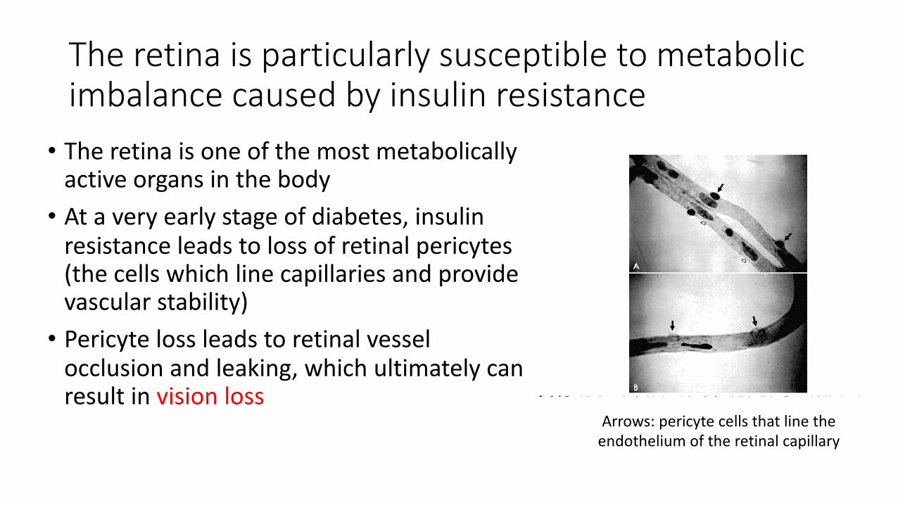
The retina is particularly susceptible to metabolic imbalance caused by insulin resistance
• The retina is one of the most metabolically active organs in the body• At a very early stage of diabetes, insulin
resistance leads to loss of retinal pericytes (the cells which line capillaries and provide vascular stability)• Pericyte loss leads to retinal vessel
occlusion and leaking, which ultimately can result in vision loss
Arrows: pericyte cells that line the endothelium of the retinal capillary

Diabetic retinopathy severity levels are evidence-based categories which help determine follow-up, management and treatment strategies in order to preserve vision
• The EyePACS system uses internationally-accepted grading guidelines and referral recommendations, based on the American Academy of Ophthalmology’s Preferred Practice Guidelines• Our certified consultant graders, however, consider that each patient
has a unique presentation of findings and rate of progression, which requires an individualized approach to management
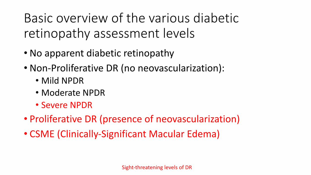
Basic overview of the various diabetic retinopathy assessment levels• No apparent diabetic retinopathy• Non-Proliferative DR (no neovascularization):• Mild NPDR• Moderate NPDR• Severe NPDR
• Proliferative DR (presence of neovascularization)• CSME (Clinically-Significant Macular Edema)
Sight-threatening levels of DR

No Apparent Diabetic Retinopathy
• Normal-appearing retina• Patient should have another
retinal exam within 12 months
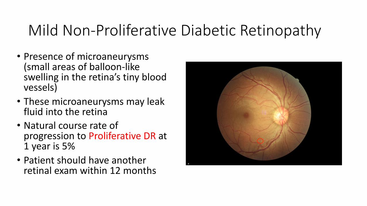
Mild Non-Proliferative Diabetic Retinopathy• Presence of microaneurysms
(small areas of balloon-like swelling in the retina’s tiny blood vessels) • These microaneurysms may leak
fluid into the retina• Natural course rate of
progression to Proliferative DR at 1 year is 5%• Patient should have another
retinal exam within 12 months

Moderate Non-Proliferative Diabetic Retinopathy
• Presence of microaneurysms, intraretinal hemorrhages, soft exudates (‘cotton wool spots’), venous beading (a ‘string of sausage’ appearance in the veins) and intraretinal microvascular abnormalities• Natural course rate of progression to
Proliferative DR at 1 year is 12 to 27%• Patient should have another retinal
exam within 6 to 12 months
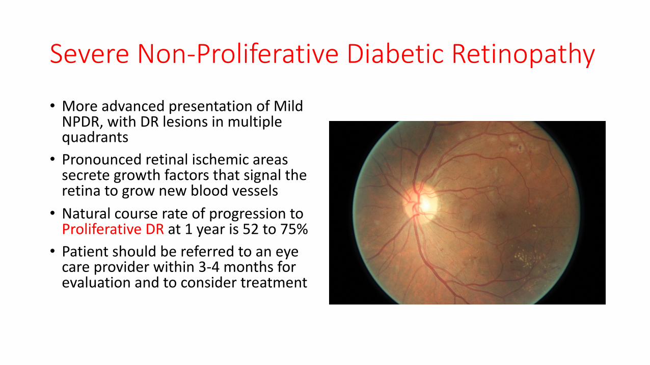
Severe Non-Proliferative Diabetic Retinopathy
• More advanced presentation of Mild NPDR, with DR lesions in multiple quadrants• Pronounced retinal ischemic areas
secrete growth factors that signal the retina to grow new blood vessels• Natural course rate of progression to
Proliferative DR at 1 year is 52 to 75%• Patient should be referred to an eye
care provider within 3-4 months for evaluation and to consider treatment

Proliferative Diabetic Retinopathy
• High risk of vision loss• New blood vessels grow inside the
retina. They are prone to bleedingand can lead to a retinal detachment• Patient should be referred for
treatment to an eye care specialist within 1-4 months• Most likely treatment is with laser
pan-retinal photocoagulation
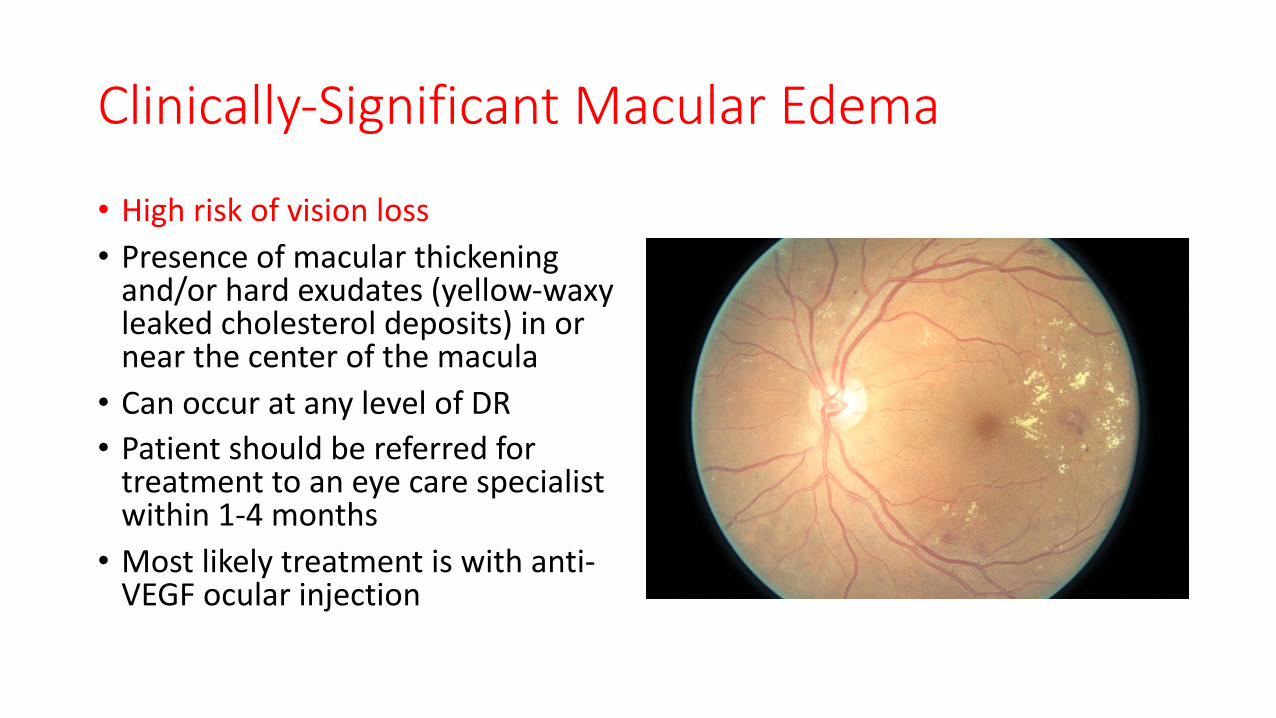
Clinically-Significant Macular Edema
• High risk of vision loss• Presence of macular thickening
and/or hard exudates (yellow-waxy leaked cholesterol deposits) in or near the center of the macula• Can occur at any level of DR• Patient should be referred for
treatment to an eye care specialist within 1-4 months• Most likely treatment is with anti-
VEGF ocular injection
Standards of Medical Care in Diabetes—2020 Abridged for Primary Care Providers• For further reference:
https://clinical.diabetesjournals.org/content/38/1/10