diabetes mellitus: insights from epidemiology
TRANSCRIPT
Review
Diabetes Mellitus: Insights from Epidemiology, Biochemistry,Risk Factors, Diagnosis, Complications andComprehensive Management
Saruar Alam 1 , Md. Kamrul Hasan 2,3 , Sharif Neaz 2, Nazmul Hussain 2, Md. Faruk Hossain 4 andTania Rahman 1,*
�����������������
Citation: Alam, S.; Hasan, M.K.;
Neaz, S.; Hussain, N.; Hossain, M.F.;
Rahman, T. Diabetes Mellitus:
Insights from Epidemiology,
Biochemistry, Risk Factors, Diagnosis,
Complications and Comprehensive
Management. Diabetology 2021, 2,
36–50. https://doi.org/10.3390/
diabetology2020004
Academic Editor: Peter Clifton
Received: 29 November 2020
Accepted: 23 March 2021
Published: 16 April 2021
Publisher’s Note: MDPI stays neutral
with regard to jurisdictional claims in
published maps and institutional affil-
iations.
Copyright: © 2021 by the authors.
Licensee MDPI, Basel, Switzerland.
This article is an open access article
distributed under the terms and
conditions of the Creative Commons
Attribution (CC BY) license (https://
creativecommons.org/licenses/by/
4.0/).
1 Department of Biochemistry and Molecular Biology, University of Dhaka, Dhaka 1000, Bangladesh;[email protected]
2 Department of Biochemistry and Molecular Biology, Tejgaon College, National University of Bangladesh,Gazipur 1704, Bangladesh; [email protected] (M.K.H.); [email protected] (S.N.);[email protected] (N.H.)
3 Department of Public Health, School of Health and Life Sciences, North South University, Bashundhara,Dhaka 1229, Bangladesh
4 Department of Biological Sciences, St John’s University, Queens, NY 11439, USA; [email protected]* Correspondence: [email protected]; Tel.: +880-2-9661900-7670
Abstract: Diabetes mellitus has become a serious and chronic metabolic disorder that results from acomplex interaction of genetic and environmental factors, principally characterized by hyperglycemia,polyuria, and polyphagia. Uncontrolled high blood sugar can result in a host of diabetic complications.Prolonged diabetes leads to serious complications some of which are life-threatening. The prevalenceof diabetes patients is rising at epidemic proportions throughout the world. Every year, a majorportion of the annual health budget is spent on diabetes and related illnesses. Multiple risk factors areinvolved in the etiopathogenesis of the disease and turning the disease into an epidemic. Diabetes,for which there is no cure, apparently can be kept under control by maintaining self-care in dailyliving, effective diabetes education, with comprehensive improvements in knowledge, attitudes,skills, and management. In this review, we focused on the biochemical aspects of diabetes, riskfactors including both environmental and genetic, disease complications, diagnosis, management,and currently available medications for the treatment of diabetes.
Keywords: diabetes mellitus; epidemiology; biochemistry; metabolic disorder; risk factors; diagnosis;complications; management of diabetes
1. Introduction
Diabetes mellitus (DM) is primarily characterized by high blood glucose levels (hyper-glycemia), polydipsia, and polyphagia. DM is one of the most common metabolic disordersthat is increasing at an alarming rate all over the world [1–3]. The number of patients withDM has quadrupled (from 108 million in 1980 to 422 million in 2014) within 34 years only,while the worldwide incidence of diabetes among adults over 18 years of age has risen to8.5% (2014) from 4.7% (1980) [1]. The WHO estimates that diabetes will be the 7th primarycause of fatality by 2030 [2]. There are mainly four common types of DM. Type 1 DM(T1DM) is caused by the autoimmune annihilation of the pancreatic-β cell with no insulinproduction [4]. This type is also called insulin-dependent diabetes mellitus (IDDM) [5,6].This type of DM is seen in childhood and includes 5–10% of total diabetes patients [1].The major type of diabetes is Type 2 DM (T2DM), which is caused due to insufficientproduction of insulin or desensitization of insulin receptors that precludes the entry ofglucose into the cell [7,8]. The type is predominantly seen in 90–95% of cases. There isanother type of diabetes called gestational diabetes mellitus (GDM) that occurs only duringpregnancy. GDM occurs in approximately 5–15% of pregnant women varying in ethnicity
Diabetology 2021, 2, 36–50. https://doi.org/10.3390/diabetology2020004 https://www.mdpi.com/journal/diabetology
Diabetology 2021, 2 37
and regions [1–3]. Multifarious factors including genetic defects, pancreatic obstruction,surgery, organ transplantation contribute to the onset of this type of diabetes [9]. In thecase of 40–60%, women having GDM can develop DM after 5–10 years of pregnancy. Im-paired glucose tolerance is potent to be expressed as T2DM whereas uncontrolled diabetesis the potential threat for the onset of other diseases like cardiovascular disease (CVD),blindness, renal failure, neurological disorder, the imbalanced osmolality of blood, hyper-tension, peripheral neuropathy, and many other diseases [10–14]. Monogenic diabetes,which is often misdiagnosed as T1DM or T2DM is caused by a mutation in a single geneor a cluster of genes [15,16]. It is an autosomal-dominant disease and patients with thishave varying signs, symptoms, and clinical courses. The two categories of monogenicdiabetes are neonatal DM and familial DM (also known as maturity-onset diabetes of theyoung (MODY)) [17]. Neonatal DM which is usually developed before 6 months of agecan be transient or permanent [18]. The development of familial DM commonly occursfrom late childhood through early adulthood, although it has been diagnosed in adultsin their 50s [19,20]. Mutations in genes encoding transcription factors are most commonin familial DM. The most common form of familial DM is MODY3 [21]. Clinically, thesepatients generally have a family history of diabetes, are non-insulin-dependent, and havea low renal threshold for glucose [17]. At least 10 genes have been linked to familial DM,and more than 20 genes may cause neonatal diabetes. Table 1 lists genes that have beenlinked to familial DM as well as variations of neonatal DM. Diabetes can be treated and itscomplications can be reduced by maintaining diet, physical activity, and proper medicationand by regular monitoring of the complications [15,16,22–25].
Table 1. Genes behind monogenic diabetes and their clinical features.
Features of Diabetes Gene Involved Clinical Outcome
MODY
GCK [26] GCK-MODY; stable, nonprogressive, normally does not requiretreatment. Microvascular complications developed in rare cases
HNF1A [27] HNF1A-MODY; progressive defect in insulin secretion, requires a lowdose of sulfonylurea therapy
HNF4A [28] HNF4A-MODY; progressive insulin secretory defect, sensitive tosulfonylurea
HNF1B [29] HNF1B-MODY; atrophy of the pancreas, renal disease, hyperuricemia,and gout may develop
Kir6.2 [30] Requires high dose of sulfonylurea therapy
Neonatal diabetes
KCNJ11 [30] Maybe permanent or transient intrauterine growth restriction, delaydevelopment, sensitive to sulfonylurea
INS [31] Require insulin therapy
ABCC8 [32] Sensitive to sulfonylurea therapy, in some cases delay development
6q24 (PLAG1, HYMA1) [33] Transient in nature, intrauterine growth restriction, macroglossia;umbilical hernia, treatment possible other than insulin
GATA6 [34] Pancreatic hypoplasia and exocrine insufficiency requireinsulin therapy
EIF2AK3 [35] Develop Wolcott–Rallison syndrome, requires insulin therapy
FOXP3 [36]Immunodysregulation, polyendocrinopathy, enteropathy X-linked
(IPEX) syndrome; autoimmune diabetes; autoimmune thyroid disease;exfoliative dermatitis; insulin-requiring
2. Epidemiology of Diabetes
The disease burden related to diabetes is high and rising in every country, fueled bythe global rise in the prevalence of obesity and unhealthy lifestyles. The latest estimatesshow a prevalence rate of 11.1% with diabetes in 2019, expected to rise to 13% by 2045 in
Diabetology 2021, 2 38
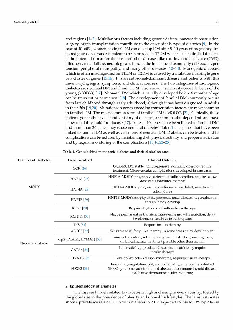
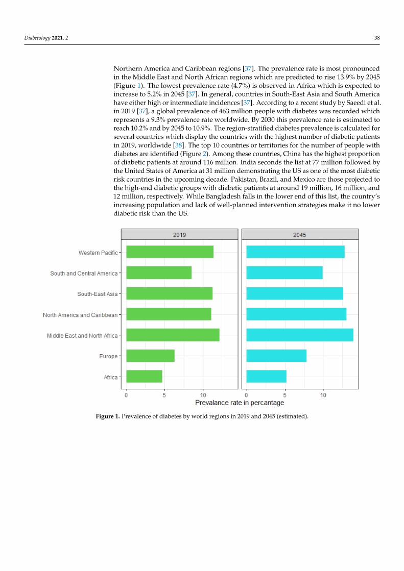
Northern America and Caribbean regions [37]. The prevalence rate is most pronouncedin the Middle East and North African regions which are predicted to rise 13.9% by 2045(Figure 1). The lowest prevalence rate (4.7%) is observed in Africa which is expected toincrease to 5.2% in 2045 [37]. In general, countries in South-East Asia and South Americahave either high or intermediate incidences [37]. According to a recent study by Saeedi et al.in 2019 [37], a global prevalence of 463 million people with diabetes was recorded whichrepresents a 9.3% prevalence rate worldwide. By 2030 this prevalence rate is estimated toreach 10.2% and by 2045 to 10.9%. The region-stratified diabetes prevalence is calculated forseveral countries which display the countries with the highest number of diabetic patientsin 2019, worldwide [38]. The top 10 countries or territories for the number of people withdiabetes are identified (Figure 2). Among these countries, China has the highest proportionof diabetic patients at around 116 million. India seconds the list at 77 million followed bythe United States of America at 31 million demonstrating the US as one of the most diabeticrisk countries in the upcoming decade. Pakistan, Brazil, and Mexico are those projected tothe high-end diabetic groups with diabetic patients at around 19 million, 16 million, and12 million, respectively. While Bangladesh falls in the lower end of this list, the country’sincreasing population and lack of well-planned intervention strategies make it no lowerdiabetic risk than the US.
Diabetology 2021, 2, FOR PEER REVIEW 3
FOXP3 [36]
Immunodysregulation, polyendocrinopathy, enteropathy X-linked (IPEX) syndrome; autoimmune diabetes; autoim-
mune thyroid disease; exfoliative dermatitis; insulin-re-quiring
2. Epidemiology of Diabetes The disease burden related to diabetes is high and rising in every country, fueled by
the global rise in the prevalence of obesity and unhealthy lifestyles. The latest estimates show a prevalence rate of 11.1% with diabetes in 2019, expected to rise to 13% by 2045 in Northern America and Caribbean regions [37]. The prevalence rate is most pronounced in the Middle East and North African regions which are predicted to rise 13.9% by 2045 (Figure 1). The lowest prevalence rate (4.7%) is observed in Africa which is expected to increase to 5.2% in 2045 [37]. In general, countries in South-East Asia and South America have either high or intermediate incidences [37]. According to a recent study by Saeedi et al. in 2019 [37], a global prevalence of 463 million people with diabetes was recorded which represents a 9.3% prevalence rate worldwide. By 2030 this prevalence rate is esti-mated to reach 10.2% and by 2045 to 10.9%. The region-stratified diabetes prevalence is calculated for several countries which display the countries with the highest number of diabetic patients in 2019, worldwide [38]. The top 10 countries or territories for the number of people with diabetes are identified (Figure 2). Among these countries, China has the highest proportion of diabetic patients at around 116 million. India seconds the list at 77 million followed by the United States of America at 31 million demonstrating the US as one of the most diabetic risk countries in the upcoming decade. Pakistan, Brazil, and Mex-ico are those projected to the high-end diabetic groups with diabetic patients at around 19 million, 16 million, and 12 million, respectively. While Bangladesh falls in the lower end of this list, the country’s increasing population and lack of well-planned intervention strategies make it no lower diabetic risk than the US.
Figure 1. Prevalence of diabetes by world regions in 2019 and 2045 (estimated). Figure 1. Prevalence of diabetes by world regions in 2019 and 2045 (estimated).
Diabetology 2021, 2 39Diabetology 2021, 2, FOR PEER REVIEW 4
Figure 2. Countries with the highest number of diabetic patients worldwide in 2019.
3. Risk Factors for Diabetes Globally, the prevalence of DM has increased and therefore has grown in severity as
a public health problem. Multiple risk factors are involved in the actual onset of the dis-ease. Genetics, atmosphere, loss of very first phase associated with insulin launch, seden-tary way of life, lack of physical exercise, smoking, alcoholic beverages, dyslipidemia, re-duced β-cell sensitivity, hyperinsulinemia, improved glucagon activity are the primary risk elements for prediabetes and DM [39–46]. These factors appear to play a significant role in insulin resistance or insulin nonfunctionality resulting in disease advancement. Based on WHO (2011), approximately 90% of patients develop T2DM, mostly related to excess body weight. Obstructive sleep apnea and sleep disorder that are seen among over-weight adult individuals are a common risk factor for insulin resistance and glucose sen-sitivity which collectively progresses to prediabetes and then T2DM. The diet containing low fiber but a high glycemic index (GI) is thought to be positively related to the onset of diabetes [47,48]. There is evidence that free fatty acids are one important link between insulin resistance and T2DM. Soft drinks of which the most deleterious feature is the high fructose load and consequent metabolic effects on the liver can cause obesity and increase body mass index (BMI) that may lead to T2DM.
Excess weight increases the mass of adipose tissue together with elevated secretion of adipokines and resistins, dysregulation of which leads to the development of T2DM [48,49]. The association of hyperuricemia and the development of T2DM have been inves-tigated in several studies. In subjects with hyperuricemia and insulin resistance, β cell function is triggered from its compensatory state [50]. In another study, while comparing four study groups (control, T2DM: with and without obesity and T1DM), C-peptide levels were found to increase in patients who had T2DM and obesity. It appears that uric acid behavior is closely related to β cell function [51]. In another follow-up study, it was found that high serum uric acid was associated with a higher risk of T2DM independent of obe-sity, dyslipidemia, and hypertension [52]. Nevertheless, uric acid plays a role in cytokine secretion and has been identified as a mediator of endothelial dysfunction and systemic inflammation [53]. In a study among the Chinese population, a positive association was
Figure 2. Countries with the highest number of diabetic patients worldwide in 2019.
3. Risk Factors for Diabetes
Globally, the prevalence of DM has increased and therefore has grown in severityas a public health problem. Multiple risk factors are involved in the actual onset of thedisease. Genetics, atmosphere, loss of very first phase associated with insulin launch,sedentary way of life, lack of physical exercise, smoking, alcoholic beverages, dyslipidemia,reduced β-cell sensitivity, hyperinsulinemia, improved glucagon activity are the primaryrisk elements for prediabetes and DM [39–46]. These factors appear to play a significant rolein insulin resistance or insulin nonfunctionality resulting in disease advancement. Based onWHO (2011), approximately 90% of patients develop T2DM, mostly related to excess bodyweight. Obstructive sleep apnea and sleep disorder that are seen among overweight adultindividuals are a common risk factor for insulin resistance and glucose sensitivity whichcollectively progresses to prediabetes and then T2DM. The diet containing low fiber but ahigh glycemic index (GI) is thought to be positively related to the onset of diabetes [47,48].There is evidence that free fatty acids are one important link between insulin resistanceand T2DM. Soft drinks of which the most deleterious feature is the high fructose load andconsequent metabolic effects on the liver can cause obesity and increase body mass index(BMI) that may lead to T2DM.
Excess weight increases the mass of adipose tissue together with elevated secretion ofadipokines and resistins, dysregulation of which leads to the development of T2DM [48,49].The association of hyperuricemia and the development of T2DM have been investigatedin several studies. In subjects with hyperuricemia and insulin resistance, β cell functionis triggered from its compensatory state [50]. In another study, while comparing fourstudy groups (control, T2DM: with and without obesity and T1DM), C-peptide levelswere found to increase in patients who had T2DM and obesity. It appears that uric acidbehavior is closely related to β cell function [51]. In another follow-up study, it wasfound that high serum uric acid was associated with a higher risk of T2DM independentof obesity, dyslipidemia, and hypertension [52]. Nevertheless, uric acid plays a rolein cytokine secretion and has been identified as a mediator of endothelial dysfunctionand systemic inflammation [53]. In a study among the Chinese population, a positiveassociation was found between plasma concentration of uric acid and the incidence of
Diabetology 2021, 2 40
T2DM. The association was somewhat attenuated after adjustment for metabolic syndrome,suggesting that the association between hyperuricemia and diabetes was partly mediatedthrough metabolic syndrome particularly insulin resistance.
It is shown by other studies that in the context of the complex cellular environmentof metabolic syndrome which is associated with oxidative stress, antioxidant propertiesof uric acid might convert to a pro-oxidant state owing to reactive oxygen species (ROS)accumulation [54]. This may also lead to adverse effects on endothelial function and aproinflammatory response, both of which are known to be associated with new-onset ofT2DM [55]. Various studies of others reported that there is a positive association betweenhigh serum uric acid level and diabetes [54–58], whereas another study by Barzilay et al.shows no association, and studies of Hu et al. show an inverse association between uricacid and T2DM [59].
Some classes of drugs (i.e., antipsychotics, diuretics, immune suppressants, beta-blockers) also can induce diabetes [60,61]. Immunoglobin E (IgE) and chymase interactwith other risk factors and both put a positive impact on the release of protease frommast cells and act as significant risk factors for prediabetes [44]. ROS and reactive nitro-gen species (RNS) which increase the oxidative stress level in the body also induce thedevelopment of vascular diseases and diabetes. Increased iron in blood makes an environ-ment for the Fenton reaction and Haber–Weiss reaction [62,63] which decreases the abilityof antioxidants and detoxifying enzymes. Low expression of catalase and superoxidedismutase 2 (SOD2) makes pancreatic β-cells sensitive to oxidative stress and decreasedexpression of a transcription factor responsible for low insulin production [64]. In T1DM,the destruction of insulin-producing pancreatic β-cells is caused by an autoimmune re-action that is activated by T-lymphocytes reacting against β-cells. CD4+ helper T cellscause tissue injury by activating macrophages and CD8+ cytotoxic T cells destroy β-cellsdirectly. CD8+ cytotoxic T cells also release cytokines and activate macrophages. Thisresults in the pancreatic lesion and causes cellular necrosis and lymphocyte infiltration.Locally produced cytokines (i.e., interferon-γ by T-cells, macrophage-mediated cytokinesTNF-α, IL-1) in the pancreas can damage β-cells through apoptosis. In 80% of T1DM cases,glutamic acid decarboxylase (GAD) autoantibodies are developed within the pancreaticβ-cells [65]. Several infectious diseases including coxsackievirus, nonenveloped linearsingle-stranded RNA virus, rotavirus, and cytomegalovirus also trigger autoimmunity toT1DM. A couple of pancreatic diseases occur due to MMR (measles, mumps, and rubella)which can contribute to the development of T1DM producing autoantibodies [65,66]. Thus,diabetes can occur due to a complex interaction among many different risk factors.
4. Diagnosis of Diabetes
As untreated DM can lead to serious complications, early diagnosis of diabetes mayprevent serious consequences due to the illness. Primary symptoms of diabetes includehigh blood glucose levels over a prolonged period, frequent urination, increased thirst,and elevated hunger. Some biochemical tests are routinely carried out to make a diagnosisof prediabetes or diabetes. Glycosylated hemoglobin (HbA1c) and oral glucose tolerancetests (OGTT) are commonly demonstrated for screening diabetes. OGTT test measures howwell body cells can absorb glucose after consuming a specific amount of sugar. Usually,the suspected individual is treated with 75 g glucose orally and the plasma glucose levelis measured 2 h after ingestion. If the plasma glucose level is found ≥11.1 mmol/L, thenthe individual is diagnosed as diabetic [63]. Fasting plasma glucose test is another reliableroutine method for the diagnosis of diabetes. Diabetes patients usually have a fastingglucose level of ≥7.0 mmol/L. If a person has a plasma glucose level ≥7.8 mmol/L after 2 hof ingesting 75 g glucose, then it is said that the person has impaired glucose tolerance [67].HbA1c is also widely used as a diagnostic test for diabetes. Patients with T2DM havea glycosylated hemoglobin level of ≥48 mmol/mol (≥6.5 DCCT%) [68]. Random bloodsugar monitoring is yet another prognostic marker for determining diabetes.
Diabetology 2021, 2 41
5. Complications of Diabetes
Long-term exposure to DM shows many other complications of diabetes. Diabeticnephropathy, diabetic retinopathy is caused due to prolonged high blood sugar level overtime. When the level of blood glucose is high for a long period, it can increase myo-inositoloxygenase (MIOX) enzyme activity and enhances myo-inositol catabolism. The enzymaticdegradation of myo-inositol alters the activity of Na+/K+ ATPase and phosphatidyli-nositol synthases, the very important molecules in the secondary signaling pathway [69].Thus, high blood glucose levels due to DM results in diabetic nephropathy, retinopathy,neuropathy, and diabetic cataracts. In uncontrolled diabetes, the high blood glucose levelin the delicate vessels of the retina increases osmotic pressure, and the vessels get leakedor rupture in some instances resulting in an impaired supply of blood to the retina. Tocompensate for the ruptured retinoid vessels, collateral blood vessels grow out of theretina and cause scar tissue to form resulting in impaired vision [70,71]. Uncontrolleddiabetes can affect kidneys, damaging the basement of glomerular capillaries, disruptingprotein crosslinking, and allowing proteins in the urine to leak through, a process knownas diabetic nephropathy [72]. Ketoacidosis is common in diabetic patients due to the contin-uous production of ketone bodies [73]. Diabetic ketoacidosis (DKA) is a feature of insulininsufficiency rather than resistance, which characterizes T2DM. In a case report conductedin China, it was found that a 52-year-old diabetic patient had impulsive gas gangrene of thescrotum due to severe DKA [74]. The phenomenon is also known as Fournier’s gangreneis a very rare condition whereas diabetes makes it more common. T1DM patients maydevelop obstructive pancreatitis because of inflammation in the pancreas, hyperplasia ofthe pancreatic duct gland resulting in obstructive pancreatitis [75]. Diabetic patients arealso at risk of free radical associated damage which is higher in diabetic patients than thatof normal leading to atherosclerosis, cardiovascular disease, and hypertension. Prevalenceof coronary artery disease (CAD), heart disease, and sudden cardiac death are elevated indiabetic patients. The high blood glucose level in diabetic patients stimulates superoxideproduction by the Maillard reaction. Several studies indicated that cognitive dysfunctionoccurs in T2DM affecting intelligence, attention, memory, learning, and perception. Dia-betes is also diagnosed as a potent risk factor for cancer because both share some commonrisk factors including age, sex, obesity, diet, smoking, and alcohol [14,76–81]. The con-tributing factor of cancer due to DM is that in T2DM the hepatic production of IGF bindingprotein becomes low and circulating IGF-1 becomes high. In T2DM, mTOR is overactivatedwhich phosphorylates IRS-1 and attenuates the metabolic pathway of insulin signaling.In T2DM, IRS-2 is upregulated and this activates the mitogen-activated protein kinase(MAPK) pathway and enhances cell proliferation [82,83]. Other studies also indicated thatdifferent forms of cancer including liver cancer, pancreatic cancer, and non-Hodgkin’slymphoma are predominant in diabetic patients. Diabetes and chronic hepatitis C (CHC)alter the immune system concurrently. Diabetic CHC patients have an augmented risk ofprogression of cirrhosis and hepatocellular carcinoma [84,85]. T2DM also affects the bloodmagnesium level. High blood glucose level decreases tubular reabsorption of magnesiumresulting in lower blood Mg [86]. Decreased Mg level in blood shows symptoms of manydiseases caused by magnesium deficiency.
6. Management of Diabetes
It was thought that once a patient is diabetic, he/she is diabetic for a lifetime; however,DM can go into remission [87]. Diabetes can be controlled by changing diet, doing physicalexercise, maintaining reasonable body weight, monitoring lipid profile, and having appro-priate medication when necessary. Changing diet is effective in controlling diabetes. Takinglow glycemic food, complex carbohydrate, protein, and polyunsaturated fatty acid (PUFA)and fiber can help to maintain normal blood sugar. Moderate exercise which decreasesobesity helps lowering blood glucose levels through insulin-independent glucose transportinto the muscle. GDM is a high-risk factor for the development of postpartum T2DM.Breastfeeding for ≥3 months downgrades the risk of postpartum T2DM by an extent of
Diabetology 2021, 2 42
≥40%. Evidence suggests that breastfeeding improves early postpartum glucose tolerance(PGT) [88]. During lactation, estrogen levels dwindle which results in improved glucoseand lipid metabolism as well as fat distribution. In this way, lactation for a long timedecreases the long-term postpartum risk of T2DM in women with GDM [89]. Medicationis the last step in the management of diabetes. T1DM and 25–30% T2DM diabetic patientsneed insulin [5,6]. An inappropriate insulin dosing can sometimes lead to hypoglycemiawhich is even more serious than hyperglycemia. To overcome this phenomenon oftendiabetic patients having insulin are advised to keep some sugar or chocolate with them.Vibration therapy may have a positive effect in treating patients with diabetic peripheralneuropathy (DPN) [90]. To reduce the risk of coronary heart diseases and atherosclerosisdue to diabetes maintaining a lipid profile is important as it might result from diabeticdyslipidemia [91]. Antioxidant therapy is effective as it lowers plasma insulin and HbA1clevel. Vitamin C has the antioxidant capacity and β-carotene has been exhibited to reduceoxidative LDL and prevent atherosclerosis [16,22]. Physical activity has always been pri-oritized by physician’s advice for DM’s management and many studies have suggestedthem effective in not only diabetic management but also in reducing the risk of age-onsetdiabetes alongside other age-onset diseases including cardiovascular diseases [92–94].Sedentary lifestyle is a critical risk factor of DM, especially via insulin resistance andevidence suggests that a healthy exercise routine reduces insulin resistance while alsoboosting cardiovascular health [95,96]. Aerobic exercise plays a vital role in reducing therisk of mortality rate in type 1 and type 2 diabetes [97]. Walking, cycling, jogging, andswimming are several examples of aerobic exercise [97]. Evidence also suggests that swim-ming can have a beneficial role for both normal diabetic and gestational diabetic (GDM)mothers, alongside drug therapies [98,99]. Wang et al. found that running at leisure timereduces the risk of developing type 2 diabetes by 28% [100]. Riiser et al. found that cyclingas transportation is inversely associated with self-reported diabetes [101]. Alternatively,high-intensity interval training (HIIT) exalts insulin sensitivity and glycemic control inadults with type 2 diabetes [102].
There are some drugs that are often prescribed to treat T2DM (Table 2). The most com-monly used drugs are a class of biguanides, thiazolidinediones, α-glucosidase inhibitors,and glucagon-like peptide-1 agonist. It is estimated that proton pump inhibitors havecertain effects in managing diabetes [25]. The different classes of drugs work in a differentway although all of these help to maintain normal blood glucose levels. Nevertheless, someof these drugs have some metabolic side effects. For example, TZDs have a well-defined setof side effects associated with their action as a PPAR-gamma agonist in adipocytes. Hence,the proper use of these drugs under prescription will exhibit a good impact in controllingdiabetes [103].
In Table 2, seven classes of drugs are listed that have been utilized in pharmacologicaltherapies of DM. These drugs are prescribed based on the etiological difference of diabeticpatients, for example, insulin secretagogues increase insulin level which in insulin-resistantpatients is not very effective and since they are only functional in the case of healthyβ-cells [104], these drugs are not applicable for T1DM patients either. Insulin secretagoguesare one of the earliest developed antidiabetic drugs and a major disadvantage of themis that there is always a chance of causing hypoglycemia. GLP-1 agonists which are thelatest in the line of antidiabetic drugs can also increase insulin level without any risk ofhypoglycemia and they are reported to be more potent than DPP4 inhibitors which performby inhibiting the GLP-1 breakdown, also a major concerning issue of the DPP4 inhibitors isthat they increase the risk of pancreatitis thus acting as a risk factor for pancreatic cancer ifuse is prolonged [105]. α–glucosidase inhibitors act by lowering glucose absorption in thegastrointestinal region and their natural origin makes them less harmful than their syntheticcounterparts [106], Unfortunately, their potency is also comparatively low and high dosescan cause gastrointestinal irritations. SGLT-2 inhibitors, on the other hand, reduce bloodglucose level by increasing glucose excretion in kidneys and some studies even suggest thatSGLT2 inhibitors may have a renoprotective effect [107,108]. Biguanide (metformin) and
Diabetology 2021, 2 43
Thiazolidinedione type drugs are most frequently used against insulin-resistant T2DM andin some cases, they are even used together in combination [109]. Speaking of combinationdrugs, metformin is reportedly the most commonly used antidiabetic combination drug,and it is used in combination with either α–glucosidase inhibitors or GLP-1 agonists, SGLT-2 inhibitors, DPP4 inhibitors, or thiazolidinedione depending on the patient’s medicalcondition [110].
Table 2. Currently used drugs targeting different sites.
Drug Class Example Mode of Action Common Side Effects References
Insulin secretagogues Sulfonylureas Inhibit β-cell K+ ATP channel andfacilitate insulin secretion
Hypoglycemia [111]Meglitinides Weight gain
GLP-1 agonistsLiraglutide Increase glucose-dependent insulin
secretion, reduces glucagon secretion,and delays gastric emptying
[112,113]Exenatide PancreatitisLixisenatide
α–glucosidase inhibitorsAcarbose Reduces the rate of digestion of
carbohydrate in the intestine henceless glucose absorption
Abdominal pain [114]Miglitol Diarrhea
Voglibose Flatulence
CanagliflozinIncreases glucose excretion in urine
Hypotension[115]SGLT-2 inhibitors Dapagliflozin Urinary tract infection
Empagliflozin
VildagliptinIncreases endogenous GLP-1 and
GIP levels
Respiratory tract infection
[116,117]DPP4 inhibitors Linagliptin Nasopharyngitis
Saxagliptin HeadacheSitagliptin
Activate AMPK, decreasing glucoseproduction and insulin resistance
Lactic acidosis [118]Biguanides Metformin Gastrointestinal irritation
Pioglitazone Activate PPAR-γ, decreasing insulinresistance
Fluid retention [119]Thiazolidinediones Rosiglitazone Weight gain
7. Recent Technologies for Combating Diabetes
The complex nature of diabetes makes it difficult to develop effective treatmentstrategies as the etiology of the disease can differ from patient to patient. A universal curefor the disease is highly sought after in studies of the etiology of diabetes, as the cure ishardly feasible.
In type 2 diabetes the inability of cells to intake glucose from the blood can be dueto genetic mutations or desensitization of receptors which makes developing treatmentstrategies harder compared to type 1 patients. Unfortunately, recent studies proposethat insulin therapy in type 1 patients can lead to developing type 2 diabetes due todesensitization [120], which makes it an important time for finding a cure or alternativetreatment methods. Several treatments have been developed so far for diabetes, with amajority of them being focused on type 1 diabetes. As the most common etiology of type 1diabetes is damaged or dysfunctional islets of Langerhans cells, various treatment methodsprioritized reconstructing or inserting functional islets of Langerhans cells in the patient’sbody which will liberate the patient from needing external insulin and terminate anypossibility of developing type 2 diabetes from type 1 and any other side effect of externalinsulin ingestion.
Advancement of stem cell technology has been a boon for researchers in developing asubsequent cure for type 1 diabetes [121]. Mesenchymal stem cell (MSC) therapy has shownconsiderable improvement in β-cell function in newly diagnosed type 1 diabetic patientwhile also showing considerable immunomodulatory effect [122–127]. Additionally, thepluripotent nature of embryonic stem cells (ESC) makes therapy using ESCs an appealingapproach for treating type 1 diabetes [128,129]. Induced stem cell therapy could becomea universal cure for type 1 diabetes if the technology can be made clinically available forgeneral patients [130].
Diabetology 2021, 2 44
Other promising diabetic treatment methods include VEGF inhibitors [131], SGLT2inhibitors [132], SiRNA therapy [133], miRNA therapy [134], phytochemicals [135], andsomatic gene therapy. While VEGF inhibition is not an absolute cure for diabetes, ithas shown considerable effect against Diabetic Macular Oedema (DMO) [131,136], on theother hand, SGLT2 is a major renal protein responsible for glucose reabsorption, so SGLT2inhibitors reduce the symptoms and risk factors of diabetes by lowering blood glucose levelvia inhibiting the glucose reabsorption pathway in kidneys [132]. Several studies on SiRNAand MiRNA therapy have suggested to be impactful against diabetic nephropathy [133,134]and delayed wound healing [137]. By concluding these factors it can be suggested that thecombination of VEGF inhibitors, SGLT2 inhibitors, SiRNA therapy, MiRNA therapy couldbe an effective management strategy for diabetic patients alongside being a reference fordeveloping novel treatment and management methods.
Since time immemorial, naturopathic therapies have been applied for a number ofhealth ailments and in recent times scientists have gone back to nature to look for cures forvarious ailments [138]. Several studies have suggested several plants to carry antidiabeticproperties too (e.g., Murraya koenigii, Allium sativum, Withania somnifera, Gymnema sylvestre,Allium cepa, Ferula foetida, etc.) [139–142]. The bioactive phytochemicals found in theseplant species can be extracted for direct use as drug-like compounds, or pharmacologicalagents [143]. This traditional approach could offer a natural key to unlock a suitable curefor diabetes.
In the case of somatic gene therapy, studies suggest this technique to have a consid-erable affinity for treating both type 1 and type 2 diabetes. While ex-vivo somatic genetherapy generates efficient β-cells, thus treating insulin deficiency [129,144], in-vivo somaticgene therapy prioritizes lowering blood glucose via generating both insulin [145,146] andnoninsulin genes [147–149] (Gck, PTG etc.). While sharing a somewhat common strategywith metformin therapy, most importantly it does not share the risk factors of metformintherapy [150]. Somatic gene therapy shares a common disadvantage with stem cell tech-nology as both these technologies are in the development phase and thus unavailable forgeneral clinical practice. However, it is expected that DM would become globally extinctwhile these technologies become available for the general public.
8. Conclusions and Future Perspectives
This review focused on the epidemiology of diabetes, the complications of diabetes,and the appropriate management of the disease. As diabetes has become a serious chronicillness, the number of diabetic patients is increasing at an alarming rate which makes it animportant topic of research. The finding of the relationship between diabetes and genetics,environmental factors, ethnicity, and lifestyle could be very helpful for the future develop-ment of the management of the disease. Based on the current data, it can be concluded thatchanging lifestyle, doing sufficient physical exercise, maintaining body weight, controllinglipid profile, and minimizing glucose level are very effective. As uncontrolled diabetescould lead to serious complications, research focusing on the early detection of diabetes, de-veloping various tools for understanding pancreatic β-cell destruction, developing targeteddrugs are the appropriate steps to control the disease.
Author Contributions: Idea and Conceptualization: S.A., M.K.H., T.R.; Literature search: S.A.,M.K.H., M.F.H., T.R.; Data analysis: S.A., N.H., M.F.H.; Original draft preparation: S.A., M.K.H.,S.N., M.F.H.; Critical revision: T.R. All authors have read and agreed to the published version of themanuscript.
Funding: This research received no external funding.
Institutional Review Board Statement: Not applicable as the study does not involve humans oranimals.
Informed Consent Statement: Not applicable as the study does not involve humans.
Data Availability Statement: Not applicable.
Diabetology 2021, 2 45
Conflicts of Interest: The authors declare no conflict of interest.
References1. World Health Organization (WHO). Global Report on Diabetes; WHO: Geneva, Switzerland, 2017. Available online: http://www.
who.int/diabetes/global-report/en/ (accessed on 22 September 2018).2. Mathers, C.D.; Loncar, D. Projections of global mortality and burden of disease from 2002 to 2030. PLoS Med. 2006, 3, e442.
[CrossRef]3. World Health Organization; International Diabetes Federation. Definition and Diagnosis of Diabetes Mellitus and Intermediate
Hyperglycaemia: Report of a WHO/IDF Consultation; WHO: Geneva, Switzerland, 2006.4. Morran, M.P.; Vonberg, A.; Khadra, A.; Pietropaolo, M. Immunogenetics of type 1 diabetes mellitus. Mol. Asp. Med. 2015, 42,
42–60. [CrossRef]5. Martin, B.C.; Warram, J.H.; Krolewski, A.S.; Soeldner, J.S.; Kahn, C.R.; Bergman, R.N. Role of glucose and insulin resistance in
development of type 2 diabetes mellitus: Results of a 25-year follow-up study. Lancet 1992, 340, 925–929. [CrossRef]6. Tisch, R.; McDevitt, H. Insulin-dependent diabetes mellitus. Cell 1996, 85, 291–297. [CrossRef]7. Reinehr, T. Type 2 diabetes mellitus in children and adolescents. World J. Diabetes 2013, 4, 270–281. [CrossRef]8. Reaven, G.M. Insulin-independent diabetes mellitus: Metabolic characteristics. Metabolism 1980, 29, 445–454. [CrossRef]9. Zimmet, P.; Alberti, K.G.M.M.; Shaw, J. Global and societal implications of the diabetes epidemic. Nat. Cell Biol. 2001, 414,
782–787. [CrossRef]10. Mokdad, A.H.; Ford, E.S.; Bowman, B.A.; Dietz, W.H.; Vinicor, F.; Bales, V.S.; Marks, J.S. Prevalence of obesity, diabetes, and
obesity-related health risk factors. JAMA 2003, 289, 76–79. [CrossRef] [PubMed]11. Razmaria, A.A. Diabetic neuropathy. JAMA 2015, 314, 2202. [CrossRef] [PubMed]12. Cahill, M.; Halley, A.; Codd, M.; O’Meara, N.; Firth, R.; Mooney, D.; Acheson, R.W. Prevalence of diabetic retinopathy in patients
with diabetes mellitus diagnosed after the age of 70 years. Br. J. Ophthalmol. 1997, 81, 218–222. [CrossRef] [PubMed]13. Giovannucci, E.; Harlan, D.M.; Archer, M.C.; Bergenstal, R.M.; Gapstur, S.M.; Habel, L.A.; Pollak, M.; Regensteiner, J.G.; Yee, D.
Diabetes and cancer: A consensus report. Diabetes Care 2010, 33, 1674–1685. [CrossRef]14. Aronson, D.; Edelman, E.R. Coronary artery disease and diabetes mellitus. Cardiol. Clin. 2014, 32, 439–455. [CrossRef]15. Cholesterol Treatment Trialists Collaborators; Reith, C.; Staplin, N.; Herrington, N.G.; Stevens, R.; Emberson, J.; Haynes, R.;
Mafham, M.; Armitage, J.; Cass, A.; et al. Efficacy of cholesterol-lowering therapy in 18,686 people with diabetes in 14 randomisedtrials of statins: A meta-analysis. Lancet 2008, 371, 117–125. [CrossRef]
16. Ceriello, A.; Testa, R. Antioxidant anti-inflammatory treatment in type 2 diabetes. Diabetes Care 2009, 32, S232–S236. [CrossRef][PubMed]
17. Fajans, S.S.; Bell, G.I. MODY. Diabetes Care 2011, 34, 1878–1884. [CrossRef] [PubMed]18. Lemelman, M.B.; Letourneau, L.; Greeley, S.A.W. Neonatal diabetes mellitus. Clin. Perinatol. 2018, 45, 41–59. [CrossRef] [PubMed]19. Jali, M.V.; Kambar, S.; Jali, S.M.; Gowda, S. Familial early onset of type-2 diabetes mellitus and its complications. N. Am. J. Med.
Sci. 2009, 1, 377–380. [PubMed]20. Alyafei, F.; Soliman, A.; Alkhalaf, F.; Sabt, A.; De Sanctis, V.; Elsayed, N.; Waseef, R. Clinical and biochemical characteristics of
familial type 1 diabetes mellitus (FT1DM) compared to non-familial type 1 DM (NFT1DM). Acta Bio Med. Atenei Parm. 2018, 89,27–31.
21. Waterhouse, C.; Keilson, J. Cori cycle activity in man. J. Clin. Investig. 1969, 48, 2359–2366. [CrossRef]22. Maritim, A.C.; Sanders, R.A.; Watkins, J.B., III. Diabetes, oxidative stress, and antioxidants: A review. J. Biochem. Mol. Toxicol.
2003, 17, 24–38. [CrossRef] [PubMed]23. Marín-Peñalver, J.J.; Martín-Timón, I.; Sevillano-Collantes, C.; Del Cañizo-Gómez, F.J. Update on the treatment of type 2 diabetes
mellitus. World J. Diabetes 2016, 7, 354–395. [CrossRef]24. Kim, K.S.; Kim, S.K.; Sung, K.M.; Cho, Y.W.; Park, S.W. Management of type 2 diabetes mellitus in older adults. Diabetes Metab. J.
2012, 36, 336–344. [CrossRef] [PubMed]25. Takebayashi, K.; Inukai, T. Effect of proton pump inhibitors on glycemic control in patients with diabetes. World J. Diabetes 2015,
6, 1122–1131. [CrossRef]26. Velho, G.; Froguel, P.; Clement, K.; Pueyo, E.M.; Rakotoambinina, B.; Zouali, H.; Passa, P.; Cohen, D.; Robert, J.J. Primary
pancreatic beta-cell secretory defect caused by mutations in glucokinase gene in kindreds of maturity onset diabetes of the young.Lancet 1992, 340, 444–448. [CrossRef]
27. Bellanné-Chantelot, C.; Carette, C.; Riveline, J.-P.; Valéro, R.; Gautier, J.-F.; Larger, E.; Reznik, Y.; Ducluzeau-Fieloux, P.; Sola-Gazagnes, A.; Hartemann-Heurtier, A.; et al. The type and the position of HNF1A mutation modulate age at diagnosis of diabetesin patients with maturity-onset diabetes of the young (MODY)-3. Diabetes 2007, 57, 503–508. [CrossRef] [PubMed]
28. Staník, J.; Dusatkova, P.; Cinek, O.; Valentínová, L.; Hucková, M.; Škopková, M.; Dusatkova, L.; Stanikova, D.; Pura, M.; Klimeš,I.; et al. De novo mutations of GCK, HNF1A and HNF4A may be more frequent in MODY than previously assumed. Diabetologia2013, 57, 480–484. [CrossRef]
29. Haldorsen, I.S.; Vesterhus, M.; Rder, H.; Jensen, D.K.; Svik, O.; Molven, A.; Njlstad, P.R.; Ræder, H.; Søvik, O.; Njølstad, P.R. Lackof pancreatic body and tail inHNF1Bmutation carriers. Diabet. Med. 2008, 25, 782–787. [CrossRef]
Diabetology 2021, 2 46
30. Sagen, J.V.; Raeder, H.; Hathout, E.; Shehadeh, N.; Gudmundsson, K.; Baevre, H.; Abuelo, D.; Phornphutkul, C.; Molnes, J.; Bell,G.I.; et al. Permanent neonatal diabetes due to mutations in KCNJ11 encoding Kir6.2: Patient characteristics and initial responseto sulfonylurea therapy. Diabetes 2004, 53, 2713–2718. [CrossRef] [PubMed]
31. Støy, J.; Edghill, E.L.; Flanagan, S.E.; Ye, H.; Paz, V.P.; Pluzhnikov, A.; Below, J.E.; Hayes, M.G.; Cox, N.J.; Lipkind, G.M.; et al.Insulin gene mutations as a cause of permanent neonatal diabetes. Proc. Natl. Acad. Sci. USA 2007, 104, 15040–15044. [CrossRef]
32. Babenko, A.P.; Polak, M.; Cavé, H.; Busiah, K.; Czernichow, P.; Scharfmann, R.; Bryan, J.; Aguilar-Bryan, L.; Vaxillaire, M.; Froguel,P. Activating Mutations in theABCC8Gene in neonatal diabetes mellitus. N. Engl. J. Med. 2006, 355, 456–466. [CrossRef]
33. Naylor, R.N.; Greeley, S.A.W.; I Bell, G.; Philipson, L.H. Genetics and pathophysiology of neonatal diabetes mellitus. J. DiabetesInvestig. 2011, 2, 158–169. [CrossRef] [PubMed]
34. Catli, G.; Abaci, A.; Flanagan, S.; De Franco, E.; Ellard, S.; Hattersley, A.; Guleryuz, H.; Bober, E. A novel GATA6 mutation leadingto congenital heart defects and permanent neonatal diabetes: A case report. Diabetes Metab. 2013, 39, 370–374. [CrossRef]
35. Brickwood, S.; Bonthron, D.T.; Al-Gazali, I.L.; Piper, K.; Hearn, T.; Wilson, I.D.; Hanley, A.N. Wolcott-Rallison syndrome:Pathogenic insights into neonatal diabetes from new mutation and expression studies of EIF2AK. J. Med. Genet. 2003, 40, 685–689.[CrossRef]
36. Wildin, R.S.; Ramsdell, F.; Peake, J.; Faravelli, F.; Casanova, J.-L.; Buist, N.; Levy-Lahad, E.; Mazzella, M.; Goulet, O.; Perroni, L.;et al. X-linked neonatal diabetes mellitus, enteropathy and endocrinopathy syndrome is the human equivalent of mouse scurfy.Nat. Genet. 2001, 27, 18–20. [CrossRef] [PubMed]
37. Saeedi, P.; Petersohn, I.; Salpea, P.; Malanda, B.; Karuranga, S.; Unwin, N.; Colagiuri, S.; Guariguata, L.; Motala, A.A.; Ogurtsova,K.; et al. Global and regional diabetes prevalence estimates for 2019 and projections for 2030 and 2045: Results from theInternational Diabetes Federation Diabetes Atlas, 9th edition. Diabetes Res. Clin. Pract. 2019, 157, 107843. [CrossRef] [PubMed]
38. International Diabetes Federation, IDF Diabetes Atlas, 9th ed.; IDF: Brussels, Belgium, 2019.39. Lee, I.-M.; Shiroma, E.J.; Lobelo, F.; Puska, P.; Blair, S.N.; Katzmarzyk, P.T. Effect of physical inactivity on major non-communicable
diseases worldwide: An analysis of burden of disease and life expectancy. Lancet 2012, 380, 219–229. [CrossRef]40. Knott, C.; Bell, S.; Britton, A. Alcohol consumption and the risk of type 2 diabetes: A systematic review and dose-response
meta-analysis of more than 1.9 million individuals from 38 observational studies. Diabetes Care 2015, 38, 1804–1812. [CrossRef]41. Akter, S.; Goto, A.; Mizoue, T. Smoking and the risk of type 2 diabetes in Japan: A systematic review and meta-analysis. J.
Epidemiol. 2017, 27, 553–561. [CrossRef]42. Danaei, G.; Finucane, M.M.; Lu, Y.; Singh, G.M.; Cowan, M.J.; Paciorek, C.J.; Lin, J.K.; Farzadfar, F.; Khang, Y.-H.; Stevens, A.G.;
et al. National, regional, and global trends in fasting plasma glucose and diabetes prevalence since 1980: Systematic analysis ofhealth examination surveys and epidemiological studies with 370 country-years and 2·7 million participants. Lancet 2011, 378,31–40. [CrossRef]
43. Cornier, M.-A.; Donahoo, W.T.; Pereira, R.; Gurevich, I.; Westergren, R.; Enerbäck, S.; Eckel, P.J.; Goalstone, M.L.; Hill, J.O.; Eckel,R.H.; et al. Insulin sensitivity determines the effectiveness of dietary macronutrient composition on weight loss in obese women.Obes. Res. 2005, 13, 703–709. [CrossRef] [PubMed]
44. Chen, L.; Chen, R.; Wang, H.; Liang, F. Mechanisms linking inflammation to insulin resistance. Int. J. Endocrinol. 2015, 2015, 1–9.[CrossRef]
45. Maj, M.; Ilhan, A.; Neziri, D.; Gärtner, W.; Berggård, T.; Attems, J.; Base, W.; Wagner, L. Age related changes in pancreatic betacells: A putative extra-cerebral site of Alzheimer’s pathology. World J. Diabetes 2011, 2, 49–53. [CrossRef] [PubMed]
46. Schulze, M.B.; Hoffmann, K.; E Manson, J.; Willett, W.C.; Meigs, J.B.; Weikert, C.; Heidemann, C.; A Colditz, G.; Hu, F.B. Dietarypattern, inflammation, and incidence of type 2 diabetes in women. Am. J. Clin. Nutr. 2005, 82, 675–684. [CrossRef]
47. Li, D.-W.; Lu, T.-F.; Hua, X.-W.; Dai, H.-J.; Cui, X.-L.; Zhang, J.-J.; Xia, Q. Risk factors for new onset diabetes mellitus after livertransplantation: A meta-analysis. World J. Gastroenterol. 2015, 21, 6329. [CrossRef] [PubMed]
48. Sears, B.; Perry, M. The role of fatty acids in insulin resistance. Lipids Health Dis. 2015, 14, 1–9. [CrossRef]49. Hossain, M.; Faruque, M.O.; Kabir, G.; Hassan, N.; Sikdar, D.; Nahar, Q.; Ali, L. Association of serum TNF-α and IL-6 with insulin
secretion and insulin resistance in IFG and IGT subjects in a Bangladeshi population. Int. J. Diabetes Mellit. 2010, 2, 165–168.[CrossRef]
50. Jia, S.-D.; Wang, Y.-G.; Li, J. An analysis of islet beta-cell function in hyperuricemia. Zhonghua Nei Ke Za Zhi 2006, 45, 456–458.51. Lytvyn, Y.; Perkins, B.A.; Cherney, D.Z. Uric acid as a biomarker and a therapeutic target in diabetes. Can. J. Diabetes 2015, 39,
239–246. [CrossRef] [PubMed]52. Dehghan, A.; Van Hoek, M.; Sijbrands, E.J.; Hofman, A.; Witteman, J.C. High serum uric acid as a novel risk factor for type 2
diabetes. Diabetes Care 2007, 31, 361–362. [CrossRef]53. Kanellis, J.; Kang, D.-H. Uric acid as a mediator of endothelial dysfunction, inflammation, and vascular disease. Semin. Nephrol.
2005, 25, 39–42. [CrossRef]54. Pradhan, A.D.; Manson, J.E.; Rifai, N.; Buring, J.E.; Ridker, P.M. C-reactive protein, interleukin 6, and risk of developing type 2
diabetes mellitus. JAMA 2001, 286, 327–334. [CrossRef]55. Nabipour, I.; Vahdat, K.; Jafari, S.; Pazoki, R. Concurrent increased high sensitivity C-reactive protein and chronic infections are
associated with coronary artery disease: A population-based study. Indian J. Med. Sci. 2007, 61, 135–143. [CrossRef]56. Hayden, M.R.; Tyagi, S.C. Uric acid: A new look at an old risk marker for cardiovascular disease, metabolic syndrome, and type 2
diabetes mellitus: The urate redox shuttle. Nutr. Metab. 2004, 1, 10. [CrossRef]
Diabetology 2021, 2 47
57. Thorand, B.; Baumert, J.; Chambless, L.; Meisinger, C.; Kolb, H.; Döring, A.; Löwel, H.; Koenig, W. Elevated markers of endothelialdysfunction predict type 2 diabetes mellitus in middle-aged men and women from the general population. Arter. Thromb. Vasc.Biol. 2006, 26, 398–405. [CrossRef] [PubMed]
58. Trayhurn, P.; Beattie, J.H. Physiological role of adipose tissue: White adipose tissue as an endocrine and secretory organ. Proc.Nutr. Soc. 2001, 60, 329–339. [CrossRef] [PubMed]
59. Hu, F.B.; Meigs, J.B.; Li, T.Y.; Rifai, N.; Manson, J.E. Inflammatory markers and risk of developing type 2 diabetes in women.Diabetes 2004, 53, 693–700. [CrossRef]
60. Penfornis, A.; Kury-Paulin, S. Immunosuppressive drug-induced diabetes. Diabetes Metab. 2006, 32, 539–546. [CrossRef]61. Fathallah, N.; Slim, R.; Larif, S.; Hmouda, H.; Ben Salem, C. Drug-induced hyperglycaemia and diabetes. Drug Saf. 2015, 38,
1153–1168. [CrossRef] [PubMed]62. Papanikolaou, G.; Pantopoulos, K. Iron metabolism and toxicity. Toxicol. Appl. Pharmacol. 2005, 202, 199–211. [CrossRef]63. Vijan, S. Type 2 diabetes. Ann. Intern. Med. 2010, 152, ITC3-1. [CrossRef]64. Simcox, J.A.; McClain, D.A. Iron and diabetes risk. Cell Metab. 2013, 17, 329–341. [CrossRef]65. Atkinson, M.A. The pathogenesis and natural history of type 1 diabetes. Cold Spring Harb. Perspect. Med. 2012, 2, a007641.
[CrossRef] [PubMed]66. Atkinson, M.A.; Von Herrath, M.; Powers, A.C.; Clare-Salzler, M. Current concepts on the pathogenesis of type 1 diabetes—
Considerations for attempts to prevent and reverse the disease. Diabetes Care 2015, 38, 979–988. [CrossRef] [PubMed]67. Siu, A.L.; US Preventive Services Task Force. Screening for abnormal blood glucose and type 2 diabetes mellitus: U.S. Preventive
Services Task Force recommendation statement. Ann. Intern. Med. 2015, 163, 861–868. [CrossRef] [PubMed]68. American Diabetes Association. Summary of revisions for the 2010 clinical practice recommendations. Diabetes Care 2009, 33, S3.
[CrossRef]69. Yang, B.; Hodgkinson, A.; Millward, B.A.; Demaine, A.G. High glucose-induced DNA-binding activities of nuclear factor of
activated T cells 5 and carbohydrate response element binding protein to the myo-inositol oxygenase gene are inhibited bysorbinil in peripheral blood mononuclear cells from patients with type 1 diabetes mellitus and nephropathy. Int. J. Diabetes Mellit.2010, 2, 169–174. [CrossRef]
70. Tarr, J.M.; Kaul, K.; Chopra, M.; Kohner, E.M.; Chibber, R. Pathophysiology of diabetic retinopathy. ISRN Ophthalmol. 2013, 2013,1–13. [CrossRef]
71. Antonetti, D.A.; Klein, R.; Gardner, T.W. Diabetic retinopathy. N. Engl. J. Med. 2012, 366, 1227–1239. [CrossRef]72. Lim, A.K. Diabetic nephropathy—Complications and treatment. Int. J. Nephrol. Renov. Dis. 2014, 7, 361–381. [CrossRef]73. Onyiriuka, A.N.; Ifebi, E. Ketoacidosis at diagnosis of type 1 diabetes in children and adolescents: Frequency and clinical
characteristics. J. Diabetes Metab. Disord. 2013, 12, 47. [CrossRef]74. Zhang, C.; Ma, L.; Peng, F.; Wu, Y.; Chen, Y.; Zhan, Y.; Zhang, X. Spontaneous gas gangrene of the scrotum in patient with severe
diabetic ketoacidosis. Int. J. Diabetes Mellit. 2010, 2, 196–198. [CrossRef]75. Moin, A.S.M.; Butler, P.C.; Butler, A.E. Increased proliferation of the pancreatic duct gland compartment in type 1 diabetes. J. Clin.
Endocrinol. Metab. 2016, 102, 200–209. [CrossRef] [PubMed]76. Satija, A.; Spiegelman, D.; Giovannucci, E.; Hu, F.B. Type 2 diabetes and risk of cancer. BMJ 2014, 350, g7707. [CrossRef]77. Shah, A.D.; Langenberg, C.; Rapsomaniki, E.; Denaxas, S.; Pujades-Rodriguez, M.; Gale, C.P.; Deanfield, J.; Smeeth, L.; Timmis, A.;
Hemingway, H. Type 2 diabetes and incidence of cardiovascular diseases: A cohort study in 1.9 million people. Lancet DiabetesEndocrinol. 2015, 3, 105–113. [CrossRef]
78. Zoungas, S.; Arima, H.; Gerstein, H.C.; Holman, R.R.; Woodward, M.; Reaven, P.; A Hayward, R.; Craven, T.; Coleman, R.L.;Chalmers, J. Effects of intensive glucose control on microvascular outcomes in patients with type 2 diabetes: A meta-analysis ofindividual participant data from randomised controlled trials. Lancet Diabetes Endocrinol. 2017, 5, 431–437. [CrossRef]
79. Leon, B.M. Diabetes and cardiovascular disease: Epidemiology, biological mechanisms, treatment recommendations and futureresearch. World J. Diabetes 2015, 6, 1246–1258. [CrossRef]
80. Marseglia, A.; Fratiglioni, L.; Laukka, E.J.; Santoni, G.; Pedersen, N.L.; Bäckman, L.; Xu, W. Early cognitive deficits in type 2diabetes: A POPULATION-based study. J. Alzheimer’s Dis. 2016, 53, 1069–1078. [CrossRef]
81. Wojciechowska, J.; Krajewski, W.; Bolanowski, M.; Krecicki, T.; Zatonski, T. Diabetes and cancer: A review of current knowledge.Exp. Clin. Endocrinol. Diabetes 2016, 124, 263–275. [CrossRef]
82. Denduluri, S.K.; Idowu, O.; Wang, Z.; Liao, Z.; Yan, Z.; Mohammed, M.K.; Ye, J.; Wei, Q.; Wang, J.; Zhao, L.; et al. Insulin-likegrowth factor (IGF) signaling in tumorigenesis and the development of cancer drug resistance. Genes Dis. 2015, 2, 13–25.[CrossRef]
83. Laplante, M.; Sabatini, D.M. mTOR signaling in growth control and disease. Cell 2012, 149, 274–293. [CrossRef]84. Vigneri, P.; Frasca, F.; Sciacca, L.; Pandini, G.; Vigneri, R. Diabetes and cancer. Endocr. Relat. Cancer 2009, 16, 1103–1123. [CrossRef]
[PubMed]85. Antonelli, A. Hepatitis C virus infection and type 1 and type 2 diabetes mellitus. World J. Diabetes 2014, 5, 586–600. [CrossRef]86. Barbagallo, M. Magnesium and type 2 diabetes. World J. Diabetes 2015, 6, 1152–1157. [CrossRef] [PubMed]87. Panunzi, S.; Carlsson, L.M.S.; De Gaetano, A.; Peltonen, M.; Rice, T.; Sjöström, L.; Mingrone, G.; Dixon, J.B. Determinants of
diabetes remission and glycemic control after bariatric surgery. Diabetes Care 2016, 39, 166–174. [CrossRef]
Diabetology 2021, 2 48
88. Ziegler, A.-G.; Wallner, M.; Kaiser, I.; Rossbauer, M.; Harsunen, M.H.; Lachmann, L.; Maier, J.; Winkler, C.; Hummel, S. Long-termprotective effect of lactation on the development of type 2 diabetes in women with recent gestational diabetes mellitus. Diabetes2012, 61, 3167–3171. [CrossRef] [PubMed]
89. Hong, J.; Barnes, M.; Kessler, N. Case study: Use of vibration therapy in the treatment of diabetic peripheral small fiber neuropathy.J. Bodyw. Mov. Ther. 2013, 17, 235–238. [CrossRef] [PubMed]
90. Hong, J.; Barnes, M.J.; Kessler, N.J. Case study: Use of vibration therapy in the treatment of diabetic peripheral small fiberneuropathy. Int. J. Diabetes Mellit. 2015, 3, 72–75. [CrossRef]
91. Verma, S.; Hussain, M.E. Obesity and diabetes: An update. Diabetes Metab. Syndr. Clin. Res. Rev. 2017, 11, 73–79. [CrossRef]92. Ruegsegger, G.N.; Booth, F.W. Health benefits of exercise. Cold Spring Harb. Perspect. Med. 2017, 8, a029694. [CrossRef]93. Rasmussen, M.G.; Grøntved, A.; Blond, K.; Overvad, K.; Tjønneland, A.; Jensen, M.K.; Østergaard, L. Associations between
recreational and commuter cycling, changes in cycling, and type 2 diabetes risk: A cohort study of Danish men and women. PLoSMed. 2016, 13, e1002076. [CrossRef] [PubMed]
94. Riddell, M.C.; Gallen, I.W.; Smart, E.C.; Taplin, E.C.; Adolfsson, P.; Lumb, A.N.; Kowalski, A.; Rabasa-Lhoret, R.; McCrimmon,R.J.; Hume, C.; et al. Exercise management in type 1 diabetes: A consensus statement. Lancet Diabetes Endocrinol. 2017, 5, 377–390.[CrossRef]
95. Kumar, A.S.; Maiya, A.G.; Shastry, B.; Vaishali, K.; Ravishankar, N.; Hazari, A.; Gundmi, S.; Jadhav, R. Exercise and insulinresistance in type 2 diabetes mellitus: A systematic review and meta-analysis. Ann. Phys. Rehabil. Med. 2019, 62, 98–103.[CrossRef] [PubMed]
96. Carbone, S.; Del Buono, M.G.; Ozemek, C.; Lavie, C.J. Obesity, risk of diabetes and role of physical activity, exercise training andcardiorespiratory fitness. Prog. Cardiovasc. Dis. 2019, 62, 327–333. [CrossRef] [PubMed]
97. Colberg, S.R.; Sigal, R.J.; Yardley, J.E.; Riddell, M.C.; Dunstan, D.W.; Dempsey, P.C.; Horton, E.S.; Castorino, K.; Tate, D.F. Physicalactivity/exercise and diabetes: A position statement of the American Diabetes Association. Diabetes Care 2016, 39, 2065–2079.[CrossRef] [PubMed]
98. Huang, L.; Yue, P.; Wu, X.; Yu, T.; Wang, Y.; Zhou, J.; Kong, D.; Chen, K. Combined intervention of swimming plus metforminameliorates the insulin resistance and impaired lipid metabolism in murine gestational diabetes mellitus. PLoS ONE 2018, 13,e0195609. [CrossRef]
99. Ghiasi, R.; Naderi, R.; Sheervalilou, R.; Alipour, M.R. Swimming training by affecting the pancreatic Sirtuin1 (SIRT1) andoxidative stress, improves insulin sensitivity in diabetic male rats. Horm. Mol. Biol. Clin. Investig. 2019, 40, 1–12. [CrossRef]
100. Wang, Y.; Lee, D.-C.; Brellenthin, A.G.; Eijsvogels, T.M.; Sui, X.; Church, T.S.; Lavie, C.J.; Blair, S.N. Leisure-time running reducesthe risk of incident type 2 diabetes. Am. J. Med. 2019, 132, 1225–1232. [CrossRef]
101. Riiser, A.; Solbraa, A.; Jenum, A.K.; Birkeland, K.I.; Andersen, L.B. Cycling and walking for transport and their associations withdiabetes and risk factors for cardiovascular disease. J. Transp. Health 2018, 11, 193–201. [CrossRef]
102. Jelleyman, C.; Yates, T.L.; O’Donovan, G.; Gray, L.J.; King, J.A.; Khunti, K.; Davies, M.J. The effects of high-intensity intervaltraining on glucose regulation and insulin resistance: A meta-analysis. Obes. Rev. 2015, 16, 942–961. [CrossRef]
103. Lai, S.-W.; Chen, P.-C.; Liao, K.-F.; Muo, C.-H.; Lin, C.-C.; Sung, F.-C. Risk of hepatocellular carcinoma in diabetic patients andrisk reduction associated with anti-diabetic therapy: A population-based cohort study. Am. J. Gastroenterol. 2012, 107, 46–52.[CrossRef]
104. Raptis, S.A.; Dimitriadis, G.D. Oral hypoglycemic agents: Insulin secretagogues, α-glucosidase inhibitors and insulin sensitizers.Exp. Clin. Endocrinol. Diabetes 2001, 109, S265–S287. [CrossRef]
105. Zhang, Z.; Chen, X.; Lu, P.; Zhang, J.; Xu, Y.; He, W.; Li, M.; Zhang, S.; Jia, J.; Shao, S.; et al. Incretin-based agents in type 2 diabeticpatients at cardiovascular risk: Compare the effect of GLP-1 agonists and DPP-4 inhibitors on cardiovascular and pancreaticoutcomes. Cardiovasc. Diabetol. 2017, 16, 31. [CrossRef] [PubMed]
106. Yin, Z.; Zhang, W.; Feng, F.; Zhang, Y.; Kang, W. α-Glucosidase inhibitors isolated from medicinal plants. Food Sci. Hum. Wellness2014, 3, 136–174. [CrossRef]
107. El Mouhayyar, C.; Riachy, R.; Khalil, A.B.; Eid, A.; Azar, S. SGLT2 inhibitors, GLP-1 agonists, and DPP-4 inhibitors in diabetesand microvascular complications: A review. Int. J. Endocrinol. 2020, 2020, 1762164. [CrossRef] [PubMed]
108. Margonato, D.; Galati, G.; Mazzetti, S.; Cannistraci, R.; Perseghin, G.; Margonato, A.; Mortara, A. Renal protection: A leadingmechanism for cardiovascular benefit in patients treated with SGLT2 inhibitors. Heart Fail. Rev. 2021, 26, 337–345. [CrossRef]
109. Strowig, S.M.; Raskin, P. Combination therapy using metformin or thiazolidinediones and insulin in the treatment of diabetesmellitus. Diabetes Obes. Metab. 2004, 7, 633–641. [CrossRef]
110. Maruthur, N.M.; Tseng, E.; Hutfless, S.; Wilson, L.M.; Suarez-Cuervo, C.; Berger, Z.; Chu, Y.; Iyoha, E.; Segal, J.B.; Bolen, S.Diabetes medications as monotherapy or metformin-based combination therapy for type 2 diabetes. Ann. Intern. Med. 2016, 164,740–751. [CrossRef]
111. Type 2 diabetes and insulin secretagogues. J. Clin. Endocrinol. Metab. 2012, 97, 37A. [CrossRef]112. Garber, A.J. Long-acting glucagon-like peptide 1 receptor agonists: A review of their efficacy and tolerability. Diabetes Care 2011,
34, S279–S284. [CrossRef]113. Filippatos, T.D.; Panagiotopoulou, T.V.; Elisaf, M.S. Adverse Effects of GLP-1 Receptor Agonists. Rev. Diabet. Stud. 2014, 11,
202–230. [CrossRef]
Diabetology 2021, 2 49
114. Kalra, S.; Bhutani, J.; Mohan, V.; Unnikrishnan, R.; Kunju, P. Alpha-glucosidase inhibitors. Diabetol. Type 2 Diabetes Mellit. 2014,64, 55. [CrossRef]
115. Hsia, D.S.; Grove, O.; Cefalu, W.T. An update on sodium-glucose co-transporter-2 inhibitors for the treatment of diabetes mellitus.Curr. Opin. Endocrinol. Diabetes Obes. 2016, 24, 1. [CrossRef]
116. Sebokova, E.; Christ, A.D.; Boehringer, M.; Mizrahi, J. Dipeptidyl peptidase iv inhibitors: The next generation of new promisingtherapies for the management of type 2 diabetes. Curr. Top. Med. Chem. 2007, 7, 547–555. [CrossRef]
117. Pathak, R.; Bridgeman, M.B. Dipeptidyl peptidase-4 (DPP-4) inhibitors in the management of diabetes. J. Formul. Manag. 2010, 35,509–513.
118. Strack, T. Metformin: A review. Drugs Today 2008, 44, 303–314. [CrossRef] [PubMed]119. Nanjan, M.; Mohammed, M.; Kumar, B.P.; Chandrasekar, M. Thiazolidinediones as antidiabetic agents: A critical review.
Bioorganic Chem. 2018, 77, 548–567. [CrossRef] [PubMed]120. Petrovski, G.; Zivkovic, M.; Milenkovic, T.; Ahmeti, I.; Bitovska, I. Successful desensitization in patient with type 2 diabetes with
an insulin allergy using insulin pump and glargine. Acta Diabetol. 2014, 51, 1073–1075. [CrossRef] [PubMed]121. Maehr, R.; Chen, S.; Snitow, M.; Ludwig, T.; Yagasaki, L.; Goland, R.; Leibel, R.L.; Melton, D.A. Generation of pluripotent stem
cells from patients with type 1 diabetes. Proc. Natl. Acad. Sci. USA 2009, 106, 15768–15773. [CrossRef]122. Abdi, R.; Fiorina, P.; Adra, C.N.; Atkinson, M.; Sayegh, M.H. Immunomodulation by mesenchymal stem cells: A potential
therapeutic strategy for type 1 diabetes. Diabetes 2008, 57, 1759–1767. [CrossRef] [PubMed]123. Spaggiari, G.M.; Abdelrazik, H.; Becchetti, F.; Moretta, L. MSCs inhibit monocyte-derived DC maturation and function by
selectively interfering with the generation of immature DCs: Central role of MSC-derived prostaglandin E2. Blood 2009, 113,6576–6583. [CrossRef]
124. Wu, J.; Sun, Z.; Sun, H.-S.; Wu, J.; Weisel, R.D.; Keating, A.; Li, Z.-H.; Feng, Z.-P.; Li, R.-K. Intravenously administered bonemarrow cells migrate to damaged brain tissue and improve neural function in ischemic rats. Cell Transpl. 2007, 16, 993–1005.[CrossRef]
125. Tyndall, A.; Walker, A.U.; Cope, A.; Dazzi, F.; De Bari, C.; Fibbe, W.; Guiducci, S.; Jones, S.; Jorgensen, C.; Le Blanc, K.; et al.Immunomodulatory properties of mesenchymal stem cells: A review based on an interdisciplinary meeting held at the KennedyInstitute of Rheumatology Division, London, UK, 31 October 2005. Arthritis Res. Ther. 2007, 9, 301. [CrossRef]
126. Ianus, A.; Holz, G.G.; Theise, N.D.; Hussain, M.A. In vivo derivation of glucose-competent pancreatic endocrine cells from bonemarrow without evidence of cell fusion. J. Clin. Investig. 2003, 111, 843–850. [CrossRef]
127. Ghannam, S.; Bouffi, C.; Djouad, F.; Jorgensen, C.; Noël, D. Immunosuppression by mesenchymal stem cells: Mechanisms andclinical applications. Stem Cell Res. Ther. 2010, 1, 1–7. [CrossRef] [PubMed]
128. Chien, K.R.; Moretti, A.; Laugwitz, K.-L. Development: ES cells to the rescue. Science 2004, 306, 239–240. [CrossRef] [PubMed]129. Zalzman, M.; Gupta, S.; Giri, R.K.; Berkovich, I.; Sappal, B.S.; Karnieli, O.; Zern, M.A.; Fleischer, N.; Efrat, S. Reversal of
hyperglycemia in mice by using human expandable insulin-producing cells differentiated from fetal liver progenitor cells. Proc.Natl. Acad. Sci. USA 2003, 100, 7253–7258. [CrossRef] [PubMed]
130. Meier, J.J.; Bhushan, A.; Butler, P.C. The potential for stem cell therapy in diabetes. Pediatr. Res. 2006, 59, 65R–73R. [CrossRef]131. Zechmeister-Koss, I.; Huic, M. Vascular endothelial growth factor inhibitors (anti-VEGF) in the management of diabetic macular
oedema: A systematic review. Br. J. Ophthalmol. 2011, 96, 167–178. [CrossRef]132. Chao, E.C.; Henry, R.R. SGLT2 inhibition—A novel strategy for diabetes treatment. Nat. Rev. Drug Discov. 2010, 9, 551–559.
[CrossRef]133. Zhang, Q.; Shi, Y.; Wada, J.; Malakauskas, S.M.; Liu, M.; Ren, Y.; Du, C.; Duan, H.; Li, Y.; Li, Y.; et al. In vivo delivery of gremlin
siRNA plasmid reveals therapeutic potential against diabetic nephropathy by recovering bone morphogenetic protein-7. PLoSONE 2010, 5, e11709. [CrossRef]
134. Kato, M.; Natarajan, R. MicroRNAs in diabetic nephropathy: Functions, biomarkers, and therapeutic targets. Ann. N. Y. Acad. Sci.2015, 1353, 72–88. [CrossRef]
135. Ledley, F.D. Pharmaceutical approach to somatic gene therapy. Pharm. Res. 1996, 13, 1595–1614. [CrossRef]136. Gupta, N.; Mansoor, S.; Sharma, A.; Sapkal, A.; Sheth, J.; Falatoonzadeh, P.; Kuppermann, B.; Kenney, M. Diabetic retinopathy
and VEGF. Open Ophthalmol. J. 2013, 7, 4–10. [CrossRef] [PubMed]137. Randeria, P.S.; Seeger, M.A.; Wang, X.-Q.; Wilson, H.; Shipp, D.; Mirkin, C.A.; Paller, A.S. siRNA-based spherical nucleic acids
reverse impaired wound healing in diabetic mice by ganglioside GM3 synthase knockdown. Proc. Natl. Acad. Sci. USA 2015, 112,5573–5578. [CrossRef] [PubMed]
138. Thattet, U.M.; Dahanukar, S.A. Immunotherapeutic modification of experimental infections by Indian medicinal plants. Phytother.Res. 1989, 3, 43–49. [CrossRef]
139. Grover, J.; Yadav, S.; Vats, V. Medicinal plants of India with anti-diabetic potential. J. Ethnopharmacol. 2002, 81, 81–100. [CrossRef]140. Pastors, J.G.; Warshaw, H.; Daly, A.; Franz, M.; Kulkarni, K. The evidence for the effectiveness of medical nutrition therapy in
diabetes management. Diabetes Care 2002, 25, 608–613. [CrossRef]141. Prasad, S.; Kulshresht, A.; Qureshi, T.N. Antidiabetic activity of some herbal plants in streptozotocin induced diabetic albino rats.
Pak. J. Nutr. 2009, 8, 551–557. [CrossRef]142. Bnouham, M.; Ziyyat, A.; Mekhfi, H.; Tahri, A.; Legssyer, A. Medicinal plants with potential antidiabetic activity—A review of
ten years of herbal medicine research (1990–2000). Int. J. Diabetes Metab. 2006, 14, 1–25. [CrossRef]
Diabetology 2021, 2 50
143. Jung, M.; Park, M.; Lee, H.C.; Kang, Y.-H.; Kang, E.S.; Kim, S.K. Antidiabetic agents from medicinal plants. Curr. Med. Chem.2006, 13, 1203–1218. [CrossRef] [PubMed]
144. Muzzin, P.; Eisensmith, R.C.; Copeland, K.C.; Woo, S.L.C. Hepatic insulin gene expression as treatment for type 1 diabetesmellitus in rats. Mol. Endocrinol. 1997, 11, 833–837. [CrossRef]
145. Auricchio, A.; Gao, G.-P.; Yu, Q.; Raper, S.; Rivera, V.; Clackson, T.; Wilson, J. Constitutive and regulated expression of processedinsulin following in vivo hepatic gene transfer. Gene Ther. 2002, 9, 963–971. [CrossRef] [PubMed]
146. Short, D.K.; Okada, S.; Yamauchi, K.; Pessin, J.E. Adenovirus-mediated transfer of a modified human proinsulin gene reverseshyperglycemia in diabetic mice. Am. J. Physiol. Metab. 1998, 275, E748–E756. [CrossRef]
147. O’Doherty, R.M.; Lehman, D.L.; Telemaque-Potts, S.; Newgard, C.B. Metabolic impact of glucokinase overexpression in liver:Lowering of blood glucose in fed rats is accompanied by hyperlipidemia. Diabetes 1999, 48, 2022–2027. [CrossRef]
148. Morral, N.; McEvoy, R.; Dong, H.; Meseck, M.; Altomonte, J.; Thung, S.; Woo, S.L. Adenovirus-mediated expression of glucokinasein the liver as an adjuvant treatment for type 1 diabetes. Hum. Gene Ther. 2002, 13, 1561–1570. [CrossRef] [PubMed]
149. O’Doherty, R.M.; Jensen, P.B.; Anderson, P.; Jones, J.G.; Berman, H.K.; Kearney, D.; Newgard, C.B. Activation of direct and indirectpathways of glycogen synthesis by hepatic overexpression of protein targeting to glycogen. J. Clin. Investig. 2000, 105, 479–488.[CrossRef] [PubMed]
150. Inzucchi, S.E.; Lipska, K.J.; Mayo, H.; Bailey, C.J.; McGuire, D.K. Metformin in patients with type 2 diabetes and kidney disease.JAMA 2014, 312, 2668–2675. [CrossRef] [PubMed]