di-carboxylic acid cross-linking interactions improves thermal stability and mechanical strength of...
TRANSCRIPT
Di-carboxylic acid cross-linking interactions improves thermalstability and mechanical strength of reconstituted type I collagen
Part I. Oxalic acid
Tapas Mitra • G. Sailakshmi • A. Gnanamani •
A. B. Mandal
Received: 13 October 2010 / Accepted: 9 March 2011 / Published online: 31 March 2011
� Akademiai Kiado, Budapest, Hungary 2011
Abstract This study emphasizes, cross-linking potential
of a simple di-carboxylic acid, namely, oxalic acid with
type I collagen for the preparation of collagen based bio-
material for clinical applications. Further the study dis-
cusses the characteristics features of the cross-linked
material in comparison with the standard cross-linker. In
addition, the study also demonstrates the role of ionic
interactions in providing the thermal stability and tensile
strength to the cross-linked biopolymer material. Type I
collagen from rat tail tendon treated with oxalic acid at
optimized concentrations provided a biopolymer material
without changing the triple helical pattern of collagen (CD
spectrum) and also with 6–7 fold increase in tensile
strength than native collagen. FTIR spectral details dem-
onstrate the ionic interactions between collagen and oxalic
acid. Thermal stability analyses of oxalic acid cross-linked
biopolymer revealed, high thermal stability compared to
materials of glutaraldehyde cross-linked. The results of the
study suggest oxalic acid as a suitable cross-linker for
collagen and it cross-link with collagen through ionic
interactions.
Keywords Type I collagen � Oxalic acid � Cross-linker �Thermal stability � Circular dichroism
Introduction
Preparation of collagen based biopolymer materials for
clinical applications was always been a great challenge,
due to the problems encountered mainly with the cross-
linkers and in additional with the thermal stability,
mechanical strength, biocompatible, and biodegradable
properties. Nevertheless, in body system, stabilization of
synthesized collagen was established by natural cross-
linkers (lysyl oxidase) under in situ condition, which pro-
vide proteolytic resistance and high mechanical (tensile
strength) properties. But, during the course of extraction of
collagen and reconstitution, the expected stability and
strength could not be achieved. Thus it is often necessary to
confer mechanical firmness and collagenase resistance (to
the material of collagen based) by introduction of exoge-
nous cross-linker into the molecular structure of collagen.
Till date exogenous cross-linking agents such as chro-
mium [1], aldehydes [2], hexamethylene diisocyanate [3],
carbodiimide [4], acylazides [5], citric acid, maleic acid
derivatives [6], usnic acid [7], alginic acid [8], dialdehyde
starch [9], and various other physical treatments like UV
[10] and gamma irradiation [11] were used for cross-linking
of collagen. In general all the said exogenous cross-linkers,
cross-linked with collagen through; (i) covalent amide
linkage (between activated –COOH functional group of
cross-linker with e-NH2 group of collagen); (ii) covalent
imine linkage (between –CHO functional group of cross-
linker with e-NH2 group of collagen); (iii) co-ordinate bond
formation (chromium cross-linking with collagen); (iv)
H-bond formation (between polyphenolic –OH group with
different type of amino acids of collagen molecule), etc.
With all these bonding patterns, one can easily get more
stable collagen based biopolymer materials. However,
recent realization on demerits (non-biodegradable, toxicity,
T. Mitra � G. Sailakshmi � A. Gnanamani (&)
Microbiology Division, Central Leather Research Institute
(Council of Scientific and Industrial Research), Adyar,
Chennai 600020, Tamil Nadu, India
e-mail: [email protected]
A. B. Mandal
Chemical Laboratory, Central Leather Research Institute
(Council of Scientific and Industrial Research), Adyar,
Chennai 600020, Tamil Nadu, India
123
J Therm Anal Calorim (2011) 105:325–330
DOI 10.1007/s10973-011-1472-2

and a weak mechanical property) of some of the cross-
linkers (for example glutaraldehyde), demands new types of
cross-linkers with all the expected properties. Moreover, in
all the above said cross-linkers, ionic interactions with
collagen were not discussed in reports.
In order to alleviate the said demerits, we attempted to
have a suitable cross-linker for the preparation of collagen
based biopolymer material with required properties. By
chance, if the cross-linker contains two active sites, it may
cross-link with e-NH2 group of two different collagen triple
helices (like hand-shake), which may provide expected
stability to the resultant biopolymer. The better choice was
a di-carboxylic acid, which has two –COOH group and
these two –COOH group able to interact ionically with two
collagen helices as expected. Melina et al. [12] reported
oxalic acid interacts with –NH2 group of chitosan and
formed a hydrogel. Results of his study suggested oxalic
acid, a simplest dicarboxylic acid also can cross-linked
with –NH2 residue of amino group of collagen.
Oxalic acid, a chemical substance; found naturally
occurring component in plants. Dark-green leafy foods
were rich source of oxalic acid. Based on our knowledge
and literature survey, till date no reports were available on
the cross-linking interactions of collagen with oxalic acid.
Thus, keeping in mind the highest demand for collagen
based biopolymer materials for wide applications (bio-
medical [13, 14], pharmaceutical [15, 16], food packaging
[17, 18]), we attempted to prepare and characterize oxalic
acid cross-linked collagen biopolymer and discussed in
detail the hypothetical explanation for the thermal stability
and mechanical strength of the resultant biopolymer. In
addition, comparison on tensile strength, thermal stability
was made with biopolymer material prepared using glu-
taraldehyde as cross-linker.
Materials and methods
Materials
Oxalic acid (OA) and glutaraldehyde were obtained from
Sigma-Aldrich (USA). Type I collagen extracted from rat
tail tendon was used as the collagen source. All other
reagents were of Analytical Reagent grade and used
without further purification.
Methods
Extraction of rat tail collagen and characterization
(SDS-PAGE)
Rat tail collagen (RTC) fibers were teased out from tail of
6-month-old male albino rat (Wistar strain) and collagen
was extracted according to Chandrasekaran et al. [19].
Collagen obtained from the above step was further sub-
jected to SDS-PAGE analysis to assess the purity, type, and
molecular profile. In brief, electrophoresis was carried out
using 8% polyacrylamide gel. Followed by electrophoresis,
gel was stained with coomassie blue and destained with the
mixture of methanol and acetic acid. Molecular mass
marker from Sigma (USA) was used to measure the
molecular mass of the bands appeared.
Preparation of cross-linked biopolymer material
Different concentrations (0.05, 0.1, 0.2, 0.3, 0.4, and 0.5%
w/v) of OA in solution form were prepared by dissolving
the required quantities in 70 mM sodium phosphate buffer
(pH 6.5) at room temperature.
About 0.5% type I collagen (dissolved in 0.005 M acetic
acid) was mixed with different concentrations of OA
(Table 1) and the homogenized solution obtained upon
stirring for 30 min at 20 �C was incubated for 48 h at 4 �C.
Followed by incubation, the reaction mixture was trans-
ferred to polypropylene plate (Tarson, India) and air-dried
at 37 �C for 12 h. The polymer material in the form of
sheets obtained from the above process was further des-
ignated as OA cross-linked collagen (OACC). Cross-link-
ing of collagen with glutaraldehyde was also carried out for
comparisons. Glutaraldehyde at respective concentration
was mixed with 0.5% of collagen and sheet was prepared
according to the procedure summarized above. In addition,
a separate collagen sheet material without cross-linker
(native) was also made accordingly and used for compar-
ative study. The dried polymer sheets of both native and
cross-linked biopolymer materials (OACC and GCC) were
stored at 4 �C in airtight containers and used for the fol-
lowing analyses.
Characterization of biopolymer material
Infrared spectrum for Oxalic acid, native collagen and
OACC samples were recorded using Spectrum One
Table 1 Preparation of OACC biopolymer material using various
concentrations of oxalic acid
Collagen/
mL
Oxalic acid
concentration/%
Volume of
oxalic acid/mL
Total volume of
reaction mixture/mL
10 0.05 4 14
10 0.1 4 14
10 0.2 4 14
10 0.3 4 14
10 0.4 4 14
10 0.5 4 14
326 T. Mitra et al.
123
(Perkin-Elmer Co., USA model) instrument in the range of
4000–400 cm-1 with 20 scans. Thermal behavior, TG and
DSC properties of the native as well as cross-linked bio-
polymer materials (GCC and OACC), evaluated by a TGA
Q 50(V20.6 build 31) instrument. In brief, samples were
heated from 25 to 800 �C under N2 flow (40 and
60 mL min-1) using a heating rate of 20 �C min-1, and on
a DSC Q 200(V 23.10 Build 79) calorimeter, using a
heating rate of 10 �C min-1 from 0 to 300 �C under
nitrogen (50 mL min-1) atmosphere using standard mode.
Tensile strength was measured using Universal testing
machine (INSTRON model 1405). Experiments were car-
ried out at constant cross-head speed of 5 mm min-1 with
relative humidity of 65% at 20 �C. Before subjecting the
samples to analysis, dumbbell shaped samples of uniform
width and thickness were made and analyzed. With regard
to circular dichroism (CD) analysis, type I collagen solu-
tion was incubated in the presence of 0.1% of oxalic acid at
4 �C for 24 h and recorded the spectrum at 25 �C using a
Jasco 715 circular dichroism spectropolarimeter. A scan
speed of 20 nm per min was used with an average of three
scans per sample. A slit width of 1 nm and a time constant
of 1 s were used. A 1 mm cell was used for the experi-
ments. A reference spectrum containing phosphate buffer
was also recorded. The CD spectra of the samples were
obtained after subtracting the reference spectrum. Change
in the conformation of collagen on addition of oxalic acid
was recorded.
Results and discussion
Molecular profile
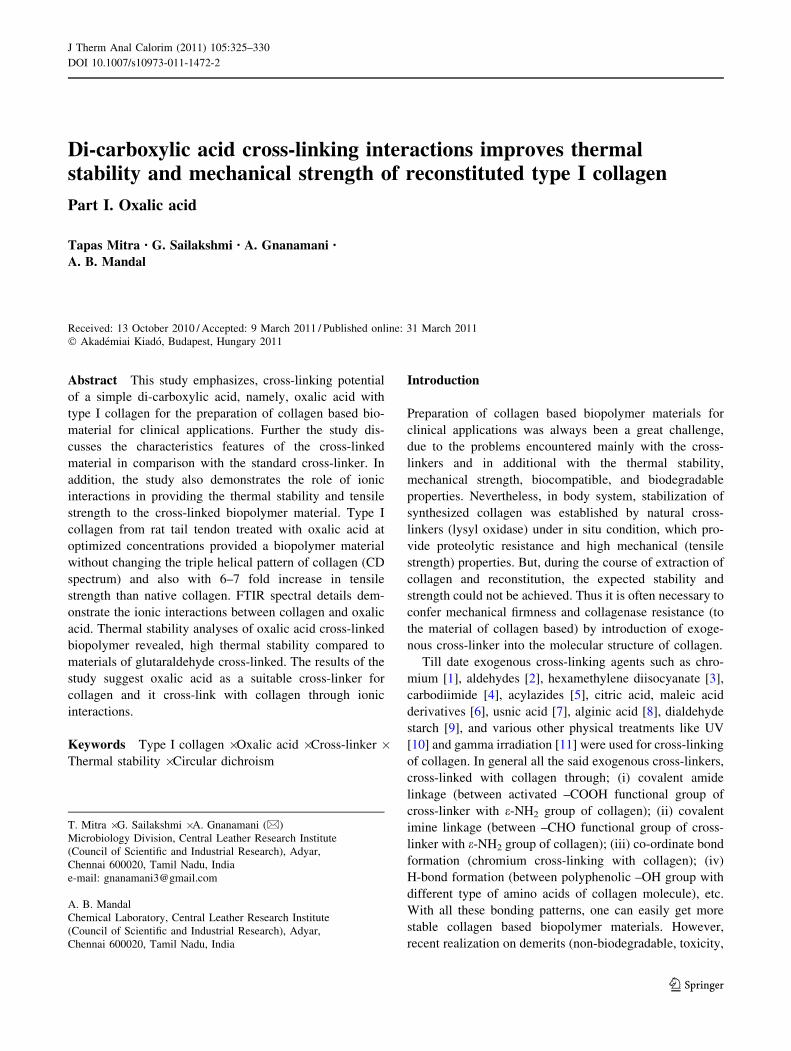
The electrophoretogram of type I collagen shown in Fig. 1a
demonstrates presence of [(a1) (I)2 (a2) (I)1] as an intense
band near 100 kDa followed by a faint band, corresponds
to a1 and a2 chains respectively. In addition, a band
observed near 200 kDa corresponds to b chain. The pat-
terns of a and b isomers of collagen compares favorably
with the standard values reported earlier [20]. Figure 1b
depicts the physical appearance of OACC biopolymer
material. Thickness of the resultant material was measured
using screw gauge.
Optimization of OA concentration with collagen for the
required mechanical strength and thermal stability studies
revealed 0.1% of OA with 0.5% of collagen (w/v) provided
the requisite properties as evidenced through the charac-
teristic features.
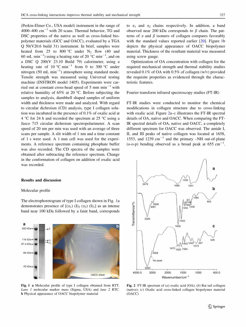
Fourier transform infrared spectroscopy studies (FT-IR)
FT-IR studies were conducted to monitor the chemical
modifications in collagen structure due to cross-linking
with oxalic acid. Figure 2a–c illustrates the FT-IR spectral
details of OA, native and OACC. When comparing the FT-
IR spectral details of OA, native and OACC, a completely
different spectrum for OACC was observed. The amide I,
II, and III peaks of native collagen was located at 1658,
1553, and 1239 cm-1 and the primary –NH out-of-plane
(o-o-p) bending observed as a broad peak at 655 cm-1.
200 kDa β
α1α2
a b
116 kDa
97.4 kDa
66 kDa
45 kDa
OACC sheet
Fig. 1 a Molecular profile of type I collagen obtained from RTT.
Lane 1 molecular marker mass (Sigma, USA) and lane 2 RTC.
b Physical appearance of OACC biopolymer material
4000.0 3000
3320
No peak
2928
1675 1625 1564
1453 12421073
947528
3326
c
b
a
30812928
1658
1553
1455 1239
1082
655
125116963479
1435
14001369
2000 1500
Wavenumber/cm–1
Tran
smitt
ance
/%
1000 400.0
Fig. 2 FT-IR spectrum of (a) oxalic acid (OA); (b) Rat tail collagen
(native); (c) Oxalic acid cross-linked collagen biopolymer material
(OACC)
DCA cross-linking interactions improves thermal stability and mechanical strength 327
123

However, after cross-linking with oxalic acid, a sharp
intense peak at 1658 disappears with the appearance of two
new peaks at 1675 and 1625 cm-1 in OACC. This might be
due to the ionic interaction between –COO- of oxalic acid
(1625) and –NH3? (1675) of collagen [21]. In addition,
deep and sharp intense peak observed at 1553 cm-1 in
native and the related overtone of 1553 at 3081 cm-1 was
reduced without any overtone peaks in OACC. However,
no change in the amide III was observed in OACC. Table 2
demonstrates the FT-IR peak assignment of OA, native and
OACC.
Thermal analysis
The thermal stability of the native and OACC biopolymer
material was evaluated by thermo gravimetric analysis
(TG). As can be seen in Fig. 3a the decomposition patterns
of the OA, native and OACC were presented in two steps;
at temperatures below 100 �C, the mass loss can probably
be attributed to the volatilization of low molecular mass
compounds (e.g., adsorbed water) [22] and post-curing
processes. The degradation of the OACC took place at
temperatures higher than 290 �C with 25% mass loss. The
main degradation step occurred at 400–500 �C, with a mass
loss in the range of 50–55%, whereas in GCC 25 and 50%
mass loss was observed at 215 and 353 �C and in native
this was observed at 190 and 373 �C, respectively.
Differential scanning calorimetry (DSC) detection was
used to study the thermal behavior of OA, native, and
OACC. The higher transition temperatures indicate colla-
gen had higher stability in a high temperature environment.
The thermal stability also influences on the durability of the
collagen based biopolymer materials. DSC studies recor-
ded melting temperature differences among OA (103 �C),
native (99 �C), OACC (137 �C), and GCC (151 �C) as
shown Fig. 3b.
Tensile strength
The mechanical property of native collagen was studied to
ensure that the sheet was intact during clinical applications.
Results showed that OACC exhibit very high tensile
strength 40.63 MPa than the native (6.8 MPa) and GCC
(1.95). About 6–7 fold increase in tensile strength was
observed after cross-linking with OA compared to native.
Circular dichroism
Collagen exhibits a unique CD spectrum with a small
positive peak between 220 and 225 nm and a large nega-
tive at 197 nm [23, 24]. The CD spectrum of native
Table 2 FT-IR analysis of OA, native (RTC), and OACC
Wave number/
cm-1Assignment
655 1� amide d(N–H) very broad band
1553 Strong 2� amide d(N–H)
1658 3� amide m(C=O) strong
3081 Weaker band at about 3100 in 2� amide is attributed
to a Fermi resonance overtone of 1550 band
3326 Strong 2� amide m(N–H)
1696 Broad (C=O) m of oxalic acid
1251 (C–O) m medium intensity of oxalic acid
3479 Very broad (O–H) m of oxalic acid
m stretching, d bending
0
0
2
0
–2
–4
–6
–8
–10
20
40
60
80
100
a
b
100 200 300
184 °C (50%)
117 °C (75%) 232 °C (75%)
329 °C (50%)
423 °C (50%)
352 °C (50%)
295 °C (75%)
136 °C (75%)
400 500 600
Temperature/°C
300250
CollagenOxalic acidOACC
GCC
Collagen
Oxalic acidOACCGCC
200150
103 °C
137 °C
98 °C
151 °C
100500
Temperature/°C
Hea
t flo
w/W
g–1
Mas
s/%
Fig. 3 a TG of oxalic acid (OA), native (in figure indicated as
‘collagen’) and oxalic acid cross-linked collagen (OACC), and GCC
biopolymer materials. b DSC analysis of oxalic acid (OA), native (in
figure indicated as ‘collagen’) and oxalic acid cross-linked collagen
(OACC), and GCC
328 T. Mitra et al.
123
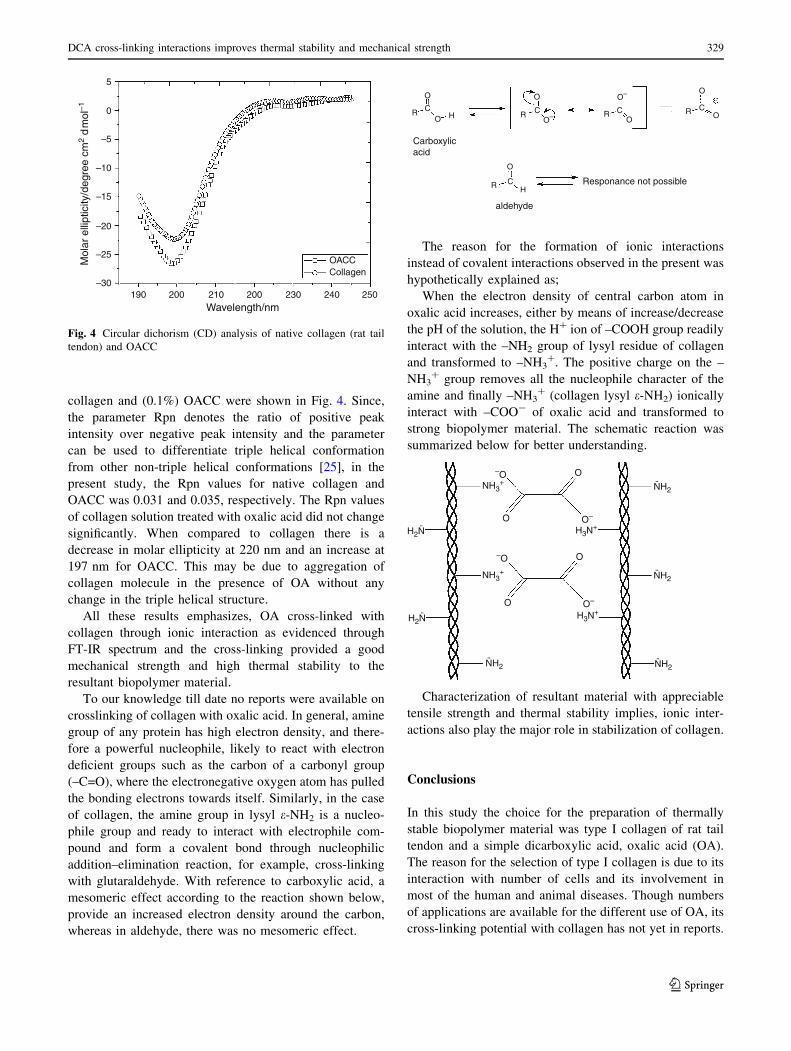
collagen and (0.1%) OACC were shown in Fig. 4. Since,
the parameter Rpn denotes the ratio of positive peak
intensity over negative peak intensity and the parameter
can be used to differentiate triple helical conformation
from other non-triple helical conformations [25], in the
present study, the Rpn values for native collagen and
OACC was 0.031 and 0.035, respectively. The Rpn values
of collagen solution treated with oxalic acid did not change
significantly. When compared to collagen there is a
decrease in molar ellipticity at 220 nm and an increase at
197 nm for OACC. This may be due to aggregation of
collagen molecule in the presence of OA without any
change in the triple helical structure.
All these results emphasizes, OA cross-linked with
collagen through ionic interaction as evidenced through
FT-IR spectrum and the cross-linking provided a good
mechanical strength and high thermal stability to the
resultant biopolymer material.
To our knowledge till date no reports were available on
crosslinking of collagen with oxalic acid. In general, amine
group of any protein has high electron density, and there-
fore a powerful nucleophile, likely to react with electron
deficient groups such as the carbon of a carbonyl group
(–C=O), where the electronegative oxygen atom has pulled
the bonding electrons towards itself. Similarly, in the case
of collagen, the amine group in lysyl e-NH2 is a nucleo-
phile group and ready to interact with electrophile com-
pound and form a covalent bond through nucleophilic
addition–elimination reaction, for example, cross-linking
with glutaraldehyde. With reference to carboxylic acid, a
mesomeric effect according to the reaction shown below,
provide an increased electron density around the carbon,
whereas in aldehyde, there was no mesomeric effect.
The reason for the formation of ionic interactions
instead of covalent interactions observed in the present was
hypothetically explained as;
When the electron density of central carbon atom in
oxalic acid increases, either by means of increase/decrease
the pH of the solution, the H? ion of –COOH group readily
interact with the –NH2 group of lysyl residue of collagen
and transformed to –NH3?. The positive charge on the –
NH3? group removes all the nucleophile character of the
amine and finally –NH3? (collagen lysyl e-NH2) ionically
interact with –COO- of oxalic acid and transformed to
strong biopolymer material. The schematic reaction was
summarized below for better understanding.
H2N
H2N H3N+
H3N+
¨
NH2¨ NH2
¨
NH2¨
NH2¨
NH3+
–O
–O
O–
O–
O
O
O
O
NH3+
Characterization of resultant material with appreciable
tensile strength and thermal stability implies, ionic inter-
actions also play the major role in stabilization of collagen.
Conclusions
In this study the choice for the preparation of thermally
stable biopolymer material was type I collagen of rat tail
tendon and a simple dicarboxylic acid, oxalic acid (OA).
The reason for the selection of type I collagen is due to its
interaction with number of cells and its involvement in
most of the human and animal diseases. Though numbers
of applications are available for the different use of OA, its
cross-linking potential with collagen has not yet in reports.
200 230 240
OACCCollagen
250210200190–30
–25
–20
–15
–10
–5
0
5
Wavelength/nm
Mol
ar e
llipt
icity
/deg
ree
cm2
d m
ol–1
Fig. 4 Circular dichorism (CD) analysis of native collagen (rat tail
tendon) and OACC
R R
R
R RC C
C
C C
O O
O
O O OO–
O– O
H
H
Carboxylicacid
aldehyde
Responance not possible
DCA cross-linking interactions improves thermal stability and mechanical strength 329
123

The cross-linking ability of OA with collagen was assessed
using FT-IR, TG, DSC, and CD. The results obtained and
the schematic representation of the reaction mechanism
summarized suggests the suitability of OA as a cross-
linking agent and the biopolymer material prepared upon
crosslinking of collagen with OA, suitable for clinical
applications without posing any toxicity to cells.
Acknowledgements All authors thank Department of Biotechnol-
ogy, Ministry of Science and Technology, New Delhi, for the finan-
cial assistance provided in the form of project in vide sanction no. BT/
PR10179/AAQ/03/385/2007.
References
1. Usha R, Ramasami T. Effect of crosslinking agents (basic chro-
mium sulfate and formaldehyde) on the thermal and thermome-
chanical stability of rat tail tendon collagen fibre. Thermochim
Acta. 2000;356(1–2):59–66.
2. Sheu M-T, Huang J-C, Yeh G-C, Ho H-O. Characterization of
collagen gel solutions and collagen matrices for cell culture.
Biomaterials. 2001;22(13):1713–9.
3. Miles CA, Avery NC, Rodin VV, Bailey AJ. The increase in
denaturation temperature following cross-linking of collagen is
caused by dehydration of the fibres. J Mol Biol. 2005;346(2):
551–6.
4. Nam K, Kimura T, Kishida A. Controlling coupling reaction of
EDC and NHS for preparation of collagen gels using ethanol/
water co-solvents. Macromol Biosci. 2008;8:32–7.
5. Petite H, Rault I, Huc A, Menasche P, Herbage D. Use of the acyl
azide method for cross-linking collagen-rich tissues such as
pericardium. J Biomed Mater Res. 1990;24(2):179–87.
6. Saito H, Murabayashi S, Mitamura Y, Taguchi T. Characteriza-
tion of alkali-treated collagen gels prepared by different cross-
linkers. J Mater Sci Mater Med. 2008;19:1297–305.
7. Nunes PS, Bezerra MLS, Costa LP, Cardoso JC, Albuquerque
RLC Jr, Rodrigues MO, Barin GB, Silva FAD, Jo AASA.
Thermal characterization of usnic acid/collagen-based films.
J Therm Anal Calorim. 2010;99:1011–4.
8. Mitra T, Sailakshmi G, Gnanamani A, Raja ST, Thiruselvi T,
Mangala Gowri V, Selvaraj NV, Ramesh G, Mandal AB. Prep-
aration and characterization of a thermostable and biodegradable
biopolymers using natural cross-linker. Int J Biol Macromol.
2011;48(2):276–85.
9. Langmaier F, Mokrejs P, Mladek M. Heat-treated biodegradable
films and foils of collagen hydrolysate crosslinked with dialde-
hyde starch. J Therm Anal Calorim. 2010;102:37–42.
10. Weadock KS, Miller EJ, Bellincampi LD, Zawadsky JP, Dunn
MG. Physical crosslinking of collagen fibers: comparison of
ultraviolet irradiation and dehydrothermal treatment. J Biomed
Mater Res. 1995;29(11):1373–9.
11. Olde Damink LHH, Dijkstra PJ, van Luyn MJA, van Wachem
PB, Nieuwenhuis P, Feijen J. Influence of ethylene oxide gas
treatment on the in vitro degradation behavior of dermal sheep
collagen. J Biomed Mater Res. 1995;29(2):149–55.
12. Melina H, Heuzey MC, Begin A. Viscoelastic properties of
phosphoric and oxalic acid-based chitosan hydrogels. Rheol Acta.
2006;45:659–75.
13. Skotak M, Leonov AP, Gustavo L, Sandra N, Anuradha S. Bio-
compatible and biodegradable ultrafine fibrillar scaffold materials
for tissue engineering by facile grafting of L-lactide onto chitosan.
Biomacromolecules. 2008;9(7):1902–8.
14. Jaklenec A, Wan E, Murray ME, Mathiowitz E. Novel scaffolds
fabricated from protein-loaded microspheres for tissue engi-
neering. Biomaterials. 2008;29(2):185–92.
15. Ghaffari A, Navaee K, Oskoui M, Bayati K, Rafiee-Tehrani M.
Preparation and characterization of free mixed-film of pectin/
chitosan/Eudragit RS intended for sigmoidal drug delivery. Eur J
Pharm Biopharm. 2007;67(1):175–86.
16. Wei X, Sun N, Wu B, Yin C, Wu W. Sigmoidal release of
indomethacin from pectin matrix tablets: effect of in situ cross-
linking by calcium cations. Int J Pharm. 2006;318(1–2):132–8.
17. Tharanathan RN. Biodegradable films and composite coatings:
past, present and future. Trends Food Sci Technol. 2003;14(3):
71–7.
18. Weber CJ, Haugaard V, Festersen R, Bertelsen G. Production and
applications of biobased packaging materials for the food
Industry. Food Addit Contam. 2002;19(4):172–7.
19. Chandrasekaran G, Torchia DA, Piez KA. Preparation of intact
monomeric collagen from tail, aorta and skin and the structure of
the nonhelical ends in solution. J Biol Chem. 1976;251:6062–7.
20. Lin YK, Liu DC. Comparison of physical–chemical properties of
type I collagen from different species. Food Chem. 2006;99:
244–51.
21. Pavia DL, Lampman GM, Kriz GS. Introduction to spectroscopy.
3rd ed. USA: Thomson Learning, Inc; 2001.
22. Puett D. DTA and heals of hydration of some polypeptides.
Biopolymers. 1967;5(3):327–30.
23. Venugopal MG, Ramshaw JAM, Braswell E, Zhu D, Brodsky B.
Electrostatic interactions in collagen-like triple-helical peptides.
Biochemistry. 1994;33(25):7948–56.
24. Sacca B, Renner C, Moroder L. The chain register in heterotri-
meric collagen peptides affects triple helix stability and folding
kinetics. J Mol Biol. 2002;324(2):309–18.
25. Madhan B, Subramanian V, Rao JR, Nair BU, Ramasami T.
Stabilization of collagen using plant polyphenol: role of catechin.
Int J Biol Macromol. 2005;37(1–2):47–53.
330 T. Mitra et al.
123