developmentofpolylactic/glycolicacid(plga…downloads.hindawi.com/journals/joph/2017/1598218.pdf ·...
TRANSCRIPT

Research ArticleDevelopment of Poly Lactic/Glycolic Acid (PLGA)Microspheres forControlled Release of Rho-Associated Kinase Inhibitor
Sho Koda,1,2 Naoki Okumura,1 Junji Kitano,1 Noriko Koizumi,1 and Yasuhiko Tabata2
1Department of Biomedical Engineering, Faculty of Life and Medical Sciences, Doshisha University, Kyotanabe 610-0321, Japan2Laboratory of Biomaterials, Department of Regeneration Science and Engineering, Institute for Frontier Life and Medical Sciences,Kyoto University, 53 Kawara-cho Shogoin, Sakyo-ku, Kyoto 606-8507, Japan
Correspondence should be addressed to Yasuhiko Tabata; [email protected]
Received 7 February 2017; Revised 21 June 2017; Accepted 9 July 2017; Published 27 July 2017
Academic Editor: Vishal Jhanji
Copyright © 2017 Sho Koda et al. This is an open access article distributed under the Creative Commons Attribution License, whichpermits unrestricted use, distribution, and reproduction in any medium, provided the original work is properly cited.
Purpose. The purpose of this study was to investigate the feasibility of poly lactic/glycolic acid (PLGA) as a drug delivery carrier ofRho kinase (ROCK) inhibitor for the treatment of corneal endothelial disease. Method. ROCK inhibitor Y-27632 and PLGA weredissolved in water with or without gelatin (W1), and a double emulsion [(W1/O)/W2] was formed with dichloromethane (O) andpolyvinyl alcohol (W2). Drug release curve was obtained by evaluating the released Y-27632 by using high performance liquidchromatography. PLGA was injected into the anterior chamber or subconjunctiva in rabbit eyes, and ocular complication wasevaluated by slitlamp microscope and histological analysis. Results. Y-27632 incorporated PLGA microspheres with differentmolecular weights, and different composition ratios of lactic acid and glycolic acid were fabricated. A high molecular weight andlow content of glycolic acid produced a slower and longer release. The Y-27632 released from PLGA microspheres significantlypromoted the cell proliferation of cultured corneal endothelial cells. The injection of PLGA did not induce any evident eyecomplication. Conclusions. ROCK inhibitor-incorporated PLGA microspheres were fabricated, and the microspheres achievedthe sustained release of ROCK inhibitor over 7–10 days in vitro. Our data should encourage researchers to use PLGAmicrospheres for treating corneal endothelial diseases.
1. Introduction
The corneal endothelium maintains corneal transparency bypump and leaky barrier functions [1]. The corneal endothe-lium is located at the anterior chamber side of the posteriorcornea at a cell density of 2000–3000 cells/mm2 in a healthysubject. A phenotypical feature of corneal endothelial cells(CECs) is a severely limited proliferative ability [2]. Whenthe corneal endothelium is impaired by any pathological con-ditions, trauma, or aging, the residual remaining CECsmigrate and spread out to cover the impaired area, resultingin a cell density drop and increased polymorphism. Theremaining CECs compensate the function to maintain cor-neal transparency, but once cell density drops to critical level,which is usually less than 500–1000mm2/cells, the cornealoses its transparency [1].
Corneal transplantation using a donor cornea is theonly therapeutic choice for treating corneal endothelial
decompensation [3]. Researchers, including our group, havesearched pharmaceutical agents such as EGF [4], PDGF [5],FGF-2 [6], lithium [7], and Rho kinase (ROCK) inhibitor [8],which can promote the proliferation of CECs, for the treat-ment of corneal endothelial diseases. Indeed, we performedpilot clinical research and showed that ROCK inhibitor eyedrops have a potency for treating early stages of Fuchsendothelial corneal dystrophy and severe corneal endothelialdamage inducedby cataract surgeryby enhancing theprolifer-ation of residual CECs [9–11].
Poly lactic/glycolic acid (PLGA), a copolymer of polylactic acid (PLA) and poly glycolic acid (PGA), has beenintensively researched as a drug delivery carrier [12, 13].During the degradation of PLGA for drug release, lactic acidand glycolic acid are coreleased and are both biologicallyremoved by the surrounding cells through normal metabolicpathways [14]. The biocompatibility, low toxicity, and drugencapsulation capabilities of PLGA-based drug delivery
HindawiJournal of OphthalmologyVolume 2017, Article ID 1598218, 9 pageshttps://doi.org/10.1155/2017/1598218

systems have attracted the attention of researchers [12, 13].Indeed, several therapies using PLGA have recently enteredpreclinical development [13, 15].
In the current study, we designed the PLGA micro-spheres, which incorporate ROCK inhibitor Y-27632, toenable efficient drug delivery for targeting corneal endothe-lium. We also evaluated the safety and stability of ROCKinhibitor released from PLGA microspheres throughin vitro experiments. In addition, the safety of PLGA micro-sphere injection into the anterior chamber or subconjunctivawas evaluated in a rabbit model.
2. Materials and Methods
2.1. Microsphere Preparation. A selective ROCK inhibitor, Y-27632 (1mg, Wako Pure Chemical Industries Ltd., Osaka,Japan), was dissolved in double-distilled water (200μL) withor without gelatin (20μg, Nitta Gelatin, Osaka, Japan) (W1)and poured into dichloromethane (2mL, DCM, NacalaiTesque, Kyoto, Japan) in which PLGA or poly (L-lactic acid)(PLA) (90mg) was dissolved (O). The first inner W1/Oemulsion was prepared by agitating for 60 secs using a vortexmixer at room temperature, followed by sonication for300 secs using a UD-21P ultrasonic generator (Tomy SeikoCo. Ltd., Tokyo, Japan). The first emulsion was then pouredinto 1% polyvinyl alcohol (2mL, PVA, Japan Vam & PovalCo. Ltd., Osaka, Japan) solution (W2), followed by vigorousmixing using a vortex mixer for 60 sec. This procedure per-mitted the formation of the double emulsion [(W1/O)/W2],in which the W1 phase was homogeneously dispersed inthe O phase. The resulting double emulsion was poured into1wt% PVA solution (300mL) and continuously stirred for 3hours at 4°C until DCM was completely evaporated. Themicrospheres were washed several times with double-distilled water by centrifugation and freeze-dried intopowdered microspheres. The microspheres with differentcomposition ratios of lactic acid/glycolic acid were preparedsimilarly (Table 1).
The shape and size of the microspheres was evaluatedusing light microscopy (CKX41, Olympus Corp., Tokyo,Japan) and a micro scale (OB1, 1mm scale length, 100 divi-sions, 0.01mm pitch, MeCan Imaging Inc., Saitama, Japan).For scanning electron microscopy analysis, microsphereswere fixed on an aluminum support with carbon-adhesiveglue and coated with a 10 nm thick coating of gold-palladium (30mA, 40 sec) (JSM 6701F; JEOL, Tokyo, Japan).
The samples were observed using a scanning electron micro-scope (S-2380N, Hitachi Ltd., Tokyo, Japan).
2.2. Release Study. The Y-27632 release profiles from thePLGA microspheres were determined in vitro. PLGA micro-spheres in which Y-27632 (10mg) was incorporated wereincubated in a tube containing 1.0mL phosphate-bufferedsaline solution (PBS; Nissui Pharmaceutical Co. Ltd., Tokyo,Japan) (pH: 7.4) at 37°C in a heat block (ALB-221, ScinicsCo., Tokyo, Japan). PBS including released Y-27632 was eval-uated at 1, 3, and 6 hour(s) and 1 to 30 day(s) after incubation.The concentration of Y-27632 in the samples was determinedby high performance liquid chromatography (HPLC; Promi-nence, Shimazu Corp., Kyoto, Japan) equipped with onepump, an auto sampler, and an ultra violet (UV) detector. Cal-ibration curves were prepared for Y-27632 based on the UVabsorbance peak area at 269nm.These calibration curveswereused for the estimation of in vitro drug release. The Y-27632release profile was calculated as follows: cumulative amountof Y‐27632 released / total incorporated Y‐27632 × 100
2.3. Cell Cultures. Eight corneas from four cynomolgus mon-keys (3–5 years of age; estimated equivalent human age, 5–20years) housed at Nissei Bilis Co. Ltd. (Otsu, Japan) were usedfor the study. The corneas were harvested at the time of theeuthanization of the monkeys for other research purposes.CECs were cultured according to a modified protocolreported previously [16]. Briefly, Descemet’s membraneincluding CECs was stripped from the cornea and incubatedwith0.6U/mLofDispase II (RocheAppliedScience,Penzberg,Germany) for 60minutes at 37°C.CECswere then seeded ontoa culture plate (CECs from one cornea to one well of a 12-wellculture plate) with a culture medium composed of Dulbecco’smodified Eagle’s medium (DMEM, Life Technologies Corp.,Carlsbad, CA) supplemented with 10% fetal bovine serum50U/mL penicillin, 50μg/mL streptomycin, and 2ng/mLbasic fibroblast growth factor (bFGF; Life TechnologiesCorp.). ROCK inhibitor was not added to the culturemediumwhenpreparingCECs for experiments.When thecells reachedconfluence in 10 to 14 days, they were trypsinized with 0.05%trypsin-EDTA (Life Technologies Corp.) for 5 minutes at37°C and passaged at ratios of 1 : 2 to 4. CECs obtained afterthe 4–7th passage that stillmaintained ahexagonal andmono-layer sheet-like structure were used for the experiments.
2.4. In Vitro Assessment of Y-27632 Released fromMicrospheres. To evaluate the stability and safety of Y-27632
Table 1: Summary of poly lactic/glycolic acid (PLGA) or poly lactic acid (PLA) microspheres.
Type Molecular weightComposition ratio
Drug incorporation rate (%) Particle size (μm)Lactic acid : glycolic acid
5005 5000 50 : 50 12.5 12.63± 5.765010 10000 50 : 50 1.38 11.83± 6.857505 5000 75 : 15 1.17 14.30± 5.130005 5000 100 : 0 9.33 16.44± 6.090020 20000 100 : 0 2.11 13.94± 7.325005 + gelatin 5000 50 : 50 18.1 11.80± 7.74
2 Journal of Ophthalmology

released from the PLGA/PLA microspheres, the effect ofreleased Y-27632 on cell growth of CECs was evaluated.Y-27632 incorporating PLGA microspheres (0020) wereincubated in PBS, and PBS was recovered at 3 and 7 daysafter incubation. The concentration of Y-27632 was thenevaluated by HPLC, and the recovered PBS including thereleased Y-27632 was added to the culture medium at afinal concentration of Y-27632 of 10μM. Cultured monkeyCECs were seeded at a density of 5.0× 10³ cells/cm2 perwell on a 96-well plate for 24 hours and then subjectedto serum starvation for an additional 24 hours in the pres-ence or absence of fresh Y-27632 (10μM) or Y-27632released from PLGA microspheres after 3 or 7 days(10μM). The number of viable cells was determined byuse of the CellTiter-Glo® Luminescent Cell Viability Assay(Promega, Fitchburg, Wisconsin) performed in accordancewith the manufacturer’s protocol. The number of CECs at24 hours after treatment with Y-27632 was measuredusing the Veritas™ Microplate Luminometer (Promega,Fitchburg, Wisconsin).
2.5. Safety Assessment of Microspheres in Rabbit. To evaluatethe safety of PLGA/PLA microspheres, 1 or 10mg PLGAmicrospheres (0020) suspended in 200μL PBS were injectedinto the anterior chamber and 1mg PLGA microspheres sus-pended in 200μL PBS were injected into the subconjunctivaof the 9 right eyes of 9 rabbits (n = 3). The left eyes were usedas a control. The corneal appearance of the 9 rabbits wasexamined at 1, 2, 3, 5, 7, 10, and 14 days using a slitlampmicroscope, and corneal opacification and conjunctivalhyperemia were evaluated according to the grading system(Supplemental Table 1 available online at https://doi.org/10.1155/2017/1598218) modified from the previous report[17]. Intraocular pressure was determined with a Tonovet®
(Icare Finland, Vantaa, Finland) instrument. Corneal thick-ness was determined through the use of an ultrasound pachy-meter (SP-2000; Tomey, Nagoya, Japan).
2.6. Histological Examination of Rabbit Eyes after Poly Lactic/Glycolic Acid (PLGA) Microsphere Injection. After 14 days ofPLGA microsphere injection, rabbits were euthanized, theeye balls were enucleated, and sclerocorneal specimens wereprepared using ophthalmic surgical scissors. Samples werefixed in 4% formaldehyde and incubated for 30 minutes in1% bovine serum albumin to block nonspecific binding.The samples were then incubated overnight at 4°C with anti-bodies against Na+/K+-ATPase (1 : 300, Upstate Biotechnol-ogy, Lake Placid, NY), ZO-1 (1 : 300, Life TechnologiesCorp.), and connexin 43 (1 : 300, Life Technologies Corp.).Alexa Fluor® 488-conjugated goat anti-mouse (Life Technol-ogies Corp.) was used as a secondary antibody at a 1 : 1000dilution. Cell morphology was evaluated after actin stainingwith a 1 : 400 dilution of Alexa Fluor 594-conjugated phalloi-din (Life Technologies Corp.). Nuclei were stained with 4′,6-diamidino-2-phenylindole (DAPI, Dojindo Laboratories,Kumamoto, Japan). The samples were examined with a fluo-rescence microscope (TCS SP2 AOBS; Leica Microsystems,Wetzlar, Germany).
2.7. Statistical Analysis. The statistical significance (p value)of differences in the mean values of the two-sample compar-ison was determined with the Student’s t-test. The statisticalsignificance of comparisons of multiple sample sets was ana-lyzed with Dunnett’s multiple-comparisons test. p < 0 05 wasconsidered as statistically significant. Results were expressedas mean± standard error of the mean.
3. Results
3.1. Characteristics of Poly Lactic/Glycolic Acid (PLGA) orPoly Lactic Acid (PLA) Microspheres for Y-27632 Release.Y-27632-incorporated PLGA or PLA microspheres withdifferent molecular weights and different composition ratiosof lactic acid and glycolic acid were prepared (Table 1).All PLGA or PLA microspheres exhibited similar morphol-ogy and size. SEM showed that representative images ofPLGA microspheres were spherical with a smooth surface(Figure 1(a), upper panel). The inside of the microsphereswas porous core due to double emulsion, which is designedto incorporate Y-27632 (Figure 1(a), lower panel). We firstevaluated the effect of molecular weight on the release pro-file in PLGA or PLA microspheres. PLGA5005 had released65.5± 0.9% (approximately 8.22μg) of the Y-27632 by 1 dayand 98.7± 0.6% (approximately 12.4μg) by 14 days, whilePLGA5010, which has a higher molecular size thanPLGA5005, released 40.8± 1.0% (approximately 0.56μg) ofY-27632 by 1 day and 99.1± 1.7% (approximately 1.37μg)by 14 days (p < 0 01 at 1 day). PLA0005 released 62.8± 0.8%(approximately 5.86μg) of Y-27632 by 1 day and 77.7± 0.5%(approximately 7.25μg) by 14 days, while PLA0020, whichhas a higher molecular size than PLA0005, released 30.2± 0.9% (approximately 0.64μg) of Y-27632 by 1 day and 55.3± 0.6% (approximately 1.16μg) by 14 days (p < 0 01 at both 1day and 14 days) (Figure 1(b)). These data showed that a lowermolecular weight increased the rate of drug release at a fixedcomposition ratio of lactic acid andglycolic acid.Wenext eval-uated the effect of the composition of lactic acid and glycolicacid on the release profile. PLGA7505 released 58.9% ofY-27632 at 1 day and 97.4% at 14 days, and PLA0005released 62.4% of Y-27632 at 1 day and 77.5% at 14 days.This data showed that a smaller amount of glycolic acidmade the release slower and a smaller amount of lacticacid made the release of Y-27632 faster (Figure 1(c)).
The mean incorporation rate of Y-27632 was 8.74%in PLGA microspheres, though PLGA microspheres pre-paredwith gelatin enabled a significantly higher incorporationof Y-27632 at 18.1%, showing that gelatin reduced the loss ofROCK inhibitor during preparation of the PLGA micro-spheres (Figure 1(d)). Characteristic release curves showedthat both PLGA with or without gelatin have the potency torelease Y-27632 and that the initial burst release of Y-27632tended to decrease formicrosphereswith gelatin (Figure 1(e)).
3.2. In Vitro Evaluation of Y-27632 Released fromMicrospheres. The effect of Y-27632 released from PLGAmicrospheres on CEC proliferation was evaluated. Phasecontrast images showed that CECs cultured with a culturemedium supplemented with fresh Y-27632 (10μM) showed
3Journal of Ophthalmology

higher numbers of cells than the control. The Y-27632released from the PLGA microspheres was recovered afterincubation and was added to a growth medium at a finalconcentration of 10μM. Higher numbers of CECs wereobserved than those in the control after 24 hours of cultiva-tion with those growth media that were supplemented with
Y-27632 released from PLGA microspheres (Figure 2(a)).The cell numbers of CECs were significantly increased bythe supplementation of fresh Y-27632 or the Y-27632recovered from PLGA microspheres after an incubation of 3and 7 days (118.8%, 110.6%, and 111.4%, resp.) (Figure 2(b)).These results showed that Y-27632 incorporated in PLGA
(a)
0
20
40
60
80
100
0 10 20 30
50055010
00050020
Time (day)
Rele
ase o
f Y-2
7632
(%)
(b)
0
20
40
60
80
100
0 10 20 30
50057505
0005
Rele
ase o
f Y-2
7632
(%)
Time (day)
(c)
0
5
10
15
20
PLGA5005 PLGA5005 +gelatin
Inco
rpor
atio
n ra
te (%
)
⁎⁎
(d)
0
20
40
60
80
100
0 5 10 15
50055005 + gelatin
Rele
ase o
f Y-2
7632
(%)
Time (day)
(e)
Figure 1: Characteristics of poly lactic/glycolic acid (PLGA) or poly lactic acid (PLA) microspheres for Y-27632 release. (a) PLGAmicrospheres were examined with a scanning electron microscope. The upper panel shows the surface of the microsphere, and the lowerpanel shows the internal structure of the microsphere. (b) PLGA or PLA microspheres were incubated in a PBS at 37°C for 14 days, and theamount of Y-27632 released was determined by HPLC. The effect of the molecular weight on the release profile for PLGA or PLAmicrospheres was evaluated. The cumulative amount of released Y-27632 was plotted in the graph (n = 3). (c) PLGA or PLA microsphereswere incubated in PBS at 37°C, and the amount of Y-27632 released was determined by HPLC. The effect of the composition of lactic acidand glycolic acid on the release profile was evaluated (n = 3). The PLGA5005 release curve plotted is the same as that shown in Figure 1(b).(d) Y-27632 was dissolved in ultrapure water with or without gelatin, and PLGA5005 microspheres were prepared (n = 3). Theincorporation rate was evaluated by HPLC. ∗∗p < 0 01. (e) PLGA5005 with or without gelatin that incorporated Y-27632 was incubated atPBS at 37°C for 14 days, and the amount of Y-27632 released was determined by HPLC. Three independent experiments were performed.
4 Journal of Ophthalmology
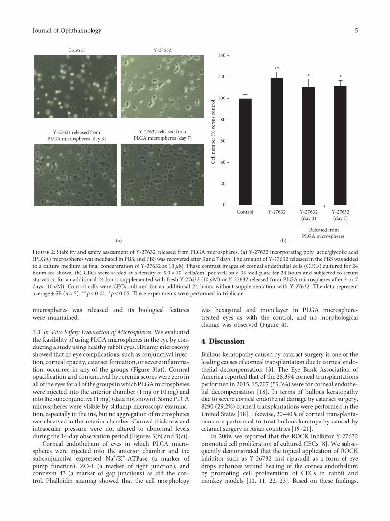
microspheres was released and its biological featureswere maintained.
3.3. In Vivo Safety Evaluation of Microspheres. We evaluatedthe feasibility of using PLGAmicrospheres in the eye by con-ducting a study using healthy rabbit eyes. Slitlampmicroscopyshowed that no eye complications, such as conjunctival injec-tion, corneal opacity, cataract formation, or severe inflamma-tion, occurred in any of the groups (Figure 3(a)). Cornealopacification and conjunctival hyperemia scores were zero inall of the eyes for all of thegroups inwhichPLGAmicrosphereswere injected into the anterior chamber (1mg or 10mg) andinto the subconjunctiva (1mg) (data not shown). Some PLGAmicrospheres were visible by slitlamp microscopy examina-tion, especially in the iris, but no aggregation of microsphereswas observed in the anterior chamber. Corneal thickness andintraocular pressure were not altered to abnormal levelsduring the 14-day observation period (Figures 3(b) and 3(c)).
Corneal endothelium of eyes in which PLGA micro-spheres were injected into the anterior chamber and thesubconjunctiva expressed Na+/K+-ATPase (a marker ofpump function), ZO-1 (a marker of tight junction), andconnexin 43 (a marker of gap junctions) as did the con-trol. Phalloidin staining showed that the cell morphology
was hexagonal and monolayer in PLGA microsphere-treated eyes as with the control, and no morphologicalchange was observed (Figure 4).
4. Discussion
Bullous keratopathy caused by cataract surgery is one of theleading causes of corneal transplantation due to corneal endo-thelial decompensation [3]. The Eye Bank Association ofAmerica reported that of the 28,394 corneal transplantationsperformed in 2015, 15,707 (55.3%) were for corneal endothe-lial decompensation [18]. In terms of bullous keratopathydue to severe corneal endothelial damage by cataract surgery,8290 (29.2%) corneal transplantations were performed in theUnited States [18]. Likewise, 20–40% of corneal transplanta-tions are performed to treat bullous keratopathy caused bycataract surgery in Asian countries [19–21].
In 2009, we reported that the ROCK inhibitor Y-27632promoted cell proliferation of cultured CECs [8]. We subse-quently demonstrated that the topical application of ROCKinhibitor such as Y-26732 and ripasudil as a form of eyedrops enhances wound healing of the cornea endotheliumby promoting cell proliferation of CECs in rabbit andmonkey models [10, 11, 22, 23]. Based on these findings,
Control Y-27632
Y-27632 released fromPLGA microspheres (day 3)
Y-27632 released fromPLGA microspheres (day 7)
(a)
0
20
40
60
80
100
120
140
Control Y-27632 Y-27632(day 3)
Y-27632(day 7)
⁎⁎
⁎ ⁎
Cell
num
ber (
% v
ersu
s con
trol
)
Released fromPLGA microspheres
(b)
Figure 2: Stability and safety assessment of Y-27632 released from PLGA microspheres. (a) Y-27632 incorporating poly lactic/glycolic acid(PLGA) microspheres was incubated in PBS, and PBS was recovered after 3 and 7 days. The amount of Y-27632 released in the PBS was addedto a culture medium as final concentration of Y-27632 as 10 μM. Phase contrast images of corneal endothelial cells (CECs) cultured for 24hours are shown. (b) CECs were seeded at a density of 5.0× 10³ cells/cm2 per well on a 96-well plate for 24 hours and subjected to serumstarvation for an additional 24 hours supplemented with fresh Y-27632 (10 μM) or Y-27632 released from PLGA microspheres after 3 or 7days (10 μM). Control cells were CECs cultured for an additional 24 hours without supplementation with Y-27632. The data representaverage± SE (n = 5). ∗∗p < 0 01, ∗p < 0 05. These experiments were performed in triplicate.
5Journal of Ophthalmology

we performed pilot clinical research in which patients withcorneal decompensation were treated with Y-27632 eyedrops following to a transcorneal freezing procedure to par-tially remove the damaged cornea endothelium. Y-27632eye drops showed effectiveness in reducing the central cor-neal thickness in patients with early stage Fuchs endothelialcorneal dystrophy [9, 10]. In addition, we demonstrated thatROCK inhibitor eye drops can enhance the wound healing of
damaged corneal endothelium caused by cataract surgery,thus successfully avoiding the need for corneal transplanta-tion [11]. Although further randomized clinical trials arenecessary, ROCK inhibitor currently might be one of themost promising pharmaceutical agents that can be appliedfor corneal decompensation.
Possible complications of the injection of PLGA into theanterior chamber are direct mechanical damage to the
PLGA 1 mganterior chamber injection
PLGA 10 mganterior chamber injection
Day
1D
ay 2
Day
14
PLGA 1 mgsubconjunctival injection
(a)
0
200
400
600
0 5 10 15Time (day)
Cor
neal
thic
knes
s (�휇
m)
1 mg PLGA in AC10 mg PLGA in AC1 mg PLGA in SC
::
:
(b)
1 mg PLGA in AC10 mg PLGA in AC1 mg PLGA in SC
::
:
0
5
10
15
20
0 5 10 15
IOP
(mm
Hg)
Time (day)
(c)
Figure 3: Safety assessment of poly lactic/glycolic acid (PLGA) microspheres in a rabbit eye. (a) One or 10mg PLGAmicrospheres suspendedin 200μL phosphate buffer solution (PBS) was injected into the anterior chamber, and 1mg PLGA microspheres suspended in 200 μL PBSwas injected into the subconjunctiva. Nine right eyes of 9 rabbits were used for the experiments (n = 3). Anterior segments were evaluatedby slitlamp microscopy for 14 days. (b, c) Corneal thickness and intraocular pressure were evaluated for 14 days and are shown in thegraph. The central corneal thickness was evaluated by ultrasound pachymetry. Intraocular pressure was determined with a Tonovet.
6 Journal of Ophthalmology

corneal endothelium or lens, indirect damage to the cornealendothelium by biodegradation products from PLGA, andintraocular pressure elevation by inhibiting aqueous humoroutflow. We therefore conducted a safety assessment usinghealthy rabbit eyes to evaluate the feasibility of applyingPLGA microspheres for intracameral injection. Here, weshowed that the injection of PLGA into the anterior chamberor the subconjunctiva did not induce any evident complica-tions. No inflammatory response was observed, in agreementwith the reported biocompatibility safety profile of PLGA.PLGA is capable of encapsulating a wide range of drugs ofalmost any molecular size [12]. Our in vitro study consis-tently showed that ROCK inhibitor released from PLGA pro-moted the proliferation of CECs in the same manner asunencapsulated ROCK inhibitor, suggesting that ROCKinhibitor was incorporated into and released from PLGA
without damage to the molecular properties of the inhibitor.Further study is needed, but it is suggested that PLGAmight be applicable as a drug carrier that is injected intothe anterior chamber.
In our clinical research, we suggested that a ROCKinhibitor has the potency to promote the cell proliferationof residual relatively healthy CECs [9–11] and that there isa “golden time” for a ROCK inhibitor, when the woundedspace has not yet been covered by the migration and spread-ing of residual CECs associated with a cell density drop [24].We showed that ROCK inhibitor eye drops restored cornealclarity within one week in patients with Fuchs endothelialcorneal dystrophy following a transcorneal freezing proce-dure and within 1-2 months in patients with severe cornealendothelial damage due to cataract surgery. Therefore, in thisstudy, we aimed to fabricate a PLGA system that would
ControlPh
allo
idin
Na+ /
K+ -A
TPas
eZO
-1Co
nnex
in 4
3
PLGA 1 mganterior chamber
injection
PLGA 10 mganterior chamber
injection
PLGA 1 mgsubconjunctival
injection
Figure 4: Histological evaluation of corneal endothelium in rabbit eyes injected with poly lactic/glycolic acid (PLGA) microspheres. One or10mg PLGA microspheres was injected into the anterior chamber, and 1mg PLGA microspheres was injected into the subconjunctiva. After14 days, corneal endothelium was evaluated by immunofluorescence staining and phalloidin staining. Na+/K+-ATPase (a marker of pumpfunction), ZO-1 (a marker of tight junction), and connexin 43 (a marker of gap junctions) were used to evaluate the functional propertyof corneal endothelium. Phalloidin staining was used for morphological analysis. Nuclei were stained with DAPI. Scale bar: 50 μm.
7Journal of Ophthalmology

release Y-27632 for 7 to 10 days to improve the therapeuticefficacy during this window of opportunity; however, therelease time should be optimized by further studies. The drugrelease from PLGA with degradation is influenced by variousfactors such as the initial molecular weight, the monomercomposition rate of PLA and PGA, the drug type, and thepH of the surrounding circumstances [12, 25]. Among thesefactors, the molecular weight and the composition rate ofPLA and PGA are recognized as important and are also easilycontrolled during fabrication [12]. Here, we showed thatPLGA5010 exhibited a slower release than PLGA5005 andthat PLA0020 exhibited slower release than PLA 0005, show-ing that PLGA or PLA microspheres with a high molecularweight produce slower and more prolonged Y-27632 release.This may be because polymers with lower molecular weightsare less hydrophobic, which increases the rate of waterabsorption, consequent hydrolysis, and erosion [26]. Thecomposition of PLA and PGA in PLGA is also an importantfactor influencing polymer degradation. In this study,PLGA7505 showed a slower release of Y-27632 thanPLGA5005, and PLA0005 showed the slowest and longestrelease, although it exhibited an initial burst in release. Thissuggests that a lower content of GA in PLGA results in aslower and longer-term drug release from the PLGA micro-spheres. It has been demonstrated that the PGA : PLA ratioof 50 : 50 make PLGA the most hydrophilic and amorphouswhen compared to other ratios, resulting in faster degrada-tion and drug release [12].
Although the further optimization of the release profileof the ROCK inhibitor is needed before clinical application,our results offers fundamental information for modifyingthe molecular weights and composition rates of PLA andPGA. Various mathematical, computational, and theoreticalmodels may be applicable for the optimization of the releaseprofile [27–29]. However, drug release from microspheresdiffers in the in vitro versus in vivo conditions, in part dueto immunological responses and the plasticizing effects ofbiological substances [12]. For instance, the release of thy-mosin alpha 1 from PLGA was slightly faster in an in vivoassay than in an in vitro assay [30]. Conversely, otherresearchers have shown that PLGA degradation was slowerin vivo than in vitro [31]. Therefore, pharmacokineticsstudies are needed on the release of ROCK inhibitor in theanterior chamber. The drawback of this study is the lack ofin vivo experiments that show an enhancement of cornealendothelial wound healing by ROCK inhibitor incorporatedinto PLGA. The fate of the PLGA injected into the anteriorchamber is also not well characterized, and further in vivoexperiments are needed prior to initiating clinical applica-tions. In the clinical setting, however, the treatment of cor-neal endothelial damage induced by cataract surgery wouldseem to be an important target for this PLGA therapy.
In conclusion, we fabricated ROCK inhibitor-incorporatedPLGA microspheres, and those microspheres act as carrierfor the sustained release of the ROCK inhibitor over 7–10days. In addition, the PLGA microspheres did not exhibitany evident complication in the eyes of a rabbit model.Although further optimization of the release profile of PLGAand in vivo experiments for safety and effectiveness
assessment are required, PLGA microspheres would appearto represent a promising drug delivery carrier for ROCKinhibitor.
Ethical Approval
In all experiments, animals were housed and treated in accor-dance with the ARVO Statement for the Use of Animals inOphthalmic and Vision Research. The rabbit experimentswere performed at Doshisha University (Kyoto, Japan)according to the protocol approved by the University’sAnimal Care and Use Committee (Approval no. 1315).
Conflicts of Interest
Noriko Koizumi is listed as inventor of the patent regardingthe application of the ROCK inhibitor for corneal endothe-lium (Registration no. 5657252).
Acknowledgments
The authors would like to acknowledge the grant from theProgram for the Strategic Research Foundation at PrivateUniversities from MEXT (Noriko Koizumi and NaokiOkumura). The authors are grateful to Dr. Takashi Saitofor the valuable advice for the PLGA experiments.
References
[1] T. Nishida and S. Saika, “Cornea and sclera: anatomy andphysiology,” Cornea Third Edition, vol. 1, pp. 3–24, 2011.
[2] N. C. Joyce, “Proliferative capacity of corneal endothelial cells,”Experimental Eye Research, vol. 95, no. 1, pp. 16–23, 2012.
[3] D. T. Tan, J. K. Dart, E. J. Holland, and S. Kinoshita,“Corneal transplantation,” Lancet, vol. 379, no. 9827,pp. 1749–1761, 2012.
[4] V. P. Hoppenreijs, E. Pels, G. F. Vrensen, J. Oosting, and W. F.Treffers, “Effects of human epidermal growth factor onendothelial wound healing of human corneas,” InvestigativeOphthalmology & Visual Science, vol. 33, no. 6, pp. 1946–1957, 1992.
[5] V. P. Hoppenreijs, E. Pels, G. F. Vrensen, and W. F. Treffers,“Effects of platelet-derived growth factor on endothelialwound healing of human corneas,” Investigative Ophthalmol-ogy & Visual Science, vol. 35, no. 1, pp. 150–161, 1994.
[6] J. Lu, Z. Lu, P. Reinach et al., “TGF-beta2 inhibits AKTactivation and FGF-2-induced corneal endothelial cell prolif-eration,” Experimental Cell Research, vol. 312, no. 18,pp. 3631–3640, 2006.
[7] E. C. Kim, H. Meng, and A. S. Jun, “Lithium treatmentincreases endothelial cell survival and autophagy in a mousemodel of Fuchs endothelial corneal dystrophy,” The BritishJournal of Ophthalmology, vol. 97, no. 8, pp. 1068–1073, 2013.
[8] N. Okumura, M. Ueno, N. Koizumi et al., “Enhancement onprimate corneal endothelial cell survival in vitro by a ROCKinhibitor,” Investigative Ophthalmology & Visual Science,vol. 50, no. 8, pp. 3680–3687, 2009.
[9] N. Koizumi, N. Okumura, M. Ueno, H. Nakagawa, J. Hamuro,and S. Kinoshita, “Rho-associated kinase inhibitor eye droptreatment as a possible medical treatment for fuchs cornealdystrophy,” Cornea, vol. 32, no. 8, pp. 1167–1170, 2013.
8 Journal of Ophthalmology

[10] N. Okumura, N. Koizumi, E. P. Kay et al., “The ROCK inhib-itor eye drop accelerates corneal endothelium wound healing,”Investigative Ophthalmology & Visual Science, vol. 54, no. 4,pp. 2493–2502, 2013.
[11] N. Okumura, R. Inoue, Y. Okazaki et al., “Effect of the Rhokinase inhibitor Y-27632 on corneal endothelial wound heal-ing,” Investigative Ophthalmology & Visual Science, vol. 56,no. 10, pp. 6067–6074, 2015.
[12] Y. Xu, C. S. Kim, D. M. Saylor, and D. Koo, “Polymer degrada-tion and drug delivery in PLGA-based drug-polymer applica-tions: a review of experiments and theories,” Journal ofBiomedical Materials Research Part B: Applied Biomaterials,vol. 105, no. 6, pp. 1692–1716, 2016.
[13] R. J. Bose, S. H. Lee, and H. Park, “Lipid-based surface engi-neering of PLGA nanoparticles for drug and gene deliveryapplications,” Biomaterials Research, vol. 20, p. 34, 2016.
[14] A. Gopferich, “Mechanisms of polymer degradation and ero-sion,” Biomaterials, vol. 17, no. 2, pp. 103–114, 1996.
[15] F. Danhier, E. Ansorena, J. M. Silva, R. Coco, A. Le Breton, andV. Préat, “PLGA-based nanoparticles: an overview of biomed-ical applications,” Journal of Controlled Release, vol. 161, no. 2,pp. 505–522, 2012.
[16] N. Koizumi, Y. Sakamoto, N. Okumura et al., “Cultivated cor-neal endothelial cell sheet transplantation in a primate model,”Investigative Ophthalmology & Visual Science, vol. 48, no. 10,pp. 4519–4526, 2007.
[17] C. Sotozono, L. P. Ang, N. Koizumi et al., “New grading systemfor the evaluation of chronic ocular manifestations in patientswith Stevens-Johnson syndrome,” Ophthalmology, vol. 114,no. 7, pp. 1294–1302, 2007.
[18] Eye Bank Association of America, Eye Banking StatisticalReport, Washington, D.C., 2015.
[19] L. Dandona, T. J. Naduvilath, M. Janarthanan, K. Ragu, and G.Rao, “Survival analysis and visual outcome in a large series ofcorneal transplants in India,” The British Journal of Ophthal-mology, vol. 81, no. 9, pp. 726–731, 1997.
[20] J. Shimazaki, S. Amano, T. Uno, N. Maeda, N. Yokoi, andJapan Bullous Keratopathy Study Group, “National surveyon bullous keratopathy in Japan,” Cornea, vol. 26, no. 3,pp. 274–278, 2007.
[21] D. T. Tan, P. Janardhanan, H. Zhou et al., “Penetrating kerato-plasty in Asian eyes: the Singapore corneal transplant study,”Ophthalmology, vol. 115, no. 6, pp. 975–982, 2008, e1.
[22] N. Okumura, N. Koizumi, M. Ueno et al., “Enhancement ofcorneal endothelium wound healing by Rho-associated kinase(ROCK) inhibitor eye drops,” The British Journal of Ophthal-mology, vol. 95, no. 7, pp. 1006–1009, 2011.
[23] N. Okumura, Y. Okazaki, R. Inoue et al., “Effect of the Rho-associated kinase inhibitor eye drop (Ripasudil) on cornealendothelial wound healing,” Investigative Ophthalmology &Visual Science, vol. 57, no. 3, pp. 1284–1292, 2016.
[24] N. Okumura, S. Kinoshita, and N. Koizumi, “The role of Rhokinase inhibitors in corneal endothelial dysfunction,” CurrentPharmaceutical Design, vol. 23, no. 4, pp. 660–666, 2017.
[25] H. K. Makadia and S. J. Siegel, “Poly lactic-co-glycolic acid(PLGA) as biodegradable controlled drug delivery carrier,”Polymers (Basel), vol. 3, no. 3, pp. 1377–1397, 2011.
[26] S. Fredenberg, M. Wahlgren, M. Reslow, and A. Axelsson,“The mechanisms of drug release in poly(lactic-co-glycolicacid)-based drug delivery systems—a review,” InternationalJournal of Pharmaceutics, vol. 415, no. 1-2, pp. 34–52, 2011.
[27] D. Y. Arifin, L. Y. Lee, and C. H. Wang, “Mathematical model-ing and simulation of drug release from microspheres:implications to drug delivery systems,” Advanced DrugDelivery Reviews, vol. 58, no. 12-13, pp. 1274–1325, 2006.
[28] C. K. Sackett and B. Narasimhan, “Mathematical modelingof polymer erosion: consequences for drug delivery,” Inter-national Journal of Pharmaceutics, vol. 418, no. 1,pp. 104–114, 2011.
[29] M. A. Lauzon, E. Bergeron, B. Marcos, and N. Faucheux,“Bone repair: new developments in growth factor delivery sys-tems and their mathematical modeling,” Journal of ControlledRelease, vol. 162, no. 3, pp. 502–520, 2012.
[30] Q. Liu, H. Zhang, G. Zhou et al., “In vitro and in vivo study ofthymosin alpha1 biodegradable in situ forming poly(lactide-co-glycolide) implants,” International Journal of Pharmaceu-tics, vol. 397, no. 1-2, pp. 122–129, 2010.
[31] A. K. Mohammad and J. J. Reineke, “Quantitative detection ofPLGA nanoparticle degradation in tissues following intrave-nous administration,” Molecular Pharmaceutics, vol. 10,no. 6, pp. 2183–2189, 2013.
9Journal of Ophthalmology

Submit your manuscripts athttps://www.hindawi.com
Stem CellsInternational
Hindawi Publishing Corporationhttp://www.hindawi.com Volume 2014
Hindawi Publishing Corporationhttp://www.hindawi.com Volume 2014
MEDIATORSINFLAMMATION
of
Hindawi Publishing Corporationhttp://www.hindawi.com Volume 2014
Behavioural Neurology
EndocrinologyInternational Journal of
Hindawi Publishing Corporationhttp://www.hindawi.com Volume 2014
Hindawi Publishing Corporationhttp://www.hindawi.com Volume 2014
Disease Markers
Hindawi Publishing Corporationhttp://www.hindawi.com Volume 2014
BioMed Research International
OncologyJournal of
Hindawi Publishing Corporationhttp://www.hindawi.com Volume 2014
Hindawi Publishing Corporationhttp://www.hindawi.com Volume 2014
Oxidative Medicine and Cellular Longevity
Hindawi Publishing Corporationhttp://www.hindawi.com Volume 2014
PPAR Research
The Scientific World JournalHindawi Publishing Corporation http://www.hindawi.com Volume 2014
Immunology ResearchHindawi Publishing Corporationhttp://www.hindawi.com Volume 2014
Journal of
ObesityJournal of
Hindawi Publishing Corporationhttp://www.hindawi.com Volume 2014
Hindawi Publishing Corporationhttp://www.hindawi.com Volume 2014
Computational and Mathematical Methods in Medicine
OphthalmologyJournal of
Hindawi Publishing Corporationhttp://www.hindawi.com Volume 2014
Diabetes ResearchJournal of
Hindawi Publishing Corporationhttp://www.hindawi.com Volume 2014
Hindawi Publishing Corporationhttp://www.hindawi.com Volume 2014
Research and TreatmentAIDS
Hindawi Publishing Corporationhttp://www.hindawi.com Volume 2014
Gastroenterology Research and Practice
Hindawi Publishing Corporationhttp://www.hindawi.com Volume 2014
Parkinson’s Disease
Evidence-Based Complementary and Alternative Medicine
Volume 2014Hindawi Publishing Corporationhttp://www.hindawi.com