developmental biology - ubdiposit.ub.edu/dspace/bitstream/2445/41864/1/article1.pdf ·...
TRANSCRIPT
Developmental BiologyArticle No. 6x1734
Copy of e-mail Notification
Your article (# 0660 ) from Developmental Biology is available for download=====Developmental Biology Published by Elsevier Science Inc. (Ephrata)
Dear Sir or Madam,
Page proofs for your article DBIO0660 are available for download at http://mothra.cadmus.com/cgi-bin/s-proof/login?520661 To access the . . .
Login: your e-mail addressPassword: ----
The site contains 1 file. You will need to have Adobe Acrobat® Reader software to read these files. This is free software and is available for user downloading at http://www.idealibrary.com/help/acrobat.jsp.
This file contains:
Proofreading and Reprints InstructionsProofreading Marks GuideReprint Order formA copy of your page proofs for your article
Within 24 hours, please return the following to the address given below:
1) original PDF set of page proofs, 2) print quality hard copy figures for corrections (we CANNOT accept figures on disk at this stage), 3) Reprint Order form.
Academic Press Susan Ikeda 525 B. St., Suite 1900 San Diego, CA 92101-4495
If you have any problems or questions, please contact Academic Press at [email protected]. PLEASE ALWAYS INCLUDE YOUR ARTICLE NO. ( 0660 ) WITH ALL CORRESPONDENCE.
The proof contains 12 pages.
Thank you!

Developmental Biology, ACADEMIC PRESS � 525 B Street � Suite 1900 � San Diego, CA 92101-4495
Production Office: Phone 619-699-6793 � Fax 619-699-6700 � email: [email protected]
We are just a few days from sending the next issue of Developmental Biology to press!
We are sending you the page proofs of your article as a PDF file readable in Adobe
Acrobat Reader (if you do not have Acrobat Reader [at minimum version 3.0], refer to
http://www.idealibrary.com/help/acrobat.jsp). Once the file has been accessed, please
print out a hard-copy and make your corrections directly on the proof.
Corrections should be kept to a minimum. Excessive alterations and revisions may incur
costs that will be charged to you and may delay publication of the article to a later issue.
The only proofreading your article will receive is yours. Therefore, it is essential that
you read the accompanying PDF proofs carefully. Please return a hard copy of the proofs
by fax or express mail with 24 hours to the address noted below. Any questions or
concerns should be directed to Academic Press at [email protected] or the address listed
below. If for any reason you cannot download the PDF file, please contact us as soon as
possible and a proof will be faxed/mailed to you.
FIGURE CORRECTIONS. Indicate the correction to the figure on the proof. If the
correction is substantial, it will require rescanning the figure and thus revised original art
should be supplied via express mail in care of the journal at the address below.
Attached herein you will find the necessary materials for ordering reprints. Please fill out
the reprint order form and mail it to the address below. Pay special attention to the
highlighted area of the form to ensure proper processing. Please allow 16 weeks to
elapse before claiming reprints or invoices. If you have any questions, please do not
hesitate to contact us.
Many thanks!
Academic Press

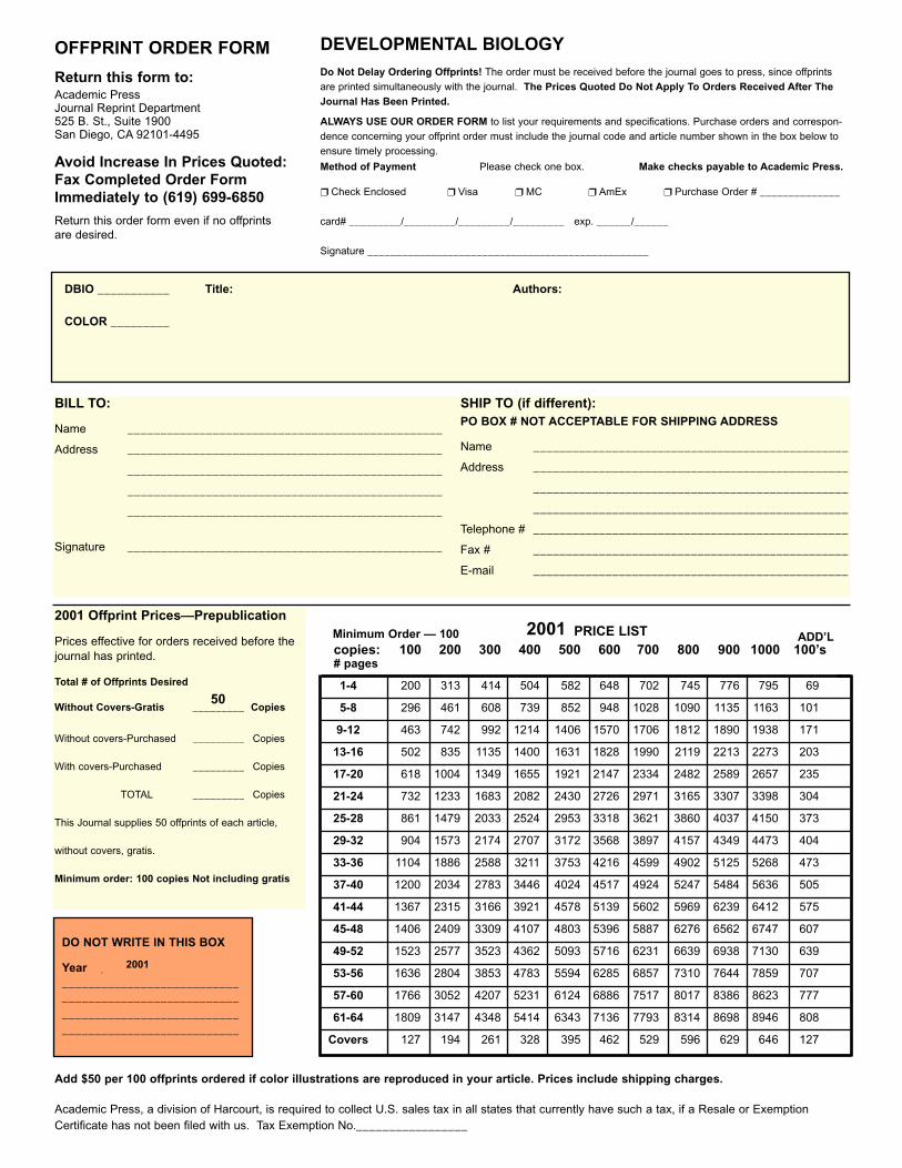
OFFPRINT ORDER FORM
Return this form to:Academic PressJournal Reprint Department525 B. St., Suite 1900San Diego, CA 92101-4495
Avoid Increase In Prices Quoted:Fax Completed Order FormImmediately to (619) 699-6850
Return this order form even if no offprintsare desired.
BILL TO:
Name ________________________________________________
Address ________________________________________________
________________________________________________
________________________________________________
________________________________________________
Signature ________________________________________________
SHIP TO (if different):PO BOX # NOT ACCEPTABLE FOR SHIPPING ADDRESS
Name ________________________________________________
Address ________________________________________________
________________________________________________
________________________________________________
Telephone # ________________________________________________
Fax # ________________________________________________
E-mail ________________________________________________
2001 Offprint Prices—Prepublication
Prices effective for orders received before thejournal has printed.
Total # of Offprints Desired
Without Covers-Gratis _________ Copies
Without covers-Purchased _________ Copies
With covers-Purchased _________ Copies
TOTAL _________ Copies
This Journal supplies 50 offprints of each article,
without covers, gratis.
Minimum order: 100 copies Not including gratis
DEVELOPMENTAL BIOLOGYDo Not Delay Ordering Offprints! The order must be received before the journal goes to press, since offprintsare printed simultaneously with the journal. The Prices Quoted Do Not Apply To Orders Received After TheJournal Has Been Printed.
ALWAYS USE OUR ORDER FORM to list your requirements and specifications. Purchase orders and correspon-dence concerning your offprint order must include the journal code and article number shown in the box below toensure timely processing.
Method of Payment Please check one box. Make checks payable to Academic Press.
❒ Check Enclosed ❒ Visa ❒ MC ❒ AmEx ❒ Purchase Order # ______________
card# _________/_________/_________/_________ exp. ______/______
Signature _________________________________________________
ADD’Lcopies: 100 200 300 400 500 600 700 800 900 1000 100’s# pages
1-4 200 313 414 504 582 648 702 745 776 795 69
5-8 296 461 608 739 852 948 1028 1090 1135 1163 101
9-12 463 742 992 1214 1406 1570 1706 1812 1890 1938 171
13-16 502 835 1135 1400 1631 1828 1990 2119 2213 2273 203
17-20 618 1004 1349 1655 1921 2147 2334 2482 2589 2657 235
21-24 732 1233 1683 2082 2430 2726 2971 3165 3307 3398 304
25-28 861 1479 2033 2524 2953 3318 3621 3860 4037 4150 373
29-32 904 1573 2174 2707 3172 3568 3897 4157 4349 4473 404
33-36 1104 1886 2588 3211 3753 4216 4599 4902 5125 5268 473
37-40 1200 2034 2783 3446 4024 4517 4924 5247 5484 5636 505
41-44 1367 2315 3166 3921 4578 5139 5602 5969 6239 6412 575
45-48 1406 2409 3309 4107 4803 5396 5887 6276 6562 6747 607
49-52 1523 2577 3523 4362 5093 5716 6231 6639 6938 7130 639
53-56 1636 2804 3853 4783 5594 6285 6857 7310 7644 7859 707
57-60 1766 3052 4207 5231 6124 6886 7517 8017 8386 8623 777
61-64 1809 3147 4348 5414 6343 7136 7793 8314 8698 8946 808
Covers 127 194 261 328 395 462 529 596 629 646 127
Minimum Order — 100
Academic Press, a division of Harcourt, is required to collect U.S. sales tax in all states that currently have such a tax, if a Resale or ExemptionCertificate has not been filed with us. Tax Exemption No._________________
DO NOT WRITE IN THIS BOX
Year ______________________________________________________________________________________________________________________
2001 PRICE LIST
2001
Add $50 per 100 offprints ordered if color illustrations are reproduced in your article. Prices include shipping charges.
DBIO ___________
COLOR _________
Title: Authors:
50
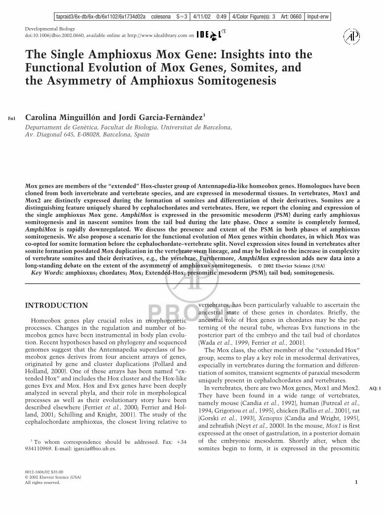
Developmental Biology 246, 000–000 (2002)doi:10.1006/dbio.2002.0660, available online at http://www.idealibrary.com on
Fn1
tapraid3/6x-db/6x-db/6x1102/6x1734d02a colesona S�3 4/11/02 0:49 4/Color Figure(s): 3 Art: 0660 Input-erw
The Single Amphioxus Mox Gene: Insights into theFunctional Evolution of Mox Genes, Somites, andthe Asymmetry of Amphioxus Somitogenesis
Carolina Minguillon and Jordi Garcia-Fernandez1
Departament de Genetica, Facultat de Biologia, Universitat de Barcelona,Av. Diagonal 645, E-08028, Barcelona, Spain
Mox genes are members of the “extended” Hox-cluster group of Antennapedia-like homeobox genes. Homologues have beencloned from both invertebrate and vertebrate species, and are expressed in mesodermal tissues. In vertebrates, Mox1 andMox2 are distinctly expressed during the formation of somites and differentiation of their derivatives. Somites are adistinguishing feature uniquely shared by cephalochordates and vertebrates. Here, we report the cloning and expression ofthe single amphioxus Mox gene. AmphiMox is expressed in the presomitic mesoderm (PSM) during early amphioxussomitogenesis and in nascent somites from the tail bud during the late phase. Once a somite is completely formed,AmphiMox is rapidly downregulated. We discuss the presence and extent of the PSM in both phases of amphioxussomitogenesis. We also propose a scenario for the functional evolution of Mox genes within chordates, in which Mox wasco-opted for somite formation before the cephalochordate–vertebrate split. Novel expression sites found in vertebrates aftersomite formation postdated Mox duplication in the vertebrate stem lineage, and may be linked to the increase in complexityof vertebrate somites and their derivatives, e.g., the vertebrae. Furthermore, AmphiMox expression adds new data into along-standing debate on the extent of the asymmetry of amphioxus somitogenesis. © 2002 Elsevier Science (USA)
Key Words: amphioxus; chordates; Mox; Extended-Hox; presomitic mesoderm (PSM); tail bud; somitogenesis.
AQ: 1
INTRODUCTION
Homeobox genes play crucial roles in morphogeneticprocesses. Changes in the regulation and number of ho-meobox genes have been instrumental in body plan evolu-tion. Recent hypotheses based on phylogeny and sequencedgenomes suggest that the Antennapedia superclass of ho-meobox genes derives from four ancient arrays of genes,originated by gene and cluster duplications (Pollard andHolland, 2000). One of these arrays has been named “ex-tended Hox” and includes the Hox cluster and the Hox-likegenes Evx and Mox. Hox and Evx genes have been deeplyanalyzed in several phyla, and their role in morphologicalprocesses as well as their evolutionary story have beendescribed elsewhere (Ferrier et al., 2000; Ferrier and Hol-land, 2001; Schilling and Knight, 2001). The study of thecephalochordate amphioxus, the closest living relative to
1 To whom correspondence should be addressed. Fax: �34934110969. E-mail: [email protected].
0012-1606/02 $35.00© 2002 Elsevier Science (USA)All rights reserved.
vertebrates, has been particularly valuable to ascertain theancestral state of these genes in chordates. Briefly, theancestral role of Hox genes in chordates may be the pat-terning of the neural tube, whereas Evx functions in theposterior part of the embryo and the tail bud of chordates(Wada et al., 1999; Ferrier et al., 2001).
The Mox class, the other member of the “extended Hox”group, seems to play a key role in mesodermal derivatives,especially in vertebrates during the formation and differen-tiation of somites, transient segments of paraxial mesodermuniquely present in cephalochordates and vertebrates.
In vertebrates, there are two Mox genes, Mox1 and Mox2.They have been found in a wide range of vertebrates,namely mouse (Candia et al., 1992), human (Futreal et al.,1994; Grigoriou et al., 1995), chicken (Rallis et al., 2001), rat(Gorski et al., 1993), Xenopus (Candia and Wright, 1995),and zebrafish (Neyt et al., 2000). In the mouse, Mox1 is firstexpressed at the onset of gastrulation, in a posterior domainof the embryonic mesoderm. Shortly after, when the
somites begin to form, it is expressed in the presomitic1

mesoderm (PSM), in the epithelial and differentiatingsomites, and in the lateral plate mesoderm. In contrast,Mox2 is not expressed before somite formation, and there-after is expressed all over the epithelial somites. Duringsomite differentiation, Mox2 expression becomes restrictedto the sclerotome and migrating myoblasts and their deriva-tive muscles in the limb buds (Candia et al., 1992; Candiaand Wright, 1996). Moreover, knockout mice for the Mox2gene show limb muscle defects, which led the authors toregard this gene as a component of the genetic hierarchycontrolling limb muscle development (Mankoo et al.,1999). In Xenopus, a single Mox gene has been isolated(XMox2; Candia and Wright, 1995). As for mouse Mox1, themesodermal-specific expression of this gene is found inundifferentiated dorsal, lateral, and ventral mesoderm, inthe posterior part of neurula/tail bud embryos, and is moreanteriorly detected in the dermatomes. In the tail budtadpole, XMox2 is expressed in tissues of the tail bud itself,a site of continuous gastrulation-like processes resulting inmesoderm formation. In chicken, two Mox genes have beenisolated. cMox2 is expressed in the somites of developingembryos, in presumptive migrating myoblasts from thedermomyotome to the limb buds, and in the ventral anddorsal parts of limb buds (Rallis et al., 2001).
In invertebrate protostomes, Mox orthologues are alsorelated to mesodermal derivatives. In Drosophila, the Moxgene buttonless (btn) is specifically expressed in 20 cells ofa single type during embryonic development, the dorsalmedial (DM) cells. These cells are located along the dorsalmidline of the bridge that links the two halves of themesoderm, which points to their mesodermal origin. Theabsence of btn gene function entails the initial commit-ment to the DM cell fate, but differentiation does not occurand DM cells are lost (Chiang et al., 1994). In the gastropodmollusc Haliotis rufescens, the Mox gene orthologue(Hrox1) is not expressed in the early embryo, and transcriptsare most prevalent during larval morphogenesis from tro-chophore to veliger. In many gastropods, the larval refractormuscle cells differentiate from the mesoderm of the earlytrochophore larva. Analysis of muscle-specific tropomyosingene expression indicates that muscle differentiation andearly Hrox1 gene expression overlap in time (Degnan et al.,1997), and may thus be linked. Mox homologues have beenfound in flatworms but there is no expression reported(Lukianov et al., 1994) and no clear Mox gene orthologuesare present in the Caenorhabditis elegans genome (The C.elegans Sequencing Consortium, 1998). A bona fide Moxgene orthologue has been reported in the cnidarian Hydramagnipapillata (Hm-Cnox5; Naito et al., 1993), but noexpression data are available.
It is now clear that numerous gene families expanded bygene duplication on the vertebrate stem lineage. Its phylo-genetic position as the sister group of vertebrates (Wada andSatoh, 1994; Cameron et al., 2000), its simple and proto-typical vertebrate-like body plan, and the preduplicative
state of its genome situate amphioxus in a privilegedposition to trace the history of a given gene family inchordates. Mox genes are particularly relevant under achordate evolutionary perspective, as they may be involvedin somite formation and differentiation in vertebrates, andsomites are an evolutionary innovation that originated justbefore the divergence of cephalochordates and vertebrates(Pourquie, 2001). Thus, the isolation and study of Mox inamphioxus may shed light on whether the ancestral role ofMox in chordates is linked to somite origin.
We isolated the single and prototypical amphioxus Moxgene. AmphiMox is the pro-orthologue of vertebrate Mox1and Mox2, and is expressed during somite formation, in thePSM and in nascent somites from the tail bud. Once asomite is completely formed, AmphiMox is rapidly down-regulated. Our results suggest that Mox was co-opted forsomite formation before the cephalochordate–vertebratesplit. Novel expression sites found in vertebrates aftersomite formation may be linked to the increase in complex-ity of vertebrate somites and their derivatives, e.g., thevertebrae.
MATERIALS AND METHODS
Gene Cloning
A 117-bp DNA fragment with similarity to the Mox homeoboxwas isolated from Branchiostoma floridae cDNA by PCR, using thedegenerate primers SO1 and SO2, which recognize the first andthird helix, respectively, of the Antennapedia-superclass ho-meobox sequence (Garcia-Fernandez and Holland, 1994). Genomicand cDNA clones containing the AmphiMox gene were isolated bya combination of library screenings and PCR. Screenings of asingle-animal genomic library (Ferrier et al., 2000) and of a larvalcDNA library (a gift of Linda Holland, Scripps Institute of Ocean-ography, San Diego) were performed in medium stringency condi-tions (60°C) in Church’s buffer (Shifman and Stein, 1995). Intronslength were determined by PCR.
Phylogenetic Analysis
The sequences used for the phylogenetic comparisons with theAmphiMox gene reported here (Accession No. AF490355) wereobtained from public databases and aligned by using ClustalX. Thehomeodomain sequences were subjected to neighbor-joining byusing the MEGA 2.0 package (www.megasoftware.net). The param-eters used underwent pairwise deletion and Poisson correction.Topology robustness was assessed by 1000 bootstrap resampling ofthe data.
Obtaining Embryos
Ripe adults of the Florida lancelet were collected from OldTampa Bay (Florida) during the summer breeding season. Males andfemales were spawned electrically in the laboratory and in vitrofertilization was performed on petri dishes. The selected develop-mental stages were raised following Holland and Holland (1993).
2 Minguillon and Garcia-Fernandez
© 2002 Elsevier Science (USA). All rights reserved.
tapraid3/6x-db/6x-db/6x1102/6x1734d02a colesona S�3 4/11/02 0:49 4/Color Figure(s): 3 Art: 0660 Input-erw

Whole-Mount in Situ Hybridization and Sectioning
In situ whole-mount hybridizations on amphioxus embryos andlarvae were performed as described by Holland et al. (1996). A1200-bp EcoRI fragment of the AmphiMox cDNA (lacking most ofthe trailer sequence) was used for in vitro transcription withdigoxigenin-labeled dUTP. After hybridization and whole-mountphotography, selected embryos were contrasted in 1% Poinceau S,1% acetic acid, dehydrated through an ethanol series, and embed-ded in LR White medium (TAAB) resin. Serial 3-�m sections wereobtained with a glass knife, mounted in DePeX, and photographedunder Nomarski optics.
RESULTS
Isolation and Characterization of AmphiMox
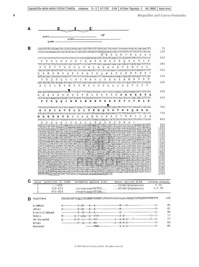
A PCR survey of amphioxus embryo cDNA with “uni-versal” Antennapedia-like homeobox primers yielded theisolation of Hox and ParaHox genes, plus a fragment similarto the Mox class of homeobox genes. The AmphiMox genewas further characterized by cDNA and genomic screenings(Fig. 1).
The genomic screening with the PCR fragment wasperformed in medium stringency conditions to isolate anygene belonging to the same or related subfamilies of ho-meoboxes. Three of six positive clones contained otherhomeobox genes, while the strongest positives encom-passed the genomic region of AmphiMox (Fig. 1A). cDNAscreening of a larval library gave two positive clones of 2.6kb. The whole cDNA sequence and the predicted proteinsequence of AmphiMox are shown in Fig. 1B. Further PCRstrategies and selected genomic sequencing allowed thecharacterization of the genomic organization of the gene.AmphiMox putatively codes for a 240-amino-acid proteinand is organized in three exons and two large introns ofabout 6 and 5.5 kb (Fig. 1C). For the species of knowngenomic organization but for Drosophila, this structure isconserved (Chiang et al., 1994). The second intron is withinthe homeobox, between residues 44 and 45 of the home-odomain (Fig. 1B, triangles). This position bears an intron inmany homeobox genes, which may reflect the ancestralcondition of all homeobox genes or particular families(Burglin, 1995). Alternatively, the specific nucleotide se-quence of the homeobox here (residues QV, encoded byCARGTN) may be a hot spot for intron insertion.
Southern blots of genomic DNA obtained from singleindividuals were hybridized with a cDNA probe that de-tects exons 1 and 2 of the AmphiMox gene. One or two largebands were detected in each individual DNA, consistentwith AmphiMox being a single copy gene (data not shown).
Blast searches against databases and comparison of thededuced amino acid sequence of AmphiMox with otherMox proteins confirmed that it belongs to the Mox familyof homeobox-containing genes (Fig. 1D). Beyond the home-odomain, sequence similarities of AmphiMox to vertebrate
and invertebrate sequences are restricted to scattered resi-dues (data not shown).
Phylogenetic Analysis
Several Mox genes have been isolated from various spe-cies. The homeodomain of AmphiMox is more closelyrelated to that of vertebrate Mox genes (85–90%) than tothose of other invertebrate genes (77–84%) (Fig. 1D). Togain more insight into the relationships among Mox genes,we conducted a molecular phylogenetic analysis by theneighbor-joining method on the homeodomain of Moxproteins, using Hox-4 sequences as outgroups (Fig. 2). Ver-tebrate Mox proteins fell into two groups: Mox1 and Mox2.AmphiMox branches immediately outside these groups (astheir sister group), between the vertebrate genes and therest of invertebrate genes. The particular grouping of proto-stome genes does not agree with current ecdysozoa/lophotrocozoa clades, but the bootstrap values supportingthese invertebrate protein groupings are very low. Thepositioning of AmphiMox, before the origin of the verte-brate groups 1 and 2, is supported by a high bootstrap value(74%; see Fig. 4), in agreement with the hypothesis thatvertebrate genes have originated by duplication after thecephalochordate–vertebrate divergence. When we omittedthe Drosophila and flatworm Mox homeodomains (as diver-gent and relatively long-branched sequences can disrupt thetree topology), the position of AmphiMox as the sistergroup of vertebrate Mox genes was further supported (boot-strap value raises to 88%; data not shown). Thus, Amphi-Mox may well represent a prototypical direct descendant ofthe preduplicative gene prior to vertebrate-specific duplica-tions.
AmphiMox Gene Expression
AmphiMox expression is intimately linked to somiteformation. Somitogenesis in amphioxus can be divided intoan early phase, in which somites originate from the out-pocketing of the archenteron (Fig. 3G), and a late phase, inwhich somites arise as solid blocks directly from theproliferating tail bud (Fig. 3J) (Schubert et al., 2001; andreferences therein).
In whole-mount in situ hybridizations, no signal wasdetected prior to hatching. The signal was first visible in theearly posthatching neurula (5-somite stage; Fig. 3A), in thenewly formed somite (s5). At this stage, AmphiMox wasalso expressed in the most anterior part of the PSM, in aregion comprising about one somite in length.
Somitogenesis in amphioxus is particularly asymmetri-cal. Left somites are formed earlier than right somites andtherefore become located slightly rostral (Cerfontaine,1906). This asymmetry was mirrored by AmphiMox expres-sion. In a dorsal view of 5- to 6-somite embryos (Fig. 3B),somites number 5 on each side are morphologically visible(s5) and the posterior boundaries are clearly discernible
3AmphiMox Gene
F1
F2
F3
© 2002 Elsevier Science (USA). All rights reserved.
tapraid3/6x-db/6x-db/6x1102/6x1734d02a colesona S�3 4/11/02 0:49 4/Color Figure(s): 3 Art: 0660 Input-erw
4 Minguillon and Garcia-Fernandez
© 2002 Elsevier Science (USA). All rights reserved.
tapraid3/6x-db/6x-db/6x1102/6x1734d02a colesona S�3 4/11/02 0:49 4/Color Figure(s): 3 Art: 0660 Input-erw

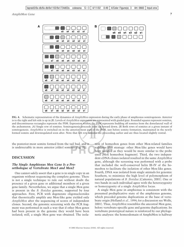
(arrowheads). However, the AmphiMox signal is asym-metrical: Mox RNA is detected in the right somite 5, but nolonger in the left one, which has been formed earlier. Leftand right somites 5 are morphologically similar, but the leftone is older and has already shut-down Mox expression,whereas the right one has not silenced Mox yet. In amagnification (Fig. 3C), the posterior border of left somite 6can be observed (arrowhead). Nevertheless, somite 6 is notyet morphologically visible on the right side. AmphiMoxwas expressed both in the newly formed left somite 6 butalso in the anterior right PSM, which gives rise to rightsomite 6. The picture emerging is that Mox is expressed inthe anterior-most part of the PSM, maintained in theforming somite, and downregulated shortly after the overtdifferentiation of the somite (schematized in Fig. 4A).
To further localize the expression of AmphiMox, weperformed transverse sections of prestained embryos of an
age similar to that of the specimen shown in Fig. 3B.Section through level i in Fig. 3C shows AmphiMox expres-sion in the formed left somite 6 and in the forming rightsomite 6 (Fig. 3D; schematized in Fig. 3E). Section througha more posterior level (ii in Fig. 3C) reveals the outpocket-ing of the right side of the archenteron, to give rise a newsomite. Note that Mox is already expressed there (Fig. 3F;schematized in Fig. 3G). In contrast, in the left side, thearchenteron has not yet initiated the outpocketing to formthe next left somite. Accordingly, Mox is still not expressed(Fig. 3G; schematized in Fig. 4B).
In 18-h embryos (11- to 12-somite stage), somites areformed directly from the tail bud. Mox is strongly expressedin the newly formed somite 11 on the right side of theembryo (Fig. 3H). The posterior border of the somite isdiscernible (arrowhead). In addition, residual levels of ex-pression were detected in somite 10. An optical sectionthrough level x in Fig. 3H distinguishes between right andleft sides of the tail bud area (Fig. 3I; schematized in Fig. 3J).Left somite 12 has just formed (s12l) and strongly expressesMox. Right somite 11 (s11r, which is slightly older) stillexpresses Mox. The future right somite 12 begins to detachfrom the right side of the tail bud (asterisk), and alsoexpresses Mox, but to a lesser extent than its left counter-part (s12l), that was built in advance.
From our data, AmphiMox was not expressed in the tailbud itself, but in nascent somites deriving from its mostanterior part. Late phase somite formation in amphioxus isslowed down with respect to the early phase. If AmphiMoxis not expressed in the tail bud itself, but in nascentsomites, in a certain moment neither a right nor a leftsomite will be forming. In that particular instant, Moxsignal would then be confined to already formed somites onboth sides, but it would not be detectable in-between. Thisholds true, as shown by the dorsal view of an embryoslightly older than that in Fig. 3I, in which two somites arelabeled for Mox expression in the right side (the anterior oneweakly), and only one somite in the left side (Fig. 3K).Remarkably, no signal was detected in any midline struc-ture.
In larval stages, AmphiMox behaves according to thedynamics described above. The signal is detected only in
FIG. 1. (A) Genomic organization of the B. floridae AmphiMox gene. The coding region is shown in gray and the homeobox is shown inblack. The introns are represented by dotted lines. Horizontal lines represent the phage clones encompassing the genomic region. (B)Nucleotide and deduced amino acid sequence of AmphiMox cDNA (GenBank Accession No. AF490355). The first in-frame methionine andthe noncanonical polyadenylation signal are underlined. The 60 residues of the homeodomain are shown in bold, and the intron positionsare indicated by black triangles. (C) Conservation of the acceptor/donor splice sites. Exon positions are shown according to the cDNAnucleotide numeration. Capital and small letters correspond to exon and intron sequences, respectively. (D) Alignment of the AmphiMoxhomeodomain with other Mox homeodomains. Dashes indicate identity to the AmphiMox sequence. The numbers on the right representthe percentage of identity to the AmphiMox sequence. Identity to the partial cnidarian Mox homeodomain refers to the available residues.The abbreviations used for species and genes are: Dutarh2, Girardia tigrina; Hm-cnox5, Hydra magnipapillata; Hrox1, Haliotis rufescens;m, mouse; r, rat; h, human; X, Xenopus laevis; c, chicken; Dm, Drosophila melanogaster. Sequences were obtained from public databases.
FIG. 2. Neighbor-joining phylogenetic tree relating the homeodo-main of AmphiMox protein with that of other available Moxproteins. Sequence and species abbreviations are the same as in Fig.1. The tree is rooted by using the AmphiHox4 and the mouseHoxC4 homeodomain sequences. The numbers refer to bootstrapvalues over 1000 replicates.
5AmphiMox Gene
F4
© 2002 Elsevier Science (USA). All rights reserved.
tapraid3/6x-db/6x-db/6x1102/6x1734d02a colesona S�3 4/11/02 0:49 4/Color Figure(s): 3 Art: 0660 Input-erw
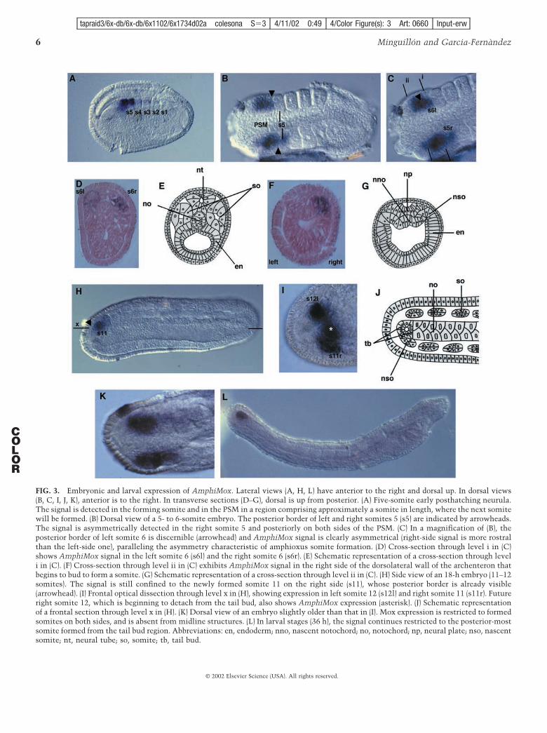
FIG. 3. Embryonic and larval expression of AmphiMox. Lateral views (A, H, L) have anterior to the right and dorsal up. In dorsal views(B, C, I, J, K), anterior is to the right. In transverse sections (D–G), dorsal is up from posterior. (A) Five-somite early posthatching neurula.The signal is detected in the forming somite and in the PSM in a region comprising approximately a somite in length, where the next somitewill be formed. (B) Dorsal view of a 5- to 6-somite embryo. The posterior border of left and right somites 5 (s5) are indicated by arrowheads.The signal is asymmetrically detected in the right somite 5 and posteriorly on both sides of the PSM. (C) In a magnification of (B), theposterior border of left somite 6 is discernible (arrowhead) and AmphiMox signal is clearly asymmetrical (right-side signal is more rostralthan the left-side one), paralleling the asymmetry characteristic of amphioxus somite formation. (D) Cross-section through level i in (C)shows AmphiMox signal in the left somite 6 (s6l) and the right somite 6 (s6r). (E) Schematic representation of a cross-section through leveli in (C). (F) Cross-section through level ii in (C) exhibits AmphiMox signal in the right side of the dorsolateral wall of the archenteron thatbegins to bud to form a somite. (G) Schematic representation of a cross-section through level ii in (C). (H) Side view of an 18-h embryo (11–12somites). The signal is still confined to the newly formed somite 11 on the right side (s11), whose posterior border is already visible(arrowhead). (I) Frontal optical dissection through level x in (H), showing expression in left somite 12 (s12l) and right somite 11 (s11r). Futureright somite 12, which is beginning to detach from the tail bud, also shows AmphiMox expression (asterisk). (J) Schematic representationof a frontal section through level x in (H). (K) Dorsal view of an embryo slightly older than that in (I). Mox expression is restricted to formedsomites on both sides, and is absent from midline structures. (L) In larval stages (36 h), the signal continues restricted to the posterior-mostsomite formed from the tail bud region. Abbreviations: en, endoderm; nno, nascent notochord; no, notochord; np, neural plate; nso, nascentsomite; nt, neural tube; so, somite; tb, tail bud.
6 Minguillon and Garcia-Fernandez
COLOR
© 2002 Elsevier Science (USA). All rights reserved.
tapraid3/6x-db/6x-db/6x1102/6x1734d02a colesona S�3 4/11/02 0:49 4/Color Figure(s): 3 Art: 0660 Input-erw
the posterior-most somite formed from the tail bud, and itis undetectable in more anterior (older) somites (Fig. 3L).
DISCUSSION
The Single Amphioxus Mox Gene Is a Pro-orthologue of Vertebrate Mox1 and Mox2
One cannot safely assert that a gene is in single copy in anorganism without sequencing the complete genome. Thereis not a single technique to rule out without doubt thepresence of a given gene or additional members of a givengene family. Nevertheless, we argue that a single Mox geneis present in the B. floridae genome, supported by fourapproaches. First, PCR with degenerate oligonucleotidesthat theoretically amplify any Mox-like gene yielded onlyAmphiMox after the sequencing of scores of independentclones. Second, the genomic screening with the PCR frag-ment was performed in such a way that if other Mox geneshad been present in the genome they would have beenisolated; still, a single Mox gene was obtained. The isola-
tion of homeobox genes from other Mox-related familiesvalidates this strategy: other Mox-like genes would havebeen isolated as they would be more similar to the probeused (Mox homeobox fragment). Third, the two indepen-dent cDNA clones isolated resulted in the same AmphiMoxgene, although the screening was performed with a probethat included the well-conserved helix III–IV of the ho-meobox to facilitate the isolation of other Mox-like genes.Fourth, DNA was isolated from single animals for genomicSouthern, to minimize the high level of polymorphism ofnatural populations of B. floridae (Canestro, 2001). One ortwo bands in each individual agree with the heterozygosityor homozygosity of a single AmphiMox locus.
A single Mox gene in amphioxus is consistent with thepresumed preduplicative state of the amphioxus genome,which preceded genome duplications at the base of verte-brate origin (Holland et al., 1994; for a discussion see Wolfe,2001). Thus, AmphiMox resembles the ancestral Mox gene,before vertebrate-specific gene and genome duplications. Itsvertebrate prototypical nature is reinforced by our phyloge-netic analyses: the homeodomain of AmphiMox is halfway
FIG. 4. Schematic representation of the dynamics of AmphiMox expression during the early phase of amphioxus somitogenesis. Anterioris to the right and left side is up in (B). Levels of AmphiMox expression are represented with graded gray. Rounded squares represent somites,and discontinuous rectangles represent the PSM. Expression within the PSM represents budding off somites from the dorsolateral wall ofthe archenteron. (A) Single row of somites. Somitogenesis proceeds from up toward down. (B) Both rows of somites at a given instant ofsomitogenesis. AmphiMox is switched on in the anterior most part of the PSM, just before somite formation, maintained in the newlyformed somite and downregulated soon after. Note that left row somites are proceeding earlier and are thus located slightly rostral.
7AmphiMox Gene
© 2002 Elsevier Science (USA). All rights reserved.
tapraid3/6x-db/6x-db/6x1102/6x1734d02a colesona S�3 4/11/02 0:49 4/Color Figure(s): 3 Art: 0660 Input-erw
between those of vertebrates and other invertebrates, andclearly emerges as the sister group of vertebrate Mox1 andMox2. Hence, AmphiMox may well be a direct descendantof the ancestral Mox gene before vertebrate gene duplica-tion and shed light on the ancestral function of Mox genesin the lineage leading to vertebrates.
Amphioxus Somitogenesis and the Extentof Amphioxus PSM
The muscular segments of amphioxus are the mostevident segmented structures of the adult. They comprise asingle row of muscular somites along either side of thenotochord. The coelomic cavities of the most anteriorsomites of amphioxus are formed by enterocoely by out-pocketing of the dorsolateral wall of the archenteron (Fig.3G). This early paraxial mesoderm is formed during gastru-lation and is used until the end of early somitogenesis. Thenumber of somite pairs formed in this phase ranges from 8in B. floridae (Holland et al., 1997) to 14 in B. lanceolatum(Conklin, 1932). More caudal somites are formed by schizo-coely (more similarly to vertebrates), by a splitting processof solid blocks from the proliferating tail bud. Early am-phioxus somitogenesis is reminiscent of the formation ofthe trunk anterior somites of fish and amphibians, from thesequential segmentation of a preexisting territory that in-volutes during gastrulation, and not from dividing stemcells in the node/primitive streak of amniotes (for furtherdiscussion and views, see Pourquie, 2001). The second-phase somites of amphioxus originate from proliferatingstem cells in the tail bud, as in vertebrates.
AmphiMox is expressed before the formation of thesomite, from somite 5 onwards, maintained in the formingsomite and downregulated shortly after the overt segmen-tation of the somite, regardless of their origin. VertebrateMox genes are conspicuously expressed in the PSM. Verte-brate PSM comprises several somites in length. In chicken,between the entry of a cell from the stem cell population ofthe rostral primitive streak or the tail bud to the PSM andits incorporation into a somite, it experiences 12 oscilla-tions of cycling gene expression (Palmeirim et al., 1997;reviewed in Maroto and Pourquie, 2001). During the earlyphase of amphioxus somitogenesis, the dorsolateral wall ofthe archenteron is the PSM itself, where AmphiMox isexpressed in an anteroposterior sequence (schematized inFig. 4A). In contrast, no such territory is found in am-phioxus tail bud-derived somites, since Mox-positive cellsarise directly from the tail bud. This is consistent withAmphiWnt5 expression in the tail bud and nascent somites(Schubert et al., 2001) and suggests that the patterningphenomena that occur in vertebrate PSM should take placein the amphioxus tail bud itself, or in small compartmentsof it.
AmphiMox is expressed similarly in both anterior-mostand posterior-most somites, regardless of their origin, withthe exception of the first four somites, in which Mox
expression is not detected. These somites are unique in thatthey bud off at once from the archenteron, and not in ananteroposterior sequence. Their appearance has also beendiscussed by classical morphologists. Hatschek (1878) ob-served embryos with a single pair of somites, whereasConklin (1932) never detected them and claimed thatalways more than one somite was present in all the em-bryos he analyzed. Mox may not be involved in theseparticular somites. Alternatively, we may have missed theshort period between Mox activation and silencing in thesesomites, although we carefully examined large numbers ofyounger embryos. In summary, our data indicate that Am-phiMox is expressed before the formation of the somite atleast from somite 5 onwards, suggesting that the mecha-nisms involved in the early specification of the somites areshared by both types regardless of their origin, be it gut wallor tail bud.
Asymmetry of Amphioxus Somitogenesis fromSomite 5 Onwards, as Revealed by AmphiMoxGene Expression
Another subject of discussion among classical amphioxusmorphologists was the asymmetry of amphioxus somites.In this regard, Conklin and Hatschek agreed that somitesare roughly symmetrical in position until the 7- or 8-somitestage. Notwithstanding, Cerfontaine (1906) postulated thatthe asymmetry is present from the first pair, with a slightdelay in the development of the right side with respect tothat of the left side. Our results do not resolve the issue ofasymmetry in the very early somites (1–4), but they do forsomite 5 onwards, in agreement with Cerfontaine observa-tions, as the asymmetry is already detected in a 5- to6-somite embryo (Fig. 3B). In this embryo, left-side somitesare slightly further forward with respect to the right-sideones and the AmphiMox expression signal is asymmetrical:left somite 5 has switched off Mox, whereas transcripts arestill detectable on the right somite 5, indicating that theformer has been formed slightly earlier, and is thus locatedmore anteriorly.
A Proposed Scenario for Mox Functional Evolutionwithin the Chordates
The use of molecular markers for tracing homologies andgaining evolutionary insights is controversial, especiallywhen comparisons concern distantly related taxa. More-over, the data on protostome Mox gene expression arescarce and do not allow sensible conjectures concerning aconserved role of this gene class across metazoans. The onlycommon feature is a loose relation of Mox genes to meso-dermal derivatives (Chiang et al., 1994; Degnan et al.,1997).
In contrast, when the overall body plans of two animalsare relatively similar, body part homologies can be con-firmed by developmental gene expression domains, which
8 Minguillon and Garcia-Fernandez
© 2002 Elsevier Science (USA). All rights reserved.
tapraid3/6x-db/6x-db/6x1102/6x1734d02a colesona S�3 4/11/02 0:49 4/Color Figure(s): 3 Art: 0660 Input-erw
have properties of special quality and relative position(Holland and Holland, 1999). This reasoning harmonizeswell with the case of cephalochordates vs vertebrates. Theprototypical vertebrate-like body plan of amphioxus and itspreduplicative vertebrate-like genome have fashioned theuse of amphioxus as a reference for establishing the ances-tral role of a given gene within chordates. This has been afruitful venture (Holland, 1999). In addition, segmentationwithin the paraxial mesoderm to form somites is a distin-guishing feature uniquely shared by cephalochordates andvertebrates (Saga and Takeda, 2001). In the latter, mesoder-mal expression is also characteristic of Mox genes, whichare mainly involved in somitogenesis and the formation ofsomite derivatives. Thus, Mox genes may participate in theearly specification of somites, before overt and terminaldifferentiation of their derivatives. However, neither Mox1nor Mox2 knockout mice confirm this hypothesis. Mox2knockout mice show alterations only in limb musculature(Mankoo et al., 1999) and no alterations in the axialskeleton. Mox1 knockout mice show vertebral abnormali-ties, like hemi-vertebrae, tail kinks and craniovertebralfusions (cited in Stamataki et al., 2001). Thus, these genesdo not seem to be critical for early somite determinationbut rather for late somite differentiation. Interestingly,AmphiMox is turned on in the PSM, just before the forma-tion of a new somite, but is switched off soon after thesomite border is formed (Fig. 4A). This points to similaritiesbut also differences with respect to vertebrate Mox genes.The amphioxus data help unravel the ancestral functionand co-option of Mox genes within vertebrates. First, ourexpression data suggest that the original function in thecommon ancestor of cephalochordates and vertebrates wasat the onset of somite specification and formation. It istempting to speculate that activation of Mox in a suitableterritory of the embryonic mesoderm was coincident withor causal to the evolutionary design of somites. Second, theexpression of Mox genes in vertebrates during somite dif-ferentiation is not detected in amphioxus, which may bedue to the simpler structure of cephalochordate somitescompared with the increased level of complexity of verte-brate somites and their derivatives.
Vertebrate somites undergo complex transformations,like epithelial to mesenchyme transition, delamination ofcells from the dermamyotome and migration of those toform the limb musculature. In contrast, amphioxus is apredominantly epithelial animal (Whittaker, 1997). Earlysomites arise as evaginations from the gut walls that pinchoff, resulting in a single-layered epithelium surrounding acavity, the myocoel. The somites of amphioxus are subdi-vided into distinct regions, although there is no histologicalevidence for sclerotome and no cells migrate away from it(Shimeld and Holland, 2000). On the contrary, the largemedial compartment of each somite is the myotome, whichretains its epithelial organization throughout development.All myotomal cells differentiate in place and become the
striated muscle cells that constitute the segmental muscleblocks along the body length (Holland et al., 1995a). Moxgenes were probably co-opted after duplication at the originof vertebrates, leading to or facilitating the acquisition ofvertebrate-specific roles after somite formation. This co-option and diversification was refined independently oneach Mox gene (Mox1 and Mox2), giving rise to theirspecific and unique roles, highlighted in their respectiveknockout phenotypes. Genes other than Mox are alsoexpressed in the already formed somites in vertebrates butnot in amphioxus [e.g., Amphisnail (Langeland et al., 1998)and AmphiPax1/9 (Holland et al., 1995b)]. Notably, theMox1 protein binds to the Pax1 protein in vitro (Stamatakiet al., 2001). The expression pattern of these genes invertebrates and the phenotype of the mutants are alsoconsistent with the hypothesis of functional association ofthe gene products in vivo. However, AmphiPax1/9 andAmphiMox cannot cooperate in vivo, as they are notcoexpressed at any developmental stage. Therefore, theassembly of both gene products occurred after gene dupli-cations in the vertebrate stem lineage. This is compatiblewith the view that complete gene networks are co-opted,and with new networks being built up after gene or genomeduplications, thus increasing the complexity of vertebratesomites.
The above proposed scenario for Mox functional evolu-tion can be tested experimentally. First, it predicts thatMox1/Mox2 double knockout mice will overcome verte-brate gene redundancy and reveal the ancestral function ofMox genes. Thus, somitogenesis will be severely disruptedin these mutants. Second, the sister group of cephalochor-dates and vertebrates, urochordates, would not express Moxin the progenitors of muscle cells. Ascidians do not formproper somites but muscle cells determined by maternalfactors (Satoh, 1994). The absence of overt segmentation inascidians may be a secondary derived condition, since inanother group of urochordates, larvaceans, each muscle cellis innervated by reiterated neuronal cells and so mayrepresent one segment. We failed to find Mox-like genes inascidian genome project public databases. From tunicates tocephalochordates to vertebrates, there is an increasing levelof mesodermal complexity, from muscle iteration, to properbut simple somites, to highly differentiated somites, andformation of vertebral tissue and other somite derivatives.Mox may well have been involved in this gradation: a singleMox gene co-opted in suitable mesodermal territories, andduplicated genes individually co-opted after gene duplica-tion at vertebrate origins.
ACKNOWLEDGMENTS
We thank Linda Holland for the cDNA library gift and RicardAlbalat and Roser Gonzalez-Duarte for sharing Southern blots. Wealso thank Ignasi Alcon, Cristian Canestro, Inaki Ruiz, SebShimeld, Dave Ferrier, and the “Alter-Epi” team for help and
9AmphiMox Gene
© 2002 Elsevier Science (USA). All rights reserved.
tapraid3/6x-db/6x-db/6x1102/6x1734d02a colesona S�3 4/11/02 0:49 4/Color Figure(s): 3 Art: 0660 Input-erw

discussions, and Robin Rycroft and Ivana Mino for checking theEnglish version of the manuscript. We are indebted to the Depart-ment of Biology at the University of South Florida, Tampa, and toRay Martınez for hosting and help during the amphioxus breedingseason. This study was supported by a grant from DGESIC PB98-1261-C02-02 (Ministerio de Ciencia y Tecnologıa, Spain). C.M.holds a CIRIT (Generalitat de Catalunya) predoctoral fellowship.Collaboration between J.G-F. and Seb Shimeld was facilitated bygrants from the Acciones Integradas of the Bristish Council/Ministerio de Ciencia y Tecnologıa.
REFERENCES
Burglin, T. R. (1995). The evolution of homeobox genes. In “Biodi-versity and Evolution” (R. Arai, M. Kato, and Y. Doi, Eds.), pp.291–336. The National Science Museum Foundation, Tokyo.
Cameron, C. B., Garey, J. R., and Swalla, B. J. (2000). Evolution ofthe chordate body plan: New insights from phylogenetic analysesof deuterostome phyla. Proc. Natl. Acad. Sci. USA 97, 4469–4474.
Candia, A. F., Hu, J., Crosby, J., Lalley, P. A., Noden, D., Nadeau,J. H., and Wright, C. V. E. (1992). Mox-1 and Mox-2 define a novelhomeobox gene subfamily and are differentially expressed duringearly mesodermal patterning in mouse embryos. Development116, 1123–1136.
Candia, A. F., and Wright, C. V. E. (1995). The expression pattern ofXenopus Mox-2 implies a role in initial mesodermal differentia-tion. Mech. Dev. 52, 27–36.
Candia, A. F., and Wright, C. V. E. (1996). Differential localizationof Mox-1 and Mox-2 proteins indicates distinct roles duringdevelopment. Int. J. Dev. Biol. 40, 1179–1184.
Canestro, C. (2001). Caracteritzacio molecular i evolutiva de lafamılia de les Alcohol Deshidrogenases de cadena mitjana alscordats. Ph. D. Thesis. University of Barcelona.
Cerfontaine, P. (1906). Recherches sur le developpement del’Amphioxus. Arch. de Biol., T. 22.
Chiang, C., Patel, N. H., Young, K. E., and Beachy P. A. (1994). Thenovel homeodomain gene buttonless specifies differentiationand axonal guidance functions of Drosophila dorsal median cells.Development 120, 3581–3593.
Conklin, E. G. (1932). The embryology of amphioxus. J. Morphol.54, 69–151.
Degnan, B. M., Degnan, S. M., Fentenany G., and Morse, D. E.(1997). A Mox homeobox gene in the gastropod mollusc Haliotisrufescens is differentially expressed during larval morphogenesisand metamorphosis. FEBS Lett. 411, 119–122.
Ferrier, D. E. K., Minguillon, C., Holland P. W. H., and Garcia-Fernandez, J. (2000). The amphioxus Hox cluster: Deuterostomeposterior Flexibility and Hox 14. Evol. Dev. 5, 284–293.
Ferrier, D. E. K., and Holland, P. W. H. (2001). Ancient origin of theHox gene cluster. Nat. Rev. Genet. 2, 33–38.
Ferrier, D. E. K., Minguillon, C., Cebrian, C., and Garcia-Fernandez,J. (2001). Amphioxus. Evx genes: Implications for the evolutionof the Midbrain-Hindbrain Boundary and the chordate tailbud.Dev. Biol. 237, 270–281.
Futreal, P. A., Cochran, C., Rosenthal, J., Miki, Y., Swenson, J.,Hobbs, M., Bennett, L. M., Haugen-Strano, A., Marks, J., Barrett,J. C., Tavtigian, S. V., Shattuck-Eidens, D., Kamb, A., Skolnick,M., and Wiseman, R. W. (1994). Isolation of a divergent ho-
meobox gene, MOX1, from the BRCA1 region on 17q21 bysolution hybrid capture. Hum. Mol. Genet. 3, 1359–1364.
Garcia-Fernandez, J., and Holland, P. W. H. (1994). Archetypalorganization of the amphioxus Hox gene cluster. Nature 370,563–566.
Gorski, D. H., LePage, D. F., Patel, C. V., Copeland, N. G., Jenkins,N. A., and Walsh, K. (1993). Molecular cloning of a divergedhomeobox gene that is rapidly down-regulated during the G0/G1transition in vascular smooth muscle cells. Mol. Cell. Biol. 13,3722–3733.
Grigoriou, M., Kastrinaki, M. C., Modi, W. S., Theodorakis, K.,Mankoo, B., Pachnis, V., and Karagogeos, D. (1995). Isolation ofthe human MOX2 homeobox gene and localization to chromo-some 7p22.1-p21.3. Genomics 26, 550–555.
Hatschek, B. (1893). The amphioxus and its development. SwanSonnenschein, London.
Holland, L. Z., Pace, D. A., Blink, M. L., Kene, M., and Holland,N. D. (1995a). Sequence and expression of amphioxus alkalimyosin light chain (AmphiMLC-alc) throughout development:Implications for vertebrate myogenesis. Dev. Biol. 171, 665–676.
Holland, L. Z., Holland, P. W. H., and Holland, N. D. (1996).Revealing homologies of distantly related animals by in situhybridization to developmental genes: Amphioxus versus verte-brates. In “Molecular Zoology: Advances, Strategies and Proto-cols” (J. D. Ferraris and S. R. Palumbi, Eds.), pp. 267–282,473–483. Wiley, New York.
Holland, L. Z., Kene, M., Williams, N. A., and Holland, N. D.(1997). Sequence and embryonic expression of the amphioxusengrailed gene (AmphiEn): The metameric pattern of transcrip-tion resembles that of its segment-polarity homolog in Drosoph-ila. Development 124, 1723–1732.
Holland, N. D., and Holland, L. Z. (1993). Embryos and larvae ofinvertebrate deuterostomes. In “Essential Developmental Biol-ogy: A Practical Approach” (C. D. Stern and P. W. H. Holland,Eds.) pp. 21–32. IRL Press, Oxford.
Holland, N. D., Holland, L. Z., and Kozmik, Z. (1995b). Anamphioxus Pax gene, AmphiPax-1, expressed in embryonicendoderm, but not in mesoderm: Implications for the evolutionof class I paired box genes. Mol. Mar. Biol. Biotechnol. 4,206–214.
Holland, N. D., and Holland, L. Z. (1999). Amphioxus and theutility of molecular genetic data for hypothesizing body parthomologies between distantly related animals. Am. Zool. 39,630–640.
Holland, P. W. H., Garcia-Fernandez, J., Williams, N. A., andSidow, A. (1994). Gene duplications and the origins of vertebratedevelopment. Dev. Suppl., 125–133.
Holland, P. W. H. (1999). The future of evolutionary developmentalbiology. Nature 402(Suppl), C41–C44.
Langeland, J. A., Tomsa, J. M., Jackman, W. R, and Kimmel, C. B.(1998). An amphioxus snail gene: Expression in paraxial meso-derm and neural plate suggests a conserved role in patterning thechordate embryo. Dev. Genes Evol. 208, 569–577.
Lukianov, K. A., Tarabykin, V. S., Potapov, V. K., Bekman, E. P.,and Lukianov, S. A. (1994). The cloning of fragments of ho-meobox genes expressed during planarian regeneration. Onto-genez 25, 28–32.
Mankoo, B. S., Collins, N. S., Ashby, P., Grigorieva, E., Pevny,L. H., Candia, A., Wright, C. V. E., Rigby, P. W. J., and Pachnis,V. (1999). Mox2 is a component of the genetic hierarchy control-ling limb muscle development. Nature 400, 69–73.
10 Minguillon and Garcia-Fernandez
© 2002 Elsevier Science (USA). All rights reserved.
tapraid3/6x-db/6x-db/6x1102/6x1734d02a colesona S�3 4/11/02 0:49 4/Color Figure(s): 3 Art: 0660 Input-erw

Maroto, M., and Pourquie, O. (2001). A molecular clock involved insomite segmentation. Curr. Top. Dev. Biol. 51, 221–248.
Naito, M., Ishiguro, H., Fujisawa, T., and Kurosawa, Y. (1993).Presence of eight distinct homeobox-containing genes in cnidar-ians. FEBS Lett. 333, 271–274.
Neyt, C., Jagla, K., Thisse, B, Haines, L., and Currie, P. D. (2000).Evolutionary origins of vertebrate appendicular muscle. Nature408, 82–86.
Palmeirim, I., Henrique, D., Ish-Horowicz, D., and Pourquie, O.(1997). Avian hairy gene expression identifies a molecular clocklinked to vertebrate segmentation and somitogenesis. Cell 91,639–648.
Pollard, S. L., and Holland, P. W. H. (2000). Evidence for 14homeobox gene clusters in human genome ancestry. Curr. Biol.10, 1059–1062.
Pourquie, O. (2001). Vertebrate somitogenesis. Annu. Rev. CellDev. Biol. 17, 311–350.
Rallis, C., Stamataki, D., Pontikakis, S., Mankoo, B. S., andKaragogeos, D. (2001). Isolation of the avian homologue of thehomeobox gene Mox2 and analysis of its expression pattern indeveloping somites and limbs. Mech. Dev. 104, 121–124.
Saga, Y., and Takeda, H. (2001). The making of the somite:Molecular events in vertebrate segmentation. Nat. Rev. Genet. 2,835–845.
Satoh, N. (1994). “Developmental Biology of Ascidians. Develop-mental and Cell Biology Series 29” (P. W. Barlow, D. Bray, andJ. M. W. Slack, Eds.). Cambridge Univ. Press, Cambridge.
Schilling T. F., and Knight R. D. (2001). Origins of anteroposteriorpatterning and Hox gene regulation during chordate evolution.Philos. Trans. R. Soc. Lond. B Biol. Sci. 356, 1599–1613.
Schubert, M., Holland, L. Z., Stokes, M. D., and Holland N. D.(2001). Three amphioxus Wnt genes (AmphiWnt3, AmphiWnt5,
and AmphiWnt6) associated with the Tail Bud: The evolution ofsomitogenesis in chordates. Dev. Biol. 240, 262–273.
Shifman, M. I., and Stein, D. G. (1995). A reliable and sensitivemethod for non-radioactive northern blot analysis of nervegrowth factor mRNA from brain tissues. J. Neurosci. Methods59, 205–208.
Shimeld, S. M., and Holland, P. W. H. (2000). Vertebrate innova-tions. Proc. Natl. Acad. Sci. USA 97, 4449–4452.
Stamataki, D., Kastrinaki, M-C., Mankoo, B. S., Pachnis, V., andKaragogeos, D. (2001). Homeodomain proteins Mox1 and Mox2associate with Pax1 and Pax3 transcription factors. FEBS Lett.499, 274–278.
The C. elegans Sequencing Consortium. (1998). Genome sequenceof the nematode C. elegans: A platform for investigating biology.Science 282, 2012–2018.
Wada, H., and Satoh, N. (1994). Details of the evolutionary historyfrom invertebrates to vertebrates, as deduced from the sequencesof 18S rDNA. Proc. Natl. Acad. Sci. USA 91, 1801–1804.
Wada, H., Garcia-Fernandez, J., and Holland, P. W. H. (1999).Colinear and segmental expression of amphioxus Hox genes.Dev. Biol. 213, 131–141.
Whittaker, J. R. (1997). Cephalochordates, the Lancelets. In “Em-bryology: Constructing the Organism” (S. F. Gilbert and A. M.Raunio, Eds.) pp. 365–381. Sinauer Associates Inc. Publishers,Sunderland.
Wolfe, K. H. (2001). Yesterday’s polyploids and the mystery ofdiploidization. Nat. Rev. Genet. 5, 333–341.
Received for publication February 18, 2002Revised March 15, 2002
Accepted March 15, 2002Published online
11AmphiMox Gene
© 2002 Elsevier Science (USA). All rights reserved.
tapraid3/6x-db/6x-db/6x1102/6x1734d02a colesona S�3 4/11/02 0:49 4/Color Figure(s): 3 Art: 0660 Input-erw

AQ1: Should these genes be italicized?
tapraid3/6x-db/6x-db/6x1102/6x1734d02a colesona S�3 4/11/02 0:49 4/Color Figure(s): 3 Art: 0660 Input-erw