development'08 - columbia university in the city of new york · ‘area cerebrovasculosa’ in...
TRANSCRIPT

12/2/08
1
EARLY
Anterior closure E26
Posterior closure E28
Anencephaly E16-E26
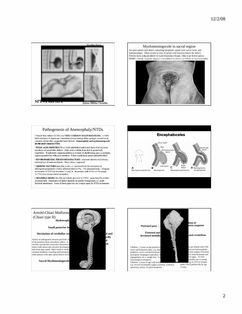
Spina Bifida
Holoprosencephaly (anterior midline closure)
MID-GESTATION
Neuronal migration
Gyral formation
Heterotopias
Macro/Microgyria
Lissencephaly
Anecephaly
A failure of anterior neural tube closure due to injury/defect at 18-26 days of gestation.
Results in total or partial absence of the cranial vault and cerebral hemisphere.
‘Area Cerebrovasculosa’ in anencephaly The cranial contents consist of a mass of disorganized neuroepithelial tissue covered
by a highly vascular meninges.
12/2/08
2
Myelomeningocele Myelomeningocele in sacral region An open spinal cord defect containing dysplastic spinal cord, nerve roots, and
leptomeninges. Often results in lack of spinal cord function below the defect.
Patients have reduced ability to walk/wheelchair bound, little or no bowel and/or
bladder control, frequent surgical interventions to minimize effects of hydrocephalus.
Pathogenesis of Anencephaly/NTDs
Hydrocephalus
Small posterior fossa
Herniation of cerebellar vermis
Sacral Myelomeningocele
Distorted and
downwardly
displaced
brainstem
Variety of pathogenetic theories put forth: ?Primary dysgenesis
of the posterior fossa mesoderm, others. A ‘unified theory’
invokes concept that ventricular distension is required to
induce both neural and calvarial development. Chronic CNS
leak from open spinal defect leads to small posterior fossa,
eventual hindbrain crowding and herniations. ??But why only
some patients with open spinal defects develop ACII?
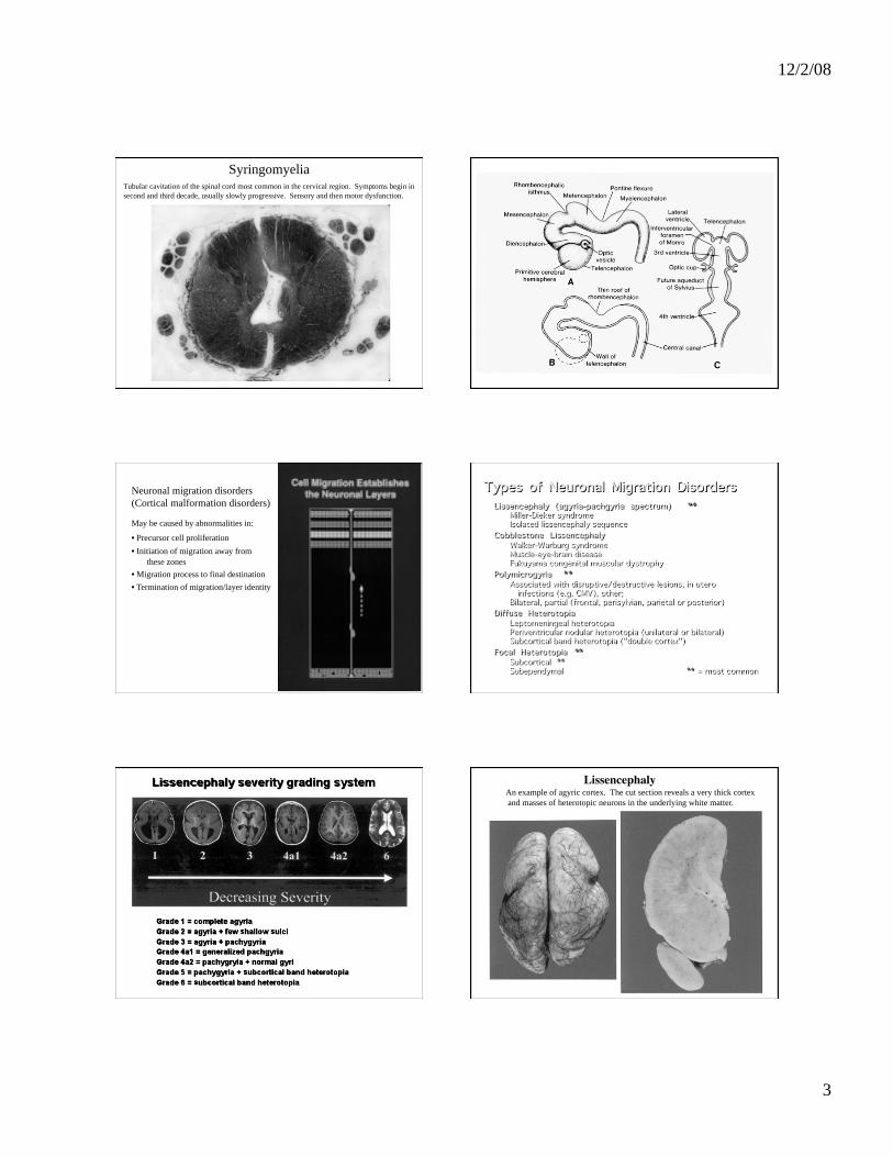
Arnold-Chiari Malformation
(Chiari type II)
Flattened pons
Flattened and
herniated medulla
Position of foramen magnum
Herniated cerebellum
Children < 2 years of age present with cranial
nerve and brainstem signs, e.g. respiratory
problems, vocal cord paresis/paralysis, apnea,
neurogenic dysphagia (aspiration, choking, nasal
regurgitation, etc.), weight loss. Can be a
neurosurgical emergency.
Children >2 years of age with insidious symptoms,
e.g. cervical myelopathy (upper extremity weakness,
spasticity), ataxia, occipital headache.
All patients are treated with CNS
shunts to prevent hydrocephalus.
Decompression surgery if become
symptomatic from brainstem and
cranial nerve signs - 15-23%
surgical mortality rate in young
children. Up to 15% of sympto-
matic CM II patients die by age
3 years.
12/2/08
3
Syringomyelia Tubular cavitation of the spinal cord most common in the cervical region. Symptoms begin in
second and third decade, usually slowly progressive. Sensory and then motor dysfunction.
Neuronal migration disorders
(Cortical malformation disorders)
May be caused by abnormalities in:
• Precursor cell proliferation
• Initiation of migration away from
these zones
• Migration process to final destination
• Termination of migration/layer identity
LissencephalyAn example of agyric cortex. The cut section reveals a very thick cortex
and masses of heterotopic neurons in the underlying white matter.

12/2/08
4
LissencephalyPredominantly agyric cortex but also a few large gyri (macrogyria). Most
lissencephalies have a variable combination of agyria-macrogyria and represent
a spectrum of disorders with varying clinical severity.
Laminar HetertopiaBilateral masses of heteropic neurons in white matter underly an apparently normal cerebral
al cortex. When this is diffuse throughout the cerebrum it is called “Double cortex” syndrome.
Nodular HeterotopiaLarge masses of heterotopic neurons in white matter near the cerebral ventricle.
May be bilateral or unilateral.
PolymicrogyriaGyri that are too small and too numerous give a very cobbled appearance to the surface of the
brain. This condition is often sporadic, may be focal, and is associated with in utero
disruptive/destructive lesions (e.g. infarcts, infections). Rare inherited disorders are recognized.
PolymicrogyriaNumerous small gyri with fusion of molecular layer between adjacent microgyri
(absence of vascular space along length of many gyri).
PERINATAL
Aquired due to hypoxia, ischemia, trauma
Germinal matrix hemorrhage
Periventricular leukomalacia
Infarcts (arterial territories or
watershed infarcts in
hypotension)
12/2/08
5
Germinal matrix hemorrhageHemorrhage limited to germinal matrix, here overlying the caudate nucleus. These lesions are seen in premature infants born before ~32-33 weeks EGA. Thin-walled vessels in this region are prone to rupture in association with hypoxia and poor cerebral blood flow autoregulation at this age.
Germinal matrix hemorrhage
Large hemorrhage has extended
into the adjacent brain.
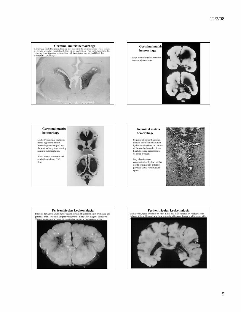
Germinal matrix hemorrhage
Marked ventricular dilatation
due to a germinal matrix
hemorrhage that erupted into
the ventricular system, causing
an acute hydrocephalus.
Blood around brainstem and
cerebellum follows CSF
flow.
Germinal matrix hemorrhage
Sequelae of hemorrhage may
include a non-communicating
hydrocephalus due to occlusion
of the cerebral aqueduct from
breakdown and organization
of blood products.
May also develop a
communicating hydrocephalus
due to organization of blood
products in the subarachnoid
space.
Periventricular LeukomalaciaBilateral damage to white matter during periods of hypotension in premature and
perinatal brain. Vascular congestion is present in the acute stage of the lesion.
The developing white matter is a watershed region in these young brains.
Periventricular LeukomalaciaChalky white, cystic cavities in the white matter next to the ventricle are residua of prior
ischemic lesions. Histologically, there is actually widespread damage to white matter with
astrocytosis and loss of oligodendrocytes and axons.
12/2/08
6
Porencephalic cystLarge destructive cerebral lesion in territory of MCA resulting in communication between
the cerebral ventricle and subarachnoid space. Often see polymicrogyria in adjacent cortex.
Porencephalic cystA 56 year old female with a history of breast carcinoma tripped and fell, sustained facial
fractures and developed a subdural hematoma. This large porencephalic cyst was an
incidental finding and related to known history of birth trauma. This is an unusual clinical
history but demonstrates the plasticity of immature brain which may compensate for the defect.
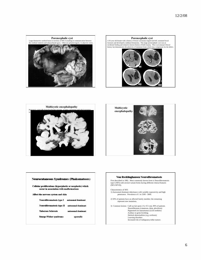
Multicystic encephalopathyPerinatal, cortically based ischemic lesions. Cystic cavitation and glial response
in brain, as seen in adults.
Multicystic encephalopathy
First described in 1882. Most commonly known form is Neurofibromatosis
type I (NFI) with several variant forms having different clinical features
(NF2-NFVII).
Characteristics of NFI:
1) Autosomal dominant inheritance with variable expressivity and high
penetrance. Prevalence of 1 in 2500 - 3000.
2) 50% of patients have an affected family member; the remaining
represent new mutations.
3) Common lesions: Café-au-lait spots (>6,>0.5 cm), 90% of patients
Neurofibromas (cutaneous, deep, plexiform)
Pigmented iris hamartomas (Lisch nodules)
Axillary or groin freckling
Skeletal abnormalities (e.g. scoliosis)
Learning disorders
Increased risk of malignancy/other tumors

12/2/08
7
Café-au-lait spots Neurofibromas
Multiplicity and size of lesions are
important for diagnosis of neurofibromatosis.
A tumor of peripheral nerve. Usually benign but may become malignant, particularly those in deeper nerves and plexuses. Plexiform neurofibromas are pathognomonic for NFI.
NF-type 2: Bilateral acoustic schwannomasSchwannomas are a benign tumor of peripheral nerve and are most often sporadic. When
present bilaterally on both VIIIth nerves, this is pathognomonic for NF-type 2.
Numerous meningiomas ( a tumor of arachnoid cells) lining the arachnoid under
the skull in this patient with NF-2.
Both NF-1 and NF-2 patients
also develop primary CNS
gliomas with increased
frequency.
Tuberous sclerosisVariable clinical presentations but may present in the first year of life with seizures. Mental retardation and behavioral problems also common.
Facial angiofibromas (adenoma sebaceum) are a common skin manifestion and appear between 2 and 5 years of age. Form a butterfly rash over the cheeks, nose, lower lip and chin.
Tuberous sclerosisCortical tubers
The tuber is a developmental
cortical malformation containing
an abnormal mixture of neurons
and glial cells. On cut surface it
has a gritty and firm texture.
These are commonly seizure foci
these patients.
Tuberous sclerosis Cortical tubers
Bizzarre giant cells (have both neuronal and astrocytic features) admixed with
neurons and astrocytes in a cortical tuber.

12/2/08
8
Tuberous sclerosis Subependymal nodules (‘candle gutterings’)
Benign astrocytic proliferations beneath the ependyma, most commonly in the lateral ventricles but also in third & fourth ventricle and aqueduct. Often calcified.
Sturge-Weber syndrome
Large ‘port-wine stain’ [naevus]
in the trigeminal territory
Ocular angioma, glaucoma
Leptomeningeal angiomatosis
and cerebral atrophy ipsilateral to side of naevus
Symptoms such as hemiparesis, hemiplegia, epilepsy and mental retardation generally begin within the first year of life or early childhood
Sporadic disorder, pathogenesis
is poorly understood.