development of polyhipe grafts for guided bone
TRANSCRIPT

DEVELOPMENT OF POLYHIPE GRAFTS FOR GUIDED BONE REGENERATION
A Dissertation
by
MICHAEL EDWARD WHITELY JR
Submitted to the Office of Graduate and Professional Studies of
Texas A&M University
in partial fulfillment of the requirements for the degree of
DOCTOR OF PHILOSOPHY
Chair of Committee, Elizabeth Cosgriff-Hernandez
Co-Chair of Committee, Akhilesh Gaharwar
Committee Members, Roland Kaunas
William B. Saunders
Head of Department, Michael McShane
August 2018
Major Subject: Biomedical Engineering
Copyright 2018 Michael Edward Whitely Jr

ii
ABSTRACT
A pressing need exists to develop an improved bone replacement to treat the millions
of non-union fractures that occur each year as a result of severe trauma, tumor resection,
spinal fusions, and joint replacements. Current bone grafts are often hindered by a lack of
biodegradability, porosity or innate ability to promote regeneration. This work employs
tissue engineering to design a novel bone replacement that combines the regenerative
potential of autologous tissue with the tunability of synthetic grafts. This is accomplished
by engineering a biodegradable scaffold with physical and mechanical properties
emulating those of cancellous bone and combining this scaffold with technologies that
allow for the controlled delivery of stem cells and osteoinductive factors.
In this work, polymerized high internal phase emulsions (polyHIPEs) were developed
as an injectable, high porosity bone graft. Thiol-methacrylate polyHIPEs were
investigated to increase resistance to oxygen inhibition and improve scaffold function
under clinically relevant conditions. Methods were established to modulate and
characterize scaffold porosity, cure rate, compressive properties, and degradation rates.
Furthermore, cell-laden poly(ethylene glycol)-dithiothreitol hydrogels were developed to
improve loading and distribution of human mesenchymal stem cells (hMSCs) within 3D
printed polyHIPEs. This approach allowed for increased cell retention and supported
critical markers of osteoblastic differentiation. Finally, to confer additional osteoinductive
character, porous microspheres with tunable release kinetics and requisite compressive
properties were fabricated using a solvent-free, in-line loading approach. Bioactivity
iii
retention of encapsulated bone morphogenetic protein-2, along with its ability to promote
osteoblastic differentiation of hMSCs, was explored.
Overall, these studies highlight the strong potential of polyHIPE scaffolds to serve as
an improved bone replacement with the ability to actively guide bone regeneration. Key
technologies have been developed that allow for fabrication of a bone graft with improved
function in a clinically relevant setting, efficient seeding with mesenchymal stem cells,
and targeted delivery of osteoinductive factors. Fundamentally, this work will be an
invaluable tool in identifying and evaluating critical design requirements for future bone
graft design.

iv
DEDICATION
To all who helped me see that where you start doesn’t have to determine where you
finish.

v
ACKNOWLEDGEMENTS
I can’t adequately express the appreciation I feel for all those who have helped me
over the past five years. Graduate school has been one of the most transformative periods
of my life, not just professionally, but personally as well. These experiences have opened
up opportunities and ways of thinking that I could have never imagined even a few short
years ago, and helped me prove to myself that our success in life is not determined by our
beginnings.
First, thank you to my advisor, Dr. Elizabeth Cosgriff-Hernandez, for taking a chance
and offering me an opportunity in her lab, even before I truly knew what it meant to be a
graduate student. You have helped me see and appreciate my strengths and challenged me
to grow and overcome my weaknesses. I will always be grateful for your patience and
guidance in helping me achieve something that I had never imagined possible.
Thank you to my committee, Dr. Akhilesh Gaharwar, Dr. Roland Kaunas, and Dr.
Brian Saunders, for your wonderful insight and guidance through this process. I have
greatly enjoyed learning from and working alongside you. Thank you to the LSAMP
community for your support, encouragement, and constant reminder of what is possible.
As challenging and rewarding as this adventure has been, it has been made so much
more enjoyable being surrounded and supported by great lab mates. I’m forever indebted
to Bobby Moglia and Jenny Robinson for your patience and generosity in teaching me all
the basics of research. To the original IBT crew, Alysha Kishan and Allison Post, for the
laughs, encouragement and research support during a time when we were all trying to

vi
figure out how to survive in graduate school. To Nick Sears, for being a great source of
knowledge and inspiring me to not just think outside the box, but to build a new box. To
Thomas Wilems, for your insightful advice and mentoring as I finished up my studies. To
Prachi Dhavalikar, for your tremendous help with the encapsulation work and always
making me feel far more confident than I actually was. To Sahar Mohiuddin, for your
steadfast reliability and patience with my inability to multitask. To Gabriel Rodriguez-
Rivera for your innovative ideas and willingness to explore new directions. To all the
members of the UT team, Taneidra Buie, Ziyang Lan, Siliang Wu and Megan Wancura
for your willingness to always help, give new ideas and share a kind word. And to Stacy
Cereceres, for leading Team College Station to the finish line with a joyous outlook,
unquestionable selflessness, and most importantly, lasting friendship.
Finally, to my wife, the constant that holds my life together and the person I strive to
be. I can never thank you enough for the support, sacrifice and love you have given during
this process. Anything I am able to do is only because I know that I have you beside me.

vii
CONTRIBUTORS AND FUNDING SOURCES
This work was supported by a dissertation committee consisting of Dr. Elizabeth
Cosgriff-Hernandez (chair), Dr. Akhilesh Gaharwar (co-chair) and Dr. Roland Kaunas
(member) of the Department of Biomedical Engineering, and Dr. William B. Saunders
(member) of the Department of Veterinary Small Animal Clinical Sciences.
Animal studies and histological analysis for Chapter II was performed by Dr. William
B. Saunders. Reporter cells utilized in Chapter IV were generously provided by the lab of
Dr. Daniel Alge. All other work conducted for the dissertation was completed
independently.
Graduate study was supported by The Texas A&M LSAMP NSF Bridge to the
Doctorate Fellowship and the Texas A&M Dissertation Fellowship.

viii
NOMENCLATURE
APS Ammonium Persulfate
BDMA 1,4 Butane Diol Dimethacrylate
BMP Bone Morphogenetic Protein
BPO Benzoyl Peroxide
BRITIER BMP Responsive Reporter Cell Line
DTT Dithiothreitol
ECM Extracellular Matrix
EGDMA Ethylene Glycol Dimethacrylate
FBS Fetal Bovine Serum
FGF Fibroblast Growth Factor
GM Growth Media
hMSC Human Mesenchymal Stem Cell
HQ Hydroquinone
IG Iron Gluconate Dihydrate
IGF Insulin-like Growth Factor
OM Osteogenic Media
PBS Phosphate Buffered Saline
PDGF Platelet Derived Growth Factor
PEG Poly(ethylene glycol)
PFDMA Propylene Fumarate Dimethacrylate

ix
PGPR Polyglycerol polyricinoleate
PLA Poly(lactic acid)
PLGA Poly(lactide-co-glycolide)
PMMA Poly(methyl methacrylate)
PolyHIPE Polymerized High Internal Phase Emulsion
RGD Arginine-Glycine-Aspartic Acid
TGF Transforming Growth Factor
TMA 4, N,N - Trimethylaniline
VEGF Vascular Endothelial Growth Factor

x
TABLE OF CONTENTS
Page
ABSTRACT .......................................................................................................................ii
DEDICATION .................................................................................................................. iv
ACKNOWLEDGEMENTS ............................................................................................... v
CONTRIBUTORS AND FUNDING SOURCES ............................................................vii
NOMENCLATURE ....................................................................................................... viii
TABLE OF CONTENTS ................................................................................................... x
LIST OF FIGURES ..........................................................................................................xii
LIST OF TABLES .......................................................................................................... xvi
CHAPTER I INTRODUCTION AND LITERATURE REVIEW ................................... 1
1.1. Clinical Overview ........................................................................................... 1
1.2. Current Treatment Options ............................................................................. 4 1.3. Tissue Engineered Scaffolds .......................................................................... 5 1.4. Mesenchymal Stem Cells in Bone Tissue Engineering .................................. 9
1.5. Growth Factor Delivery in Bone Tissue Engineering .................................. 17 1.6. Summary and Approach ............................................................................... 23
CHAPTER II THIOL-METHACRYLATE POLYHIPES WITH IMPROVED
RESISTANCE TO OXYGEN INHIBITION .................................................................. 25
2.1. Introduction .................................................................................................. 25 2.2. Materials and Methods ................................................................................. 29
2.3. Results and Discussion ................................................................................. 40 2.4. Conclusions .................................................................................................. 64
CHAPTER III IMPROVED IN SITU SEEDING OF 3D PRINTED BONE GRAFTS
USING CELL-RELEASING HYDROGELS .................................................................. 65
3.1. Introduction .................................................................................................. 65 3.2. Materials and Methods ................................................................................. 69 3.3. Results and Discussion ................................................................................. 75

xi
3.4. Conclusions .................................................................................................. 92
CHAPTER IV TUNABLE RELEASE OF BMP-2 FROM POROUS POLYHIPE
MICROSPHERES ............................................................................................................ 93
4.1. Introduction .................................................................................................. 93
4.2. Materials and Methods ................................................................................. 96 4.3. Results and Discussion ............................................................................... 103 4.4. Conclusions ................................................................................................ 117
CHAPTER V CONCLUSIONS .................................................................................... 118
5.1. Summary .................................................................................................... 118 5.2. Significance of Work .................................................................................. 120
5.3. Challenges and Future Directions .............................................................. 123
REFERENCES ............................................................................................................... 128

xii
LIST OF FIGURES
Page
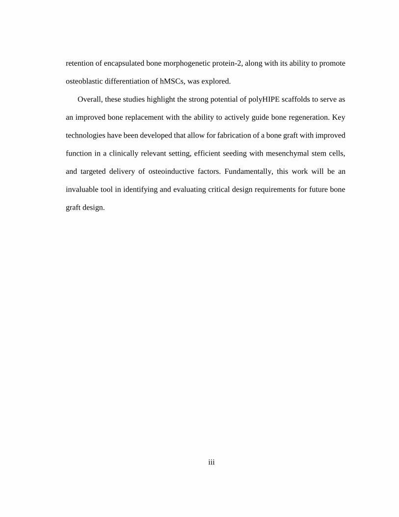
Figure 2.1. Molecular structure of propylene fumarate dimethacrylate (PFDMA) (A)
and pentaerythritol tetrakis(3-mercaptoproprionate) (tetrathiol) (B).
Reprinted from Whitely et al.146 ....................................................................... 32
Figure 2.2. Representative scanning electron microscopy (SEM) micrographs of
PFDMA (A), PFDMA-5T (B), and PFDMA-10T (C) polyHIPE pore
architecture. Reprinted from Whitely et al.146 .................................................. 41
Figure 2.3. Storage modulus during polymerization of polyHIPE (A) and work and set
times (B) of polyHIPEs cured at 37 °C with 1.0 wt% initiator and reducing
agent. Reprinted from Whitely et al.146 ............................................................ 43
Figure 2.4. The effect of increasing tetrathiol concentration on average gel fraction for
high surface-area-to-volume ratio polyHIPEs cured under ambient and low
oxygen conditions (A) and bulk cured polyHIPEs cured under ambient
conditions (B). Reprinted from Whitely et al.146 .............................................. 46
Figure 2.5. The effect of hydroquinone inhibitor (A) on average storage time (B), work
and set time (C), and gel fraction (D) of thiol-methacrylate polyHIPEs.
Reprinted from Whitely et al.146 ....................................................................... 49
Figure 2.6. The effect of increasing tetrathiol concentration on polyHIPE compressive
modulus (A) and yield strength (B). Reprinted from Whitely et al.146 ............. 50
Figure 2.7. The effect of increasing tetrathiol concentration on polyHIPE degradation
up to 4 weeks in 0.25 M NaOH (A) and 0.5 M NaOH (B). Reprinted from
Whitely et al.146 ................................................................................................ 53
Figure 2.8. hMSC viability after 24 h incubation with two concentrations of PFDMA
and PFDMA-10T extracts (1.0 and 0.5 vol%) (A). Micrographs illustrating
live (green) and dead (red) cells cultured with respective polyHIPE extracts
at 0.5 vol% (B). Reprinted from Whitely et al.146 ............................................ 55
Figure 2.9. NMR spectrum of PFDMA and PFDMA-10T polyHIPE extracts. Reprinted
from Whitely et al.146 ........................................................................................ 55
Figure 2.10. hMSC viability on PFDMA and PFDMA-10T polyHIPEs at 1, 3, and 7
days (A). Micrographs illustrating live (green) and dead (red) cells on the
respective polyHIPE sections at 7 days (B). Reprinted from Whitely et al.146 57

xiii
Figure 2.11. Proliferation of hMSCs seeded on PFDMA and PFDMA-10T polyHIPEs
at 1, 6, and 11 days as determined by DNA quantification. hMSCs were
cultured in growth media (GM) and osteogenic media (OM) with an initial
cell seeding density was 50,000 cells/cm2. Reprinted from Whitely et al.146 .. 57
Figure 2.12. Alkaline phosphatase activity of hMSCs seeded on PFDMA and PFDMA-
10T polyHIPEs at 1, 6, and 11 days. PolyHIPEs were cultured in growth
media (GM) (A) and osteogenic media (OM) (B). Reprinted from Whitely et
al.146 .................................................................................................................. 59
Figure 2.13. The effect of gelatin surface modification on collagen staining (A),
attached hMSC viability (B), hMSC adhesion (C), and hMSC morphology
and spreading (D) of polyHIPE scaffolds. ........................................................ 61
Figure 2.14. Biocompatibility of polyHIPE in rat calvarial defect model. A: Negative
control defects contain neovascularization (arrowheads), spindle shaped
fibroblasts, and collagenous matrix. B: The polyHIPE treated defect contains
similar fibrous tissue (arrowheads). C: Ordinal scoring of negative control
and polyHIPE (n = 6 tissue sections) indicated no significant differences in
the biologic response, except for lymphocyte infiltrate, which was increased
in polyHIPEs. .................................................................................................... 63
Figure 2.15. Rabbit medial femoral condyle defect with diameter 3.5 mm and depth of
5 mm (white arrow) (A). PolyHIPE injected into the defect (B). PolyHIPE
interdigitates with the bone, and solidifies (black arrow) (C). Harvested and
sectioned image of the filled defect (D), and representative SEM images of
the injected polyHIPE (E) and bone/graft interface (F). ................................... 63
Figure 3.1 Schematic illustrating hMSCs loading in hydrogel precursor solutions (A),
injection and encapsulation in 3D printed polyHIPE scaffold (B), and
protection during early stages of implantation (C). .......................................... 67
Figure 3.2. Effect of initiator concentration and reducing agent ratio on gelation onset
(A) and complete network formation (B) of hydrogel carrier. The + represents
a gelation and network formation time of greater than 30 minutes. The *
represents a gelation onset time of less than 10 seconds. All data represents
average ± standard deviation for n = 3. ............................................................ 77
Figure 3.3. Percent hMSC viability following 10 min exposure to redox agents,
ammonium persulfate and iron gluconate (A); 24h additional culture (B).
Percent hMSC viability and cell density following carrier encapsulation with
micrographs illustrating live (green) and dead (red) cells in respective
hydrogel formulations (C). All data represents average ± standard deviation
for n = 12. The * and ° represent significant difference (p<0.05) for 25 mM
concentration compared to other concentrations. ............................................. 82

xiv
Figure 3.4. Micrographs illustrating live (green) and dead (red) cells 1 day post
encapsulation and 7 days post release onto TCPS and polyHIPE substrates
(A). Percent hMSC viability 1 day and 7 days following release and
attachment onto TCPS and polyHIPE substrates (B). All data represents
average ± standard deviation for n = 12. .......................................................... 84
Figure 3.5. Distribution of hMSCs using carrier seeding onto multilayer polyHIPE
scaffolds determined by dsDNA quantification and compared to suspension
seeding control (A). Representative micrographs of top, middle and bottom
layers of scaffold (B). All data represents average ± standard deviation for n
= 3. The * represents significant difference (p<0.05) between suspension and
carrier seeding at specified layer. ..................................................................... 86
Figure 3.6. Adhesion of hMSCs released onto TCPS and polyHIPE substrates
determined by dsDNA quantification (A). Alkaline phosphatase activity of
hMSCs 7D and 14D post carrier release. Cells were cultured in growth media
(GM) and osteogenic media (OM) as positive control. All data represents
average ± standard deviation for n = 3. The * and ° represent significant
difference (p<0.05) for TCPS substrates compared to polyHIPEs at indicated
time points. The + represents significant difference (p<0.05) for ALP activity
on polyHIPE substrates at 14D. ........................................................................ 88
Figure 3.7. Alizarin red staining of hMSC cultures 4 weeks following carrier release.
hMSC mineralization post release onto TCPS substrates in growth vs
osteogenic media (A). Effect of release substrate (TCPS vs polyHIPE) and
media conditions (growth vs osteogenic) on hMSC mineralization (B). Semi-
quantitative analysis of mineralization determined by alizarin red stain
recovery (C). All data represents average ± standard deviation for n = 4. The
* represents significant difference (p<0.05) between all compositions. .......... 89
Figure 3.8. Effect of scaffold chemistry (PFDMA vs PLA films) and scaffold porosity
(PFDMA polyHIPE vs PFDMA film) on calcium deposition in cell free
conditions (A). Quantification of hMSC adhesion on varied substrate
determined by dsDNA quantification (B). Effect of substrate on hMSC
alkaline phosphatase activity after 14 days (C). Cells were cultured in growth
media (GM) and osteogenic media (OM) as positive control. All data
represents average ± standard deviation for n = 4. The * represents significant
difference (p<0.05) for indicated composition compared to all others in
respective media. .............................................................................................. 91
Figure 4.1. Schematic of microsphere fabrication. HIPE is injected through a needle
parallel to the flow of 3 wt% PVA solution and polymerized via UV
irradiation. Polymerized particles are collected and filtered prior to use. ........ 98

xv
Figure 4.2. Modulated particle diameter of model compositions with representative
SEM micrographs (A-D). Modulated pore diameter of model compositions
with representative SEM micrographs (E-H). From left to right: large
particle-small pore, large particle-large pore, small particle-small pore, small
particle-large pore. .......................................................................................... 104
Figure 4.3. Tuning release profiles of BSA-FITC from polyHIPE microspheres. Daily
and cumulative release profiles for all model compositions (A). Effect of
particle size on release kinetics for large (45um) and small (15um) pore size
(B). Effect of pore diameter on release kinetics for large (900um) and small
(300um) particle size (C). All data represents average ± standard deviation
for n = 3. ......................................................................................................... 109
Figure 4.4. Normalized FFLuc activity of BRITER cell line treated with releasates
taken from rhBMP-2 loaded microspheres. Percent bioactivity retention
determined by comparison to FFLuc of known rhBMP-2 stocks. .................. 111
Figure 4.5. Effect of rhBMP-2 loaded polyHIPE releasate on cell density (A) and
alkaline phosphatase activity (B) of hMSCs cultured with releasate for 14
days. Cells were cultured in fresh solution of stock rhBMP-2 (BMP-2) and
osteogenic media (OM) as positive control, and growth media (GM) as
negative control. All data represents average ± standard deviation for n = 4.
The * and ° represent significant difference (p<0.05) between BMP-2 or GM
and indicated compositions for density or ALP activity. ............................... 113
Figure 4.6. Effect of microsphere particle size and pore size on composite scaffold
compressive modulus and strength for 5 wt% incorporation using four model
compositions (A). Effect of increasing microsphere incorporation (5, 10 and
20 wt%) on composite scaffold compressive modulus and strength (B). All
data represents average ± standard deviation for n = 3. The * and ° represent
significant difference (p<0.05) for 20 wt% compressive modulus or strength
and other indicated compositions. .................................................................. 116

xvi
LIST OF TABLES
Page
Table 1.1. Trophic activity and therapeutic effects of mesenchymal stem cells. ............. 13
Table 1.2. Role of growth factors in bone repair.............................................................. 18
Table 3.1. Effect of initiator and reducing agent concentration on hydrogel compressive
modulus, swelling rato, and gel fraction. .......................................................... 79
Table 4.1. Summary table of fabrication parameters including tubing diameter, needle
size, flow rate, and surfactant compositions for model compositions. ............. 98
Table 4.2. Summary table of properties for model compositions including particle
diameter (n=25), pore size (n=100), and loading efficiency (n=12). All data
represents average ± standard deviation. ........................................................ 104

1
CHAPTER I
INTRODUCTION AND LITERATURE REVIEW
1.1. Clinical Overview
1.1.1. Prevalence of Nonunion Bone Defects
Treatment of musculoskeletal injury and disease are among the most common
procedures performed annually in the United States, with an estimated financial burden of
greater than $200 billion.1 Specifically, bone fractures comprise a majority of these
disorders with over seven million reported cases in the United States. These defects arise
from treatment of traumatic injury, congenital defect, tumor resection, and joint
replacements. Unfortunately, it is estimated that up to 10 percent of these fractures will be
associated with delayed or total absence of bone union during healing.2 Several factors
influence nonunion development including patient age and health, location of the injury
site, damage to surrounding tissue and vasculature, and mechanical instability.3 In cases
with significantly high loss of bone tissue, proper stabilization and control of the injury
site alone is not sufficient to promote healing. These defects, termed critical size, are
defined as osseous defects that fail to heal spontaneously with bone during the lifetime of
the patient, unless a suitable bone replacement material is placed in or onto the defect.4
Without intervention, these defects fill primarily with fibrous connective tissue and fail to
recapitulate the structure of native bone. As a result of this dependence on a secondary
substrate to facilitate bone healing, critical size defects are an ideal platform to investigate
novel bone replacement materials. Clinically, critical size may be determined through

2
radiological analysis of defect geometry. Key’s hypothesis states that a segmental long
bone defect greater than 1.5X the diaphyseal diameter will fail to undergo complete
healing in canine models.5 Furthermore, researchers have explored and identified similar
effects of segmental defect length and diaphyseal diameter on healing in other animal
models including sheep, cat and rabbit.6
1.1.2. Bone Biology and Structure
Bone is a nanocomposite of a stiff mineralized hydroxyapatite matrix and elastic
collagen fibrils. Inorganic hydroxyapatite mineral comprises approximately 70% of
calcified bone with 25% consisting of organic material (including cells), and 5% water.7
Newly synthesized osteoids that have yet to be mineralized consist primarily of collagen
(greater than 90%) with the remainder consisting of bone specific growth factors and
proteoglycans. Cortical and cancellous bone make up the two distinct tissue architectures
present in bone. The robust mechanical properties and load bearing potential is provided
by the cortical layer, a primarily non-porous architecture making up 80% of skeletal tissue.
Compressive modulus of cortical bone has been estimated at an impressive 17 GPa with
tensile properties about 20% weaker. The inner architecture of bone consists of a spongey,
honeycomb-like structure known as cancellous bone. As a result of this improved porosity
(estimated up to 90%), compressive modulus values are reduced, with an estimated
modulus of 20-500 MPa.8. Cancellous bone is typically found in the metaphysis of long
bones while cortical bone makes up the diaphysis. The cortical layer of bone is surrounded
by a thin membrane known as the periosteum, which contains many progenitor cells
responsible for bone maintenance. Bone maintenance is governed by complex interactions

3
and feedback mechanisms between matrix producing osteoblasts and tissue-resorbing
osteoclasts. The majority of cells in adult skeleton consist of osteocytes, fully specialized
osteoblasts which facilitate extracellular calcium and phosphorus concentrations and
direct osteogenesis.9 Improved understanding of the complex processes and regulators in
bone remodeling is critical to developing an optimal treatment for critical sized defects.
1.1.3. Natural Bone Healing
Bone is unique among tissues as it has the ability to heal new, healthy tissue with the
absence of scar tissue formation.10 Fracture healing occurs in multiple phases, often
occurring simultaneously. During the early stages of repair, disruption of the native
vasculature results in formation of a hematoma, providing a matrix that encloses the
wound, allows migration of inflammatory cells, fibroblasts, and progenitor cells into the
wound site, and promotes granular tissue formation.9, 11 This initial response is followed
by a rapid release of angiogenic factors and cytokines to direct new blood vessel
formation, as well as the release of osteoindutive factors from infiltrating stem cells and
extracellular matrix.12 The next stage is defined by the formation of a soft callus that
begins to stabilize the defect. Matrix composed of Type 1 collagen and proteoglycans is
produced by osteoblasts which forms the soft, internal callus, eventually undergoing
mineralization and forming the bony callus. Simultaneously, intramembranous
ossification occurs in the periosteum, creating an external callus. The ossification of the
soft callus to bridge the broken can occur in as little as 4 weeks if properly immobilized.
Finally, the hard callus is replaced with native bone, with size and vascular structure fully
returning to normal over a period of several months.13 Similar to healing in non-critical

4
defects, critical defect healing processes include formation of poorly organized endosteal
and periosteal callus formation that proceed toward bony bridging of the gap. However,
unlike non-critical healing, full bridging does not occur of the opposing callus parts,
resulting instead in formation of rounded corticies.14 This nonunion results in the loss of
load transfer and mechanical stimuli that are needed to fully initiate tissue organization.14,
15
1.2. Current Treatment Options
1.2.1. Autografts
Autologous grafts are currently the gold standard for bone grafting procedures due to
their high healing capacities. In these procedures, tissue is harvested from a secondary
location on the patient, most commonly the iliac crest of the hip or tibia, and transplanted
to the injury site.16 Advantages of autografts include high integration with surround tissue,
strong osteoinductive character, and no risk of immune rejection. Multiple trials including
vertebral fusions and treatment of tibial nonunion have demonstrated extremely high
success rates.17 Unfortunately, this procedure is not an option for many patients due to a
lack of graft availability and complications arising from a second surgical site.17-19
1.2.2. Allografts
Allograft bone provides a more readily available treatment option as it can be
harvested from cadaveric tissue donors and easily stored.20 As immune rejection and
disease transmission can be areas of concern for allogenic transplants, decellularization
processes are often introduced to reduce potential risks. Similarly to grafting with
autologous tissue, the complex geometry of bone defects often makes fitting a replacement

5
allograft extremely difficult. As a result, donor tissue can be demineralized to form a
powder or paste that allows easier implementation and is better able to match defect
geometry. This processing however, severely compromises load bearing potential, further
driving the need or a more viable bone replacement.21
1.2.3. Alloplastic Grafts
Poly(methyl methacrylate) (PMMA) bone cement is the most widely utilized synthetic
material in bone grafting procedures, commonly selected to stabilize implants, fill defects
in tumor resection, and provide minor load bearing in spinal procedures.22, 23 Despite
widespread use, PMMA cements are not an ideal treatment option as they are nonporous,
do not undergo necessary biodegradation, and often reach temperatures in excess of 100ºC
during polymerization, which can lead to necrosis of the surrounding tissue. In contrast,
calcium phosphate cements have been readily explored as a more cytocompatible and
osteoinductive bone replacement.1 Despite success, these grafts often exhibit low fracture
toughness and fail due to fracture.24 Combined, these limitations suggest that physicians
and patients would greatly benefit from an improved bone grafting option.
1.3. Tissue Engineered Scaffolds
Tissue engineering aims to provide an improved tissue replacement option by
combining the regenerative capacity of autologous grafts with the tunability and
availability of synthetic materials.25 The tissue engineering paradigm consists of a
biomaterial scaffold that temporarily replaces tissue structure, and combining this scaffold
with appropriate biochemical cues and progenitor cells to facilitate tissue regeneration.26
As a result, this strategy has numerous advantageous over standard grafting materials.
6
1.3.1. Scaffold Requirements
Success as a tissue engineered scaffold is dictated by the ability to support critical cell
and tissue functions. To achieve this, it is necessary to have sufficiently interconnected
and porous architecture to allow cellular infiltration, vascularization, and nutrient and
waste transport. Multiple groups have explored optimal pore sizes needed to encourage
ingrowth and osteogenesis. Broadly, a minimum pore size of 100 microns is desired, with
larger pores demonstrating increased osteoblastic activity.27-29 Native bone is unique in its
ability to achieve a porous architecture while maintaining the ability to withstand high
physiological loads. Typical physiological stresses have been estimated up to 50 MPa with
highest stresses occurring in the femur during strenuous activities such as running.30
Achieving this combination of porosity and strength in a synthetic material, without
introducing negative stress-shielding effects, has been a major hurdle in bone grafting.31
It is generally accepted that scaffolds with compressive properties approaching those of
cancellous bone (100 MPa modulus; 10 MPa strength) are capable of supporting bone
regeneration.32 Finally, an ideal tissue engineered scaffold should degrade at a rate similar
to de novo tissue formation, and appropriately transfer responsibility back to the healing
tissue.26 Femoral defect models in rabbits have illustrated improved healing when tissue
engineered scaffolds are tuned to degrade at a rate that better matches healing.33 Matching
regeneration rates can be particular challenging however, as patient age, health, and
severity of injury effect healing rates.

7
1.3.2. Scaffold Fabrication Techniques
A multitude of fabrication techniques have been developed by tissue engineers to
generate highly porous scaffolds for repair of musculoskeletal injuries. The most
prominent of these include electrospinning, gas foaming, porogen leaching, and thermally
induced phase separations. Electrospinning is a method utilized to generate 2-dimensional
sheets of fibrous meshes that contain high SA/V ratios.34 Tunable porosities, diameters,
and alignment patterns are achieved through modulation of polymer solution and system
parameters.35 Lack of suitable compressive properties and inability to generate large
constructs limit its potential in orthopedic applications. Gas foaming, or blowing,
techniques are utilized to produce highly porous scaffolds with a range of pore sizes.36
Pores are generated through gas production, typically CO2, in a polymer melt, utilizing
chemical or physical processes. Elevated temperatures and post fabrication processes
needed to remove a non-porous skin relegates this method to solely prefabrication
applications.37
Dispersion, and subsequent leaching, of porogens (often salts) from a polymer solution
after network formation have also yielded porous, interconnected scaffolds.38 A common
drawback to salt leached scaffolds is the presence of stress concentrations resulting from
the uncontrolled shape of porogens, which can result in decreased mechanical
properties.39, 40 Inducing a change in solubility of a polymer solution can be used create
polymer-rich and polymer-poor regions, that upon subsequent sublimation, yields a
porous polymer scaffold. Modulating parameters in this phase separation method,
including polymer type and temperature, can be used to obtain a variety of pore

8
geometries.41, 42 Despite the ability of these techniques to yield porous scaffolds,
fabrication parameters often introduce concerns or preclude use for in situ delivery,
incorporation of cells, or use in growth factor delivery.
1.3.3. Polymerized High Internal Phase Emulsions (PolyHIPEs)
Emulsion templating is a unique fabrication technique that is currently being
studied for use in tissue engineering.43, 44 High internal phase emulsions are defined by an
internal droplet phase (greater than 74%), and an organically soluble prepolymer outer
phase. Polymerization of the continuous phase locks in the internal architecture dictated
by the droplet phase resulting in highly porous foams. Historically, numerous groups have
reported on processing variables than can be tuned to achieve a diverse range of properties,
(75-99% porosity; 1-100 µm pore size; 2 kPa- 60 MPa compressive moduli).43, 45-49
Recently we developed a polyHIPE scaffold suitable for bone regeneration based on the
biodegradable macromer, propylene fumarate dimethacrylate (PFDMA). This
hydrophobic, low viscosity macromer contains ester linkages that allow for hydrolytic
degradation, and reactive methacrylate end groups that undergo rapid polymerization in
physiological conditions. Uniquely, we have demonstrated these injectable materials are
suitable for in situ injection, facilitate osteogenic activity, and adaptable as 3D printable
emulsion inks.
1.3.4. Thiol-Methacrylate Polymerization for Improved Resistance to Oxygen Inhibition
Although traditional free radical polymerization is a robust mechanism for scaffold
fabrication, use of only methacrylated macromers in our system renders it susceptible to
oxygen inhibition. When polymerized under oxygen rich conditions, initiating radicals are

9
scavenged by molecular oxygen and converted to peroxy radicals.50, 51 These radicals are
no longer able to reinitiate polymerization of methacrylate functional groups, essentially
terminating cure. This often results in reduced cure rates, poor monomer conversion, and
reduced mechanical properties. Although many researchers have developed techniques to
prevent oxygen inhibition in commercial settings (e.g. purging with inert gases), limited
progress has been made in addressing this problem in an injectable system. Researchers
have probed Michael addition-type reactions to create ideal, step-growth crosslinked
networks that provide resistance to oxygen inhibition. Specifically, thiol-acrylate and
thiol-methacrylate systems have been reported to have improved resistance to oxygen
inhibition. O’Brient et al. illustrated that increasing thiol concentration in diacrylate
systems resulted in reduced levels of oxygen inhibition.52 In contrast to the traditional free-
radical polymerization of unsaturated vinyls, thiol-methacrylate polymerization can be
initiated by both cleave-type initiators and hydrogen abstraction of the thiol monomer,
rendering it less susceptible to oxygen inhibition. Propagation then proceeds via thiol or
methacrylic/acrylic radical addition to methacrylate/acrylate functional groups. This
reduction in oxygen inhibition provided by a mixed mode polymerization mechanism
could prove a major benefit in an injectable polyHIPE system.
1.4. Mesenchymal Stem Cells in Bone Tissue Engineering
1.4.1. MSC Discovery and Isolation
One of the most exciting developments over the past 60 years in regenerative medicine
is the growth of mesenchymal stem cell therapies.53 Discovered in the 1960’s through the
ground breaking work of Friedenstein and colleagues, a population of stromal cells that

10
could be isolated from bone marrow, were plastic adherent, and could promote formation
of de novo bone tissue were discovered.54 Over the next two decades, significant emphasis
was placed on better understanding these potent sources of healing, termed mesenchymal
stem cells, and developing markers for their identification.55, 56 Although mesenchymal
stem cells represent a small portion (less than 0.01%) of total cells present in bone marrow,
Pittenger et al. demonstrated that these cells could be expanded with high efficiency in
vitro while retaining their multipotency.57 These studies demonstrated that mesenchymal
stem cells were capable of differentiating into adipcytic, chondrocytic, and osteocytic
lineages. Exponential increase in interest led to the release of a position statement by The
International Society for Cellular Therapy defining minimal criteria for classification of
multipotent mesenchymal stromal cells as plastic-adherent, positive for expression of
CD105, CD73, and CD90, ability to differentiate into osteoblasts, adipocytes, and
chondrocytes.58
Harvesting of MSCs from bone marrow remains the most widely studied isolation
method. In humans, the superior iliac crest of the pelvis is commonly selected, with
additional isolation compartments being those of the femur, tibia, and thoracic and lumbar
spine.59-61 Large animal models typically utilize similar methods with small animal models
focusing on harvest from the mid-diaphysis of the tibia or femur. Primary harvests are
typically kept in culture for two weeks to allow for adherence and removal of non-adherent
hematopoetic cells.61 More recently, additional stem cell niches have been discovered,
providing additional sources for tissue-specific stem cells including the periosteum,
adipose tissue, and umbilical cord blood.62-64 Yoshimura et al. demonstrated that solid

11
tissues could provide higher concentrations of mesenchymal stem cells than marrow
harvests.
1.4.2. Trophic Activity
The ability of mesenchymal stem cells to differentiate into specialized cell types has
prompted many researchers to employ these cells in their grafts in the hopes of directly
replacing the cells lost to injury and responsible for tissue remodeling. Although
significant improvements in regeneration have been observed as a result of mesenchymal
stem cell presence, a new school of thought has emerged suggesting an alternative
therapeutic mechanism.53, 65 It has been evidenced that in many cases, rather than
specialized differentiation, MSCs were in fact creating a ‘trophic effect’, or that is to say,
creating a regenerative microenvironment through the secretion of growth factors and
bioactive molecules that promotes infiltration of native cells, angiogenesis, and tissue
healing. Common trophic mechanisms are summarized in Table 1.1.53 This trophic
activity is achieved through the secretion of numerous growth factors to recruit and
promote proliferation of native cells including transforming growth factors, insulin-like
growth factor-1, basic fibroblast growth factor and epithelial growth factor.66, 67
Furthermore, MSCs have been shown to facilitate angiogenesis through secretion of
vascular endothelial growth factor and stabilizing newly formed vessels.68, 69 Tateishi-
Yuyama et al. demonstrated that autologous implantation of bone marrow derived cells
could improve angiogenesis in patients suffering from unilateral ischemia of the leg.70
Another unforeseen benefit of MSC therapy is anti-inflammatory and
immunomodulatory effects. Inflammation at the site of musculoskeletal injury can result

12
in impediment of healing processes of native progenitor cells. MSCs have been shown to
secret a multitude of anti-inflammatory factors in response to injury.71 As a result, MSCs
are able to modulate function of surveilling inflammatory immune cells, including T cells,
B cells, and macrophages.72, 73 MSC delivery has also demonstrated anti-apoptotic and
antimicrobial properties. Cselenyak et al. explored the mechanism behind bone marrow
derived MSCs improving viability of ischemic cariomyoblast populations and identified
the recovery to be dependent on cell-to-cell interactions.74, 75 hCAP-18/LL-37, a peptide
commonly expressed by epithelial cells and macrophages to combat bacterial infections,
is secreted by MSCs in response to e coli and other infections. In an experimental colitis
and sepsis model, systemic infusion of adipose derived MSCs protected from severe sepsis
by reducing infiltration of inflammatory cells and by down regulation of inflammatory
mediators.76

13
Table 1.1. Trophic activity and therapeutic effects of mesenchymal stem cells.
Properties Mechanism Effect
Trophic
Release of TGF-α, TGF-β,
HGF, EGF, FDF-2, IGF-1
Release of VEGF, IGF-1, EGF,
angiopoietin-1
Release of keratinocyte growth
factor, SDF-1, macrophage
inflammatory protein-1 α and β
Direct fibroblasts,
epithelial, and endothelial
cells
Recruit endothelial cells
and facilitate
vascularization
Reduce scar tissue
formation
Immunomodulatory
Release of prostaglandin 2,
TGF-β1, HGF, SDF-1, nitrous
oxide, indoleamine 2,3-
dioxygenase, IL-4, IL-6, IL-10,
IL-11 receptor antagonist,
soluble tumor necrosis factor-α
receptor
Express HLA Class I and HLA
Class II
Inhibit function of T cells,
killer cells, B cells,
monocytes, macrophages
and dendritic cells
Avoid immune system
recognition
Apoptotic Rescue
Secretion of IGF-1, IL-6,
VEGF, HGF, TGF-β1
Increase protein kinase B
production to facilitate anti-
apoptotic pathways
Antimicrobial
Express hCAP-18/LL-37
Upregulate indoleamine 2,3-
dioxygenase
Combat Gram-positive and
negative bacterial
infections
Regulate T-cell activity
1.4.3. Clinical Efficacy of Stem Cell Therapy
The clinical benefits of marrow derived stem cell delivery have been widely
established. Early studies focused on the percutaneous grafting of autologous bone
marrow in nonunion fractures, including those of the tibia. Connolly et al. observed
improved callus formation and defect union in eighty percent of patients stabilized with

14
an external cast, and one hundred percent of patients stabilized with intramedullary nails.
Observed results were comparable to standard autologous grafting procedures.77 It was
later established by Hernigou and colleagues that efficacy of autologous bone marrow
therapies in the treatment of nonunions was related to the number of progenitors present
in the graft.78 This work articulated that bone marrow harvesting from the iliac crest may
be patient variable and not suitable for promotion of healing in the absence of
concentration, emphasizing the importance of normalizing progenitor amount to reach
efficacy. Finally, Bruder et al. demonstrated that purified and culture-expanded human
MSCs could be utilized to improve regeneration in a critical sized femoral defect treated
with porous hydroxyapatite/beta calcium phosphate scaffolds.79, 80 Stem cell loaded
constructs exhibited radiological and histological evidence of new bone formation after 8
weeks with improved biomechanical properties.
1.4.4. Natural Polymeric Cell Carriers
Although MSC delivery holds therapeutic promise, traditional delivery methods (e.g.
percutaneous grafting of stem cells via syringe or catheter) often result in limited cell
engraftment and survivability, ranging between 5%-20%.81, 82 In addition, the hostile
environment of injured or diseased tissue can deprive cells of nutrients or subject them to
removal by surveilling inflammatory cells, further reducing cell retention. As a result,
biomaterial carriers that provide an external matrix and mechanical barrier to removal
have been investigate as an improved substrate for tissue engraftment. Hydrogels, a class
of biomaterials comprised of hydrophilic, water-swollen polymer networks, have emerged
as a promising platform for delivering stem cells. Numerous natural and synthetic

15
hydrogels have been investigated for cell encapsulation.83, 84 Natural platforms, such as
those based on modified gelatin, alginate, or fibrin provide an extracellular-like matrix
that is both cytocompatible during encapsulation processes, and able to promote cell
attachment and migration after delivery. Encapsulation in these systems often occurs
through thermal induced gelation or ionic crosslinking. Awad et al. investigated
chondrogenic differentiation of adipose derived stem cells encapsulated in multiple natural
polymer systems including agarose, alginate, and gelatin scaffolds.85 Furthermore, Nichol
et al. demonstrated that methacrylated gelatin, a natural derivative of collagen, can be
utilized to encapsulate cells with high cell survival.86 These constructs were advantageous
as they retained native RGD binding sites and MMP-sensitive degradation sites while
achieving improved mechanical properties of covalently crosslinked systems.
1.4.5. Synthetic Polymeric Cell Carriers
Synthetic hydrogels, specifically those based on poly(ethylene glycol) (PEG), provide
an encapsulation platform with excellent cytocompatibility, and a blank slate nature that
can be modified for any target tissue. Most commonly, hydrophilic precursor solutions are
fabricated containing prepolymer, photoinitatior, and cell payload. These solutions are
then exposed to an external UV source to facilitate free-radical polymerization of
acrylate/methacrylate groups, and physical entrapment of the cell inside the polymer
network. Excellent encapsulation viability, as well as the ability to promote osteogenic
differentiation of MSCs was observed by Nuttleman et al.87 After one week, gene
expression for osteogenic markers osteonectin, ostoeopontin, and alkaline phosphatase
was upregulated with mineralization present after four weeks. To improve viability of cells

16
encapsulated for extended periods, RGD binding sites have been covalently inserted to
improve cell-matrix interactions and stimulate bone growth.88, 89 Synthetic hydrogel
platforms with improved biodegradation have also been explored for cell delivery and
release. A novel in situ application system based on oligo(poly(ethylene glycol) fumarate)
was investigated and shown to support osteogenic differentiation of encapsulate rat
marrow stromal cells.90 The redox initiation mechanism utilized in this system has the
advantage of not requiring external equipment to facilitate polymerization. This system
has since been modified to allow for tunable degradation and release of marrow stromal
cells from the hydrogel cell carrier through incorporation of the hydrolytically degradable
macromer acrylated poly(ethylene glycol)-dithiothreitol.91 Modulating degradation profile
was identified as a tool that can be utilized to control cell retention at the tissue site.
Finally, hydrogel carriers provide the added advantage of being able to simultaneously
deliver bioactive factors. Simmons et al. demonstrated that bone morphogenetic protein-
2 and transforming growth factor could be combined with stem cell deliver to improve
ectopic bone formation.92
1.4.6. Combination Cell Carriers
As evidenced by the success of these varied platforms, hydrogel delivery holds strong
promise in stem cell therapy. A challenge specific to bone tissue engineering is developing
a cell carrier that has suitable mechanical properties for physiological loading. Recently,
composite grafts have been investigated to deliver stem cells in more robust scaffolds by
combining traditional bone grafting materials with hydrogel cell carriers. Zhao et al.
demonstrated the promise of this approach by encapsulating umbilical cord mesenchymal

17
stem cells in alginate microbeads and incorporating them in an injectable calcium
phosphate cement paste.93 Encapsulated cells remained viable, exhibited gene markers of
osteoblastic differentiation, and facilitated mineral synthesis. Similar systems, including
those based on settable polyurethane scaffolds have been explored with similar promising
results for chondrogenic tissue repair.94 Combined, this work illustrates the benefits of
stem cell delivery and the future promise of incorporating these cells in robust scaffold
design. A primary aim of this work was to expand upon these systems by developing an
injectable cell carrier that can improve seeding in patient specific grafts prepared with
state of the art 3D printing modalities.
1.5. Growth Factor Delivery in Bone Tissue Engineering
1.5.1. Growth Factors in Bone Healing
As outlined previously in this review, the stages of bone healing are well established.
What is less established however, is the complex interactions and feedback loops
regulating these remodeling processes. Although there are physical cues that arise from
changes in mechanical properties and a loss of nutrient and oxygen supply due to fracture
and damaged vasculature, the primary initiators of healing are likely the multitude of
bioactive growth factors released into the fracture site.95 These secreted proteins act as a
signaling service that directs surrounding cells to carry out a specified action. Upon
binding of the ligand to a target receptor on the surface of the cell, a conformational change
occurs, resulting in formation of a transcription factor that travels through the cell. This
transcription factor then binds nuclear DNA and facilitates new gene expression and
subsequent morphological changes of the cell. The most prevalent growth factors active

18
in bone repair are summarized in Table 1.2.95, 96 One of the most widely studied of these
factors are members of the transforming growth factor-beta super family. Signaling from
these proteins is activated through a transmembrane receptor complex formed by type-I
and type-II serine/threonine kinase receptors. This leads to downstream activation of class
of transcription factors known as SMAD proteins, which facilitate the intracellular
response.97 Other regulating factors such as platelet-drived growth factor initiate response
through activation of receptor tyrosine kinases.96 Ferguson and colleagues demonstrated
that the genetic mechanisms that regulate fetal skeletal development also regulate bone
healing in adults.98 Developing platforms that allow for improved understanding of the
complex overlap and cross-communication between these signaling pathways has been a
driving area of study.
Table 1.2. Role of growth factors in bone repair.
Factor Activity Source
Transforming
growth factor beta
(TGF-β)
Stimulate ECM production,
osteoprogenitor cell
proliferation, osteogenic
differentiation
ECM, platelets,
inflammatory cells,
osteoblasts
Bone
morphogenetic
protein (BMP)
Promote osteoblast and
chondrocyte differentiation of
progenitor cells
ECM, osteoblasts,
osteoprogenitor cells
Fibroblast growth
factor (FGF)
Mitogenic effect of MSCs,
osteoblasts and chondrocytes,
promote angiogenesis
Macrophages, MSCs,
chondrocytes, osteoblasts
Insulin-like growth
factors (IGF)
Promote proliferation and
differentiation of progenitor
cells
ECM, chondrocytes,
osteoblasts
Platelet-drived
growth factor
(PDGF)
Promote chemotaxis and
proliferation of macrophages
and MSCs
Platelets, osteoblasts,
macrophages

19
1.5.2. Bone Morphogenetic Proteins
The most widely studies proteins studied for bone regeneration are members of a
subset of the TGF-Beta superfamily known as bone morphogenetic proteins (BMP).
Investigation into these proteins first began after the discovery that new bone could be
formed as a result of intramuscular implantation of demineralized bone matrix.99
Discovery of this ‘bone induction principle’ sparked numerous investigations, eventually
resulting in the genetic sequencing and identification of numerous bone morphogenetic
proteins.100 Furthermore, Cheng and colleagues established a hierarchical model of
osteogenisis for multiple BMPs, observing that BMP-2, -6, and -9 were able to induce
alkaline phosphatase activity in pluripotent cell lines.101 BMP-2, -4, -6, -7, and -9 were
able to induce alkaline phosphatase activity in preosteoblasts, while the majority of tested
factors could induce activity of differentiated osteoblasts. As a result of these collective
investigations, numerous BMPs have been explored to improve bone regeneration in a
clinical setting, the most promising of these being BMP-2. The therapeutic effect of BMP-
2 delivery has been shown to emerge from its role in the initiation of fracture healing,
chemoattactive properties, and facilitation of angiogenesis.13, 102-106 The importance of
BMP-2 in initiating fracture response is widely recognized as it is present in extracellular
matrix and released into the wound environment post injury. Tsuji et al. reported that in
the absence of BMP-2, even in the presence of other osteoindutive stimuli, limb defects in
a mouse model failed to resolve with time.102 It was critical to note that stem cell
populations with upregulated expression of BMP receptors were present at the injury site,
but lacked necessary signaling to facilitate healing. Furthermore, Fiedler et al. reported a

20
greater than 3-fold increase in migration of primary mesenchymal human porogenitor cells
in response to BMP-2 delivery, suggesting a functional role of BMP-2 factor in the
recruitment of progenitor cells in bone healing.103 Finally, it has been reported that BMPs
enhance expression of potent angiogenic factors, including vascular endothelial growth
factr (VEGF) in osteoblasts.104 This expression is not associated with osteoblastic
differentiation, but rather serves to combine critical osteogenic and angiogenic processes.
1.5.3. Clinically Available Growth Factor Delivery Systems
Combining bone morphogenetic proteins into grafting materials has been explored as
an approach to overcoming limitations in autologous bone grafting, to great success. To
date, two devices have been approved for clinical treatment of bone defects.107-110 Delivery
of recombinant expressed BMP-2 in an absorbable collagen sponge was first approved for
treatment of specific interbody spinal fusion procedures. It later gained approval for
treatment of open tibial fractures in intramedullary nail fixation and most recently for
specific craniofacial applications. In a 279 patient trial clinical trial for the treatment of
degenerative disc disease, it was reported that 2-year fusion rates for the BMP/collagen
sponge were greater than 88%, only marginally lower than autologous rates of 94.5%.108
No differences in patient pain or neurological profiles were reported. In addition,
complications related to secondary donor site pain and morbidity were expectedly
eliminated. Furthermore, use of BMP-7, clinically known as Osteogenic Protein-1, has
been granted approval by the FDA under the Humanitarian Device Exemption program
for delivery in a puddy of bovine bone collagen. A clinical trial investigating repair of
tibial nonunions reported that 75% of patients had radiographic confirmation of fracture

21
healing after nine months, only marginally lower than the autograft treatment at 84%.110
Despite the clear ability of osteoinductive factor delivery to promote regeneration, several
safety concerns have emerged. Concerns include inflammation, ectopic bone formation,
and neurological deficits.111, 112 These undesirable off-target complications often result
from a bolus release of supraphysiological dosages of the factor. As the majority of protein
is quickly released form the collagen carrier and removed from the injury site, high
concentrations of protein are required to be initially loaded to ensure a robust therapeutic
response.113 These recent complications highlight the need to develop a delivery system
that can retain the benefits of osteoinductive factor delivery, but limit safety concerns.
1.5.4. Controlled Growth Factor Delivery
Numerous platforms have been investigated to provide controlled release of growth
factors, aiming to achieve more physiologically relevant delivery profiles. The hydrophilic
nature and mild fabrication conditions of hydrogels make them intriguing platforms for
growth factor delivery. Indeed, delivery of BMPs from hydrogel platforms have been
shown to promote ectopic bone formation. However, to better control release kinetics,
affinity-based functionality is often required. Furthermore, growth factor delivery in
calcium phosphate cements has been explored. Limited porosities and poor loading of
surface coatings limits commercial potential of these systems. As a result, encapsulation
of growth factors into porous, polymeric microspheres systems has emerged as a method
to provide controlled release of bioactive factors.

22
1.5.5. Porous Microspheres for Growth Factor Delivery
Significant and wide-reaching progress has been made in the development of porous
microsphere fabrication techniques.114 The most widely studied microsphere platform for
drug delivery is the fabrication of porous microspheres based on the biodegradable
polymer pol(lactic-co-glycolide) (PLGA).115, 116 The ester linkages in these polymers
allow for hydrolytic degradation of the polymer with degradation products that can be
removed through natural processes. For fabrication, these systems often employ a double
emulsion-solvent evaporation method. In this method, polymer is first dissolved in a
volatile solvent, most often methylene chloride or chloroform. The polymer solution is
then emulsified (water-in-oil) with an aqueous solution containing the protein, added to a
secondary aqueous phase to form the double emulsion (water-oil-water), and then agitated
to form the microparticles. The final step is the removal of solvent through excess stirring
or vacuum drying. To control particle properties like size, emulsion parameters such as
stirring conditions, solvent choice, and polymer concentration are modulated. Typical
microsphere diameters fabricated with this method range from 1-200 microns with notable
size distributions, and pore sizes generally remaining below a single micron.117-119
Osmotic agents have been utilized to control pore architecture and provide porous systems
for sustained growth factor release.120 Although exposure to common solvents including
methylene chloride has demonstrated minimal effect on BMP-2, stability of growth factors
can vary greatly, and thus compatibility of a method should be determined with caution.
Other fabrication methods of porous microspheres mirror those used for general
porous scaffold fabrication. Broadly, these include porogen leaching and gas foaming.121

23
When adapted for microsphere fabrication however, it has proven difficult to adequately
control the relationship between particle and pore size. Fabrication of porous scaffolds
through porogen leaching relies on the addition of salts to polymer solutions, followed by
subsequent leaching of the porogen. This results in heterogeneous pore structures which
may result in variable scaffold properties. Additionally, the post fabrication requirements
preclude in-line loading of agents into the scaffolds. Gas foaming has also been explored
to fabricate porous microsphere by addition of effervescent salts as blowing agents.
Ammonium bicarbonate has been shown to create highly porous microspheres with pore
sizes up to twenty microns. In this report however, the relationship between particle size
and pore size was coupled, limiting the tunability of this system.36
To minimize manufacturing concerns resulting from costly post fabrication processes,
including solvent removal and porogen extraction, spray drying has emerged as a minimal
processing fabrication technique.122 For the encapsulation of proteins, the primary water
in oil emulsion is sprayed in a stream of heated air. Fabrication parameters of injection
rate, temperature, and solvent choice are modulated to dictate microsphere morphology.
Berkland and colleagues demonstrated that coaxial spray drying setups could be utilized
to fabricate core-shell microspheres with tunable shell thickness.123 Disadvantages of
spray drying systems include suboptimal yields due to difficulties with microsphere
collection and potential denaturation if elevated temperatures are utilized for fabrication.
1.6. Summary and Approach
Limited availability of autologous tissue, combined with inherent variability in
allogenic grafts, is driving the need for an improved bone replacement material. Tissue

24
engineering presents a promising strategy to combine the regenerative advantages of these
grafts with the availability of synthetic materials. Uniquely, emulsion templating provides
a platform that can be adapted to generate highly porous scaffolds suitable for bone
grafting procedure. When designing a tissue engineered polyHIPE graft, it is critical to
develop a platform that can achieve requisite physical and mechanical properties inside a
clinical setting. Use of a thiol-methacrylate polyHIPE allows for improved oxygen
resistance and network formation. Furthermore, the tunable nature of the emulsion
templating system allows for facile incorporation of injectable carrier, and controlled
growth factor delivery systems. Combined, these systems provide the tools to develop a
tissue engineered scaffold capable of actively guiding bone regeneration.

25
CHAPTER II
THIOL-METHACRYLATE POLYHIPES WITH IMPROVED RESISTANCE TO
OXYGEN INHIBITION1
2.1. Introduction
Tissue engineers have demonstrated the importance of biomaterial scaffolds in guiding
tissue regeneration.124 Ideally, these scaffolds promote neotissue formation by providing
a 3D substrate to guide cell growth, exhibiting requisite mechanical properties to restore
function, and degrading at a rate that complements the rate of neotissue formation. A
variety of fabrication strategies have been employed to achieve this diverse set of criterion
with differing levels of success.25, 27, 34, 36-39 Emulsion templating is a unique fabrication
technique that is currently being investigated for application in tissue engineering.125-130
High internal phase emulsions (HIPEs) are characterized by an internal droplet phase
volume fraction greater than 74%. Polymerization of the continuous phase secures the
architecture defined by the emulsion geometry resulting in a high porosity foam
(polyHIPE). Multiple compositional and processing variables have been investigated to
determine the effect on emulsion stability and the corollary impact on the resulting pore
architecture and mechanical properties. Through manipulation of these variables, a diverse
set of scaffolds have been fabricated with a broad range of pore sizes, porosities, and
1 Part of the data reported in this chapter is reprinted with permission from “Prevention of oxygen inhibition
of polyHIPE radical polymerization using a thiol-based cross-linker,” by Michael E. Whitely, Jennifer L.
Robinson, Melissa C. Stuebben, Hannah A. Pearce, Madison A. P. McEnery, and Elizabeth Cosgriff-
Hernandez, ACS Biomaterials Science & Engineering 2017, 3 (3), 409-414. Copyright (2017) American
Chemical Society.

26
mechanical properties that illustrate the utility of polyHIPEs for hard and soft tissue
repair.43, 49, 125, 126, 128, 131
Recently, our lab developed a polyHIPE scaffold for use as an injectable bone graft
based on propylene fumarate dimethacrylate (PFDMA).126 Unlike traditional poly(methyl
methacrylate) bone cements, fumarate based polyHIPEs do not exhibit significant
exotherms during polymerization and allow for hydrolytic degradation in vivo. Uniquely,
these injectable grafts cure in situ to compressive properties approaching cancellous bone
while also promoting osteogenic differentiation of human mesenchymal stem cells.129 No
previously investigated polyHIPE graft has displayed this combination of properties while
retaining the requisite properties to permit deployment as a space-filler with in situ cure.
Although initial in vitro testing of these scaffolds has proved promising, additional criteria
need to be addressed to permit successful implementation in the clinic. A rapidly curing
polyHIPE is desired to reduce surgical times, limit infection risk, and rapidly stabilize
defects.132 We recently reported a redox initiated polymerization route that improved upon
previous methods of fabricating injectable polyHIPE grafts.128 This system permitted
fabrication of an off-the-shelf graft with long term storage and cure rates similar to
commonly used bone cements (<15 minutes) without sacrificing porosity or compressive
properties.
The ability to achieve rapid cure with tunable polymerization profiles is a primary
advantage of our polyHIPE system. However, an injectable polyHIPE for use as a bone
graft must retain these characteristics when administered in the surgical suite, which
includes exposure to an oxygen-rich environment. The utilization of radical mediated

27
chain-growth polymerization of methacrylate-capped macromers in our scaffold design
renders it susceptible to oxygen inhibition. Under oxygen-rich conditions, high levels of
initiating and propagating radicals are scavenged and converted to peroxy radicals.50, 51
These peroxy radicals do not readily reinitiate polymerization of vinyl macromers,
terminating further network formation. This often results in reduced cure rates, elevated
levels of uncured macromer, and a reduction in mechanical properties.133 Traditional
industrial methods utilized to prevent oxygen inhibition (e.g. purging with inert gases) are
not suitable to the proposed application as an injectable graft. Several researchers have
reported reduced oxygen inhibition in thiol-ene and thiol-acrylate polymerizations. Thiol–
acrylate and thiol-methacrylate polymerization may be initiated via hydrogen abstraction
from a thiol functional group or radical addition to the acrylate/methacrylate functional
group. Propagation then proceeds via thiol or methacrylic/acrylic radical addition to
methacylate/acrylate functional groups.52, 134-136 Unlike vinyl systems where oxygen
scavenges and effectively terminates radicals, the peroxy radicals generated in the
presence of oxygen can abstract the thiol hydrogen to generate thiyl radicals that can
continue to propagate through addition or chain transfer. Thus, the mixed mode initiation
of the thiol-acrylate /methacrylate polymerizations renders them less susceptible to
oxygen inhibition. It has been reported that increasing thiol monomer content in diacrylate
systems resulted in reduced levels of oxygen inhibition.52 Furthermore, higher thiol
functionality provided a faster polymerization rate and increased viscosity, serving to
further reduce diffusion of inhibitory oxygen. We hypothesized that the addition of a thiol-
based crosslinker would confer resistance to oxygen inhibition under physiological

28
environments to our injectable polyHIPE system. Although there are previous reports of
thiol-methacrylate and thiol-ene polyHIPE systems, these were not injectable systems and
did not characterize the effect of the thiol monomer on oxygen inhibition.137-139
In addition to achieving requisite physical properties, success in vivo depends on the
ability of tissue engineered scaffolds to promote recruitment and attachment of native
osteoprogenitor cells. Attachment of cells to extracellular matrix proteins is mediated
through a class of heterodimeric surface receptors known as integrins. These linkages play
critical roles in activate biomechanical and biochemical signaling pathways responsible
for directing cell activity.140, 141 Unfortunately, synthetic polymeric materials lack these
native binding sites required for cell attachment, leading to reduced levels of cell
infiltration in unmodified systems. Furthermore, adsorption of serum proteins from media
has been shown to be heterogenous and substrate dependent during in vitro testing.142 To
combat this lack of native binding, hybrid systems containing natural polymers have been
explored as surface modifiers in tissue engineered systems.143, 144 Specifically, gelatin, a
derivative of naturally produced collagen, has been widely utilized to develop scaffolds
with improved substrates for cell adhesion and proliferation.86, 131 An injectable polyHIPE
that combined the cell attachment advantages of natural polymers with the physical and
mechanical properties of fumarate based polyHIPEs would prove a promising option in
bone grafting procedures.
In this study, we explore the use of a tetrafunctional thiol, pentaerythritol tetrakis(3-
mercaptoproprionate (tetrathiol), to provide improved resistance to oxygen inhibition to
injectable PFDMA polyHIPEs. Rheological properties were monitored to determine the

29
effect of thiol on crosslinking kinetics by characterizing work and set times. To further
probe this relationship, gel fraction was quantified to assess the impact of tetrathiol
incorporation on network formation under ambient and inert conditions. To evaluate the
potential of our system in orthopaedic applications, the effects of tetrathiol concentration
on pore architecture, compressive modulus, and yield strength were assessed. Thiol based
scaffolds have previously demonstrated improved degradation rates in vivo and could
prove a potent method for tuning polyHIPE degradation. To this end, the effect of thiol
incorporation on hydrolytic degradation rate was determined by measuring mass loss after
accelerated hydrolytic testing. Next, human mesenchymal stem cell (hMSC) activity and
scaffold-induced osteogenic differentiation were investigated using established viability,
proliferation, and alkaline phosphatase (ALP) assays. Finally, polyHIPE scaffolds were
modified with functionalized gelatin to characterize improvements in cell attachment,
viability, and spreading in surface modified grafts. This work aims to highlight the strong
potential of thiol-methacrylate polyHIPEs to serve as rapid-curing injectable bone grafts
that retain desirable properties when applied under physiological conditions.
2.2. Materials and Methods
2.2.1. Materials
Polyglycerol polyricinoleate (PGPR 4125) was donated by Palsgaard. Human
mesenchymal stem cells were provided by the Texas A&M Health Science Center College
of Medicine Institute for Regenerative Medicine at Scott & White. All other chemicals
were purchased and used as received from Sigma–Aldrich, unless otherwise noted.

30
2.2.2. PFDMA Synthesis and Purification
Propylene fumarate dimethacrylate (PFDMA) was synthesized in a two-step process
adapted from Timmer et al 145. Briefly, propylene oxide was added dropwise to a solution
of fumaric acid and pyridine in 2-butanone (2.3:1.0:0.033 mol) and refluxed at 75°C for
18 hours. Residual propylene oxide and 2-butanone were removed through a two-step
distillation procedure. Residual acidic by-products and water were removed with washing,
and the product was dried under vacuum (<0.2 millibar) at ambient temperature for 12
hours. The diester bis(1,2 hydroxypropyl) fumarate product was then end-capped with
methacrylate groups using methacryloyl chloride in the presence of triethylamine. The
molar ratios of the diester, methacryloyl chloride, and triethylamine were 1:2.1:2.1,
respectively. Hydroquinone was added at a molar ratio of 0.008:1 to inhibit crosslinking
during synthesis. The reaction was maintained below -10˚C to reduce undesirable side
reactions and stirred vigorously overnight under a nitrogen blanket. The macromer was
neutralized overnight with 2 M potassium carbonate and residual base removed with an
aluminum oxide column (7 Al2O3:1 TEA). The PFDMA product was then vacuum dried
and the structure confirmed using 1H NMR (300 MHz, CdCl3), δ 1.33 (dd, 3H, CH3), 1.92
(s, 3H, CH3), 4.20 (m, 2H, -CH2-), 5.30 (m, 1H, -CH-), 5.58 (s, 1H, -C=CH2), 6.10 (s,
1H, -C=CH2), 6.84 (m, 2H, -CH=CH-). The integration ratio of methacrylate protons to
fumarate protons in the 1H NMR spectra was used to confirm > 90% functionalization
prior to polyHIPE fabrication.

31
2.2.3. PolyHIPE Fabrication
HIPEs were prepared using a FlackTek Speedmixer DAC 150 FVZ-K following a
method adapted from Moglia et al.128 Briefly, PFDMA was mixed with a varied amount
of pentaerythritol tetrakis(3-mercaptoproprionate) (0, 5, or 10 mol%) and 200 PPM
hydroquinone as inhibitor, Figure 2.1. Two mixtures containing either 1 wt% benzoyl
peroxide as initiator or 1 wt% trimethylaniline as reducing agent were combined with the
organic phase and 10 wt% PGPR prior to emulsification. After homogenous mixing of the
organic phase, an aqueous solution of calcium chloride (1 wt%) was added to the organic
phase (75% v) in six additions and mixed at 500 rpm for 2.5 min. HIPEs were placed into
a double barrel syringe and the two components mixed upon injection using a static mixing
head into centrifuge tubes (5 mL syringe with 3 cm straight mixer, Sulzer Mixpac K-
System). HIPEs were placed in a 37°C aluminum bead bath to facilitate crosslinking
overnight.
32
Figure 2.1. Molecular structure of propylene fumarate dimethacrylate (PFDMA) (A)
and pentaerythritol tetrakis(3-mercaptoproprionate) (tetrathiol) (B). Reprinted from
Whitely et al.146
2.2.4. Rheological Analysis
Work and set times of the polyHIPEs were characterized using an Anton Paar MCR
301 rheometer following a procedure adapted from Foudazi et al.147 Storage and loss
moduli were measured every 15 seconds using a parallel-plate configuration with a 1 mm
gap and 0.5% strain. Redox initiated HIPEs were injected through a mixing head onto the
plate heated to 37ºC. Work time was determined as the onset of storage modulus increases
and set time determined as the tan δ minimum, which corresponds to the yielding of
storage modulus. Values were reported as the average of three specimens from three
different HIPEs for each composition (n = 9).
2.2.5. Gel Fraction and Sol Fraction Composition
Gel fraction was quantified to assess the impact of tetrathiol incorporation on network
formation under ambient and inert conditions. Two distinct polyHIPE specimen

33
morphologies were utilized to investigate the extent of oxygen inhibition during
polymerization. To characterize network formation at the scaffold surface, polyHIPEs
were cured into a bead morphology with a high surface-area-to-volume ratio under
ambient conditions. Scaffolds were also cured in the same morphology under a nitrogen
blanket to confirm inhibition by oxygen (Labconco Controlled Atmosphere Glovebox).
To characterize network formation under bulk-cured conditions, polyHIPEs were cured
into 15 mL centrifuge tubes and sectioned 1 mm thick from the polyHIPE bulk (Isomet®
saw). All specimens were vacuum dried for 48 hours, weighed, and then extracted in
dichloromethane (DCM) at a ratio of 1 mL DCM to 10 mg of specimen to facilitate
dissolution of uncrosslinked macromer. After extraction on a shaker table for 48 hours,
the DCM was decanted and the specimens vacuum dried for 48 hours at ambient
temperature. The gel fraction was calculated as the final weight divided by original weight
(n = 6). The fraction of mass loss attributed to surfactant was subtracted prior to gel
fraction calculation. The extractables of high surface area bead constructs cured under
ambient conditions were analyzed with 1H NMR (300 MHz, CdCl3) after vacuum removal
of the DCM from the sol fraction. The integration ratio of the PFDMA methacrylate
protons to tetramethylsilane protons in the 1H NMR spectra was used to identify
qualitative differences in residual monomer content present in 0 and 10 mol% thiol-
methacrylate polyHIPEs.
2.2.6. Scaffold Architecture Characterization
Average polyHIPE pore size was determined using scanning electron microscopy
(SEM, Phenom Pro, Nanoscience Instruments). Specimens from three separate polyHIPEs

34
were vacuum dried for 24 hours to remove water, sectioned into disks, and fractured at the
center to produce an unaltered surface for characterization. Each specimen was coated
with gold and imaged in a raster pattern yielding five images. Pore size measurements
were completed on the first ten pores that crossed the median of each 1000x magnification
micrograph. Average pore sizes for each polyHIPE composition were reported (n = 150).
A statistical correction was calculated to account for the random fracture plane through
spherical voids and pores, 2/ .148 Average diameter values were multiplied by this
correction factor to yield a more accurate pore diameter description.
2.2.7. Compressive Testing
The effect of thiol concentration on polyHIPE compressive modulus and yield strength
was investigated following ASTM D1621-04a. PolyHIPEs were cured in 15 ml centrifuge
tubes and sectioned into disks with a 3:1 diameter to height ratio (15 mm diameter, 5 mm
thick) using an Isomet® saw. PolyHIPE specimens were compressed using an Instron
3300 at a strain rate of 50 mm/s. The compressive modulus was calculated from the slope
of the linear region and the compressive strength was identified, after correcting for zero
strain, as the stress at the yield point or 10% strain, whichever point occurred first.
Reported compressive moduli and yield strength data were averages of nine specimens for
each polyHIPE composition.
2.2.8. Accelerated Degradation In Vitro
Accelerated degradation testing was performed on polyHIPE specimens that were
sectioned using an Isomet® saw into 1 mm thick sections. Specimens were vacuum dried
for 48 hours and dry weights recorded prior to incubation in base solution (0.25 and 0.5M
3

35
NaOH) at a ratio of 1 g specimen to 20 mL solution. Specimens were secured with Teflon
weights in the solution and placed on a shaker table at 37°C. The solutions were changed
every 2-3 days with time points every week for four weeks. At each time point, specimens
were washed twice with RO water, incubated for 1 hour with 1 mL RO water to remove
any salts and dried under vacuum for 48 hours before weighing (n = 3).
2.2.9. Culture of Human Mesenchymal Stem Cells
Bone marrow-derived hMSCs were obtained as passage 1 from the Center for the
Preparation and Distribution of Adult Stem Cells at Texas A&M Health Science Center
College of Medicine, Institute for Regenerative Medicine at Scott & White through NIH
Grant # P40RR017447. Cells were cultured to 80% confluency on tissue-culture
polystyrene flasks in standard growth media containing Minimum Essential Media α
(MEM α, Life Technologies) supplemented with 16.5% fetal bovine serum (FBS, Atlanta
Biologicals) and 1% L-glutamine (Life Technologies) prior to passaging. All experiments
were performed with cells at passage 3.
2.2.10. Cytocompatibility of Leachable Components from PolyHIPEs
A viability study was performed to assess the cytocompatibility of the extractables
from cross-linked polyHIPE networks immediately following scaffold injection and cure.
PolyHIPEs containing 0 or 10 mol% tetrathiol were cured into a bead morphology with a
high surface-area-to-volume ratio under ambient conditions and incubated in standard
growth media at 37°C at a ratio of 0.13 mL HIPE to 1 mL media (surface area ~975
cm2/mL). After 24 hours, the extraction media was collected and sterile filtered. To
approximate the extractables from bulk-cured specimens that have roughly 50% of the sol

36
fraction of the bead specimens, extracts were diluted to 0.5 vol% with media. hMSCs were
seeded at a density of 40,000 cells/cm2 in a 96-well plate and allowed to adhere for 24
hours prior to extract exposure. 100 µL of the extract solutions was added to hMSCs and
the cells cultured for 24 hours. Viability was assessed utilizing the LIVE/DEAD assay kit
(Molecular Probes) according to standard protocols. Briefly, cells were washed with PBS,
stained with 2 µM calcein-AM (live) and 2 µM ethidium homodimer-1 (dead) for 30
minutes at 37°C, and washed with PBS for imaging. Imaging (3 images per specimen)
was conducted on five specimens (n = 15) with a fluorescence microscope (Nikon Eclipse
TE2000-S).
2.2.11. hMSC Viability on Thiol-Methacrylate PolyHIPEs
Investigation of hMSC viability and morphology on seeded constructs was performed
to assess the effect of tetrathiol addition on cell behavior. PolyHIPEs were fabricated
utilizing 0 or 10 mol% tetrathiol and sectioned into 500 µm thick wafers using an Isomet®
saw. Specimens were sterilized for 3 hours in 70% ethanol, subjected to a progressive
wetting ladder, washed four times with PBS, and incubated overnight in MEM α
supplemented with 40 w/v% FBS at 5% CO2, 37°C. hMSCs were seeded at a density of
50,000 cells/cm2 onto the polyHIPE sections. Viability at 24 hours, 72 hours, and 1 week
was assessed utilizing the LIVE/DEAD assay kit (Molecular Probes). Cells were washed
with PBS, stained with 2 µM calcein-AM (live) and 2 µM ethidium homodimer-1 (dead)
for 30 minutes at 37°C, and washed with PBS for imaging. Imaging (3 images per
specimen) was conducted on five specimens (n = 15) with a fluorescence microscope
(Nikon Eclipse TE2000-S).

37
2.2.12. hMSC Proliferation on Thiol-Methacrylate PolyHIPEs
A Quant-iT™ PicoGreen® dsDNA Assay Kit (Molecular Probes) was utilized to
quantify dsDNA to confirm thiol-methacrylate polyHIPEs supported hMSC proliferation.
hMSCs were seeded at a density of 50,000 cells/cm2 in standard growth media and
allowed to adhere. After 24 hours, growth media or osteogenic media (growth media
supplemented with 50 μg/mL ascorbic acid, 10 mM β-glycerophosphate, and 10 nM
dexamethasone) was added and changed every 2 days for 10 additional days. PolyHIPE
sections were removed from the culture wells and placed in unused wells for the lysis
procedure prior to the PicoGreen assay to ensure only DNA from cells adhered to the
scaffolds was measured. The assay was performed according to manufacturer instructions
and fluorescence intensity was assessed using a plate reader (Tecan Infinite M200Pro)
with excitation/emission wavelengths of 480/520 nm, respectively. Average cell number
for day 1, 6, and 11 was determined by converting dsDNA values to individual cell number
using 6.9 pg DNA/cell.149 Specimens were analyzed in triplicate.
2.2.13. Alkaline Phosphatase Activity of hMSCs on Thiol-Methacrylate PolyHIPEs
Alkaline phosphatase activity of cells cultured on polyHIPE scaffolds was determined
by monitoring the conversion of p-nitrophenyl phosphate (PNPP, Thermo Scientific) to p-
nitrophenol. hMSCs were seeded at a density of 50,000 cells/cm2 in standard growth
media and allowed to adhere. After 24 hours, growth media or osteogenic media (growth
media supplemented with 50 μg/mL ascorbic acid, 10 mM β-glycerophosphate, and 10 nM
dexamethasone) was added and changed every 2 days for 10 additional days following
measurement of ALP activity. Scaffold cultures were washed with ALP reaction buffer

38
(100 mM Tris-HCl, pH 9, containing 100 mM KCl and 1 mM MgCl2) and incubated with
pNPP for 30 min. ALP activity was determined as the rate of PNPP conversion to p-
nitrophenyl by measuring the absorbance at 405 nm (Tecan Infinite M200Pro) and
normalized to cell number obtained from the PicoGreen assay. Specimens were analyzed
in triplicate.
2.2.14. Surface Modification of PolyHIPE Scaffolds
Methacrylated gelatin was synthesized by adding 2-isocyantoethyl methacrylate
(IEMA) to a 2.5% (w/v) gelatin solution in dimethyl sulfoxide. The reaction was allowed
to proceed under stirred conditions for 3 h at 40 °C. The fraction of lysine groups was
reacted at a 1x ratio with IEMA under the assumption of 11 lysines per protein molecule.
After completion, the solution was dialyzed against distilled water for 24 hours at 40 °C
to remove impurities and lyophilized. Functionalized gelatin was added as the aqueous
phase of the model HIPE (1, 4 butanediol dimethacrylate) at a 2 wt% solution and mixed
using standard fabrication methods. The collagen indicator, picrosirius red (PSR), was
used to confirm attachment of gelatin to the scaffold surface after polymerization and
sterilization. Scaffolds were stained with a 1 mg/mL solution for 30 min at room
temperature. After staining, scaffolds were rinsed with deionized water 5× and digitally
photographed.
2.2.15. hMSC Attachment on Gelatin Modified PolyHIPEs
The impact of polyHIPE surface modification on cellular behavior was determined by
characterizing adhesion, spreading, and viability of hMSCs a 24 hours. Gelatin modified
scaffolds were prepared and sterilized as described previously. hMSCs were seeded at a

39
density of 10,000 cells/cm2 onto polyHIPE sections. Viability and spreading was assessed
utilizing the LIVE/DEAD assay and imaging protocols outlined previously. Attachment
of hMSCs was quantified using ImageJ analysis on viability stained images.
2.2.16. Pilot Assessment of PolyHIPE Biocompatibility
In vivo deployment procedures and initial biocompatibility of injectable PFDMA
HIPEs was assessed in a critical sized rat calvarial defect adapted from Spicer et al.
Briefly, an incision was made through outer tissue layers of the scalp and an 8 mm
craniotomy made using a dental drilling machine with an 8 mm trephine burr. The
resulting defect was cleaned prior to the addition of the HIPE from a double-barrel syringe.
HIPE was allowed to cure for 2 minutes, followed by suturing of the periosteum,
subcutaneous tissue, and skin. Histological analysis was performed after 4 weeks to
characterize preliminary host response. Integration of thiol-methacrylate polyHIPEs with
native bone tissue was assessed using a rabbit medial femoral condyle defect with
diameter 3.5 mm and depth of 5 mm. Femur specimens with polymerized polyHIPE were
harvested, sectioned utilizing a low speed Isomet® saw and imaged utilizing a scanning
electron microscope (SEM) (JEOL 6500).
2.2.17. Statistical Analysis
The data are displayed as mean ± standard deviation for each composition. A Student’s
t-test was performed to determine any statistically significant differences between
compositions. All tests were carried out at a 95% confidence interval (p<0.05).

40
2.3. Results and Discussion
2.3.1. Effect of Tetrathiol Concentration on PolyHIPE Fabrication and Architecture
Given that successful polyHIPE fabrication is dependent on characteristics such as
macromer hydrophobicity and viscosity, the effect of the thiol additive on emulsion
stability was first assessed. The octanol-water partition coefficient (logP) was used as a
means of comparing molecular hydrophobicity. LogP values are a measure of the
differential solubility of a compound between two immiscible solvents, typically water
and a hydrophobic solvent such as octanol. LogP values range from 0-20 where 0
corresponds to a hydrophilic molecule and 20 a hydrophobic molecule. The tetrathiol has
a logP of 1.0 and a viscosity of 0.41 Pa.s compared to PFDMA with a logP of 3.4 and 0.13
Pa.s viscosity. Despite these differences in molecular hydrophobicity and viscosity, all
compositions formed stable HIPEs and cured to rigid, interconnected monoliths. Pore size
and homogeneity have been used as a relative measure of HIPE stability and strongly
influence the monolith compressive properties and cellular behavior. The addition of the
tetrathiol had a negligible effect on polyHIPE pore architecture, Figure 2.2. Likely, the
low concentration of tetrathiol in the organic phase and rapid cure of the PFDMA HIPE
limited time for phase separation and resulting effects on polyHIPE architecture. Retention
of the desired pore architecture permits investigation of these polyHIPEs as scaffolds with
reduced oxygen inhibition.

41
Figure 2.2. Representative scanning electron microscopy (SEM) micrographs of
PFDMA (A), PFDMA-5T (B), and PFDMA-10T (C) polyHIPE pore architecture.
Reprinted from Whitely et al.146
2.3.2. Effect of Tetrathiol Concentration on PolyHIPE Cure Rate
Developing a material with polymerization kinetics comparable to commonly utilized
bone cements is critical to successful translation of our device. In general, the work time
of dental cements is defined by ISO 9917 as the time at which a physician can manipulate
and inject the graft without altering any material properties. The set time is the point at
which the material has reached its gelation point and the network is set. According to ISO
5833, acrylic based cements should have a set time of 10-15 minutes. PolyHIPE cure rates
were determined utilizing rheological methods with the onset and yielding of the storage
modulus defined as the work and set times, respectively. PFDMA polyHIPE work and set
times decreased with the addition of tetrathiol, Figure 2.3. The ~1.5 minute work time of
PFDMA was reduced to 15 seconds with the addition of either 5 or 10 mol% tetrathiol.
Similar trends were observed with the set time of these polyHIPEs. No significant
difference in work and set times were observed between thiol concentrations utilizing this
method. It was hypothesized that the extent of oxygen consumption needed prior to

42
initiation provided the increased cure time of the PFDMA polyHIPE control. Increased
induction time in the presence of inhibitory oxygen has been well documented in other
vinyl mediated systems.150 It has been reported that the equilibrium dissolved oxygen in
acrylate systems is ~10-3 M.51, 151 Decker et al. reported that the dissolved oxygen
concentration in these systems must decrease by a factor of 300 to ~4 x 10⁻⁶ M prior to
polymerization proceeding.51 It was hypothesized that the addition of the thiol-based
crosslinker permitted a reduced induction period and rapid network formation under
ambient conditions due to the mixed mode chain and step growth polymerization
mechanism of the thiol-methacrylate HIPEs. In addition to the increase in methacrylate
polymerization rate with the addition of the thiol, the delay in methacrylate
homopolymerization due to oxygen inhibition is minimized because the peroxy radicals
can abstract the thiol hydrogen to generate thiyl radicals that can continue to propagate
through addition or chain transfer.152 Although the presented set times are approximately
10X faster than current bone cement standard values, the cure times can be modulated by
decreasing redox initiator concentration, initiator and reducing agent ratios, and
chemistries, as shown previously.128

43
Figure 2.3. Storage modulus during polymerization of polyHIPE (A) and work and set
times (B) of polyHIPEs cured at 37 °C with 1.0 wt% initiator and reducing agent.
Reprinted from Whitely et al.146
2.3.3. Improved Resistance to Oxygen Inhibition in PolyHIPE Scaffolds
The inhibitory effect of molecular oxygen on the polymerization of multifunctional
monomers has been widely acknowledged as a primary limitation in traditional free-
radical polymerization.150, 153, 154 Several methods have been explored to overcome this
limitation including the use of elevated concentrations of initiating agents, high-intensity
irradiation sources, and fabrication within an inert environment.155, 156 However, less
progress has been made in addressing this limitation in an injectable system intended for

44
in situ polymerization. The primary aim of this work was to fabricate an injectable,
polyHIPE bone graft that could provide more rapid and complete network formation in a
clinically relevant environment. Specifically, we aimed to better understand the effect of
oxygen inhibition on network formation in our polyHIPE system.
A major advantage of the emulsion templating platform is the ability to readily
modulate pore architecture and surface area, properties vital to the success of tissue
engineered scaffolds.157 A range of surface areas have been reported from 3-20 m2g-1 for
traditional polyHIPEs to greater than 700 m2g-1 for porogen modification scaffolds.158, 159
Although promising for promoting cell activity, the increased surface area of polyHIPEs
provides a challenge over non-porous systems as it allows for increased diffusion of
inhibitory oxygen into the HIPE surface. Near-complete network formation at these
surfaces is critical to establishing suitable integration with native tissue and providing
proper mechanical support. In order to better approximate monomer incorporation at the
outer surface of the HIPE, network formation was first characterized in polyHIPEs cured
in a high surface area morphology (flat bead) that maximized exposure to ambient oxygen.
It was expected that the outer polyHIPE surface would be unable to compensate for the
continual diffusion of oxygen into the sample and experience severely reduced
polymerization and increased surface tackiness. Gel fraction was reduced to 38% in
PFDMA control polyHIPEs cured under ambient conditions, Figure 2.4A. It follows that
in the absence of inhibitory oxygen, monomer conversion in vinyl systems should increase
to levels comparable to those of their oxygen resistant analogues. To this end, network
formation was characterized in polyHIPEs cured under an inert nitrogen blanket in the

45
high surface area morphology. PFDMA polyHIPE gel fraction increased to 60%,
confirming the inhibitory effect of oxygen on our system. It is established that
incorporation of thiol monomers mitigates the effects of oxygen exposure by acting as
chain transfer agents and restoring initiating thiyl radicals. As expected, the addition of 5
and 10 mol% tetrathiol improved network formation to greater than 60% and 70% when
cured under ambient conditions. It was hypothesized that the increase in continuous phase
viscosity provided by tetrathiol addition further served to improve inhibition resistance by
decreasing oxygen diffusion into the scaffold. The effect of monomer viscosity in reducing
oxygen inhibition has been studied in other thiol-acrylate systems.52, 160 Furthermore,
network formation in thiol-methacrylate polyHIPEs increased to a lesser extent when
cured under inert conditions as compared to PFDMA polyHIPE controls. Differences in
network formation between methacrylate and thiol-methacrylate scaffolds cured under
nitrogen-purged conditions was attributed to the presence of small amounts of residual
oxygen.

46
Figure 2.4. The effect of increasing tetrathiol concentration on average gel fraction for
high surface-area-to-volume ratio polyHIPEs cured under ambient and low oxygen
conditions (A) and bulk cured polyHIPEs cured under ambient conditions (B). Reprinted
from Whitely et al.146
Oxygen inhibition has also been modeled in acrylate systems to identify the effect of
film thickness on oxygen inhibition.136 It is accepted that as film thickness increases,
inhibitory oxygen levels decrease and allow for polymerization. The subsequent increase
in viscosity during cure serves to limit subsequent diffusion of oxygen. Therefore, it was
expected that network formation of PFDMA monomers in bulk-cured scaffolds would be
comparable to thiol-methacrylate systems. Gel fraction values were 85% for PFDMA
controls, 87% for 5 mol% tetrathiol, and 92% for 10 mol% tetrathiol, Figure 2.4B.

47
Observed increases in gel fraction for thiol-methacrylate polyHIPEs under these
conditions was attributed to the presence of dissolved oxygen prior to cure. Previous work
has demonstrated injectable, fumarate based systems with sol fraction values greater than
10% to be biocompatible and support new bone formation in vitro and in vivo.161-163
Furthermore, commercially available bone cements have been shown to exhibit site
specific network formation within the implant site.164 Our ability to improve network
formation in the presence of oxygen, combined with the success of similar fumarate
systems, illustrates the strong clinical potential of these polyHIPE bone grafts.
2.3.4. Improved Storage Stability with Hydroquinone Additive
We have previously demonstrated that redox initiated polyHIPEs have the ability to
be stored for extended periods and serve as an off-the-shelf graft. Although thiol-
methacrylate polyHIPEs demonstrated improved resistance to oxygen inhibition, an
increase in emulsion viscosity that precluded proper injection and space filling was
observed after only one week of storage. It has been reported that thiol monomers may act
as a primary reducing agent and yield high monomer conversion rates in redox initiated
thiol-ene systems.165 It was hypothesized that the loss of storage stability in our system
resulted from the uninhibited reaction of benzoyl peroxide with the tetrathiol monomer
during fabrication and storage. This allowed marginal levels of initiating radicals to form
prior to mixing, facilitate early crosslinking, and increase emulsion viscosity. This high
reactivity of thiol-ene systems often prompts the need for additional stabilizing agents to
prevent undesired polymerization.134, 166 To this end, hydroquinone was added as a
stabilizing agent to the HIPE to scavenge propagating radicals and prevent early

48
polymerization, Figure 2.5A. Quinone based inhibitors have been utilized to control the
induction period and polymerization rate of thiol-ene polymerizations initiated with
benzoyl peroxide.165 Inhibitor concentration of 200 PPM was selected as the lowest
concentration required to prevent early polymerization while retaining a reduced
activation profile to allow rapid cure during redox initiation. Storage time of stabilized
polyHIPEs with 10 mol% tetrathiol was monitored for 30 days to ensure an increase in
storage time. Cure rate and gel fraction of stabilzed polyHIPEs was characterized to ensure
the addition of inhibitor did not negatively impact key scaffold properties. Although a
minimal decrease in cure rate and gel fraction was observed in stabilized polyHIPEs as
compared to non-stabilized controls, the values still provide a marked increase over
PFDMA only and support significant resistance to oxygen inhibition, Figure 2.5.

49
Figure 2.5. The effect of hydroquinone inhibitor (A) on average storage time (B), work
and set time (C), and gel fraction (D) of thiol-methacrylate polyHIPEs. Reprinted from
Whitely et al.146
2.3.5. Retention of Compressive Properties
PFDMA/tetrathiol polyHIPE compressive modulus and yield strength were assessed
to ensure that these polyHIPEs retained appropriate compressive mechanical properties
for use as bone grafting materials. A significant decrease in compressive modulus and
yield strength was observed with an increase in tetrathiol concentration, Figure 2.6.
PFDMA polyHIPEs fabricated with greater than 10 mol% tetrathiol exhibited decreases
in compressive properties greater than 40% as compared to PFDMA controls and were
not further characterized. PFDMA polyHIPEs with 10 mol% tetrathiol resulted in an
average compressive modulus of 15 MPa and strength of 0.7 MPa. It was hypothesized
that the observed decrease in compressive properties was due to a reduction in crosslink

50
density. Increasing the number of thiol functional groups resulted in an increase in chain
transfer during polymerization with a resulting reduction in chain length and the number
of crosslinks attached to each kinetic chain. A similar decrease in compressive modulus
with increasing amount of trithiol was observed by Rydholm et al. in poly(ethylene
glycol)-based hydrogels.152
Figure 2.6. The effect of increasing tetrathiol concentration on polyHIPE compressive
modulus (A) and yield strength (B). Reprinted from Whitely et al.146
Although there was a reduction in compressive modulus and yield strength, the values
were still within the range of typical bone grafting materials. The ability to promote new
bone formation within porous and biodegradable systems has been reported with scaffold

51
compressive moduli ranging from 2-100 MPa.167-170 Introduction of porosity into these
systems is often achieved through particulate leaching or gas foaming, techniques that
may result in reduced compressive properties as porosity is increased.171-174 In contrast,
emulsion templating yields a uniform and spherical pore architecture that eliminates the
potential for stress concentrators. As a result, PFDMA and thiol-methacrylate polyHIPEs
exhibit improved mechanical properties over similar systems of >70% porosity, and retain
compressive properties within a range demonstrated suitable in vivo.171, 172 Recent work
has focused on incorporating additional methacrylate-functionalized monomers into the
HIPE organic phase to further modulate viscosity for cell encapsulation. PolyHIPEs
fabricated from these monomers alone have increased crosslink density and therefore
increased compressive properties. As a result, it is probable that modulating the molar
ratio of PFDMA:methacrylated-monomer will result in an increase in compressive
properties relative to standard PFDMA polyHIPEs.
2.3.6. Tunable Degradation Profiles
An additional goal of fabricating thiol-methacrylate polyHIPEs was to generate
scaffolds with a tunable degradation profile for future matching with in vivo neotissue
formation. Thiol-methacrylate scaffolds with tunable degradation profiles have previously
shown promise for use as tissue engineered scaffolds. Accelerated degradation scouting
studies were conducted at two sodium hydroxide concentrations (0.25 and 0.5 M NaOH)
to determine the effect of tetrathiol on PFDMA polyHIPE degradation. Values reported
reflect mass loss after accounting for the theoretical mass of the surfactant, PGPR. An
increase in mass loss was observed with an increase in tetrathiol concentration when

52
assessed in basic, accelerated conditions, Figure 2.7. In 0.5 M NaOH conditions, all thiol-
methacrylate polyHIPEs exhibited complete loss of integrity by 3 weeks whereas the
PFDMA polyHIPE control maintained ~35% mass loss after the initial mass loss at 1
week. The initial PFDMA mass loss at one week was attributed to the removal of
unreacted macromer or the formation of rapidly degrading microgels, which was further
supported with the lower gel fraction of PFDMA control polyHIPEs.175 This increase in
hydrolytic degradation rate of thiol-methacrylate polyHIPEs was attributed to the
incorporation of β-thioesters and reduction in crosslink density due to increased chain
transfer.176 Schoenmaker et al. demonstrated an increase in atomic charge on the carbon
atom of an ester as the distance from the sulfide decreased rendering it more susceptible
to hydrolytic attack. It is believed that the increased number of hydrolytically labile ester
linkages present in the fumarate backbone of PFDMA would allow for increased
degradation over similar methacrylate monomers without requiring increased thiol
content. Although an increase in degradation rate was observed in vitro, the in vivo
degradation rate of these specific polyHIPE formulations is unknown and would need to
be explored in an animal model. Degradation and cytocompatibility profiles of similar
fumarate based systems have been previously reported in in vitro and in vivo models.177-
179

53
Figure 2.7. The effect of increasing tetrathiol concentration on polyHIPE degradation up
to 4 weeks in 0.25 M NaOH (A) and 0.5 M NaOH (B). Reprinted from Whitely et al.146
2.3.7. Osteogenic Activity Supported on Thiol-Methacrylate PolyHIPEs
A primary aim in bone tissue engineering is the development of scaffolds that allow
for the recruitment and retention of stem cell populations at the site of injury. Therefore,
a main goal of this work was to create a highly porous, oxygen resistant bone graft that
would support desired cellular activity through verification of viability, proliferation and
osteogenic activity of hMSCs. Studies with poly(propylene fumarate)-based biomaterial
scaffolds with similar chemistries demonstrated in vitro cytocompatibility and in vivo
biocompatibility illustrating the potential of PFDMA based systems.39, 167, 180 Furthermore,
our lab previously demonstrated PFDMA polyHIPEs are capable of supporting hMSC

54
viability up to 2 weeks.128 Given the proposed application of polyHIPEs to be injected and
cured in situ, an extraction study was performed on cross-linked PFDMA control and 10
mol% thiol-methacrylate scaffolds to provide an initial assessment of the
cytocompatibility of the injectable polyHIPE immediately following cure. Although acute
viability of hMSCs exposed to undiluted extraction media of polyHIPE bead specimens
was poor (<30%), a significant improvement in acute viability and morphology was
observed for the thiol-methacrylate polyHIPEs at 0.5 vol% over the PFDMA control.
Viability increased to greater than 94%, with no morphological differences observed
between hMSCs cultured with the thiol-methacrylate extract compared to standard growth
media, Figure 2.8. The bead morphology was used to provide high surface-area-to-
volume ratios that would maximize the effect of oxygen inhibition and sol fraction. The
2X dilution of the extraction solution was used to estimate extractable concentrations of
bulk polymerized specimens (roughly 50% sol fraction of bead specimens, Figure 2.4),
which are expected to be more similar to bone grafting applications. It was hypothesized
that the improvement in network formation in thiol-methacrylate systems reduced
leachable monomer content present in the extraction media and resulted in improved
viability over the PFDMA control. Figure 2.9, illustrates a decrease in PFDMA macromer
content present in extraction media as determined by 1H NMR.

55
Figure 2.8. hMSC viability after 24 h incubation with two concentrations of PFDMA
and PFDMA-10T extracts (1.0 and 0.5 vol%) (A). Micrographs illustrating live (green)
and dead (red) cells cultured with respective polyHIPE extracts at 0.5 vol% (B).
Reprinted from Whitely et al.146
Figure 2.9. NMR spectrum of PFDMA and PFDMA-10T polyHIPE extracts. Reprinted
from Whitely et al.146

56
After confirming an improvement in acute cytocompatibility with tetrathiol
incorporation, hMSCs were seeded directly onto cleaned PFDMA control and 10 mol%
thiol-methacrylate polyHIPEs up to one week to further characterize cell response. Thiol-
methacrylate polyHIPE scaffolds supported hMSC viability of greater than 80%, while
PFDMA controls exhibited viability greater than 90% Figure 2.10. Minor differences in
viability between scaffold compositions was attributed to differences in initial cell
attachment as a result of altered surface chemistry and protein adsorption. In addition, a 2
and 3 fold increase in cell number was observed at 11 days on PFDMA controls and thiol-
methacrylate polyHIPEs respectively, Figure 2.11. The ability of thiol-methacrylate
polyHIPEs to support long-term proliferation permits further investigation into osteogenic
activity of seeded hMSCs. Notably, the specimens were sterilized with ethanol washes
prior to cell seeding that may remove extractables and not fully replicate the injected form,
which is a limitation of the current study. In future studies, each of the components will
be sterilized prior to HIPE formation and sterility maintained prior to injection. This
method will permit cytocompatibility and other biocompatibility assessments immediately
following injection and cure without additional processing. It is expected that possible
leachables will likely to be removed by the native vasculature, as postulated previously
for other fumarate systems.161

57
Figure 2.10. hMSC viability on PFDMA and PFDMA-10T polyHIPEs at 1, 3, and 7
days (A). Micrographs illustrating live (green) and dead (red) cells on the respective
polyHIPE sections at 7 days (B). Reprinted from Whitely et al.146
Figure 2.11. Proliferation of hMSCs seeded on PFDMA and PFDMA-10T polyHIPEs at
1, 6, and 11 days as determined by DNA quantification. hMSCs were cultured in growth
media (GM) and osteogenic media (OM) with an initial cell seeding density was 50,000
cells/cm2. Reprinted from Whitely et al.146

58
The ability to direct bone-marrow derived cells down a discrete lineage is a potent tool
for improving the regenerative capacity of our tissue engineered graft. We previously
demonstrated the ability of polyHIPE scaffolds to serve as a delivery vehicle for a
multitude of osteoinductive agents and support osteogenic activity of seeded hMSCs as
confirmed by early and late stage gene expression.129 Furthermore, unmodified PFDMA
scaffolds also reported osteogenic differentiation under standard culture conditions
demonstrating an inherent osteoinductive character of these grafts. In this study, ALP
enzyme activity was assessed as an early marker of osteoblastic differentiation of seeded
hMSCs to confirm that thiol-methacrylate polyHIPEs retained this ability to support
osteogenic activity. ALP activity increased (2-3 fold) for all scaffold compositions at 11
days for scaffolds cultured in both growth and osteogenic media, Figure 2.12. It was noted
that the rate of proliferation decreased while the rate of increase in ALP activity increased
for both scaffold compositions from day 6 to day 11, when hMSCs were cultured in
ostegenic media. No significant differences were observed between PFDMA control and
thiol-methacrylate polyHIPEs. These observations support established activity profiles for
MSC differentiation and illustrate the retained ability of these grafts to support osteogenic
activity.

59
Figure 2.12. Alkaline phosphatase activity of hMSCs seeded on PFDMA and PFDMA-
10T polyHIPEs at 1, 6, and 11 days. PolyHIPEs were cultured in growth media (GM)
(A) and osteogenic media (OM) (B). Reprinted from Whitely et al.146
Although initial activity of MSCs seeded on our polyHIPE graft provide a clear
promise for success in vitro, a future goal of this project is the development of an injectable
polyHIPE system with the potential to support encapsulation and in vivo delivery of these
cells to the injury site. The addition of tetrathiol into our injectable polyHIPE may provide

60
added benefit in this work to adapt our system as a rigid cell carrier. Roberts et al.
demonstrated that thiol-ene based polymerization mechanisms can have long-term effects
on the quality of engineered cartilage in thiol-ene systems over acrylate based PEG
hydrogel scaffolds.181 Furthermore, the role of thiol-ene chemistries in reducing
intracellular ROS damage was noted, a characteristic of thiol-methacrylate polyHIPEs that
would further support viability and retention of encapsulated cells.
2.3.8. PolyHIPE Surface Modification
In this work, methacrylated gelatin was utilized to modify polyHIPE pore surfaces
fabricated with the macromer 1,4 butanediol dimethacrylate. The collagen denaturation
process utilized to form gelatin exposes RGD binding sites that are strong promoters of
cell attachment.182 Increased intensity of Picrosirius Red staining confirmed gelatin
modification of polyHIPE pore surfaces post sectioning and sterilization procedures,
Figure 2.13. It is hypothesized that the hydrophobicity of gelatin relative to macromer
resulted in relocation of the protein closest to the aqueous phase at the pore wall. A
corollary increase in attachment, viability, and spreading of hMSCs was observed on
gelatin modified constructs as compared to untreated polyHIPEs. Viability on modified
scaffolds was greater than 90% with a 50% increase in cell attachment. A significant
increase in cell spreading was also observed. This work holds significant promise for
future polyHIPE surface modification work as this approach can be utilized to incorporate
targeted proteins capable of guiding cell activity. Specifically, a class of engineered,
streptococcal collagen-like proteins are currently being explored due to their ability to
present specific and targeted receptor binding motifs.183 Integrin mediated signaling plays

61
a prominent role in the regulation of hMSC differentiation and promotion of
mineralization through both biomechanical and biochemical pathways.184, 185 The ability
to not only control cell attachment but facilitate activation of targeted signaling pathways
has the potential to significantly improve osteogenesis.
Figure 2.13. The effect of gelatin surface modification on collagen staining (A),
attached hMSC viability (B), hMSC adhesion (C), and hMSC morphology and spreading
(D) of polyHIPE scaffolds.

62
2.3.9. Pilot Assessment of PolyHIPE Biocompatibility
Pilot studies were performed to demonstrate the ability to deploy injectable HIPEs in
vivo and characterize biocompatibility. PolyHIPE scaffolds injected into a critical sized
rat calvarial defect were evaluated for host response at 4 weeks and compared to an empty
defect control, Figure 2.14. PolyHIPE grafts supported cell attachment and migration
demonstrated by moderate lymphocytic/plasmacytic infiltrate. Ordinal scoring of negative
control and polyHIPE (n=6 evaluated tissue sections) indicated no significant differences
in the biologic response, with the exception of lymphocyte infiltrate. A rat femoral defect
model was utilized to characterize polyHIPE integration with native tissue prior to
investigation in load bearing models, Figure 2.15. Scanning electon microscopy of filled
defects demonstrates excellent, microscale integration with the native bone tissue. Success
of this procedure allows for more in depth investigation of scaffold integration through
mechanical, push out testing. Overall, these studies represent an important milestone in
the development of an injectable bone graft.

63
Figure 2.14. Biocompatibility of polyHIPE in rat calvarial defect model. A: Negative
control defects contain neovascularization (arrowheads), spindle shaped fibroblasts, and
collagenous matrix. B: The polyHIPE treated defect contains similar fibrous tissue
(arrowheads). C: Ordinal scoring of negative control and polyHIPE (n = 6 tissue
sections) indicated no significant differences in the biologic response, except for
lymphocyte infiltrate, which was increased in polyHIPEs.
Figure 2.15. Rabbit medial femoral condyle defect with diameter 3.5 mm and depth
of 5 mm (white arrow) (A). PolyHIPE injected into the defect (B). PolyHIPE
interdigitates with the bone, and solidifies (black arrow) (C). Harvested and sectioned
image of the filled defect (D), and representative SEM images of the injected polyHIPE
(E) and bone/graft interface (F).

64
2.4. Conclusions
This study demonstrates the potential of thiol-methacrylate polyHIPEs to serve as an
injectable bone graft with improved resistance to oxygen inhibition. Incorporation of
tetrathiol monomer provided more rapid and complete network formation in the presence
of oxygen with minimal impact on compressive modulus, yield strength, and pore
architecture. The introduction of β-thioesters also served to increase the rate of hydrolytic
degradation of the polyHIPE, providing a potent tool for tuning desired degradation
profiles. Finally, thiol-methacrylate polyHIPEs demonstrated strong potential as a tissue
engineered scaffold by supporting extended viability and proliferation of hMSCs and
retaining the osteoconductive character previously observed in our PFDMA system.
Overall, the investigation of thiol-methacrylate based grafts improves the translational
potential of polyHIPEs by providing a material with improved function in clinically
relevant environments and demonstrating the potential to influence cellular activity.

65
CHAPTER III
IMPROVED IN SITU SEEDING OF 3D PRINTED BONE GRAFTS USING CELL-
RELEASING HYDROGELS
3.1. Introduction
Despite the high regenerative potential of bone, treatment of large defects and
nonunions remains a significant challenge and often requires surgical intervention.
Autologous grafting serves as the current standard of care due to its high regenerative
capacity. However, this treatment is unavailable in a large number of patients due to
anatomical limitations associated with harvesting.186 Patients eligible for autologous
grafting face elevated risk of donor site morbidity, pain, and infection. Tissue engineering
aims to provide a bone replacement that combines the regenerative potential of autologous
grafts with the availability and tunability of synthetic materials. Tissue engineered bone
grafts are designed to be a porous scaffold that matches defect geometry, degrades at a
rate complementary to new tissue formation, and exhibits requisite mechanical properties
to withstand physiological loading.187, 188 It is often challenging to achieve this
combination of properties using traditional fabrication techniques. For example, highly
porous constructs that facilitate nutrient and waste transport often struggle achieving
desired mechanical properties.171, 172 Expansion of 3D printing technologies into tissue
engineering has provided researchers new tools to independently control and optimize
these properties. We recently developed a multi-modal printing system to generate tissue
engineered scaffolds that mimics the native structure of bone.189, 190 In this system,

66
fumarate-based emulsion inks with hierarchical porosity were reinforced with a poly(ε-
caprolactone) or poly(lactic acid) shell to achieve simultaneous improvements in
permeability and compressive properties.
In addition to the design of scaffold properties, success as a tissue engineered bone
graft depends on the delivery or recruitment of mesenchymal stem cells (MSC). These
multipotent progenitor cells aid regeneration through a variety of mechanisms including
serving as new centers of bone formation and secretion of trophic factors that modulate
inflammation, stimulate angiogenesis, and limit fibrosis.65, 191-193 Despite the therapeutic
advantages of MSCs, traditional cell delivery (e.g. direct injection of stem cells via syringe
or catheter) are associated with limited cell engraftment, often retaining less than 5% of
injected cells.81, 82, 194 Furthermore, the hostile environment of injured or diseased tissue
can reduce retention of transplanted cells by depriving them of nutrients or subjecting
them to early clearance by surveying inflammatory cells.
To overcome this limitation, researchers have investigated and reviewed numerous
hydrogel carriers to improve retention and viability of transplanted cells.83, 84, 195, 196
Encapsulation within an external matrix improves retention by acting as a mechanical
barrier to cell wash out and providing an improved substrate for tissue engraftment.
Multiple natural and synthetic hydrogel platforms have been investigated for cell
encapsulation based on modfied gelatin, fibrin, and poly(ethylene glycol)-based
systems.197-202 Although these have improved cell retention, use of hydrogel scaffolds
alone are not ideal for bone grafting due to their poor compressive properties. Multiple
groups have reported on the benefits of using mechanically robust scaffolds coupled with

67
microsphere mediated cell delivery to improve both cell retention and mechanical
stability. Alginate based microspheres have been utilized as cell delivery vehicles in both
calcium phosphate and polyurethane scaffolds.93, 94 Although these systems illustrate the
potential of combination delivery methods, these protocols often require significant
processing prior to use that add significant cost and regulatory hurdles. In contrast, we
propose to use an in situ curing hydrogel as a cell carrier to seed the bone graft with MSCs
at the time of surgery, Figure 3.1. Combination with advanced 3D manufacturing
technologies has the potential to generate a graft with patient specific geometries and
improved retention of stem cells.
Figure 3.1 Schematic illustrating hMSCs loading in hydrogel precursor solutions (A),
injection and encapsulation in 3D printed polyHIPE scaffold (B), and protection during
early stages of implantation (C).

68
In this study, 3D printed polyHIPE scaffolds were seeded with MSCs using a cell-
releasing hydrogel carrier that cures in situ using redox initiation. The hydrolytically
degradable macromer, poly(ethylene glycol)-dithiothreitol, was investigated as a cell
carrier and the effect of the oxidant-to-reductant ratio on network formation time, sol-gel
fraction, and swelling ratio was investigated to identify candidate cell carriers. The
viability and release profiles of MSCs encapsulated in these in situ cured hydrogels was
then characterized. To confirm the benefits of hydrogel delivery in 3D printed polyHIPEs,
MSC-loaded macromer solutions were injected into multi-layered constructs and cell
distribution compared to a traditional suspension seeding method. Mesenchymal stem cell
activity on 3D printed polyHIPEs was monitored using established alkaline phosphatase
(ALP) and mineralization assays to ensure delivered cells retained the ability to undergo
osteoblastic differentiation. We previously reported that unmodified scaffolds based on
propylene fumarate dimethacrylate (PFDMA) promoted osteoblastic differentiation under
standard culture conditions, demonstrating the inherent osteoinductive character of these
grafts.129 In the current study, we aimed better understand the mechanism behind this
osteoinductive character by isolating the effects of scaffold chemistry and surface area on
osteoblastic differentiation. Collectively, this work aims to highlight the potential of cell-
laden 3D printed scaffolds to serve as rigid cell carriers and improve the regenerative
capacity of tissue engineered bone grafts.

69
3.2. Materials and Methods
3.2.1. Materials
Polyglycerol polyricinoleate (PGPR 4125) was donated by Palsgaard. Human
mesenchymal stem cells were provided by the Texas A&M Health Science Center College
of Medicine Institute for Regenerative Medicine at Scott & White. All other chemicals
were purchased and used as received from Sigma–Aldrich, unless otherwise noted.
3.2.2. hMSC Culture
Bone marrow-derived hMSCs were obtained as passage 1 from the Center for the
Preparation and Distribution of Adult Stem Cells at Texas A&M Health Science Center
College of Medicine, Institute for Regenerative Medicine at Scott & White through NIH
Grant # P40RR017447. Cells were cultured to 80% confluency on tissue-culture
polystyrene flasks in standard growth media containing Minimum Essential Media α
(MEM α, Life Technologies) supplemented with 16.5% fetal bovine serum (FBS, Atlanta
Biologicals) and 1% L-glutamine (Life Technologies) prior to passaging. All experiments
were performed with cells at passage 3.
3.2.3. PEGDTT Synthesis
Poly(ethylene glycol)-dithiothreitol (PEGDTT) was synthesized by adding a solution
of d,l-dithiothreitol (DTT), TEA, and DCM dropwise to a solution of PEGDA 2kDa in
DCM. The molar ratios of DTT, PEGDA and TEA were 2:3:0.9. After the addition of the
DTT and TEA solution in DCM, the reaction was stirred for 24 hours at room temperature.
The resulting solution was then precipitated in cold diethyl ether, washed, filtered, and

70
dried under ambient conditions for 24 hours. The resulting macromer was placed under
vacuum to remove excess solvent until the weight was stabilized.
3.2.4. Fabrication of Redox Initiated hMSC-releasing Hydrogels
Hydrogel solutions were prepared by dissolving PEGDTT (10 wt%) in PBS containing
either ammonium persulfate as oxidant or iron gluconate as reductant. Precursor solutions
were then loaded into a double barrel syringe and injected through a mixing head to
facilitate crosslinking. In specified studies, precursor solutions were used to resuspend
hMSCs at a density of 1 million cells/mL prior to loading. Cell-laden hydrogels were
injected either into neat hydrogel constructs (5 mm diameter x 1 mm height) or used to fill
pre-fabricated polyHIPE prints and allowed to cure for 5 minutes. After the specified time,
cell-laden constructs were moved to culture media and incubated.
3.2.5. Rheological Characterization
Work and set times of the polyHIPEs were characterized using an Anton Paar MCR
301 rheometer following adapted procedures. Storage, loss, and complex moduli were
measured every 3 s using a parallel-plate configuration with a 1 mm gap and 0.5% strain.
Redox initiated hydrogels were injected through a mixing head onto the plate heated to 37
°C. Gelation onset was determined as the crossing of loss and storage modulus. Complete
network formation was determined as the fourth point after which there was a less than
1% change in complex viscosity. Values were reported as the average of three specimens.
3.2.6. Characterization of Network Formation
To characterize sol-gel fraction of redox initiated hydrogels, precursor solutions were
injected into cylindrical tubes and allowed to cure for 1h. Specimens were then sectioned

71
(8 mm diameter, 2 mm thick) and vacuum-dried for 24 h. Dry polymer mass was obtained
and placed in dichloromethane (DCM) at a ratio of 1 mL of DCM to 10 mg of specimen
to facilitate dissolution of un-cross-linked macromer. After extraction for 24 h, the DCM
was decanted and the specimens vacuum-dried for 24 h at ambient temperature. The gel
fraction was calculated as the final weight divided by original weight. Similarly, swelling
ratio was determined from bulk cured samples. Specimens were swelled in RO water to
reach equilibrium swelling mass (Ws). Then, specimens were dried under vacuum for 24
hours and weighed to assess dry (polymer) mass (Wd). The equilibrium volumetric
swelling ratio, Q, was calculated from the equilibrium mass swelling ratio: Ws/Wd.
3.2.7. Preparation of Emulsion Inks and 3D Printed PolyHIPEs
Propylene fumarate dimethacrylate (PFDMA) was synthesized in a two-step process
detailed previously. Briefly, propylene oxide was added dropwise to a solution of fumaric
acid and pyridine in 2-butanone and refluxed at 75°C to yield the diester, bis(1,2
hydroxypropyl). Following purification, the diester was end-capped with methacrylate
groups using methacryloyl chloride in the presence of trimethylamine and purified to yield
the final product. To prepare emulsion inks, propylene fumarate dimethacrylate (PFDMA)
was combined with 10 wt% surfactant (polyglycerol polyricinoleate), 1 wt% initiator
(phenylbis (2,4, 6-trimethylbenzoyl)- phosphine oxide)(BAPO), and mixed in a FlacTek
Speedmixer. An aqueous solution of calcium chloride (1 wt%) was then added to the
organic phase (w:o 75:25) and mixed. Emulsion inks were extruded layer-by-layer through
respective syringes and motor actuated plungers. The emulsions inks rapidly cured after
deposition by UV exposure.

72
3.2.8. hMSC Viability during Preparation, Encapsulation and Degradation
Investigation of hMSC viability and morphology was performed to ensure redox
initiated PEGDTT hydrogels were cytocompatible during precursor preparation,
encapsulation and carrier degradation stages. Viability was assessed utilizing the
LIVE/DEAD assay kit (Molecular Probes) after specified exposure conditions. Briefly,
cells were washed with PBS, stained with 2 µM calcein-AM (live) and 2 µM ethidium
homodimer-1 (dead) for 30 minutes at 37°C, and washed with PBS for imaging. Imaging
(3 images per specimen) was conducted on four specimens (n = 12) with a fluorescence
microscope (Nikon Eclipse TE2000-S).
3.2.9. Distribution of hMSCs in 3D Printed PolyHIPE Scaffolds
A Quant-iT™ PicoGreen® dsDNA Assay Kit (Molecular Probes) was utilized to
quantify cell distribution throughout multi-layer polyHIPE scaffolds. hMSC-PEGDTT
precursor solutions were prepared and injected 3mm tall polyHIPE prints, allowed to cure
for 5 minutes, and placed in standard growth media. Cell density was characterized at 1
and 7 days post release at depths of 1, 2, and 3mm to confirm improved distribution of
hMSCs throughout the bulk of the scaffold. At selected time points, polyHIPEs were
removed from the culture wells and placed in unused wells for thermal shock lysis
procedure to ensure only DNA from cells adhered to the scaffolds was measured. The
assay was performed according to manufacturer instructions and fluorescence intensity
was assessed using a plate reader (Tecan Infinite M200Pro) with excitation/emission
wavelengths of 480/520 nm, respectively. Average cell number was determined by

73
converting dsDNA values to individual cell number using 6.9 pg DNA/cell. Specimens
were analyzed in triplicate.
3.2.10. Alkaline Phosphatase Activity of hMSCs Released into 3D Printed PolyHIPE
Scaffolds
Alkaline phosphatase activity of cells encapsulated and released into 3D printed
polyHIPE scaffolds was determined by monitoring the conversion of p-nitrophenyl
phosphate (PNPP, Thermo Scientific) to p-nitrophenol. After 24 hours post encapsulation,
growth media or osteogenic media (growth media supplemented with 50 μg/mL ascorbic
acid, 10 mM β-glycerophosphate, and 10 nM dexamethasone) was added and changed
every 2 days for 21 additional days following measurement of ALP activity. Scaffold
cultures were lysed using thermal shock treatment and incubated with pNPP for 30 min.
ALP activity was determined as the rate of PNPP conversion to p-nitrophenyl by
measuring the absorbance at 405 nm (Tecan Infinite M200Pro) and normalized to cell
number obtained from the PicoGreen assay. Specimens were analyzed in triplicate.
3.2.11. Mineralization Activity of hMSCs Released into 3D Printed PolyHIPE Scaffolds
Alizarin red staining was performed to detect calcium phosphate mineral deposition
of hMSCs encapsulated and released into 3D printed polyHIPE scaffolds. After 24 hours
post encapsulation, growth media or osteogenic media (growth media supplemented with
50 μg/mL ascorbic acid, 10 mM β-glycerophosphate, and 10 nM dexamethasone) was
added and changed every 2 days for 28 additional days following staining of mineralized
nodules. hMSCs were fixed in 3.7% glutaraldehyde and incubated for 5 minutes in 2%
Alizarin Red S. Scaffolds were then washed with PBS to remove excess stain and

74
photographed under optical microscopy. A semi-quantitate procedure was then performed
by destaining scaffolds in 10% acetic acid solution and monitoring absorbance at 405 nm.
Specimens were analyzed in triplicate.
3.2.12. Mechanism of PolyHIPE Osteoinductivity
To investigate the underlying mechanism of polyHIPE osteoinductivity, ALP activity
of seeded hMSCs was monitored on three differing substrates, PFDMA polyHIPEs,
PFDMA films, and poly(lactic acid) (PLA) films. Briefly, poly(lactic acid) filament was
heated to 200°C, extruded into films, and allowed to cool under ambient temperatures.
PFDMA films were fabricated by mixing the pre-polymer with 1 wt% BAPO, adding
dropwise into round molds and UV cured. Finally, PFDMA polyHIPE scaffolds were
fabricated as described previously with the exception of utilizing a 100% infill density.
Scaffolds were sterilized using ethylene oxide sterilization prior to cell culture.
3.2.13. Statistical Analysis
The data are displayed as mean ± standard deviation for each composition. An analysis
of variance (ANOVA) comparison was used for multiple composition comparisons with
a Tukey’s multiple comparison to analyze the significance of the data. A Student’s t-test
was performed to determine any statistically significant differences if only two
compositions were present. All tests were carried out at a 95% confidence interval
(p<0.05).

75
3.3. Results and Discussion
3.3.1. Rheological Characterization of Redox Initiated PEGDTT Hydrogels
Photopolymerization of acrylated poly(ethylene glycol) is a widely studied delivery
platform that allows cell encapsulation in mild conditions with high cell survival.88, 203, 204
Despite these advantages, reliance on an external UV source to initiate polymerization
often results in depth dependent properties that can limit translation of these systems.153
Increased depths and irregular geometries of bone injuries make sufficient filling and
curing within these defects difficult with photopolymerization alone. In contrast, this study
aimed to develop and characterize an in situ curing hydrogel based on redox initiation of
the biodegradable macromer, poly(ethylene glycol)-dithiothreitol. An in situ curing cell
carrier would allow for facile incorporation of marrow derived cells in the surgical setting
and improve spatial distribution of cells within a tissue engineered scaffold. To assess the
feasibility of the proposed carrier, polymerization times were characterized as a function
of initiator concentration to ensure encapsulation occurred in a relevant time scale. 132
First, onset of gelation was determined by monitoring the rheological transition between
the liquid and gelled state to ensure appropriate time for injection and filling of the scaffold
was allowed, Figure 3.2A. Next, complete network formation was determined by
identifying the plateau of complex viscosity to ensure uniform property formation and
retention of cells at the defect site, Figure 3.2B. Multiple researchers have reported use of
the water-soluble initiator, ammonium persulfate (APS) in cell encapsulation platforms.90,
205 Temeneoff et al. demonstrated rapid encapsulation of rat marrow stromal cells in a
thermal initiated, oligo(poly(ethylene glycol) fumarate) hydrogel with gelation onset

76
occurring in less than 5 minutes at an initiator concentration of 25 mM. Traditional redox
carriers such as these often utilize N, N, N, N-tetramethylethylendiamine (TMED) to
accelerate radical formation. As an alternative, ferrous based reducing agents facilitate
radical formation at an elevated rate, introducing the potential to retain rapid
polymerization rates with reduced initiator concentrations.206, 207 In this study, ammonium
persulfate was added at concentrations of 1.25, 2.5, 5, 10 and 25 mM with equal molar
iron gluconate dihydrate added as reducing agent. As expected, increasing initiator
concentration resulted in more rapid gelation onset ranging from approximately 10
minutes to less than 10 seconds. Uniquely, the use of a ferrous reducing agent allowed for
gelation to occur at rates comparable to other APS systems with the benefit of a 10-fold
reduction in required concentration. Furthermore, complete network formation time was
could be tuned from approximately 15 minutes to less than 5 minutes. To further develop
tools that could be utilized to modulate cure rate independent of initiator concentration,
the effect of initator:reductant ratio on cure rate was explored by adding iron gluconate
dihydrate concentration at relative molar concentrations of 1:0.5, 1:1, 1:2 (ammonium
persulfate:iron gluconate dihydrate). Thoughtful selection of iron based reducing agent
concentration is required as excessive concentrations of ferrous ions are known to inhibit
polymerization through oxidation of propagating radicals. In this work, no inhibitory
effects of increasing iron gluconate concentration was observed and it is hypothesized that
a more rapid production of the APS-iron gluconate complex led to a tunable range of cure
rates with a single initiator concentration. A minimum of 2-fold range in polymerization
times were observed with increasing ratio of reductant to initiator. Studies investigating

77
enzyme-mediated reodox initiation of hydrogel cell carriers have also demonstrated no
inhibitory effects in similar ranges of ferrous ion concentration.208 However, the potential
impact on final double bond conversion has been noted, and as such, it is critical to ensure
requisite network formation in this system.
Figure 3.2. Effect of initiator concentration and reducing agent ratio on gelation onset
(A) and complete network formation (B) of hydrogel carrier. The + represents a gelation
and network formation time of greater than 30 minutes. The * represents a gelation onset
time of less than 10 seconds. All data represents average ± standard deviation for n = 3.

78
3.3.2. Network Formation of Redox Initiated PEGDTT Hydrogels
Complete and consistent network formation is critical to controlling carrier properties
and degradation profiles.209, 210 To this end, sol-gel fraction and swelling ratio were
characterized to assess the effect of oxidant and reductant concentration on hydrogel
network formation, Table 3.1 Sol-gel fraction increased with elevated initiator, ranging
from greater than ~95% for 25 mM concentrations, to ~70% for 1.25 mM concentrations.
For comparison, photopolymerized PEGDTT hydrogels were cured with 5mM
concentration of irgacure 2959 and exhibited a sol-gel fraction of 98%. Differences in sol-
gel fraction for redox systems versus UV systems is attributed to dissociation of the
photoinitiatior resulting in a pair of free radicals, versus only a single free radical site being
generated with the formation of each redox complex site.207 Furthermore, due to double
barrel mixing, initiator concentration is diluted in half upon mixing of the two phases.
Therefore, when matched by radical generation, sol-gel fractions were more closely
aligned at greater than 95%. Hydrogel swelling ratios further confirmed efficient network
formation with a less than 15% change in swelling for all compositions when compared
to photoinitiated control. After completing physical characterization of the hydrogel
carrier, selection criteria were implemented to identify potential carriers to investigate in
cytocompatibility studies. Initiator concentrations of 2.5 mM or greater had complete
network formation times of less than 10 minutes, sol-gel fraction greater than 80%, and
less than 10% change swelling. As such, these compositions moved forward in the testing.

79
Table 3.1. Effect of initiator and reducing agent concentration on hydrogel compressive
modulus, swelling rato, and gel fraction.
[APS] APS:IG
(molar ratio) Gel
Fraction Swelling
Ratio
1.25 mM 1:0.5 -- -- 1:1 -- -- 1:2 66 ± 6 18.2 ± 2
2.5 mM 1:0.5 -- -- 1:1 78 ± 4 18.9 ± 4 1:2 81 ± 6 17.6 ± 6
5 mM 1:0.5 81 ± 2 18.4 ± 2 1:1 88 ± 2 17.2 ± 3 1:2 90 ± 2 17.2 ± 4
10 mM 1:0.5 85 ± 2 17.1 ± 3 1:1 88 ± 7 17.1 ± 1 1:2 88 ± 4 17.5 ± 1
25 mM 1:0.5 91 ± 2 16.9 ± 3 1:1 95 ± 2 16.8 ± 3 1:2 95 ± 2 16.8 ± 2
Irgacure 2959 5 mM -- 98 ± 1 16.4 ± 1
3.3.3. Effect of Redox Initiation on Stem Cell-Loaded Hydrogel Viability
In situ delivery of marrow derived cells within a biodegradable carrier has the potential
to improve translation by eliminating timely pre-culture and reducing external equipment
required for the surgical procedure. However, these benefits must be achieved without
compromising cytocompatibility observed in photoinitiated systems.211, 212 As PEGDTT
has been demonstrated cytocompatible in previous cell encapsulation studies, the only
anticipated source of potential cytotoxicity was due to the redox initators.91, 213 Cell

80
delivery in this in situ system occurs through mixing of two distinct hydrogel suspensions
containing the cell payload and either the initiator or reducing agent. As a result, it was
critical to identify the exposure limits of these agents to ensure a formulation was selected
that optimizes cell survival during encapsulation. Previously, it has been reported that
extended exposure to elevated concentrations of redox initiators can result in poor
cytocompatibility resulting from significant changes to pH.214 However, cell carrier
studies that utilize redox initiator concentrations at the maximum end of our experimental
design demonstrate minimal pH change in buffered environments, with a return to neutral
conditions after mixing.90, 198 These studies also confirmed cells encapsulated in redox
carriers supported markers of osteoblast and chondrocyte differentiation. hMSC viability
was first assessed following ten minute exposure to initiator or reducing agent, mimicking
anticipated conditions during early stages of carrier polymerization. As shown in Figure
3.3A, all concentrations of APS and iron gluconate supported viability greater than 95%.
Next, to ensure encapsulated cells did not experience long term effect due to precursor
exposure, cells were cultured for an additional 24h at a 10-fold dilution to mimic hydrogel
swelling and dilution of excess initiator following cure, Figure 3.3B. All APS
concentrations lower than 25 mM supported high viability of greater than 95%. The slight
reduction in viability, ~70%, for the 25 mM APS is attributed to the reduction in media
pH resulting from initiator addition. After confirming hMSCs would survive exposure to
initiating agents, the effect of initiator mixing, radical formation, and resultant
encapsulation on viability was assessed, Figure 3.3C. Two critical observations were
made during the encapsulation procedure. First, as was seen in single the initiator studies,

81
all compositions below 25 mM experienced excellent viability and retained high cell
densities. Second, a cell-dependent effect was observed on the lowest, 2.5 mM
concentration, resulting in a slower cure rate than was observed during rheological
characterization. The radical scavenging effect of cell encapsulation is believed to have
caused this composition to cure slower than desired for an in situ encapsulation procedure.
As a result, the 10 mM formulation was selected for subsequent cell release and osteogenic
activity studies.

82
Figure 3.3. Percent hMSC viability following 10 min exposure to redox agents,
ammonium persulfate and iron gluconate (A); 24h additional culture (B). Percent hMSC
viability and cell density following carrier encapsulation with micrographs illustrating
live (green) and dead (red) cells in respective hydrogel formulations (C). All data
represents average ± standard deviation for n = 12. The * and ° represent significant
difference (p<0.05) for 25 mM concentration compared to other concentrations.
3.3.4. Improved Stem Cell Loading of 3D Printed PolyHIPE Scaffolds
The primary aim of this work was to develop a transient, hydrogel carrier that could
improve cell retention during early stages of delivery, then undergo biodegradation and
provide targeted stem cell release into 3D printed polyHIPE scaffolds under more

83
hospitable conditions. Degradable hydrogels formed by the Michael-type addition
reaction of dithiothreitol and poly(ethylene glycol) diacrylate have been successfully
explored for cell encapsulation and delivery applications.91, 213, 215 Biodegradation is
achieved by introducing hydrolytically labile DTT linkages into the macromer backbone.
Resultant thioether linkages change the atomic charge of adjacent acrylate ester bonds,
increasing susceptibility to nucleophilic attack and hydrolytic degradation.216 Simulated
degradation profiles for the selected hydrogel composition demonstrated complete
hydrolytic degradation after 7 days in buffered saline conditions. Cell-loaded PEGDTT
hydrogels were cultured for 2 weeks to assess hMSC loaded carrier degradation and
viability of released cells. As high encapsulation viability was observed in PEG based
systems under this target degradation time frame, any viability effects would arise from
exposure to carrier degradation products.87 Partial release of the hMSC loaded carrier was
observed after 4 days in culture, with significant cell numbers remaining entrapped in the
hydrogel network (data not shown). After 7 days, hMSCs were completely released from
the hydrogel carrier and successfully adhered to tissue culture polystyrene substrate and
polyHIPE substrates, Figure 3.4A. Viability was monitored for an additional 7 days to
ensure cells remained viable and persisted on the target substrate, Figure 3.4B. hMSCs
released onto 3D printed polyHIPE scaffolds demonstrated viability of greater than 90%
with morphology matching that of a direct-seeded polyHIPE scaffold, indicating no effects
of degradation products on cell survival.189 In this study, a single hydrogel degradation
profile was studied; however, use of the PEGDTT platform allows for facile tuning of
degradation through modulation of the number of DTT linkages.213 Modulation of carrier

84
degradation profile has been shown to play a major role in cell retention. Qui et al.
demonstrated that a range of carrier degradation profiles could be engineered utilizing the
PEGDTT macromer and that increasing carrier degradation rate resulted in a greater than
2-fold increase in cell retention in a tendon tissue explant model.91
Figure 3.4. Micrographs illustrating live (green) and dead (red) cells 1 day post
encapsulation and 7 days post release onto TCPS and polyHIPE substrates (A). Percent
hMSC viability 1 day and 7 days following release and attachment onto TCPS and
polyHIPE substrates (B). All data represents average ± standard deviation for n = 12.

85
A major hurdle to achieving the full potential of stem cell therapies is retaining cells
at the target site after transplantation. Direct injection often results in rapid dispersal of
cells away from the injury site, resulting in subtherapeutic retention levels.81, 217 Rapid
curing hydrogels act as a physical barrier to cell dispersal and 3D substrate to improve cell
engraftment. A primary hypothesis of this work was that an in situ curing hydrogel carrier
would improve spatial distribution and retention of hMSCs in our 3D printed polyHIPE
grafts. To this end, cell density was monitored after encapsulation and release onto
multilayered polyHIPE scaffolds and compared to a standard suspension seeding method,
Figure 3.5. Cell distribution was quantified at 1 mm depths with a total construct size of
3 mm. In contrast to standard suspension seeding, encapsulation loading resulted in
uniform cell distribution across the three scaffold layers. Suspension seeding
demonstrated irregular distribution with a significantly reduced number of cells observed
on the top two layers and high settling at the bottom most layer. Furthermore, a 3-fold
increase in cell density was observed on the top two layers while retaining a 2-fold increase
over the bottom layer. This improved control over seeding density across scaffold depth
is a significant advantage over standard seeding techniques as seeding density has been
shown to influence osteoblastic differentiation of transplanted marrow stromal cells on
poly(propylene fumarate) scaffolds.218 Kim et al. investigated the role of cell-cell
paracrine signaling distance, demonstrating that controlling cell seeding density resulted
in elevated osteoblastic gene expression of marrow derived cells. It is believed that in vivo
application of this system will result in greater improvements in cell retention as rapid
wash out effects are often observed during the early stages following surgery.219 Current

86
studies are investigating improved loading of printed constructs with sizes greater than 2
cm in the aim of developing a platform to explore healing in critical sized defects of more
clinically relevant large animal models.79, 220, 221
Figure 3.5. Distribution of hMSCs using carrier seeding onto multilayer polyHIPE
scaffolds determined by dsDNA quantification and compared to suspension seeding
control (A). Representative micrographs of top, middle and bottom layers of scaffold
(B). All data represents average ± standard deviation for n = 3. The * represents
significant difference (p<0.05) between suspension and carrier seeding at specified layer.
3.3.5. Osteoblastic Activity of hMSCs Released into 3D Printed PolyHIPE Scaffolds
To fully assess the potential of this in situ cell carrier to improve the regenerative
capacity of our 3D printed bone graft, it was essential to confirm encapsulated stem cells

87
retained their potency and ability to undergo osteoblastic differentiation post release. To
this end, alkaline phosphatase activity was investigated as an early marker of
differentiation at 7 and 14 days post release onto tissue culture polystyrene and polyHIPE
substrates, Figure 3.6. ALP activity of hMSCs released onto TCPS increased ~3-fold
when cultured in osteogenic conditions compared to control cells cultured in standard
growth conditions. A 7-fold increase in ALP activity was observed at 7D post release on
polyHIPE substrates, with no statistical effect of culture in osteogenic media observed.
This peak was followed by a 5-fold increase in activity at 14D for polyHIPE substrates in
osteogenic conditions. These expression profiles support conclusion of osteoblastic
differentiation as observed in other systems investigating MSC differentiation.222, 223 It is
noted that encapsulated cells also retained proliferative potential as demonstrated by an
increase in cell density during the two week period post release. Next, mineralization was
characterized as a late stage marker of differentiation. Alizarin red staining indicated
encapsulated hMSCs retained the ability to facilitate mineralization 4 weeks following
release onto TCPS substrates, Figure 3.7A. Calcium deposition was further characterized
as a function of TCPS or polyHIPE substrate, Figure 3.7B-C. A synergistic effect was
observed for hMSCs cultured on polyHIPE scaffolds in osteogenic conditions as a 5-fold
increase in calcium deposition was observed over hMSCs cultured on TCPS in same
conditions. Overall, these profiles support retention of stem cell potency after
encapsulation and promotion of osteoblastic activity after release onto 3D printed
polyHIPEs, demonstrating the strong potential of stem cell seeded polyHIPE grafts.

88
Figure 3.6. Adhesion of hMSCs released onto TCPS and polyHIPE substrates
determined by dsDNA quantification (A). Alkaline phosphatase activity of hMSCs 7D
and 14D post carrier release. Cells were cultured in growth media (GM) and osteogenic
media (OM) as positive control. All data represents average ± standard deviation for n =
3. The * and ° represent significant difference (p<0.05) for TCPS substrates compared to
polyHIPEs at indicated time points. The + represents significant difference (p<0.05) for
ALP activity on polyHIPE substrates at 14D.

89
Figure 3.7. Alizarin red staining of hMSC cultures 4 weeks following carrier release.
hMSC mineralization post release onto TCPS substrates in growth vs osteogenic media
(A). Effect of release substrate (TCPS vs polyHIPE) and media conditions (growth vs
osteogenic) on hMSC mineralization (B). Semi-quantitative analysis of mineralization
determined by alizarin red stain recovery (C). All data represents average ± standard
deviation for n = 4. The * represents significant difference (p<0.05) between all
compositions.
We previously demonstrated the ability of fumarate-based polyHIPEs to promote
osteoblastic activity in the absence of dexamethasone supplements through increased
deposition of media derived calcium ions onto the polyHIPE surface.129 In addition to
confirming osteogenic potential, this study aimed to elucidate a potential mechanism
behind this increased calcium deposition and assess its impact on osteoblastic
differentiation. Extracellular calcium is known to play a key role in bone regeneration via
direct activation of Ca-sensing receptors that result in increased osteoblast proliferation,
expression of osteoinductive factors, and matrix mineralization.224-226 To this end, three

90
substrates were tested (PFDMA polyHIPE, PFDMA film and PLA film) to isolate and
explore the effects of surface area and scaffold chemistry on calcium deposition, Figure
3.8. Alizarin red staining was performed on neat, cell-free scaffolds that had been soaked
in growth media for 2 weeks for a qualitative assessment of calcium deposition. Fumarate-
based chemistries have been shown to support surface mineralization when soaked in
concentrated solutions of simulated body fluid.227 In this study, calcium concentrations
were limited to those present in basal media, ~1.6 mM, plus calcium found in the fetal
bovine serum supplement. Despite a buffered environment conducive to mineral
deposition, minimal calcium was observed on neat PFDMA and PLA films. In contrast,
PFDMA polyHIPEs displayed significant levels of staining. Surface roughening has been
used previously in titanium implants to provide an improved substrate for apatite
precipitation.228, 229 Specifically, Chen et al. identified surface grooves approximately 3
microns wide to be ideal for surface mineralization.228 It is hypothesized that the increased
surface area of polyHIPE scaffolds over neat films, combined with a pore size on the scale
of several microns had a similar effect, providing an ideal substrate for surface deposition.
Initial investigation of hMSC activity supports this hypothesis as polyHIPE scaffolds
promoted increased levels of ALP activity in standard growth media. Elevated activity
was observed when cultured in osteogenic conditions as the substrate most likely provided
an ideal surface for nucleation and mineral growth.

91
Figure 3.8. Effect of scaffold chemistry (PFDMA vs PLA films) and scaffold porosity
(PFDMA polyHIPE vs PFDMA film) on calcium deposition in cell free conditions (A).
Quantification of hMSC adhesion on varied substrate determined by dsDNA
quantification (B). Effect of substrate on hMSC alkaline phosphatase activity after 14
days (C). Cells were cultured in growth media (GM) and osteogenic media (OM) as
positive control. All data represents average ± standard deviation for n = 4. The *
represents significant difference (p<0.05) for indicated composition compared to all
others in respective media.

92
3.4. Conclusions
This study demonstrates the ability of biodegradable poly(ethylene glycol)-
dithiothreitol hydrogels to serve as injectable stem cell carriers and improve in situ seeding
of 3D printed polyHIPE grafts. Oxidant and reductant concentration were modulated to
achieve desired cure rates while maintaining high cell viability during cure and after
release. This redox-based initator system demonstrated the ability to encapsulate stem
cells without relying on external stimuli (e.g. UV) that can be attenuated in large constructs
or tissues. As expected, the cell carrier improved cell distribution in 3D printed polyHIPE
scaffolds over standard suspension cell seeding. Finally, these polyHIPEs demonstrated
strong potential as tissue engineered scaffolds by supporting long term viability and
osteoblastic differentiation of carrier-delivered hMSCs. Overall, this approach has strong
potential to improve bone regeneration with broader applications in stem cell delivery for
regenerative medicine.

93
CHAPTER IV
TUNABLE RELEASE OF BMP-2 FROM POROUS POLYHIPE MICROSPHERES
4.1. Introduction
Limitations of current treatments for large bone defects and non unions has resulted in
significant efforts to develop novel grafting materials with improved regenerative
potential.230, 231 A promising approach has been the incorporation of bone morphogenetic
proteins (BMPs) into biomaterial scaffolds. BMPs are a family of potent osteogenic
factors active in bone tissue formation during embryonic development and skeletal
repair.232, 233 Of the numerous members of the BMP family that have demonstrated
efficacy in bone regeneration, BMP-2 has been the most extensively studied due to its
putative role in osteoblastic differentiation, angiogenesis, chemoattraction, and cell
signaling during fracture healing.101-104, 234, 235 As a result, numerous platforms have been
investigated as carriers for this potent osteoinductive factor.1, 236-240
Recently, delivery of recombinant expressed BMP-2 (rhBMP-2) in a collagen sponge
was approved for commercial use by the FDA for treatment of specific spine, tibia, and
craniofacial defects.107, 108 Since its release, this graft has demonstrated strong clinical
efficacy and become a leader in the bone grafting market. Despite its regenerative
potential, there were several safety concerns including inflammation, ectopic bone
formation and neurological deficits.111 The bolus release of rhBMP-2 from the collagen
sponge is rapidly cleared away from the injury and this necessitates the use of
supraphysiological dosages to ensure therapeutic levels.241, 242 Although this bolus release

94
of growth factor has demonstrated improved bone formation, it also leads to undesirable
off-target complications.
The dynamic role that BMP-2 plays during various stages of the healing process
suggests that a more local and sustained approach may be beneficial. It has been reported
that there is an innate upregulation of ectopically osteoinductive BMP expression for
several weeks following injury.11, 243 Sustained BMP levels over this period provides a
robust osteogenic effect, allowing appropriate time for osteoprogenitor cell recruitment,
retention, and differentiation.99, 103, 244 As such, numerous systems have been investigated
to provide more physiologically relevant growth factor delivery profiles including
hydrogels carriers, ceramic materials, and synthetic polymer scaffolds.245, 246 Although the
mild processing conditions of hydrogels make them attractive carriers for growth factors,
typical mesh sizes often result in burst release profiles with limited controls of kinetics
without the addition of an affinity-based functionality (e.g. heparin).247-249 Surface-
modified ceramics that improve regeneration may experience reduced loading efficiencies
during fabrication that raise scale-up concerns.1, 250, 251 As a result, encapsulation of BMPs
into polymeric microspheres has emerged as one of the most promising methods to
provide local and controlled delivery of these factors.
The most widely studied of these systems is the fabrication of poly(lactic-co-glycolic
acid) (PLGA) microspheres using emulsification-evaporation or porogen-leaching
methods.239, 240, 252-256 257 These established techniques yield biodegradable particles with
a range of porosities and sizes. Delivery of rhBMP-2 in these vehicles has been shown to
significantly extend delivery profiles and improve bone regeneration in numerous in vivo

95
models. Kempen et al. reported a marked increase in ectopic and orthotopic bone
formation with sustained rhBMP-2 release from a composite PLGA
microsphere/poly(propylene fumarate) scaffold.258 Furthermore, Brown et al.
demonstrated that microsphere mediated release could be used to enhance
pharmacokinetic profiles in an injectable polyurethane scaffold and improve regeneration
over current BMP-2 soaked scaffolds.259 Despite the strong potential of this traditional
microsphere fabrication method, the requisite use of toxic solvents during fabrication
poses significant challenges to bioactivity retention and commercialization. In addition to
loss of therapeutic activity, protein denaturation resulting from unsatisfactory processing
has been shown to introduce immunogenicity and toxicity concerns.260 Post fabrication
loading of growth factors has been explored to minimize processing effects, but often
results in reduced loading efficiency and elevated costs.246, 261 As a result, fabrication
strategies that eliminate harsh processing conditions and allow for more efficient loading
of costly therapeutics could offer several translational benefits for microsphere delivery
of growth factors.
We recently reported a method for solvent-free fabrication of porous microspheres
using the principles of emulsion templating and fluid dynamics.262 This new methodology
provides in-line loading of therapeutics and independent control over particle size and
pore architecture, properties known to strongly influence release kinetics of encapsulated
growth factors.117, 252, 263 Furthermore, the same macromer chemistry can be used for both
the microsphere and the porous bone graft, which is expected to improve microsphere-
scaffold integration and mechanical integrity. Incorporation of microsphere delivery

96
systems with unmatched chemistries and porosities (e.g. hydrogel microspheres in PLGA
graft) has been shown to significantly reduce mechanical properties.264, 265 As such, this
novel fabrication technique provides significant advantages over traditional microsphere
methods and has the potential to minimize commercialization and safety concerns.
The focus of the current study was to establish fundamental relationships between
microsphere properties and the resulting protein release kinetics. First, loading efficiencies
and release kinetics were investigated as a function of microsphere diameter and pore
architecture using a model protein. Bioactivity retention of encapsulated rhBMP-2 was
then confirmed by monitoring luciferase activity in a BMP-responsive osteoblast reporter
cell line. Next, the ability of rhBMP-2 loaded polyHIPEs to induce osteoblastic
differentiation of human mesenchymal stem cells (hMSCs) was assessed using alkaline
phosphatase (ALP) and mineralization assays. Finally, the effects of microsphere
incorporation on composite scaffold architecture, compressive modulus, and yield
strength were assessed to ensure no deleterious effects were observed. Collectively, this
work aims to highlight the potential of polyHIPE microspheres to serve as a tunable,
sustained release system and improve the regenerative capacity of polyHIPE bone grafts.
4.2. Materials and Methods
4.2.1. Materials
Polyglycerol polyricinoleate (PGPR 4125) was donated by Paalsgard. All other
chemicals were purchased and used as received from Sigma Aldrich unless otherwise
noted. Ethylene glycol dimethacrylate (EGDMA) was filtered through an aluminum oxide
column to remove the inhibitor monomethyl ether hydroquinone.

97
4.2.2. Fabrication of BSA-FITC Loaded Microspheres
Microspheres were fabricated via a fluidics double emulsion technique (w/o/w)
adapted from Moglia et al., Figure 4.1.262 Briefly, primary high internal phase emulsions
(HIPEs) were fabricated by combining the photocurable macromer EGDMA with PGPR
surfactant (10 or 30 wt%) and 2 wt% of the organically soluble photoinitiatior, 2,2-
Dimethoxy-2-phenylacetophenone (DMPA). Once mixed, an aqueous solution containing
calcium chloride (1 wt%) and the model protein (200 ug/mL), bovine serum albumin –
fluorescein isothiocyanate conjugate (BSA-FITC), was added to the organic phase (75%
v) and emulsified using a FlackTek Speedmixer DAC 150 FVZ-K. This primary HIPE
was then injected dropwise (KD Scientific-100 Infusion Pump) into a continuously
flowing external aqueous phase containing 3 wt% poly(vinyl alcohol) (PVA) aqueous
solution and passed through UV excitation (UVP High Performance Transilluminator 365
nm) to initiate radical crosslinking. Needle gauge, tubing diameter, aqueous flow rate, and
surfactant concentration were varied to modulate particle and pore size using previously
established relationships as outlined in Table 4.1. Collected microspheres were dried in
vacuo for a minimum of 24 hours prior to characterization. Four model compositions were
fabricated containing a particle diameter of ~900 or ~300 microns with a pore diameter of
~45 or ~15 microns.

98
Figure 4.1. Schematic of microsphere fabrication. HIPE is injected through a needle
parallel to the flow of 3 wt% PVA solution and polymerized via UV irradiation.
Polymerized particles are collected and filtered prior to use.
Table 4.1. Summary table of fabrication parameters including tubing diameter, needle
size, flow rate, and surfactant compositions for model compositions.
Composition
Tubing
ID Needle Size
External
Flow Rate
HIPE
Injection PGPR
Part L-Pore S 1.6 mm 27 Ga 1.2 ml/min 0.2 ml/hr 30 wt%
Part L-Pore L 1.6 mm 27 Ga 1.2 ml/min 0.2 ml/hr 10 wt%
Part S-Pore S 0.8 mm 30 Ga 6.0 ml/min 1.0 ml/hr 30 wt%
Part S-Pore L 0.8 mm 30 Ga 6.0 ml/min 1.0 ml/hr 10 wt%
4.2.3. SEM Analysis
Average particle and pore diameter of varying compositions was determined using
SEM (JOEL 6500) image analysis. A minimum of twenty-five particles, distributed over
three fabrication batches, were coated with gold, imaged, and particle diameter measured.

99
Pore size measurements were completed on these particles using the first ten pores that
crossed the median of each representative micrograph. Average particle (n = 25) and pore
sizes (n = 100) for each polyHIPE microsphere composition are reported.
4.2.4. Loading Efficiency Model Protein
Loading efficiency of each composition was determined using an accelerated release
protocol. Prior to incubation, microspheres were crushed to increase surface area and
minimize barriers to diffusion. Specimens were then placed in 2 mL centrifuge tubes and
incubated in 1 ml DI water with agitation at 37C. After 24 hours, specimens were
centrifuged to pellet the crushed particles and the aqueous phase removed. The pellet was
then re-agitated with 1 ml DI water and incubated an additional 24 hours to remove
residual protein. Protein concentration was determined using fluorescence spectroscopy
(Tecan Infinite 200 Pro) by referencing a standard 10-point calibration curve prepared by
measuring fluorescence of known concentrations of BSA-FITC. The concentration of
BSA-FITC successfully encapsulated in the microsphere was then calculated and
compared to the theoretical concentration loaded into the primary emulsion.
4.2.5. In Vitro Release Kinetics of Model Protein
Microspheres containing BSA-FITC were placed in 2 mL centrifuge tubes and
incubated in 1 mL DI water with agitation at 37C. At specified time points over 21 days,
microsphere releasates were collected and replaced with 1 mL of fresh DI water. Daily
protein release was determined using fluorescence spectroscopy (Tecan Infinite 200 Pro)
by referencing a standard 10-point calibration curve prepared by measuring fluorescence
of known concentrations of BSA-FITC. Cumulative release was determined by

100
normalizing each cumulative time point to the total amount released over the experiment
period.
4.2.6. Preparation of rhBMP-2 Loaded Microspheres
Similar to BSA-FITC microsphere fabrication, an aqueous solution containing
rhBMP-2 (E. coli expressed, R&D systems) was emulsified with EGDMA and surfactant
to yield the primary HIPE. The rhBMP-2 HIPE was then polymerized and immersed in
0.5 mL release medium (DMEM containing 0.1% FBS) and allowed to release for 4 days.
Releasates were collected and diluted to a concentration of 100 ng/mL for testing of
bioactivity.
4.2.7. Bioactivity Retention of Encapsulated rhBMP-2 with Reporter Cells
A rapid assessment of bioactivity retention of rhBMP-2 loaded into polyHIPE
scaffolds was performed by monitoring luciferase activity of a BMP responsive
immortalized reporter (BRITER) cell line. Reporter cells were seeded at a density of
100,000 cells/cm2 and cultured in growth media (DMEM + 10% FBS) containing 1 uM 4-
hydroxytamoxifen to minimize endogenous BMP expression. After 24 hours, culture
media was removed and replaced with polyHIPE releasate (100 ng/mL rhBMP-2) or
indicated concentration of stock rhBMP-2 solution (100, 60, 20, 0 ng/mL rhBMP-2). After
3 hours, cells were lysed and luciferase activity measured using a Dual Luciferase
Reporter Assay System (Promega). Relative luciferase activity was determined by
normalizing BMP-2 dependent firefly luciferase activity to an internal cell density control
via renilla luciferase activity.

101
4.2.8. Osteogenic Differentiation of Human Mesenchymal Stem Cells
Bone marrow-derived hMSCs were obtained as passage 1 from the Center for the
Preparation and Distribution of Adult Stem Cells at Texas A&M Health Science Center
College of Medicine, Institute for Regenerative Medicine at Scott & White through NIH
Grant # P40RR017447. Cells were cultured to 80% confluency on tissue-culture
polystyrene flasks in standard growth media containing Minimum Essential Media α
(MEM α, Life Technologies) supplemented with 16.5% fetal bovine serum (FBS, Atlanta
Biologicals) and 1% L-glutamine (Life Technologies) prior to passaging. All experiments
were performed with cells at passage 3.
Alkaline phosphatase activity of hMSCs cultured with polyHIPE releasate was
determined by monitoring the conversion of p-nitrophenyl phosphate (PNPP, Thermo
Scientific) to p-nitrophenol. hMSCs were seeded at a density of 15,000 cells/cm2 in
standard growth media and allowed to adhere. After 24 hours, polyHIPE releasate or fresh
rhBMP-2 as positive control (100 ng/mL) was added and changed every 3 days for 14
days following measurement of ALP activity. Samples were lysed using thermal shock
and incubated with PNPP Substrate for 30 min. ALP activity was determined as the rate
of PNPP conversion to p-nitrophenyl by measuring the absorbance at 405 nm (Tecan
Infinite M200Pro) and normalized to cell number obtained from dsDNA quantification
(PicoGreen, Life Technologies).
4.2.9. Composite Scaffold Fabrication and Characterization
Composite microsphere/polyHIPE scaffolds were fabricated by loading microspheres
into redox initiated polyHIPEs prior to cure. Briefly, redox HIPEs were fabricated

102
according to established protocols with an organic phase comprised of 10 wt% PGPR and
1 wt% benzoyl peroxide (BPO) initiator or trimethylaniline (TMA) as reducing agent.128
Microspheres were added at 0, 5, 10, or 20 wt% (dried microsphere/ HIPE polymer phase)
to both initiating and reducing emulsions and mixed to facilitate crosslinking. HIPEs were
placed in a 37°C aluminum bead bath to facilitate crosslinking overnight.
The effect of microsphere incorporation on polyHIPE compressive modulus and yield
strength was investigated following ASTM D1621-04a. PolyHIPEs were sectioned into
disks with a 3:1 diameter to height ratio (15 mm diameter, 5 mm thick) using an Isomet®
saw. PolyHIPE specimens were compressed using an Instron 3300 at a strain rate of 50
mm/s. The compressive modulus was calculated from the slope of the linear region and
the compressive yield strength was identified, after correcting for zero strain, as the stress
at the yield point or 10% strain.
4.2.10. Statistical Analysis
The data are displayed as mean ± standard deviation for each composition. An analysis
of variance (ANOVA) comparison was used for multiple composition comparisons with
a Tukey’s multiple comparison to analyze the significance of the data. A Student’s t-test
was performed to determine any statistically significant differences if only two
compositions were present. All tests were carried out at a 95% confidence interval
(p<0.05).

103
4.3. Results and Discussion
4.3.1. Fabrication and Characterization of PolyHIPE Microspheres
To establish polyHIPE microspheres as a platform for sustained delivery of bioactive
factors, the role of particle diameter and architecture on encapsulation efficiency and
release kinetics of the model protein, BSA-FITC, was determined. We previously
determined that a strict balance of fabrication parameters including external flow velocity
and emulsion injection rates is necessary to ensure laminar flow and uniform particle
generation.262 Prior to investigation of encapsulation efficiency and release kinetics,
particle diameter and pore architecture was characterized to ensure there were no negative
effects on emulsion stability after loading of the protein. A factorial design of two particle
diameters and two pore sizes was utilized to fabricate four distinct compositions utilizing
the parameters outlined in Table 4.1.
First, control over external flow velocity was modulated to generate two distinct
particle diameters. Tubing diameter of 1.6 mm was used with a flow rate of 1.2 ml/min to
yield a flow velocity of 15 cm/min and large particles approximately 900 microns in size.
An elevated flow velocity of 298 cm/min was achieved by decreasing tubing diameter to
0.8 mm and increasing flow rate to 6.0 ml/min. As a result of the increased shear forces at
the injection site, small diameter particles approximately 300 microns in diameter were
formed. A significant advantage of polyHIPE microsphere fabrication is the facile
modulation of particle size and architecture independent of each other. We have
previously discussed the role of surfactant concentration on internal droplet size and
resultant pore diameter in polyHIPE scaffolds. Using this knowledge, two particle

104
diameters ~45 microns to ~15 microns were fabricated by increasing surfactant
concentrations from 10 wt% and 30 wt%. Representative micrographs particle diameter
and pore size are provided in Figure 4.2 with quantification of particle and pore size
provided in Table 4.2.
Figure 4.2. Modulated particle diameter of model compositions with representative
SEM micrographs (A-D). Modulated pore diameter of model compositions with
representative SEM micrographs (E-H). From left to right: large particle-small pore,
large particle-large pore, small particle-small pore, small particle-large pore.
Table 4.2. Summary table of properties for model compositions including particle
diameter (n=25), pore size (n=100), and loading efficiency (n=12). All data represents
average ± standard deviation.
Composition Particle Size (µm) Pore Size (µm) Loading Efficiency
Part L-Pore S 908 ± 67 19 ± 5 92% ± 5%
Part L-Pore L 933 ± 76 46 ± 11 85% ± 4%
Part S-Pore S 282 ± 48 16 ± 5 83% ± 2%
Part S-Pore L 333 ± 41 42 ± 12 71% ± 5%

105
4.3.2. Improved Loading Efficiency with In-Line Encapsulation
A significant drawback to growth factor incorporation is elevated production costs
associated with inefficient loading of these therapeutics during the manufacturing process.
Microsphere delivery systems fabricated using traditional emulsion-solvent evaporation
methods often report encapsulation efficiencies below 70%.266, 267 Other systems, such as
post-loaded scaffolds soaked in concentrated growth factor solutions, require elevated
soak times and growth factor concentrations.113 Therefore, a fabrication method with the
ability to efficiently load bioactive factor into delivery vehicles would overcome a
significant barrier to clinical translation. A major advantage of combining emulsion
templating with controlled fluidics is the ability to in-line load growth factor directly into
the microsphere vehicle during fabrication. This is achieved through incorporation of a
concentrated protein solution as the internal droplet phase during formation of the primary
emulsion. After injection into the external phase, the continuous phase is then
photopolymerized, trapping the protein within the porous structure of the microsphere.
Encapsulation efficiency of the model protein, BSA-FITC, was determined in each
composition by crushing and extracting the protein. Particles with large diameters had
exceptionally high encapsulation efficiencies with 92% and 85% for small and large pore
sizes, respectively (Table 4.2). The rapid polymerization of the prepolymer phase during
fabrication trapped the in-line loaded protein, which combined with the lack of additional
purification processes allowed for high encapsulation efficiency. Microspheres with small
particle diameter experienced a minor decrease in encapsulation efficiency to 83% and
71% for small and large pore sizes, respectively. It is hypothesized that the increased

106
surface area/volume ratio of small diameter particles allowed for more rapid diffusion of
protein trapped in surface pores during fabrication. It is important to note that the
fabrication setup was optimized over several iterations to achieve this high encapsulation
efficiency. Specifically, microsphere collection was adjusted to minimize flow of the
external aqueous phase over microspheres that had already exited the tubing, minimizing
undesirable washout of protein after cure. Overall, the ability to achieve high
encapsulation efficiency of a model protein within a multitude of particle and pore
diameters demonstrates the versatility of the polyHIPE microsphere platform.
4.3.3. Effect of Microsphere Composition on Release Kinetics
A primary aim of this work was to identify key relationships between microsphere
property and release kinetics of a model protein. Achieving physiologically relevant
delivery profiles of osteogenic factors is a critical design criterion that has yet to be
sufficiently addressed in clinically available systems. Although some level of burst
release, or rapid diffusion of factor out of the vehicle immediately following implantation,
can be desirable and initiate fracture healing, it has been demonstrated that sustained
delivery of osteogenic factor is also needed to allow appropriate time for osteoprogenitor
cell recruitment and differentiation.243 To this end, delivery profiles of polyHIPE
microspheres were monitored over 21 days to assess levels of burst and sustained release,
Figure 4.3. Unlike traditional delivery of small, hydrophobic compounds, release of
growth factors from polymeric systems most often proceeds through diffusion of the
protein through water filled pores.268 Due to high molecular weight, growth factor release
is not achieved by diffusion through the hydrophobic polymer phase. Passive diffusion of

107
protein out of water filled pores is referred to as the ‘intrinsic’ or ‘true-release’ mechanism
of porous polymeric systems.268 To further understand release kinetics, extensive study
has been performed to identify rate-controlling mechanisms in these systems.268 In this
study we investigate the role of several rate-controlling mechanisms on model protein
release including surface area-to-volume ratio, path length/tortuosity, and protein
adsorption. The use of a double emulsion, controlled fluids fabrication system, allows for
independent modulation of microsphere property and more systematic study of these
mechanisms. The effect of surface area-to-volume ratio on release kinetics was first
assessed by fabricating two distinct sets of microspheres, each set containing a large and
small diameter particle of similar pore size. As expected small microspheres had a higher
level of burst release after 24 hours, ranging from ~65% to ~78%, Figure 4.3B.
Microspheres with a reduced surface area-to-volume ratio (large diameter) experienced a
decreased 24 hour burst ranging from ~25% to ~47%. We attribute this difference in burst
profile to the 3-fold increase in relative surface area for smaller particles, allowing for
more rapid diffusion of protein away from the particle surface. Furthermore, the increased
path length of large diameter particles resulted in ~80% of protein released after 7 days as
opposed to ~95% protein release for small diameter particles.
In addition to characterizing the effect of particle diameter on release kinetics, the
effect of pore architecture and tortuosity was assessed, Figure 4.3C. Tortuosity, a unit-
less measure of path length through a porous medium relative to the end-to-end path
length, is hypothesized to act as a primary tool in modulating release kinetics. Specifically,
as pore size decreases, the subsequent increase in tortuosity is predicted to reduce protein

108
diffusion out of the microsphere. To investigate this mechanism, two distinct sets of
microspheres were fabricated, each set containing two unique pore sizes for particles of
similar diameter. For both particle diameters, a decrease in pore size resulted in more
sustained release. Release after 72 hours decreased from ~80% to ~60% for large particles.
A smaller change in protein release was observed after 72 hours for small particles with
varied pore size. This was attributed to the high level of burst release discussed previously.
However, increased tortuosity delayed 95% release of the protein from day 5 to day 9 for
small diameter particles. In addition to increased tortuosity, it was hypothesized that a
decreased pore size resulted in increased protein adsorption as a result of larger surface
area present in the particle. Although protein adsorption may lead to incomplete release,
protein-material interactions have been demonstrated as a potent rate-controlling
mechanism in polymeric delivery systems.269
These studies were designed to provide introductory investigation into the potential of
polyHIPE microspheres as controlled delivery vehicles. Additional methods can be
explored to further modulate release kinetics. Given the slow degradation rates of
EGDMA scaffolds, degradation-based release was not hypothesized to play significant
role in these studies. However, selection of alternative biodegradable prepolymers
fabricated with a closed-pore architecture would allow for introduction of yet another rate-
controlling mechanism, further enhancing the tunability of this system. We have
previously demonstrated that closed pore morphologies can be achieved with this system
by changing to an aqueous initiator.130

109
Figure 4.3. Tuning release profiles of BSA-FITC from polyHIPE microspheres. Daily
and cumulative release profiles for all model compositions (A). Effect of particle size on
release kinetics for large (45um) and small (15um) pore size (B). Effect of pore diameter
on release kinetics for large (900um) and small (300um) particle size (C). All data
represents average ± standard deviation for n = 3.

110
4.3.4. Bioactivity Retention of Encapsulated rhBMP-2
A significant hurdle to translation of BMP-2 loaded scaffolds is developing a
fabrication method that retains therapeutic activity of encapsulated factors. Processes that
require extensive purification, exposure to heat, or interaction with toxic solvents are
prone to reduced bioactivity retention.270 The photopolymerization method utilized here
crosslinks the continuous phase of the primary emulsion within minutes at room
temperature and eliminates the need for additional purification. The lack of organic
solvents in this process provides the advantage of eliminating toxic leachables that may
denature rhBMP-2 and raise concerns over biocompatibility. A significant step in the
investigation of polyHIPE microspheres as a viable growth factor delivery system is to
confirm growth factor loaded into the primary emulsion retains therapeutic activity after
fabrication. To this end, a BMP responsive reporter cell was utilized as an initial test of
bioactivity, Figure 4.4. Abbreviated BRITER, these immortalized calvarial osteoblasts
are modified with a BMP responsive dual luciferase reporter construct.271 Upon exposure
to exogenous BMP-2, these cell exhibit a prompt and robust response, allowing for rapid
detection of BMP activity. Luciferase activity was first determined in response to selected
concentrations of non-encapsulated rhBMP-2 and utilized to determine a reference activity
profile. rhBMP-2 was then encapsulated in high internal phase emulsion and
photopolymerized. Encapsulated factor was extracted from the scaffold over 4 days and
exposed to reporter cells for 3h followed my measurement of luciferase activity. Protein
concentration extracted from primary emulsions was determined using established
CBQCA quantification assays and normalized to a concentration of 100 ng/mL (data not

111
shown). As shown in Figure 4.4, encapsulated rhBMP-2 retained ~60% activity compared
to non-encapsulated BMP-2 of equal concentration. BRITER cells with no exogenous
BMP-2 exposure were used as negative control and demonstrated negligible activity.
Figure 4.4. Normalized FFLuc activity of BRITER cell line treated with releasates taken
from rhBMP-2 loaded microspheres. Percent bioactivity retention determined by
comparison to FFLuc of known rhBMP-2 stocks.
4.3.5. Osteoblastic Differentiation of hMSCs Induced by rhBMP-2 Release
Next, an additional measure of bioactivity was performed to investigate if extracted
rhBMP-2 could elicit osteoblastic differentiation of human mesenchymal stem cells. In
this study, alkaline phosphatase activity was characterized as an early marker of
osteoblastic differentiation, Figure 4.5. The upregulation of ALP, an enzyme active in
mineral formation and dephosphorylation processes of osteoblasts, is a well-established
response to exposure of exogenous BMPs.235 hMSCs were cultured with rhBMP-2
encapsulated and released form polyHIPE scaffolds, with non-encapsulated rhBMP-2 or

112
osteogenic media used as positive controls. Additionally, cells cultured in standard growth
media without an osteoinductive agent were used as a negative control. After 2-week
exposure, encapsulated rhBMP-2 promoted a similar osteoblastic response as hMSCs
cultured in osteogenic (dexamethasone supplemented) conditions. Non-encapsulated
rhBMP-2 had the highest activity with a ~4 fold increase over standard growth conditions.
There are several mechanisms that may explain the observed difference in bioactivity for
factor released from polyHIPEs compared to stock rhBMP-2. Most likely, early
degradation of the factor occurred during the incubation period utilized for protein
extraction. Bone morphogenetic proteins have been shown to possess low stability with
therapeutic half-life being a function of incubation conditions such as solvent,
temperature, and time in solution.272-274 Stabilizing saccharides such as trehalose and
heparin are often utilized to improve bioactivity retention in microsphere delivery systems
studied for bone tissue engineering. Trehalose, a non-reducing disaccharide, serves as
thermal protectant and improves stability under lyophilization and storage conditions.275
Furthermore, Zhao et al. and Bramono et al. demonstrated heparin, a sulfated
polysaccharide, could be used to limit degradation and prolong half-life of BMP-2 in
media, up to 20-fold in cases.276, 277 Although this study suggests that there is some
reduction in bioactivity, the ability of encapsulated factor to promote measurable
osteoblastic differentiation after release from polyHIPE scaffolds in the absence of
additional stabilization demonstrates the utility of the polyHIPE microspheres. In addition,
exposure of polyHIPE releaseate had no deleterious effects on cell density during the
culture period, confirming no toxic leachable are present after microsphere fabrication. As

113
these preliminary studies indicate the ability to retain bioactivity of encapsulated factor
after HIPE fabrication, current focus will be placed on extending therapeutic activity
during duration of the targeted release profiles through investigation of stabilizing
molecules.
Figure 4.5. Effect of rhBMP-2 loaded polyHIPE releasate on cell density (A) and
alkaline phosphatase activity (B) of hMSCs cultured with releasate for 14 days. Cells
were cultured in fresh solution of stock rhBMP-2 (BMP-2) and osteogenic media (OM)
as positive control, and growth media (GM) as negative control. All data represents
average ± standard deviation for n = 4. The * and ° represent significant difference
(p<0.05) between BMP-2 or GM and indicated compositions for density or ALP activity.
4.3.6. Effect of Microsphere Incorporation on Composite PolyHIPE Properties
Ideally, an injectable bone graft contains a porous architecture to facilitate cellular
infiltration, and compressive properties sufficient to stabilize the injury and provide
mechanical stimuli to encourage regeneration.20, 27 PolyHIPE scaffolds are unique in their
ability to combine high porosities with uniform pore structures with mechanical properties
that approach those of cancellous bone. Therefore, it is critical that any platform utilized
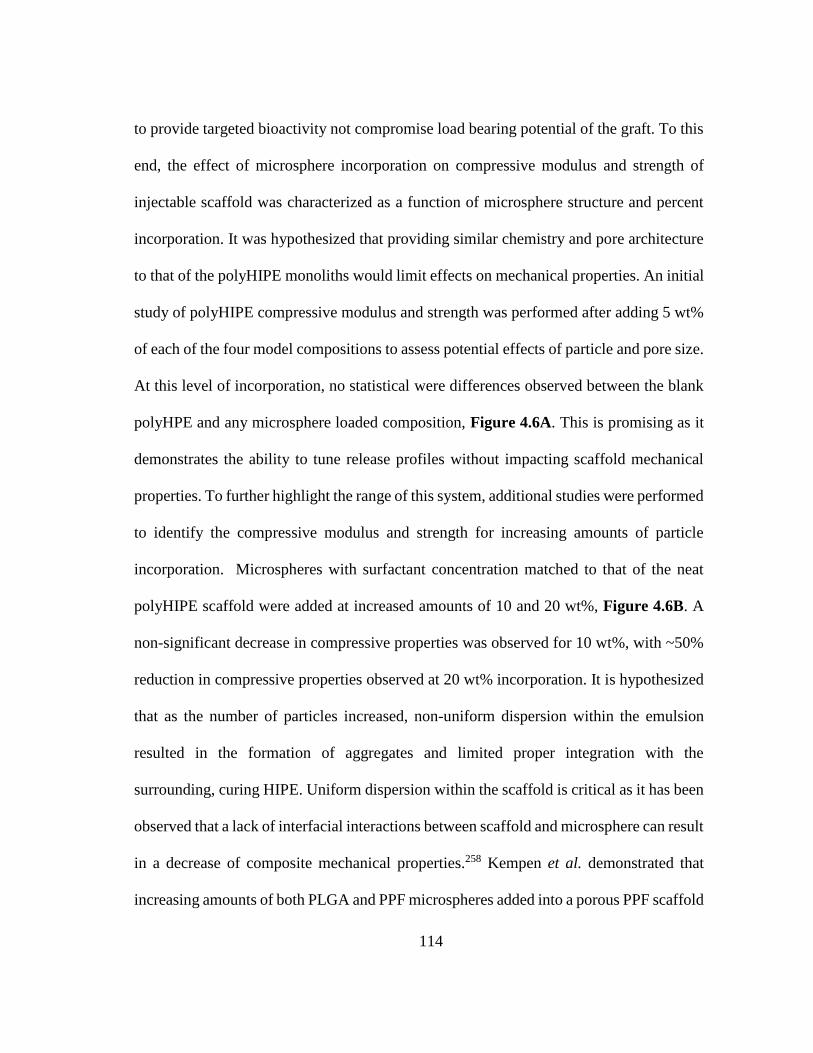
114
to provide targeted bioactivity not compromise load bearing potential of the graft. To this
end, the effect of microsphere incorporation on compressive modulus and strength of
injectable scaffold was characterized as a function of microsphere structure and percent
incorporation. It was hypothesized that providing similar chemistry and pore architecture
to that of the polyHIPE monoliths would limit effects on mechanical properties. An initial
study of polyHIPE compressive modulus and strength was performed after adding 5 wt%
of each of the four model compositions to assess potential effects of particle and pore size.
At this level of incorporation, no statistical were differences observed between the blank
polyHPE and any microsphere loaded composition, Figure 4.6A. This is promising as it
demonstrates the ability to tune release profiles without impacting scaffold mechanical
properties. To further highlight the range of this system, additional studies were performed
to identify the compressive modulus and strength for increasing amounts of particle
incorporation. Microspheres with surfactant concentration matched to that of the neat
polyHIPE scaffold were added at increased amounts of 10 and 20 wt%, Figure 4.6B. A
non-significant decrease in compressive properties was observed for 10 wt%, with ~50%
reduction in compressive properties observed at 20 wt% incorporation. It is hypothesized
that as the number of particles increased, non-uniform dispersion within the emulsion
resulted in the formation of aggregates and limited proper integration with the
surrounding, curing HIPE. Uniform dispersion within the scaffold is critical as it has been
observed that a lack of interfacial interactions between scaffold and microsphere can result
in a decrease of composite mechanical properties.258 Kempen et al. demonstrated that
increasing amounts of both PLGA and PPF microspheres added into a porous PPF scaffold

115
resulted in a significant decrease in compressive properties. Although a decrease was
observed with increasing incorporation of PPF microspheres, the availability of covalent,
interfacial interactions between scaffold and microsphere of matching chemistry resulted
in increased compressive properties over PLGA microspheres. From this, it is anticipated
that if uniform dispersion within our scaffold is maintained, allowing for appropriate
interfacial bonding between methacrylate functional groups of sphere and emulsion,
mechanical effects will be reduced. Current studies are exploring modified double barrel
syringes that allow for more homogenous mixing and distribution of microspheres in the
injectable HIPE. Furthermore, it is not anticipated that 20 wt% microsphere incorporation
will be needed to promote an osteogenic response. Microsphere incorporation ranging 1-
10 wt% has been demonstrated suitable to promote new bone formation utilizing loaded
protein concentrations similar to those presented here.118, 119, 259, 278 If a more robust
response is desired, BMP-2 concentration loaded into the primary emulsion can be easily
modulated to improve activity as has been observed in similar microsphere delivery
systems.278

116
Figure 4.6. Effect of microsphere particle size and pore size on composite scaffold
compressive modulus and strength for 5 wt% incorporation using four model
compositions (A). Effect of increasing microsphere incorporation (5, 10 and 20 wt%) on
composite scaffold compressive modulus and strength (B). All data represents average ±
standard deviation for n = 3. The * and ° represent significant difference (p<0.05) for 20
wt% compressive modulus or strength and other indicated compositions.
In addition, incorporation of microspheres into the polyHIPE scaffold will provide an
additional variable to tune release of osteoinductive factors in our system. Multiple studies
have reported on the ability to improve sustained release profiles of growth factor
delivered from microsphere vehicles by imbedding into secondary scaffolds.119, 239 These

117
studies have reported success in promoting osteogenic activity in vitro and in vivo.
Subsequent studies will assess the ability of polyHIPE scaffolds embedded with rhBMP-
2 loaded microspheres to promote osteoblastic differentatin of mesenchymal stem cells
cultured on the scaffold surface.
4.4. Conclusions
The aim of this study was to improve the osteoinductive potential of polyHIPE grafts
by utilizing a new polyHIPE microsphere-based growth factor delivery system and
establish the key relationships between microsphere properties and resultant protein
release kinetics. The independent and combined effects of particle and pore size on burst
and sustained release of proteins was determined. Bioactivity retention of the encapsulated
BMP-2 growth after emulsion fabrication and photopolymerization processes was
confirmed with demonstrated ability of the released BMP-2 to induce osteogenic
differentiation of hMSCs. Finally, microsphere loaded composites were characterized to
demonstrate that microsphere incorporation did not have deleterious effects on
compressive properties. Overall, this investigation provides key insights critical to the
design of an osteoinductive polyHIPE system capable of providing physiologically
relevant delivery profiles.

118
CHAPTER V
CONCLUSIONS
5.1. Summary
The work presented here aims to develop an improved bone replacement based on the
emulsion templating platform. These studies detail methods of fabricating injectable and
porous grafts with improved function in clinically relevant environments, cell delivery
platforms for improved loading of polyHIPE scaffolds, and porous microspheres for
targeted growth factor delivery. Together, these advancements address limitations of
current standards of care, including autologous and allogenic grafts, by providing a readily
available bone replacement that has been optimized to actively guide bone regeneration.
Injectable polyHIPEs with improved resistance to oxygen inhibition were fabricated
utilizing the thiol monomer pentaerythritol tetrakis(3-mercaptoproprionate), and the
biodegradable macomer, propylene fumarate dimethacrylate. Thiol-methacrylate
polyHIPEs exhibited rapid cure and improved network formation under oxygen rich
conditions and retained the ability to modulate key properties including porosity,
architecture, and compressive properties. Furthermore, thiol-methacrylate polymerization
introduced a potent tool to tune the hydrolytic degradation rates of these injectable
scaffolds. Matching the degradation profile of a tissue engineered scaffold with the healing
rate of the native tissue is critical component of improving defect regeneration.
To improve the regenerative capacity of polyHIPE grafts, poly(ethylene glycol)-
dithiothreitol hydrogels were utilized to improve seeding and distribution of human

119
mesenchymal stem cells (hMSCs) within 3D printed polyHIPEs. These in situ cell carriers,
fabricated from biodegradable, redox initiated hydrogels, exhibited tunable
polymerization rates, uniform cell distribution, and viable release of encapsulated stem
cells. Regenerative potential of these composite, cell-laden polyHIPEs was demonstrated
by characterizing osteoblastic differentiation of hMSCs released onto 3D printed
polyHIPEs. This in situ approach to cell seeding permits facile use of patient specific cells
that can be incorporated in tissue engineered scaffolds with minimal processing in the
surgical suite.
Finally, additional osteoinductive character was added to the graft through the solvent-
free fabrication of porous, in-line loaded microspheres capable of efficiently delivering
osteoinductive factors. Modulation of polyHIPE microsphere property provided tools for
tuning release kinetics without compromising compressive properties. Methods were
developed to characterize bioactivity retention of encapsulated BMP-2 factor using a
luminescence based reporter cell line and functional MSC differentiation assays. These
studies demonstrate the strong potential of polyHIPE microspheres to improve bone
regeneration through growth factor delivery and decrease manufacturing costs through
utilization of more efficient fabrication and loading techniques.
Collectively, these studies highlight the strong potential of polyHIPE scaffolds to serve
as an improved bone replacement with the ability to actively guide bone regeneration.
Although the technologies developed in this work hold advantages in bone tissue
engineering, the platforms utilized here are tunable in nature and be easily applied to the
engineering of other target tissues.

120
5.2. Significance of Work
To date, no system has been able to fully match the healing potential that autologous
tissue possesses due to its unique combination of osteoconductive (collagen-based ECM),
osteoinductive (growth factors and cytokines), and osteogenic (stem and osteoprogenitor
cells) properties. This work aimed to develop a polyHIPE graft that was capable of actively
guiding bone regeneration by modeling these natural processes that make autologous
grafting the current standard of care. Specifically, this work investigated PFDMA
polyHIPE scaffolds as a porous and osteoconductive substrate to support bone repair.
Furthermore, platforms to induce osteoinductive and osteogenic activity were investigated
through development of polymeric delivery vehicles that allow for incorporation of bone
morphogenetic proteins and mesenchymal stem cells. Combined, these technologies
provide the foundation for developing novel, patient specific polyHIPE bone replacements
with improved healing potential.
In Chapter 2, an alternative polymerization mechanism was explored to improve
polymerization times and network formation in clinically relevant, oxygen rich
enviornments. Redox initiated polyHIPEs have demonstrated a host of properties that are
advantageous for bone repair. However, these studies were performed in a controlled
environment that does not adequately represent all the challenges that are present in vivo.
Retaining established properties, including rapid cure, uniform pore architecture, and load
bearing potential, is dependent on the ability to obtain uniform high network formation in
oxygen rich environments. Addressing the problem of oxygen inhibition has been
discussed extensively in photopolymerized systems, however it has not been addressed

121
sufficiently in injectable systems.155 Development of an injectable, highly porous graft
that undergoes rapid polymerization in vivo is a major challenge in bone tissue
engineering. This work demonstrated a thiol-methacrylate polymerization route that
would be better immune to oxygen inhibition and provide an additional tool for
modulating scaffold degradation rate. Differences in patient demographics, including age,
health, and severity of injury, make developing a single graft capable of matching healing
rate extremely difficult. As a result, modulating thiol concentration is a critical tool as it
allows for rapid tuning of ester-based hydrolysis of the polyHIPE scaffold.
In Chapter 3, a platform for improved seeding of stem cells in tissue engineered grafts
was developed. Mesenchymal stem cells play a significant role in regeneration of
musculoskeletal tissues through recruitment and direction of progenitor cells, promotion
of angiogenesis, and modulation of immune response.192 Designing a biomaterial carrier
that improves placement, retention, and survival of transplanted stem cells is currently a
major focus in regenerative medicine as the hostile environment of injured tissue often
yields the direct injection of stem cells into the defect site ineffective.81 This work
provided a method of encapsulating stem cells in biodegradable hydrogel, protecting them
from early removal and subsequently improving the number and placement of stem cells
seeded in a 3D printed polyHIPE graft. Development of a composite polyHIPE-hydrogel
graft has the additional advantage of retaining requisite mechanical properties that are
often lacking in hydrogel carriers alone. In addition to the direct effect this platform could
have on improving regeneration, methods were developed to better characterize
osteogenic activity on polyHIPE grafts through characterization of ALP and

122
mineralization markers. These methods can be used and further adapted to investigate
osteogenic potential as additional technologies are introduced into polyHIPE grafts.
Furthermore, this work provides a platform that can be further adapted (e.g. bioactivity,
degradation profiles, and cell type) for incorporation of stem cells therapies into
engineering strategies for other target tissues, including cardiac and nerve applications.
In Chapter 4, growth factor delivery was explored as a means to improve the
osteoinductive character of our grafts. Through this work, a solvent-free method for
generating porous, growth factor loaded microspheres was established via combination of
emulsion templating and controlled fluidics fabrication techniques. Development of a
cost-efficient growth factor delivery system that has high loading efficiency and tunable
release kinetics is a significant challenge in tissue engineering. The improvements this
system presents in processing and purification offer the potential to maximize loading
efficiency while simultaneously minimizing scale up concerns. These studies produced
methods to characterize and modulate protein release, as well as assess bioactivity
retention of encapsulated factor, all protocols that can be easily adapted to a wide range
of bioactive factors. Furthermore, this system provides a baseline fabrication method that
can be further modified to deliver multiple growth factors simultaneously. Numerous
studies have probed the benefits of multiple factor delivery and worked to design systems
that better mimic the complex chemical environment of native tissue.253
Overall, this work demonstrates the ability to tune polyHIPE grafts to achieve desired
physical and mechanical properties in clinically relevant settings. Furthermore, the tunable
nature of the emulsion templating platform allows it to be easily tuned for the

123
incorporation of stem cells and bioactive factors. Although each component of this work
aimed to provide a specific advantage to our polyHIPE bone graft, the broader impact of
this work lies in the development of platforms and methods that can be easily modified to
achieve regeneration in any target tissue.
5.3. Challenges and Future Directions
This work details the development of tunable polyHIPE scaffolds with improved
healing potential through incorporation of stem cells and osteoinductive factors. Despite
the demonstrated improvements in scaffold function and osteogenic potential in vitro,
additional studies are needed to further optimize scaffold composition and assess bone
formation in vivo.
A primary focus of this work was the modification of polyHIPE scaffolds, and
development of assaying techniques, to better probe hMSC-polyHIPE interactions. It was
demonstrated that polyHIPE scaffolds support desired markers of stem cell attachment,
viability, and osteoblastic differentiation. However, these studies were performed in a
highly controlled in vitro setting that relied on the adsorption of serum proteins to facilitate
cell attachment. It is probable that cellular infiltration and migration would be reduced in
vivo due to a lack of native binding sites for host cells, a phenomenon observed in other
synthetic polymer systems.94 Developing a hybrid polyHIPE scaffold with ECM-like
surface modifiers would present an intriguing approach to encouraging native cell
infiltration, as well as a potent tool to controlling cell fate. Integrin-mediated control of
osteoblastic differentiation has been explored previously as a means of improving bone
regeneration.185 The work presented here demonstrated proof of principle protocols that

124
allow for facile incorporation of functionalized protein into polyHIPE scaffolds. Pore
surfaces of modified scaffolds stained positive for gelatin incorporation and promoted cell
attachment in serum free culture. This same approach could be utilized to crosslink a
multitude of ECM-like proteins to the pore surface. Streptococcal collagen-like proteins
are engineered proteins that form a triple helix similar to mammalian collagen, but with
the added advantage of being able to provide precise control over presented integrin
binding domains. Systematic investigation must first be completed to identify integrin-
mediated signaling pathways most active in osteoblastic differentiation, followed by
synthesis of the corresponding designer protein. Common methods for identification
include antibody blocking targeted integrin receptors and monitoring the effect on gene
expression using the osteoblastic markers, alkaline phosphatase, RUNX2, osteopontin,
osteocalcin, and collagen I. Next, attachment, viability, and differentiation protocols
discussed in this work could be implemented to ensure improvements in desired cell
markers over a two week culture period.
The ability to delivery cells within a polyHIPE scaffold was a key advancement
presented in this work. In vitro studies demonstrated significant improvement in cell
distribution and retention over standard suspension seeding methods. Despite these
improvements, it is critical that in vivo improvements be investigated as implantation into
diseased or injured tissue is known to pose significant challenges to cell retention.82 A
proof of principle study investigating cell retention could be performed utilizing a
subcutaneous model in Sprague-Dawley rats. A primary advantage of the in situ cell
carrier developed here is that it allows for facile incorporation of host specific cells in the

125
surgical setting. As such, bone marrow derived stromal cells can be isolated from femoral
limbs, expanded in vitro, and injected into a 3D printed polyHIPE. Monitoring retention
and migration of transplanted cells can be performed through RT-PCR analysis of gender
specific genes, fluorescent tagging using standard immunostaining protocols, or magnetic
resonance imaging of magnetically labelled cells. It is probable that in vivo application
may alter degradation profiles over those observed in vitro. If accelerated degradation
were to occur, it is possible that transplanted cells may be removed before they have
substantial time to adjust the regenerative microenvironment of damaged tissue. If
necessary, degradation rate can be adjusted by modulating the number of DTT linkages
present in the macromer backbone. This would allow for retention of established hydrogel
properties while ensuring cells remained protected and present long enough to produce a
robust trophic effect.
Treatment of musculoskeletal defects is a unique challenge in tissue engineering as
these injuries are often accompanied by large volumetric tissue loss, compromising native
vasculature and nutrient transport systems. As a result, it is necessary to engineer systems
that promote both osteo-and angiogenesis. BMP-2 delivery is a potent method for
improving osteogenic activity and has been shown to upregulate production of angiogenic
factors in osteoblasts, however direct delivery of angiogenic factors, such as vascular
endothelial growth factor, has been shown to provide synergistic improvements in bone
healing.253 Controlled co-delivery of these factors would provide a platform that could
better explore the complex feedback loops present in bone healing and vascularization
signaling pathways. The emulsion templating based-controlled fluidics fabrication method

126
would allow for development of microsphere delivery systems capable of co-delivery of
these factors. Core-shell systems allow for controlled, sequential delivery of multiple
therapeutic agents by encapsulating them in distinct spatial locations of the delivery
system and providing unique temporal release profiles.279 The present fluidics system
would be modified with a coaxial needle to allow dual injection of two primary emulsions,
each containing a distinct bioactive factor. These emulsions could be fabricated with
unique pore architectures to allow for independent control over release kinetics. Although
modulation of particle and pore size provides independent tools to control protein release
kinetics, it is probable that a more sustained release profiles will be desired. To extend
release, closed pore microspheres could be investigated to change the release mechanism
from solely passive diffusion, to degradation mediated release. Upon degradation of the
pore walls, encapsulated protein would be allowed to diffuse out into the surrounding
scaffold. Modulation of internal phase volume fraction, or use of a water soluble initiator
could facilitate formation of closed pore polyHIPEs. Although the methacrylated
macromer utilized for microsphere fabrication here does not degrade in a suitable time
frame, other systems explored here, such as thiol-methacrylate systems, could provide
more rapid and controlled degradation rates.
Finally, to fully assess the clinical potential of the polyHIPE system developed in this
work, in vivo osteogenesis must be monitored. The rabbit femoral condyle plug model
provides a rapid, high throughput method for investigation of biocompatibility, host
response and osteogenesis.280 It is hypothesized that the platforms developed in this work
would provide synergistic improvements in bone regeneration. To confirm this

127
hypothesis, a factorial study design must be implemented to investigate the individual and
synergistic effects of scaffold surface modification, stem cell delivery, and growth factor
delivery. MicroCT radiographic analysis would be used to monitor temporal changes in
mineralized tissue formation over 8 and 16 weeks. Next, histological analysis using H&E
and trichrome stains would be used to evaluate inflammation, vascularization, and bone
formation across various regions of each defect. Sections would be subjectively scored,
evaluated, and compared to blank defect controls and clinical calcium phosphate controls.
Finally, microscale integration of the scaffold with the host tissue will be assessed by
measuring peak loads of mechanical push out testing. Completion of this study would be
invaluable in the development of selection criteria needed to balance improvements in
healing with potential scale up and manufacturing costs.
Although additional investigation is required to confirm polyHIPE grafts are a viable
bone grafting option, the technologies detailed in this work provide a promising platform
that can be built upon to ensure all necessary criteria are met to support in vivo
regeneration. The methods established here allow for characterization of all major tenets
the tissue engineering paradigm, including scaffold properties, cell-material interactions,
and bioactive factor delivery. In summary, the presented work, combined with these
proposed studies, will provide knowledge critical to the development of a polyHIPE graft
capable of actively guiding bone regeneration.

128
REFERENCES
1. Bose, S. & Tarafder, S. Calcium phosphate ceramic systems in growth factor and
drug delivery for bone tissue engineering: a review. Acta biomaterialia 8, 1401-
1421 (2012).
2. Praemer, A., Furner, S., Rice, D.P. & Kelsey, J.L. Musculoskeletal conditions in
the United States. (1992).
3. Rodriguez-Merchan, E.C. & Forriol, F. Nonunion: general principles and
experimental data. Clinical orthopaedics and related research 419, 4-12 (2004).
4. Bosch, C., Melsen, B. & Vargervik, K. Importance of the critical-size bone defect
in testing bone-regenerating materials. The Journal of craniofacial surgery 9, 310-
316 (1998).
5. Key, J.A. The effect of a local calcium depot on osteogenesis and healing of
fractures. JBJS 16, 176-184 (1934).
6. Hollinger, J.O. & Kleinschmidt, J.C. The critical size defect as an experimental
model to test bone repair materials. The Journal of craniofacial surgery 1, 60-68
(1990).
7. Sommerfeldt, D. & Rubin, C. Biology of bone and how it orchestrates the form
and function of the skeleton. European Spine Journal 10, S86-S95 (2001).
8. Coombes, A.G.A. & Meikle, M.C. Resorbable synthetic polymers s replacements
for bone graft. Clinical Materials 17, 35-67 (1994).
9. Kalfas, I.H. Principles of bone healing. Neurosurgical Focus 10, 1-4 (2001).
10. Yaszemski, M.J., Payne, R.G., Hayes, W.C., Langer, R. & Mikos, A.G. Evolution
of bone transplantation: molecular, cellular and tissue strategies to engineer human
bone. Biomaterials 17, 175-185 (1996).
11. Dimitriou, R., Tsiridis, E. & Giannoudis, P.V. Current concepts of molecular
aspects of bone healing. Injury 36, 1392-1404 (2005).
12. Bostrom, M.P.G. Expression of bone morphogenetic proteins in fracture healing.
Clinical Orthopaedics and Related Research, S116-S123 (1998).
13. Carano, R.A. & Filvaroff, E.H. Angiogenesis and bone repair. Drug discovery
today 8, 980-989 (2003).

129
14. Hoerth, R.M. et al. Mechanical and structural properties of bone in non-critical and
critical healing in rat. Acta biomaterialia 10, 4009-4019 (2014).
15. Chamay, A. & Tschantz, P. Mechanical influences in bone remodeling.
Experimental research on Wolff's law. Journal of Biomechanics 5, 173-180
(1972).
16. Gutta, R. & Waite, P.D. Cranial bone grafting and simultaneous implants: a
submental technique to reconstruct the atrophic mandible. British Journal of Oral
and Maxillofacial Surgery 46, 477-479 (2008).
17. Samartzis, D. et al. Comparison of allograft to autograft in multilevelanterior
cervical discectomy and fusion with rigid plate fixation. The Spine Journal 3, 451-
459 (2003).
18. Chau, A. & Mobbs, R. Bone graft substitutes in anterior cervical discectomy and
fusion. European Spine Journal 18, 449-464 (2009).
19. Bauer, T.W. & Muschler, G.F. Bone graft materials - An overview of the basic
science. Clinical Orthopaedics and Related Research, 10-27 (2000).
20. Kretlow, J.D., Young, S., Klouda, L., Wong, M. & Mikos, A.G. Injectable
biomaterials for regenerating complex craniofacial tissues. Advanced Materials
21, 3368-3393 (2009).
21. Dumas, J.E. et al. Synthesis and characterization of an injectable allograft
bone/polymer composite bone void filler with tunable mechanical properties.
Tissue Engineering: Part A 16, 2505-2518 (2010).
22. Katti, K.S. Biomaterials in total joint replacement. Colloids and Surfaces B:
Biointerfaces 39, 133-142 (2004).
23. Haas, S.S., Brauer, G.M. & Dickson, G. A characterization of
polymethylmethacrylate bone cement. Journal Article 57, 380-391 (1975).
24. Zhang, J., Liu, W., Schnitzler, V., Tancret, F. & Bouler, J.-M. Calcium phosphate
cements for bone substitution: Chemistry, handling and mechanical properties.
Acta Biomaterialia 10, 1035-1049 (2014).
25. Rose, F.R. & Oreffo, R.O. Bone tissue engineering: hope vs hype. Biochemical
and biophysical research communications 292, 1-7 (2002).
26. Langer, R. & Vacanti, J.P. Tissue engineering. Science 260, 920-926 (1993).

130
27. Karageorgiou, V. & Kaplan, D. Porosity of 3D biomaterial scaffolds and
osteogenesis. Biomaterials 26, 5474-5491 (2005).
28. Ishaug-Riley, S.L., Crane-Kruger, G.M., Yaszemski, M.J. & Mikos, A.G. Three-
dimensional culture of rat calvarial osteoblasts in porous biodegradable polymers.
Biomaterials 19, 1405-1412 (1998).
29. Marcacci, M. et al. Stem cells associated with macroporous bioceramics for long
bone repair: 6-to 7-year outcome of a pilot clinical study. Tissue Engineering 13,
947-955 (2007).
30. Taylor, D. Fatigue of bone and bones: An analysis based on stressed volume.
Journal of Orthopaedic Research 16, 163-169 (1998).
31. Huiskes, R., Weinans, H. & Van Rietbergen, B. The relationship between stress
shielding and bone resorption around total hip stems and the effects of flexible
materials. Clinical orthopaedics and related research 274, 124-134 (1992).
32. Temenoff, J.S. & Mikos, A.G. Injectable biodegradable materials for orthopedic
tissue engineering. Biomaterials 21, 2405-2412 (2000).
33. Dumas, J. et al. Balancing the rates of new bone formation and polymer
degradation enhances healing of weight-bearing allograft/polyurethane
composites in rabbit femoral defects. Tissue engineering. Part A 20, 115-129
(2014).
34. Pham, Q.P., Sharma, U. & Mikos, A.G. Electrospinning of polymeric nanofibers
for tissue engineering applications: a review. Tissue engineering 12, 1197-1211
(2006).
35. Nezarati, R.M., Eifert, M.B. & Cosgriff-Hernandez, E. Effects of humidity and
solution viscosity on electrospun fiber morphology. Tissue Engineering Part C:
Methods 19, 810-819 (2013).
36. Kim, T.K., Yoon, J.J., Lee, D.S. & Park, T.G. Gas foamed open porous
biodegradable polymeric microspheres. Biomaterials 27, 152-159 (2006).
37. Harris, L.D., Kim, B.-S. & Mooney, D.J. Open pore biodegradable matrices
formed with gas foaming. Journal of biomedical materials research 42, 396-402
(1998).
38. Hacker, M. et al. Solid lipid templating of macroporous tissue engineering
scaffolds. Biomaterials 28, 3497-3507 (2007).

131
39. Mistry, A.S. et al. Fabrication and in vitro degradation of porous fumarate‐based
polymer/alumoxane nanocomposite scaffolds for bone tissue engineering. Journal
of Biomedical Materials Research Part A 89, 68-79 (2009).
40. Lin-Gibson, S., Cooper, J.A., Landis, F.A. & Cicerone, M.T. Systematic
investigation of porogen size and content on scaffold morphometric parameters
and properties. Biomacromolecules 8, 1511-1518 (2007).
41. Guan, J., Fujimoto, K.L., Sacks, M.S. & Wagner, W.R. Preparation and
characterization of highly porous, biodegradable polyurethane scaffolds for soft
tissue applications. Biomaterials 26, 3961-3971 (2005).
42. Day, R.M. et al. In vivo characterisation of a novel bioresorbable poly(lactide-co-
glycolide) tubular foam scaffold for tissue engineering applications. Journal of
Materials Science: Materials in Medicine 15, 729-734 (2004).
43. Busby, W., Cameron, N.R. & Jahoda, C.A.B. Emulsion-derived foams
(polyHIPEs) containing poly(ε-caprolactone) as matrixes for tissue engineering.
Biomacromolecules 2, 154-164 (2001).
44. Busby, W., Cameron, N.R. & Jahoda, C.A.B. Tissue engineering matrixes by
emulsion templating. Polymer International 51, 871-881 (2002).
45. Barbetta, A. et al. Scaffolds based on biopolymeric foams. Adv Funct Mater 15,
118-124 (2005).
46. Bokhari, M.A., Birch, M.A. & Akay, G. Polyhipe polymer: a novel scaffold for in
vitro bone tissue engineering. Experimental Medical Biology: Tissue Engineering,
Stem Cells, and Gene Therapies 534, 247-254 (2003).
47. Cameron, N.R., Barbetta, A. & Cooper, S.J. High internal phase emulsions
(HIPEs) containing divinylbenzene and 4-vinylbenzyl chloride and the
morphology of the resulting PolyHIPE materials. Chemical Communications,
221–222 (2000).
48. Cameron, N.R. & Sherrington, D.C. Synthesis and characterisation of poly(aryl
ether sulfone) polyHIPE materials. Macromolecules 30, 5860-5869 (1997).
49. Hayman, M.W., Smith, K.H., Cameron, N.R. & Przyborskia, S.A. Growth of
human stem cell-derived neurons on solid three-dimensional polymers. J Biochem
Biophys Methods 62, 231-240 (2005).
50. Decker, C. Effect of UV radiation on polymers. Marcel Dekker, Handbook of
Polymer Science and Technology. 3, 541-608 (1989).

132
51. Decker, C. & Jenkins, A.D. Kinetic approach of oxygen inhibition in ultraviolet-
and laser-induced polymerizations. Macromolecules 18, 1241-1244 (1985).
52. O'Brien, A.K., Cramer, N.B. & Bowman, C.N. Oxygen inhibition in thiol–acrylate
photopolymerizations. Journal of Polymer Science Part A: Polymer Chemistry 44,
2007-2014 (2006).
53. Murphy, M.B., Moncivais, K. & Caplan, A.I. Mesenchymal stem cells:
environmentally responsive therapeutics for regenerative medicine. Experimental
& molecular medicine 45, e54 (2013).
54. Friedenstein, A., Piatetzky-Shapiro, I. & Petrakova, K. Osteogenesis in transplants
of bone marrow cells. Development 16, 381-390 (1966).
55. Caplan, A.I. Mesenchymal stem cells. Journal of orthopaedic research 9, 641-650
(1991).
56. Owen, M. Marrow stromal stem cells. J Cell Sci 1988, 63-76 (1988).
57. Pittenger, M.F. et al. Multilineage potential of adult human mesenchymal stem
cells. science 284, 143-147 (1999).
58. Dominici, M. et al. Minimal criteria for defining multipotent mesenchymal stromal
cells. The International Society for Cellular Therapy position statement.
Cytotherapy 8, 315-317 (2006).
59. DiGirolamo, C.M. et al. Propagation and senescence of human marrow stromal
cells in culture: a simple colony‐forming assay identifies samples with the greatest
potential to propagate and differentiate. British journal of haematology 107, 275-
281 (1999).
60. Murphy, J.M. et al. Reduced chondrogenic and adipogenic activity of
mesenchymal stem cells from patients with advanced osteoarthritis. Arthritis &
Rheumatology 46, 704-713 (2002).
61. Barry, F.P. & Murphy, J.M. Mesenchymal stem cells: clinical applications and
biological characterization. The international journal of biochemistry & cell
biology 36, 568-584 (2004).
62. Schäffler, A. & Büchler, C. Concise review: adipose tissue‐derived stromal cells—
basic and clinical implications for novel cell‐based therapies. Stem cells 25, 818-
827 (2007).

133
63. Yoshimura, H. et al. Comparison of rat mesenchymal stem cells derived from bone
marrow, synovium, periosteum, adipose tissue, and muscle. Cell and tissue
research 327, 449-462 (2007).
64. Lee, O.K. et al. Isolation of multipotent mesenchymal stem cells from umbilical
cord blood. Blood 103, 1669-1675 (2004).
65. Caplan, Arnold I. & Correa, D. The msc: an injury drugstore. Cell Stem Cell 9, 11-
15 (2011).
66. Haynesworth, S.E., Baber, M.A. & Caplan, A.I. Cytokine expression by human
marrow‐derived mesenchymal progenitor cells in vitro: Effects of dexamethasone
and IL‐1α. Journal of cellular physiology 166, 585-592 (1996).
67. Caplan, A.I. & Bruder, S.P. Mesenchymal stem cells: building blocks for
molecular medicine in the 21st century. Trends in molecular medicine 7, 259-264
(2001).
68. Sorrell, J.M., Baber, M.A. & Caplan, A.I. Influence of adult mesenchymal stem
cells on in vitro vascular formation. Tissue Engineering Part A 15, 1751-1761
(2009).
69. Chen, L., Tredget, E.E., Wu, P.Y. & Wu, Y. Paracrine factors of mesenchymal
stem cells recruit macrophages and endothelial lineage cells and enhance wound
healing. PloS one 3, e1886 (2008).
70. Tateishi-Yuyama, E. et al. Therapeutic angiogenesis for patients with limb
ischaemia by autologous transplantation of bone-marrow cells: a pilot study and a
randomised controlled trial. The Lancet 360, 427-435 (2002).
71. Aggarwal, S. & Pittenger, M.F. Human mesenchymal stem cells modulate
allogeneic immune cell responses. Blood 105, 1815-1822 (2005).
72. Ren, G. et al. Mesenchymal stem cell-mediated immunosuppression occurs via
concerted action of chemokines and nitric oxide. Cell stem cell 2, 141-150 (2008).
73. Selmani, Z. et al. Human leukocyte antigen‐G5 secretion by human mesenchymal
stem cells is required to suppress T lymphocyte and natural killer function and to
induce CD4+ CD25highFOXP3+ regulatory T cells. Stem cells 26, 212-222
(2008).
74. Cselenyák, A., Pankotai, E., Horváth, E.M., Kiss, L. & Lacza, Z. Mesenchymal
stem cells rescue cardiomyoblasts from cell death in an in vitro ischemia model
via direct cell-to-cell connections. BMC Cell Biology 11, 29 (2010).

134
75. Uccelli, A., Moretta, L. & Pistoia, V. Mesenchymal stem cells in health and
disease. Nature reviews immunology 8, 726 (2008).
76. Gonzalez-Rey, E., Gonzalez, M.A., Rico, L., Buscher, D. & Delgado, M. Human
adult stem cells derived from adipose tissue protect against experimental colitis
and sepsis. Gut (2009).
77. Connolly, J.F., Guse, R., Tiedeman, J. & Dehne, R. Autologous marrow injection
as a substitute for operative grafting of tibial nonunions. Clinical orthopaedics and
related research, 259-270 (1991).
78. Hernigou, P., Poignard, A., Beaujean, F. & Rouard, H. Percutaneous autologous
bone-marrow grafting for nonunions: influence of the number and concentration
of progenitor cells. JBJS 87, 1430-1437 (2005).
79. Bruder, S.P., Kraus, K.H., Goldberg, V.M. & Kadiyala, S. The effect of implants
loaded with autologous mesenchymal stem cells on the healing of canine
segmental bone defects. JBJS 80, 985-996 (1998).
80. Bruder, S.P. et al. Bone regeneration by implantation of purified, culture‐expanded
human mesenchymal stem cells. Journal of orthopaedic research 16, 155-162
(1998).
81. Horwitz, E.M. et al. Transplantability and therapeutic effects of bone marrow-
derived mesenchymal cells in children with osteogenesis imperfecta. Nature
medicine 5, 309 (1999).
82. Burdick, J.A., Mauck, R.L. & Gerecht, S. To serve and protect: hydrogels to
improve stem cell-based therapies. Cell stem cell 18, 13-15 (2016).
83. Nicodemus, G.D. & Bryant, S.J. Cell encapsulation in biodegradable hydrogels for
tissue engineering applications. Tissue Engineering Part B: Reviews 14, 149-165
(2008).
84. Marquardt, L.M. & Heilshorn, S.C. Design of injectable materials to improve stem
cell transplantation. Current stem cell reports 2, 207-220 (2016).
85. Awad, H.A., Wickham, M.Q., Leddy, H.A., Gimble, J.M. & Guilak, F.
Chondrogenic differentiation of adipose-derived adult stem cells in agarose,
alginate, and gelatin scaffolds. Biomaterials 25, 3211-3222 (2004).
86. Nichol, J.W. et al. Cell-laden microengineered gelatin methacrylate hydrogels.
Biomaterials 31, 5536-5544 (2010).

135
87. Nuttelman, C.R., Tripodi, M.C. & Anseth, K.S. In vitro osteogenic differentiation
of human mesenchymal stem cells photoencapsulated in PEG hydrogels. Journal
of Biomedical Materials Research Part A 68, 773-782 (2004).
88. Burdick, J.A. & Anseth, K.S. Photoencapsulation of osteoblasts in injectable
RGD-modified PEG hydrogels for bone tissue engineering. Biomaterials 23, 4315-
4323 (2002).
89. Nuttelman, C.R., Tripodi, M.C. & Anseth, K.S. Synthetic hydrogel niches that
promote hMSC viability. Matrix biology 24, 208-218 (2005).
90. Temenoff, J.S. et al. Thermally cross-linked oligo (poly (ethylene glycol)
fumarate) hydrogels support osteogenic differentiation of encapsulated marrow
stromal cells in vitro. Biomacromolecules 5, 5-10 (2004).
91. Qiu, Y. et al. PEG-based hydrogels with tunable degradation characteristics to
control delivery of marrow stromal cells for tendon overuse injuries. Acta
biomaterialia 7, 959-966 (2011).
92. Simmons, C.A., Alsberg, E., Hsiong, S., Kim, W.J. & Mooney, D.J. Dual growth
factor delivery and controlled scaffold degradation enhance in vivo bone formation
by transplanted bone marrow stromal cells. Bone 35, 562-569 (2004).
93. Zhao, L., Weir, M.D. & Xu, H.H. An injectable calcium phosphate-alginate
hydrogel-umbilical cord mesenchymal stem cell paste for bone tissue engineering.
Biomaterials 31, 6502-6510 (2010).
94. Guo, R. et al. A transient cell-shielding method for viable MSC delivery within
hydrophobic scaffolds polymerized in situ. Biomaterials 54, 21-33 (2015).
95. Barnes, G.L., Kostenuik, P.J., Gerstenfeld, L.C. & Einhorn, T.A. Growth factor
regulation of fracture repair. Journal of Bone and Mineral Research 14, 1805-1815
(1999).
96. Lieberman, J.R., Daluiski, A. & Einhorn, T.A. The role of growth factors in the
repair of bone: biology and clinical applications. JBJS 84, 1032-1044 (2002).
97. Heldin, C.-H., Miyazono, K. & Ten Dijke, P. TGF-β signalling from cell
membrane to nucleus through SMAD proteins. Nature 390, 465 (1997).
98. Ferguson, C., Alpern, E., Miclau, T. & Helms, J.A. Does adult fracture repair
recapitulate embryonic skeletal formation? Mechanisms of development 87, 57-66
(1999).
99. Urist, M.R. Bone: formation by autoinduction. Science 150, 893-899 (1965).

136
100. Wozney, J.M. et al. Novel regulators of bone formation: molecular clones and
activities. Science 242, 1528-1534 (1988).
101. Cheng, H. et al. Osteogenic activity of the fourteen types of human bone
morphogenetic proteins (BMPs). JBJS 85, 1544-1552 (2003).
102. Tsuji, K. et al. BMP2 activity, although dispensable for bone formation, is required
for the initiation of fracture healing. Nature genetics 38, 1424 (2006).
103. Fiedler, J., Röderer, G., Günther, K.P. & Brenner, R.E. BMP‐2, BMP‐4, and
PDGF‐bb stimulate chemotactic migration of primary human mesenchymal
progenitor cells. Journal of cellular biochemistry 87, 305-312 (2002).
104. Deckers, M.M. et al. Bone morphogenetic proteins stimulate angiogenesis through
osteoblast-derived vascular endothelial growth factor A. Endocrinology 143,
1545-1553 (2002).
105. Zhang, F. et al. Sustained BMP signaling in osteoblasts stimulates bone formation
by promoting angiogenesis and osteoblast differentiation. Journal of Bone and
Mineral Research 24, 1224-1233 (2009).
106. Zhang, W. et al. VEGF and BMP-2 promote bone regeneration by facilitating bone
marrow stem cell homing and differentiation. Eur Cell Mater 27, 1-11 (2014).
107. McKay, W.F., Peckham, S.M. & Badura, J.M. A comprehensive clinical review of
recombinant human bone morphogenetic protein-2 (INFUSE® Bone Graft).
International orthopaedics 31, 729-734 (2007).
108. Burkus, J.K., Gornet, M.F., Dickman, C.A. & Zdeblick, T.A. Anterior lumbar
interbody fusion using rhBMP-2 with tapered interbody cages. Clinical Spine
Surgery 15, 337-349 (2002).
109. Vaccaro, A.R. et al. A pilot study evaluating the safety and efficacy of OP-1 Putty
(rhBMP-7) as a replacement for iliac crest autograft in posterolateral lumbar
arthrodesis for degenerative spondylolisthesis. Spine 29, 1885-1892 (2004).
110. Friedlaender, G.E. et al. Osteogenic protein-1 (bone morphogenetic protein-7) in
the treatment of tibial nonunions: a prospective, randomized clinical trial
comparing rhOP-1 with fresh bone autograft. The Journal of bone and joint
surgery. American volume 83, S151 (2001).
111. Carragee, E.J., Hurwitz, E.L. & Weiner, B.K. A critical review of recombinant
human bone morphogenetic protein-2 trials in spinal surgery: emerging safety
concerns and lessons learned. The Spine Journal 11, 471-491 (2011).

137
112. Shields, L.B. et al. Adverse effects associated with high-dose recombinant human
bone morphogenetic protein-2 use in anterior cervical spine fusion. Spine 31, 542-
547 (2006).
113. Geiger, M., Li, R. & Friess, W. Collagen sponges for bone regeneration with
rhBMP-2. Advanced drug delivery reviews 55, 1613-1629 (2003).
114. Gokmen, M.T. & Du Prez, F.E. Porous polymer particles—A comprehensive
guide to synthesis, characterization, functionalization and applications. Progress
in Polymer Science 37, 365-405 (2012).
115. Makadia, H.K. & Siegel, S.J. Poly lactic-co-glycolic acid (PLGA) as
biodegradable controlled drug delivery carrier. Polymers 3, 1377-1397 (2011).
116. Anderson, J.M. & Shive, M.S. Biodegradation and biocompatibility of PLA and
PLGA microspheres. Advanced drug delivery reviews 28, 5-24 (1997).
117. Cohen, S., Yoshioka, T., Lucarelli, M., Hwang, L.H. & Langer, R. Controlled
delivery systems for proteins based on poly (lactic/glycolic acid) microspheres.
Pharmaceutical research 8, 713-720 (1991).
118. Lee, J.W. et al. Bone regeneration using a microstereolithography-produced
customized poly (propylene fumarate)/diethyl fumarate photopolymer 3D scaffold
incorporating BMP-2 loaded PLGA microspheres. Biomaterials 32, 744-752
(2011).
119. Li, B. et al. The effects of rhBMP-2 released from biodegradable
polyurethane/microsphere composite scaffolds on new bone formation in rat
femora. Biomaterials 30, 6768-6779 (2009).
120. Lee, J., Oh, Y.J., Lee, S.K. & Lee, K.Y. Facile control of porous structures of
polymer microspheres using an osmotic agent for pulmonary delivery. Journal of
Controlled Release 146, 61-67 (2010).
121. Nam, Y.S., Yoon, J.J. & Park, T.G. A novel fabrication method of macroporous
biodegradable polymer scaffolds using gas foaming salt as a porogen additive.
Journal of biomedical materials research 53, 1-7 (2000).
122. Jain, R.A. The manufacturing techniques of various drug loaded biodegradable
poly (lactide-co-glycolide)(PLGA) devices. Biomaterials 21, 2475-2490 (2000).
123. Berkland, C., Pollauf, E., Pack, D.W. & Kim, K.K. Uniform double-walled
polymer microspheres of controllable shell thickness. Journal of controlled
release 96, 101-111 (2004).

138
124. Middleton, J.C. & Tipton, A.J. Synthetic biodegradable polymers as orthopedic
devices. Biomaterials 21, 2335-2346 (2000).
125. Akay, G., Birch, M.A. & Bokhari, M.A. Microcellular polyHIPE polymer supports
osteoblast growth and bone formation in vitro. Biomaterials 25, 3991-4000 (2004).
126. Moglia, R.S. et al. Injectable PolyHIPEs as High-Porosity Bone Grafts.
Biomacromolecules 12, 3621-3628 (2011).
127. Moglia, R.S. et al. Injectable polyMIPE scaffolds for soft tissue regeneration.
Polymer 55, 426-434 (2014).
128. Moglia, R.S. et al. Injectable Polymerized High Internal Phase Emulsions with
Rapid in Situ Curing. Biomacromolecules 15, 2870-2878 (2014).
129. Robinson, J.L. et al. Osteoinductive PolyHIPE Foams as Injectable Bone Grafts.
Tissue Engineering Part A 22, 403-414 (2016).
130. Robinson, J.L., Moglia, R.S., Stuebben, M.C., McEnery, M.A. & Cosgriff-
Hernandez, E. Achieving interconnected pore architecture in injectable polyHIPEs
for bone tissue engineering. Tissue Engineering Part A 20, 1103-1112 (2014).
131. Barbetta, A., Dentini, M., Zannoni, E.M. & De Stefano, M.E. Tailoring the
Porosity and Morphology of Gelatin-Methacrylate PolyHIPE Scaffolds for Tissue
Engineering Applications. Langmuir 21, 12333-12341 (2005).
132. Peter, S.J., Miller, M.J., Yasko, A.W., Yaszemski, M.J. & Mikos, A.G. Polymer
concepts in tissue engineering. Journal of Biomedical Materials Research 43, 422-
427 (1998).
133. Rueggeberg, F. & Margeson, D. The effect of oxygen inhibition on an
unfilled/filled composite system. Journal of Dental Research 69, 1652-1658
(1990).
134. Cramer, N.B. & Bowman, C.N. Kinetics of thiol–ene and thiol–acrylate
photopolymerizations with real-time fourier transform infrared. Journal of
Polymer Science Part A: Polymer Chemistry 39, 3311-3319 (2001).
135. Morgan, C.R., Magnotta, F. & Ketley, A.D. Thiol/ene photocurable polymers.
Journal of Polymer Science: Polymer Chemistry Edition 15, 627-645 (1977).
136. O'Brien, A.K. & Bowman, C.N. Modeling the effect of oxygen on
photopolymerization kinetics. Macromolecular Theory and Simulations 15, 176-
182 (2006).

139
137. Caldwell, S. et al. Degradable emulsion-templated scaffolds for tissue engineering
from thiol-ene photopolymerisation. Soft Matter 8, 10344-10351 (2012).
138. Lovelady, E., Kimmins, S.D., Wu, J. & Cameron, N.R. Preparation of emulsion-
templated porous polymers using thiol-ene and thiol-yne chemistry. Polym Chem
2, 559-562 (2011).
139. Langford, C., Johnson, D. & Cameron, N. Chemical functionalization of emulsion-
templated porous polymers by thiol–ene “click” chemistry. Polymer Chemistry 5,
6200-6206 (2014).
140. Hynes, R.O. Integrins: bidirectional, allosteric signaling machines. Cell 110, 673-
687 (2002).
141. Giancotti, F.G. & Ruoslahti, E. Integrin signaling. Science 285, 1028-1033 (1999).
142. Chastain, S.R., Kundu, A.K., Dhar, S., Calvert, J.W. & Putnam, A.J. Adhesion of
mesenchymal stem cells to polymer scaffolds occurs via distinct ECM ligands and
controls their osteogenic differentiation. Journal of Biomedical Materials
Research Part A 78, 73-85 (2006).
143. Bokhari, M.A., Akay, G., Zhang, S. & Birch, M.A. The enhancement of osteoblast
growth and differentiation in vitro on a peptide hydrogel—polyHIPE polymer
hybrid material. Biomaterials 26, 5198-5208 (2005).
144. Chen, G., Sato, T., Ushida, T., Ochiai, N. & Tateishi, T. Tissue engineering of
cartilage using a hybrid scaffold of synthetic polymer and collagen. Tissue
Engineering 10, 323-330 (2004).
145. Timmer, M.D., Horch, A.R., Ambrose, C.G. & Mikos, A.G. Effect of
physiological temperature on the mechanical properties and network structure of
biodegradable poly(propylene fumarate)-based networks. J Biomat Sci Polym Ed
14, 369-382 (2003).
146. Whitely, M.E. et al. Prevention of Oxygen Inhibition of PolyHIPE Radical
Polymerization Using a Thiol-Based Cross-Linker. ACS biomaterials science &
engineering 3, 409-419 (2017).
147. Foudazi, R., Gokun, P., Feke, D.L., Rowan, S.J. & Manas-Zloczower, I.
Chemorheology of Poly(high internal phase emulsions). Macromolecules 46,
5393-5396 (2013).
148. Barbetta, A. & Cameron, N.R. Morphology and surface area of emulsion-derived
(PolyHIPE) solid foams prepared with oil-phase soluble porogenic solvents: Span
80 as surfactant. Macromolecules 37, 3188-3201 (2004).

140
149. Gregory, T.R. Nucleotypic effects without nuclei: genome size and erythrocyte
size in mammals. Genome 43, 895-901 (2000).
150. Kloosterboer, J.G. in Electronic applications 1-61 (Springer, 1988).
151. Gou, L., Coretsopoulos, C.N. & Scranton, A.B. Measurement of the dissolved
oxygen concentration in acrylate monomers with a novel photochemical method.
Journal of Polymer Science Part A: Polymer Chemistry 42, 1285-1292 (2004).
152. Rydholm, A.E., Bowman, C.N. & Anseth, K.S. Degradable thiol-acrylate
photopolymers: polymerization and degradation behavior of an in situ forming
biomaterial. Biomaterials 26, 4495-4506 (2005).
153. Bowman, C.N. & Kloxin, C.J. Toward an enhanced understanding and
implementation of photopolymerization reactions. AIChE Journal 54, 2775-2795
(2008).
154. Decker, C. UV-curing chemistry: past, present, and future. JCT, Journal of
coatings technology 59, 97-106 (1987).
155. Studer, K., Decker, C., Beck, E. & Schwalm, R. Overcoming oxygen inhibition in
UV-curing of acrylate coatings by carbon dioxide inerting, Part I. Progress in
Organic Coatings 48, 92-100 (2003).
156. Ligon, S.C., Husar, B., Wutzel, H., Holman, R. & Liska, R. Strategies to reduce
oxygen inhibition in photoinduced polymerization. Chemical reviews 114, 557-
589 (2013).
157. Murphy, C.M., Haugh, M.G. & O'Brien, F.J. The effect of mean pore size on cell
attachment, proliferation and migration in collagen–glycosaminoglycan scaffolds
for bone tissue engineering. Biomaterials 31, 461-466 (2010).
158. Barbetta, A. et al. in Macromolecular Symposia, Vol. 226 203-212 (Wiley Online
Library, 2005).
159. Hainey, P., Huxham, I., Rowatt, B., Sherrington, D. & Tetley, L. Synthesis and
ultrastructural studies of styrene-divinylbenzene polyhipe polymers.
Macromolecules 24, 117-121 (1991).
160. Lee, T., Guymon, C., Jönsson, E.S. & Hoyle, C. The effect of monomer structure
on oxygen inhibition of (meth) acrylates photopolymerization. Polymer 45, 6155-
6162 (2004).

141
161. Peter, S.J., Lu, L., Kim, D.J. & Mikos, A.G. Marrow stromal osteoblast function
on a poly (propylene fumarate)/β-tricalcium phosphate biodegradable orthopaedic
composite. Biomaterials 21, 1207-1213 (2000).
162. Peter, S.J. et al. In vitro degradation of a poly (propylene fumarate)/β-tricalcium
phosphate composite orthopaedic scaffold. Tissue Engineering 3, 207-215 (1997).
163. Yaszemski, M.J. et al. The ingrowth of new bone tissue and initial mechanical
properties of a degrading polymeric composite scaffold. Tissue engineering 1, 41-
52 (1995).
164. Stańczyk, M. & Van Rietbergen, B. Thermal analysis of bone cement
polymerisation at the cement–bone interface. Journal of biomechanics 37, 1803-
1810 (2004).
165. Cole, M.A., Jankousky, K.C. & Bowman, C.N. Redox initiation of bulk thiol–ene
polymerizations. Polymer chemistry 4, 1167-1175 (2013).
166. Fouassier, J.-P. & Rabek, J.F. Radiation curing in polymer science and technology:
Practical aspects and applications, Vol. 4. (Springer Science & Business Media,
1993).
167. Mistry, A. et al. In vivo bone biocompatibility and degradation of porous fumarate-
based polymer/alumoxane nanocomposites for bone tissue engineering. J Biomed
Mater Res A 92A, 451-462 (2010).
168. Patel, Z.S. et al. Dual delivery of an angiogenic and an osteogenic growth factor
for bone regeneration in a critical size defect model. Bone 43, 931-940 (2008).
169. Kim, S.-S., Park, M.S., Jeon, O., Choi, C.Y. & Kim, B.-S. Poly (lactide-co-
glycolide)/hydroxyapatite composite scaffolds for bone tissue engineering.
Biomaterials 27, 1399-1409 (2006).
170. Dumas, J.E. et al. Injectable reactive biocomposites for bone healing in critical-
size rabbit calvarial defects. Biomedical Materials 7, 024112 (2012).
171. Kim, C.W. et al. Characterization of porous injectable poly‐(propylene fumarate)‐based bone graft substitute. Journal of Biomedical Materials Research Part A 85,
1114-1119 (2008).
172. P. Fisher, J., Holland, T.A., Dean, D., Engel, P.S. & Mikos, A.G. Synthesis and
properties of photocross-linked poly (propylene fumarate) scaffolds. Journal of
Biomaterials Science, Polymer Edition 12, 673-687 (2001).

142
173. Dumas, J.E. et al. Synthesis and characterization of an injectable allograft
bone/polymer composite bone void filler with tunable mechanical properties.
Tissue Engineering Part A 16, 2505-2518 (2010).
174. Gorna, K. & Gogolewski, S. Biodegradable porous polyurethane scaffolds for
tissue repair and regeneration. Journal of Biomedical Materials Research Part A
79, 128-138 (2006).
175. Graham, N.B. & Cameron, A. Nanogels and microgels: The new polymeric
materials playground. Pure Appl Chem 70, 1271-1275 (1998).
176. Schoenmakers, R.G., van de Wetering, P., Elbert, D.L. & Hubbell, J.A. The effect
of the linker on the hydrolysis rate of drug-linked ester bonds. J Control Release
95, 291-300 (2004).
177. Timmer, M.D., Shin, H., Horch, R.A., Ambrose, C.G. & Mikos, A.G. In vitro
cytotoxicity of injectable and biodegradable poly (propylene fumarate)-based
networks: unreacted macromers, cross-linked networks, and degradation products.
Biomacromolecules 4, 1026-1033 (2003).
178. Peter, S.J., Miller, S.T., Zhu, G., Yasko, A.W. & Mikos, A.G. In vivo degradation
of a poly(propylene fumarate)/β-tricalcium phosphate injectable composite
scaffold. J Biomed Mater Res 41, 1-7 (1998).
179. He, S. et al. Synthesis of biodegradable poly (propylene fumarate) networks with
poly (propylene fumarate)–diacrylate macromers as crosslinking agents and
characterization of their degradation products. Polymer 42, 1251-1260 (2001).
180. Shi, X.e.a. Fabrication of porous ultra-short single-walled carbon nanotube
nanocomposite scaffolds for bone tissue engineering. Biomaterials 28, 4078-4090
(2007).
181. Roberts, J.J. & Bryant, S.J. Comparison of photopolymerizable thiol-ene PEG and
acrylate-based PEG hydrogels for cartilage development. Biomaterials 34, 9969-
9979 (2013).
182. Ruoslahti, E. & Pierschbacher, M.D. New perspectives in cell adhesion: RGD and
integrins. Science 238, 491-497 (1987).
183. Cosgriff-Hernandez, E. et al. Bioactive hydrogels based on designer collagens.
Acta biomaterialia 6, 3969-3977 (2010).
184. Clover, J., Dodds, R. & Gowen, M. Integrin subunit expression by human
osteoblasts and osteoclasts in situ and in culture. Journal of cell science 103, 267-
271 (1992).

143
185. Schneider, G.B., Zaharias, R. & Stanford, C. Osteoblast integrin adhesion and
signaling regulate mineralization. Journal of dental research 80, 1540-1544
(2001).
186. Artico, M. et al. Bone autografting of the calvaria and craniofacial skeleton:
historical background, surgical results in a series of 15 patients, and review of the
literature. World Neurosurgery 60, 71-79 (2003).
187. Lanza, R., Langer, R. & Vacanti, J.P. Principles of tissue engineering. (Academic
press, 2011).
188. Vacanti, J.P. & Langer, R. Tissue engineering: the design and fabrication of living
replacement devices for surgical reconstruction and transplantation. The lancet
354, S32-S34 (1999).
189. Sears, N., Dhavalikar, P., Whitely, M. & Cosgriff-Hernandez, E. Fabrication of
biomimetic bone grafts with multi-material 3D printing. Biofabrication 9, 025020
(2017).
190. Sears, N.A., Dhavalikar, P.S. & Cosgriff‐Hernandez, E.M. Emulsion inks for 3D
printing of high porosity materials. Macromolecular rapid communications 37,
1369-1374 (2016).
191. Caplan, A. Why are MSCs therapeutic? New data: new insight. Journal of
Pathology 217, 318-324 (2009).
192. Caplan, A.I. Review: mesenchymal stem cells: cell-based reconstructive therapy
in orthopedics. Tissue engineering 11, 1198-1211 (2005).
193. Caplan, A.I. & Dennis, J.E. Mesenchymal stem cells as trophic mediators. Journal
of Cellular Biochemistry 98, 1076-1084 (2006).
194. Templin, C., Lüscher, T.F. & Landmesser, U. Cell-based cardiovascular repair and
regeneration in acute myocardial infarction and chronic ischemic
cardiomyopathy–current status and future developments. International journal of
developmental biology 55, 407-417 (2011).
195. Gómez‐Barrena, E. et al. Bone regeneration: stem cell therapies and clinical
studies in orthopaedics and traumatology. Journal of cellular and molecular
medicine 15, 1266-1286 (2011).
196. Loebel, C. & Burdick, J.A. Engineering Stem and Stromal Cell Therapies for
Musculoskeletal Tissue Repair. Cell stem cell (2018).

144
197. Hoffman, M.D., Van Hove, A.H. & Benoit, D.S. Degradable hydrogels for
spatiotemporal control of mesenchymal stem cells localized at decellularized bone
allografts. Acta biomaterialia 10, 3431-3441 (2014).
198. Park, H., Temenoff, J.S., Tabata, Y., Caplan, A.I. & Mikos, A.G. Injectable
biodegradable hydrogel composites for rabbit marrow mesenchymal stem cell and
growth factor delivery for cartilage tissue engineering. Biomaterials 28, 3217-
3227 (2007).
199. Zhou, H. & Xu, H.H. The fast release of stem cells from alginate-fibrin microbeads
in injectable scaffolds for bone tissue engineering. Biomaterials 32, 7503-7513
(2011).
200. Vo, T. et al. Injectable dual-gelling cell-laden composite hydrogels for bone tissue
engineering. Biomaterials 83, 1-11 (2016).
201. Man, Y. et al. Angiogenic and osteogenic potential of platelet-rich plasma and
adipose-derived stem cell laden alginate microspheres. Biomaterials 33, 8802-
8811 (2012).
202. Yao, R., Zhang, R., Luan, J. & Lin, F. Alginate and alginate/gelatin microspheres
for human adipose-derived stem cell encapsulation and differentiation.
Biofabrication 4, 025007 (2012).
203. Benoit, D.S., Schwartz, M.P., Durney, A.R. & Anseth, K.S. Small functional
groups for controlled differentiation of hydrogel-encapsulated human
mesenchymal stem cells. Nature materials 7, 816 (2008).
204. Williams, C.G. et al. In vitro chondrogenesis of bone marrow-derived
mesenchymal stem cells in a photopolymerizing hydrogel. Tissue engineering 9,
679-688 (2003).
205. Griffin, D.R. & Kasko, A.M. Photodegradable macromers and hydrogels for live
cell encapsulation and release. Journal of the American Chemical Society 134,
13103-13107 (2012).
206. Mawad, D., Martens, P.J., Odell, R.A. & Poole-Warren, L.A. The effect of redox
polymerisation on degradation and cell responses to poly (vinyl alcohol)
hydrogels. Biomaterials 28, 947-955 (2007).
207. Sarac, A. Redox polymerization. Progress in Polymer Science 24, 1149-1204
(1999).

145
208. Johnson, L.M., Fairbanks, B.D., Anseth, K.S. & Bowman, C.N. Enzyme-mediated
redox initiation for hydrogel generation and cellular encapsulation.
Biomacromolecules 10, 3114-3121 (2009).
209. Anseth, K.S. et al. In situ forming degradable networks and their application in
tissue engineering and drug delivery. Journal of controlled release 78, 199-209
(2002).
210. Metters, A., Anseth, K. & Bowman, C. Fundamental studies of a novel,
biodegradable PEG-b-PLA hydrogel. Polymer 41, 3993-4004 (2000).
211. Williams, C.G., Malik, A.N., Kim, T.K., Manson, P.N. & Elisseeff, J.H. Variable
cytocompatibility of six cell lines with photoinitiators used for polymerizing
hydrogels and cell encapsulation. Biomaterials 26, 1211-1218 (2005).
212. Fedorovich, N.E. et al. The effect of photopolymerization on stem cells embedded
in hydrogels. Biomaterials 30, 344-353 (2009).
213. Hudalla, G.A., Eng, T.S. & Murphy, W.L. An approach to modulate degradation
and mesenchymal stem cell behavior in poly (ethylene glycol) networks.
Biomacromolecules 9, 842-849 (2008).
214. Temenoff, J.S., Shin, H., Conway, D.E., Engel, P.S. & Mikos, A.G. In vitro
cytotoxicity of redox radical initiators for cross-linking of oligo (poly (ethylene
glycol) fumarate) macromers. Biomacromolecules 4, 1605-1613 (2003).
215. Jongpaiboonkit, L. et al. An adaptable hydrogel array format for 3-dimensional
cell culture and analysis. Biomaterials 29, 3346-3356 (2008).
216. Van De Wetering, P., Metters, A.T., Schoenmakers, R.G. & Hubbell, J.A. Poly
(ethylene glycol) hydrogels formed by conjugate addition with controllable
swelling, degradation, and release of pharmaceutically active proteins. Journal of
Controlled Release 102, 619-627 (2005).
217. Malliaras, K., Kreke, M. & Marban, E. The stuttering progress of cell therapy for
heart disease. Clinical Pharmacology & Therapeutics 90, 532-541 (2011).
218. Kim, K., Dean, D., Lu, A., Mikos, A.G. & Fisher, J.P. Early osteogenic signal
expression of rat bone marrow stromal cells is influenced by both hydroxyapatite
nanoparticle content and initial cell seeding density in biodegradable
nanocomposite scaffolds. Acta biomaterialia 7, 1249-1264 (2011).
219. Xu, H.H., Takagi, S., Quinn, J.B. & Chow, L.C. Fast‐setting calcium phosphate
scaffolds with tailored macropore formation rates for bone regeneration. Journal
of Biomedical Materials Research Part A 68, 725-734 (2004).

146
220. Arinzeh, T.L. et al. Allogeneic mesenchymal stem cells regenerate bone in a
critical-sized canine segmental defect. JBJS 85, 1927-1935 (2003).
221. Ball, A.N. et al. The Challenges of Promoting Osteogenesis in Segmental Bone
Defects and Osteoporosis. Journal of Orthopaedic Research (2017).
222. Mygind, T. et al. Mesenchymal stem cell ingrowth and differentiation on coralline
hydroxyapatite scaffolds. Biomaterials 28, 1036-1047 (2007).
223. Jaiswal, N., Haynesworth, S.E., Caplan, A.I. & Bruder, S.P. Osteogenic
differentiation of purified, culture‐expanded human mesenchymal stem cells in
vitro. Journal of cellular biochemistry 64, 295-312 (1997).
224. Maeno, S. et al. The effect of calcium ion concentration on osteoblast viability,
proliferation and differentiation in monolayer and 3D culture. Biomaterials 26,
4847-4855 (2005).
225. Marie, P.J. The calcium-sensing receptor in bone cells: a potential therapeutic
target in osteoporosis. Bone 46, 571-576 (2010).
226. Hoppe, A., Güldal, N.S. & Boccaccini, A.R. A review of the biological response
to ionic dissolution products from bioactive glasses and glass-ceramics.
Biomaterials 32, 2757-2774 (2011).
227. Lan, P.X., Lee, J.W., Seol, Y.-J. & Cho, D.-W. Development of 3D PPF/DEF
scaffolds using micro-stereolithography and surface modification. Journal of
Materials Science: Materials in Medicine 20, 271-279 (2009).
228. Chen, X. et al. Effect of surface roughness of Ti, Zr, and TiZr on apatite
precipitation from simulated body fluid. Biotechnology and bioengineering 101,
378-387 (2008).
229. Chen, X.-B., Li, Y.-C., Du Plessis, J., Hodgson, P.D. & Wen, C.e. Influence of
calcium ion deposition on apatite-inducing ability of porous titanium for
biomedical applications. Acta biomaterialia 5, 1808-1820 (2009).
230. Burchardt, H. The biology of bone graft repair. Clinical orthopaedics and related
research, 28-42 (1983).
231. Khan, Y., Yaszemski, M.J., Mikos, A.G. & Laurencin, C.T. Tissue engineering of
bone: material and matrix considerations. JBJS 90, 36-42 (2008).
232. Chen, D., Zhao, M. & Mundy, G.R. Bone morphogenetic proteins. Growth factors
22, 233-241 (2004).

147
233. Wozney, J.M. & Rosen, V. Bone morphogenetic protein and bone morphogenetic
protein gene family in bone formation and repair. Clinical orthopaedics and
related research, 26-37 (1998).
234. Kanczler, J. & Oreffo, R. Osteogenesis and angiogenesis: the potential for
engineering bone. Eur Cell Mater 15, 100-114 (2008).
235. Rawadi, G., Vayssiere, B., Dunn, F., Baron, R. & Roman‐Roman, S. BMP‐2
controls alkaline phosphatase expression and osteoblast mineralization by a Wnt
autocrine loop. Journal of Bone and Mineral Research 18, 1842-1853 (2003).
236. Lee, K., Silva, E.A. & Mooney, D.J. Growth factor delivery-based tissue
engineering: general approaches and a review of recent developments. Journal of
the Royal Society Interface 8, 153-170 (2011).
237. Young, S., Wong, M., Tabata, Y. & Mikos, A.G. Gelatin as a delivery vehicle for
the controlled release of bioactive molecules. Journal of controlled release 109,
256-274 (2005).
238. Li, C., Vepari, C., Jin, H.-J., Kim, H.J. & Kaplan, D.L. Electrospun silk-BMP-2
scaffolds for bone tissue engineering. Biomaterials 27, 3115-3124 (2006).
239. Kempen, D.H. et al. Retention of in vitro and in vivo BMP-2 bioactivities in
sustained delivery vehicles for bone tissue engineering. Biomaterials 29, 3245-
3252 (2008).
240. Kretlow, J.D., Klouda, L. & Mikos, A.G. Injectable matrices and scaffolds for drug
delivery in tissue engineering. Advanced drug delivery reviews 59, 263-273
(2007).
241. Uludag, H., D'Augusta, D., Palmer, R., Timony, G. & Wozney, J. Characterization
of rhBMP‐2 pharmacokinetics implanted with biomaterial carriers in the rat
ectopic model. Journal of Biomedical Materials Research: An Official Journal of
The Society for Biomaterials, The Japanese Society for Biomaterials, and The
Australian Society for Biomaterials 46, 193-202 (1999).
242. Seeherman, H. & Wozney, J.M. Delivery of bone morphogenetic proteins for
orthopedic tissue regeneration. Cytokine & growth factor reviews 16, 329-345
(2005).
243. Cho, T.J., Gerstenfeld, L.C. & Einhorn, T.A. Differential temporal expression of
members of the transforming growth factor β superfamily during murine fracture
healing. Journal of Bone and Mineral Research 17, 513-520 (2002).

148
244. Celeste, A.J. et al. Identification of transforming growth factor beta family
members present in bone-inductive protein purified from bovine bone.
Proceedings of the National Academy of Sciences 87, 9843-9847 (1990).
245. Uludag, H., Gao, T., Porter, T.J., Friess, W. & Wozney, J.M. Delivery systems for
BMPs: factors contributing to protein retention at an application site. JBJS 83,
S128-S135 (2001).
246. Vo, T.N., Kasper, F.K. & Mikos, A.G. Strategies for controlled delivery of growth
factors and cells for bone regeneration. Advanced drug delivery reviews 64, 1292-
1309 (2012).
247. Brazel, C.S. & Peppas, N.A. Mechanisms of solute and drug transport in relaxing,
swellable, hydrophilic glassy polymers. Polymer 40, 3383-3398 (1999).
248. Jeon, O., Powell, C., Solorio, L.D., Krebs, M.D. & Alsberg, E. Affinity-based
growth factor delivery using biodegradable, photocrosslinked heparin-alginate
hydrogels. Journal of Controlled Release 154, 258-266 (2011).
249. Bhakta, G. et al. Hyaluronic acid-based hydrogels functionalized with heparin that
support controlled release of bioactive BMP-2. Biomaterials 33, 6113-6122
(2012).
250. Crouzier, T. et al. The performance of BMP-2 loaded TCP/HAP porous ceramics
with a polyelectrolyte multilayer film coating. Biomaterials 32, 7543-7554 (2011).
251. Kuboki, Y. et al. BMP‐induced osteogenesis on the surface of hydroxyapatite with
geometrically feasible and nonfeasible structures: topology of osteogenesis.
Journal of Biomedical Materials Research: An Official Journal of The Society for
Biomaterials, The Japanese Society for Biomaterials, and the Australian Society
for Biomaterials 39, 190-199 (1998).
252. Crotts, G. & Park, T.G. Protein delivery from poly (lactic-co-glycolic acid)
biodegradable microspheres: release kinetics and stability issues. Journal of
microencapsulation 15, 699-713 (1998).
253. Kempen, D.H. et al. Effect of local sequential VEGF and BMP-2 delivery on
ectopic and orthotopic bone regeneration. Biomaterials 30, 2816-2825 (2009).
254. Arnold, M.M., Gorman, E.M., Schieber, L.J., Munson, E.J. & Berkland, C.
NanoCipro encapsulation in monodisperse large porous PLGA microparticles.
Journal of Controlled Release 121, 100-109 (2007).
255. Yang, Y. et al. Development of highly porous large PLGA microparticles for
pulmonary drug delivery. Biomaterials 30, 1947-1953 (2009).

149
256. Cai, Y., Chen, Y., Hong, X., Liu, Z. & Yuan, W. Porous microsphere and its
applications. International journal of nanomedicine 8, 1111 (2013).
257. Gentile, P., Chiono, V., Carmagnola, I. & Hatton, P.V. An overview of poly
(lactic-co-glycolic) acid (PLGA)-based biomaterials for bone tissue engineering.
International journal of molecular sciences 15, 3640-3659 (2014).
258. Kempen, D.H. et al. Controlled drug release from a novel injectable biodegradable
microsphere/scaffold composite based on poly (propylene fumarate). Journal of
Biomedical Materials Research Part A 77, 103-111 (2006).
259. Brown, K.V. et al. Improving bone formation in a rat femur segmental defect by
controlling bone morphogenetic protein-2 release. Tissue Engineering Part A 17,
1735-1746 (2011).
260. Cleland, J.L., Powell, M.F. & Shire, S.J. The development of stable protein
formulations: a close look at protein aggregation, deamidation, and oxidation.
Critical reviews in therapeutic drug carrier systems 10, 307-377 (1993).
261. Schliephake, H. Application of bone growth factors—the potential of different
carrier systems. Oral and maxillofacial surgery 14, 17-22 (2010).
262. Moglia, R. et al. Solvent‐Free Fabrication of polyHIPE Microspheres for
Controlled Release of Growth Factors. Macromolecular rapid communications 35,
1301-1305 (2014).
263. Freiberg, S. & Zhu, X. Polymer microspheres for controlled drug release.
International journal of pharmaceutics 282, 1-18 (2004).
264. Santoro, M., Tatara, A.M. & Mikos, A.G. Gelatin carriers for drug and cell
delivery in tissue engineering. Journal of controlled release 190, 210-218 (2014).
265. Tan, H., Huang, D., Lao, L. & Gao, C. RGD modified PLGA/gelatin microspheres
as microcarriers for chondrocyte delivery. Journal of Biomedical Materials
Research Part B: Applied Biomaterials 91, 228-238 (2009).
266. Yang, Y.-Y., Chia, H.-H. & Chung, T.-S. Effect of preparation temperature on the
characteristics and release profiles of PLGA microspheres containing protein
fabricated by double-emulsion solvent extraction/evaporation method. Journal of
Controlled Release 69, 81-96 (2000).
267. Kirby, G.T. et al. PLGA-based microparticles for the sustained release of BMP-2.
Polymers 3, 571-586 (2011).

150
268. Fredenberg, S., Wahlgren, M., Reslow, M. & Axelsson, A. The mechanisms of
drug release in poly (lactic-co-glycolic acid)-based drug delivery systems—a
review. International journal of pharmaceutics 415, 34-52 (2011).
269. Crotts, G., Sah, H. & Park, T.G. Adsorption determines in-vitro protein release
rate from biodegradable microspheres: quantitative analysis of surface area during
degradation. Journal of controlled release 47, 101-111 (1997).
270. van de Weert, M., Hennink, W.E. & Jiskoot, W. Protein instability in poly (lactic-
co-glycolic acid) microparticles. Pharmaceutical research 17, 1159-1167 (2000).
271. Yadav, P.S., Prashar, P. & Bandyopadhyay, A. BRITER: a BMP responsive
osteoblast reporter cell line. PloS one 7, e37134 (2012).
272. Hsieh, C.-Y., Hsieh, H.-J., Liu, H.-C., Wang, D.-M. & Hou, L.-T. Fabrication and
release behavior of a novel freeze-gelled chitosan/γ-PGA scaffold as a carrier for
rhBMP-2. Dental Materials 22, 622-629 (2006).
273. Takada, T. et al. Sulfated polysaccharides enhance the biological activities of bone
morphogenetic proteins. Journal of Biological Chemistry 278, 43229-43235
(2003).
274. Seto, S.P., Miller, T. & Temenoff, J.S. Effect of selective heparin desulfation on
preservation of bone morphogenetic protein-2 bioactivity after thermal stress.
Bioconjugate chemistry 26, 286-293 (2015).
275. Zhao, J. et al. Trehalose maintains bioactivity and promotes sustained release of
BMP-2 from lyophilized CDHA scaffolds for enhanced osteogenesis in vitro and
in vivo. PLoS One 8, e54645 (2013).
276. Bramono, D.S. et al. Bone marrow-derived heparan sulfate potentiates the
osteogenic activity of bone morphogenetic protein-2 (BMP-2). Bone 50, 954-964
(2012).
277. Zhao, B. et al. Heparin potentiates the in vivo ectopic bone formation induced by
bone morphogenetic protein-2. Journal of Biological Chemistry 281, 23246-23253
(2006).
278. Kempen, D.H. et al. Effect of autologous bone marrow stromal cell seeding and
bone morphogenetic protein-2 delivery on ectopic bone formation in a
microsphere/poly (propylene fumarate) composite. Tissue Engineering Part A 15,
587-594 (2008).
279. Perez, R.A. & Kim, H.-W. Core–shell designed scaffolds for drug delivery and
tissue engineering. Acta biomaterialia 21, 2-19 (2015).

151
280. Link, D.P. et al. Bone response and mechanical strength of rabbit femoral defects
filled with injectable CaP cements containing TGF-β1 loaded gelatin
microparticles. Biomaterials 29, 675-682 (2008).