development of microtitre plates for electrokinetic assays
TRANSCRIPT

Development of microtitre plates for electrokinetic assays
This content has been downloaded from IOPscience. Please scroll down to see the full text.
Download details:
IP Address: 136.159.235.223
This content was downloaded on 28/09/2013 at 10:15
Please note that terms and conditions apply.
2007 J. Micromech. Microeng. 17 250
(http://iopscience.iop.org/0960-1317/17/2/010)
View the table of contents for this issue, or go to the journal homepage for more
Home Search Collections Journals About Contact us My IOPscience
INSTITUTE OF PHYSICS PUBLISHING JOURNAL OF MICROMECHANICS AND MICROENGINEERING
J. Micromech. Microeng. 17 (2007) 250–257 doi:10.1088/0960-1317/17/2/010
Development of microtitre plates forelectrokinetic assaysJ P H Burt1,2, A D Goater2, A Menachery1, R Pethig1 andN H Rizvi2
1 School of Informatics, University of Wales, Bangor, Dean Street, Bangor,Gwynedd LL57 1UT, UK2 UK Laser Micromachining Centre, Dean Street, Bangor, Gwynedd LL57 1UT, UK
E-mail: [email protected]
Received 8 October 2006, in final form 21 November 2006Published 27 December 2006Online at stacks.iop.org/JMM/17/250
AbstractElectrokinetic processes have wide ranging applications in microsystemstechnology. Their optimum performance at micro and nano dimensionsallows their use both as characterization and diagnostic tools and as a meansof general particle manipulation. Within analytical studies, measurement ofthe electrokinesis of biological cells has the sensitivity and selectivity todistinguish subtle differences between cell types and cells undergoingchanges and is gaining acceptance as a diagnostic tool in high throughputscreening for drug discovery applications. In this work the development andmanufacture of an electrokinetic-based microtitre plate is described. Theplate is intended to be compatible with automated sample loading andhandling systems. Manufacturing of the microtitre plate, which employsindium tin oxide microelectrodes, has been entirely undertaken usingexcimer and ultra-fast pulsed laser micromachining due to its flexibility inmaterials processing and accuracy in microstructuring. Lasermicromachining has the ability to rapidly realize iterations in deviceprototype design while also having the capability to be scaled up for largescale manufacture. Device verification is achieved by the measurement ofthe electrorotation and dielectrophoretic properties of yeast cells while theflexibility of the developed microtitre plate is demonstrated by the selectiveseparation of live yeast from polystyrene microbeads.
(Some figures in this article are in colour only in the electronic version)
1. Introduction
Microtitre plates, or microtitration trays as they are sometimescalled, are a standard tool in analytical biochemical researchand clinical diagnostic testing laboratories. They essentiallyconsist of a disposable, flat plastic plate, into which multiple‘wells’ have been fabricated. These wells are used as smalltest tubes, and a plate typically has 6, 24, 96, 384, 1536or even 6144 sample wells arranged in a 2:3 rectangularmatrix. So, for example, 96 different reactions can beperformed simultaneously on a single platform. The formatis flexible and allows any combination of different or repeatreactions on any part of the plate. Reaction progress or endresults monitoring is usually based on a colour or fluorescence
change allowing each well to be optically interrogated usingautomated plate reading equipment, which, when combinedwith robotic plate loading equipment allows experiments tobe completely automated and capable of high throughputanalysis. Increasingly, researchers are using non-colorimetricmonitoring of reactions or are examining samples containingcells or cell populations. This has led to an evolution in themicrotitre platform to include custom plates and make useof different monitoring technologies, but employ the sameautomated sample handling and processing equipment.
An area of biotechnology that has seen significant growthin the past decade is that of ac electrokinetics [1] whereac electric fields are used to manipulate, characterize andanalyse biological cells. The two most common electrokinetic
0960-1317/07/020250+08$30.00 © 2007 IOP Publishing Ltd Printed in the UK 250

Development of microtitre plates for electrokinetic assays
techniques are dielectrophoresis and electrorotation. Indielectrophoresis non-uniform ac electric fields are used toselectively attract or repel cells from microelectrode arrays.In electrorotation a uniform, rotating, field is used to imparta rotational motion on cells. The direction and speed ofelectrokinesis is a function of the electrical permittivity andconductivity of the cell and its suspending medium alongwith the electric field geometry and strength. Simplistically,biological cells can be thought of as comprising of definedelectrical regions such as the membrane, cytoplasm andnucleus. Since each region will have different electricalproperties, interfacial polarization and relaxation processescause the magnitude and direction of both dielectrophoreticand electrorotational motion to be electric field frequencydependent. Particle characterization and manipulation canbe achieved by examining or exploiting the motion ofcells under the influence of electric fields of one or morefrequencies. An example of this is the separation of yeastcells from polystyrene microbeads. In low conductivity media(<50 µS cm−1) and mid-frequency electric fields (50 kHz to∼50 MHz), the high effective conductivity of the yeast cellwall makes it more polarizable than its suspending media andso experiences a positive force towards regions of high fieldstrength, which generally occur at the edges of electrodes.In contrast, the low conductivity of the polymer microbeadscauses them to be less polarizable than the suspending mediaand so they experience a negative force away from high fieldregions. The net effect is the physical separation of thetwo particle types into defined locations within an electrodesystem. While dielectrophoresis can be used for both particlecharacterization and manipulation, electrorotation is generallyused as a characterization and diagnostic tool. Frequency-dependent changes in the polarizability of the particle andmedium cause particles to either rotate in a co-field or anti-fielddirection. Each particle type will exhibit characteristic peaksin its rotation spectrum along with crossover frequencies fromco-field to anti-field rotation. By examining changes in therotation spectrum of cells changes in their physico-chemicalproperties can be detected [2].
Laser micromachining [3], as a manufacturing technique,emerged from the development of micro and nanotechnologiesover the past decade. While laser micromachining is stillconsidered a new process in many areas of microengineering,it has become an established manufacturing method in nicheapplications areas such as printer nozzle drilling [4] and flatpanel display patterning [5]. Laser micromachining exploitsthe absorption of controlled amounts of photon energy bymaterials. The absorption mechanism is a complex, materialand photon energy dependent, process. At low photonenergies, such as those found in infra-red radiation, molecularvibration and rotation occurs leading to local heating ofexposed areas of the material. Higher energies, such asthose found at ultra-violet and smaller wavelengths, tendto lead to molecular dissociation and ionization processes.Accurate laser micromachining tends to use pulsed lasersat wavelengths where heating and melting-based surfacedisruption is minimal. By controlling the number oflaser pulses, and hence the total incident radiation, precisemachining depths can be achieved while minimal thermaldistortion occurs at the edge of the exposed region.
In this work laser micromachining using nanosecondpulsed, ultra-violet (248 nm) excimer and femtosecond pulsedinfrared (800 nm) lasers were used for the manufacturingstages in the creation of a microtitre array plate forelectrokinetic assays capable of use with automated loadingand measurement systems.
2. Methods
2.1. Design
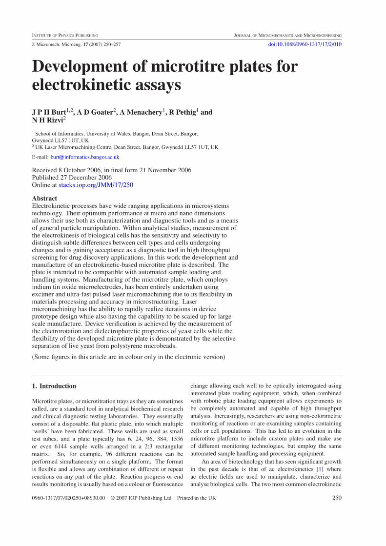
As with conventional microtitre plates, the electrokineticplatform comprised of a number of identical units arrayedon a single substrate. In this case six electrokinesis (combineddielectrophoresis and eletrorotation) units were arrayed ona 75 mm × 25 mm substrate compatible with standardautomated plate handling and sample loading systems. Thestarting point in designing the electrokinesis plate was thedetermination of a suitable microelectrode system that allowedboth dielectrophoresis and electrorotation to be carried outin the same unit. A suitable electrode configuration is thequadrature polynomial electrode design of Huang et al [6].Here four identical electrodes with a defined, curved, outlineare arranged in a planar quadrature configuration. Whenadjacent electrodes are energized with sinusoidal voltages 180◦
out of phase with each other the electrodes form a non-uniformelectric field in the central region of the inter-electrode spacewith a constant gradient in field strength. Energizing theelectrodes with sinusoidal signals 90◦ out of phase with eachother creates a rotating electric field where the central regionof the inter-electrode space has a near uniform electric fieldstrength. To allow a suitably large number of cells to beexamined at the same time in a single unit electrode system,while using easy to generate ac voltages of less than 20 Vpp,the inter-electrode distance between opposite electrodes waschosen to be 400 µm. Such electrodes can be manufacturedusing a range of materials and methods. However, it has beenfound that fabrication using thin films produces an optimalelectric field distribution both in the plane of the electrodes andextending away from the electrodes into the bulk sample liquid[7]. Figure 1(A) shows a diagram of the quadrature polynomialelectrode configuration. Within the developed electrokinesisplate six identical microelectrodes and fluidic chamber unitsare linearly arranged equidistant along the major axis of thesubstrate. This simplifies automatic processing and also assistsin the routing of electrical connection tracks between the activeregion of the microelectrodes and a single, 0.1 inch pitch,edge connector to give individual access to each electrode onthe plate. Since the electrode components consisted of thinfilms, attention was given to the electrical resistance of eachconnection track. It is essential that each electrode in thequadrature arrangement is energized with an identical voltageto produce a rotationally symmetric electric field. Figure 1(B)shows the routing layout for the connection tracks of theelectrokinetic microtitre plate. The edge connector contactsare 1.25 mm wide on a 2.5 mm pitch. The widths of tracksfrom the contacts to the active region of the microelectrodeshave been reduced for electrodes closer to the edge connectorto compensate for the shorter distance travelled and hencemaintain a constant electrical resistance for all tracks.
251

J P H Burt et al
(A) (B)
Va Vb
Vd Vc
Figure 1. (A) A close up view of the quadrature polynomial microelectrode design with four energizing voltages Va, Vb, Vc and Vdindicated. (B) Electrokinetic microtitre plate layout showing six identical units on a single substrate. In both diagrams conducting regionsare shown in grey. The diagrams are not to scale.
Glass Glass
ITO coated glass
Polymer Polymer
Adhesive
Figure 2. A cross sectional illustration of a single unit of the electrokinetic microtitre plate. Diagram not to scale.
The microelectrode array element of the electrokinesisplates takes the form of a planar substrate onto whichelectrodes typically <1 µm high have been produced. Toallow the individual units to be used independently, anopen fluidic chamber system is required around each activeelectrode region. Such a chamber could be filled andpossibly drained using either manual or automated dispensingsystems. The active area of the electrodes is approximately0.125 mm2. Finite element analysis of the usable electricfield produced by the thin film polynomial electrodes showsthat it extends approximately 300 µm above the electrodeswhen an aqueous sample occupies the electrokinesis unit.This gives an active volume of the unit of approximately40 nl. For accurate imaging-based analysis of electrorotationa maximum of approximately 100 cells should occupy thisvolume, so allowing the electrokinesis chamber to be usedwith sample concentrations up to 2.6 × 10⁶ cells per ml.Accurate dispensing of such small volumes is still extremelydifficult for both manual and automated systems. Therefore,the electrokinesis plate was required to hold significantly largersample volumes for each unit but to physically constrain a 40 nlvolume over the electrode active region. This was achieved bythe creation of a larger fluidic holding chamber above a smaller40 nl constraining chamber enclosing the active volume of theelectrokinesis unit as shown in cross section in figure 2. Theholding chamber is circular in nature with a 4 mm diameterand 1 mm depth allowing sample volumes of up to 12.5 µl tobe held. Such a volume is easily dispensed both by hand andautomatically.
2.2. Materials
A recognized advantage of laser micromachining over manyother microfabrication methods is its ability to structure analmost unlimited range of materials with the correct choiceof laser. A range of substrate and electrode materials couldbe used for manufacturing the electrokinetic microtitre plates,
all of which are suitable for laser micromachining. In thiswork it was intended that the movement of cells within eachelectrokinesis unit would be quantified by optical imagingtechniques and may use fluorescence to highlight cells ordistinguish between different cell populations. Therefore, animportant consideration in choosing materials for the substrateand electrodes is maintaining a low background of UV-inducedfluorescence. Glass was chosen as the substrate material forits transparent optical qualities, low fluorescence, mechanicalstrength and biocompatibility. Typically, glass substrate-basedmicroelectrodes for electrokinesis have been manufacturedfrom multilayer films such as gold on a chrome adhesion layer[8]. To assist in the imaging of the electrokinesis chamberit was desirable to have transparent electrodes. To this end,the electrodes employed in the electrokinesis platform wereformed in a film of low resistance (10 � sq−1) indium tin oxide(ITO) film precoated onto a flat 1 mm thick glass substrate(Delta Technologies Ltd, USA). ITO films have successfullybeen selectively laser micromachined on a glass substrateusing excimer lasers for a number of applications such asdisplay devices [5] and other electrokinetic cell analysisprocesses [9].
In fabricating and integrating the fluidic chamber overeach unit, both chamber formation and bonding issues neededto be considered. The chamber must form a strong seal aroundthe active region of the polynomial electrodes. To ensure anidentical electrical path for the electric fields in each unit onthe platform, fluid needed to be prevented from travellingbetween the ITO/glass substrate surface and the chamberforming layer surface by capillary action or any other means.To achieve this, a bilayer approach was adopted for formingthe fluidic chambers. The upper holding chamber was formedfrom a single, 1 mm thick glass sheet. As well as providinga biocompatible microfluidic chamber, the glass plate alsoprovides additional mechanical rigidity. The constrainingchambers were formed in a single, adhesive backed, 250 µmthick polymer sheet (Melinex A, Katco Ltd, UK).
252

Development of microtitre plates for electrokinetic assays
(A) (B)
WorkpieceSample
Mask
ExcimerLaser Beam
Y directionX direction
Static
WorkpieceSample
Mask
StaticExcimerLaserBeam
Y directionX direction
Figure 3. (A) An illustration of the principles of mask scanning laser micromachining. Mask motion is continuously synchronized withworkpiece motion. (B) An illustration of direct writing excimer laser micromachining. The uniform large area beam is apertured by a staticmask while the workpiece is moved. In both cases the laser beam is stationary and laser firing is triggered by incremental movement of theworkpiece in either the X or Y direction.
2.3. Manufacture
Manufacture of the electrokinetic microtitre plates was carriedout in two parts. The first used an excimer laser forthe high resolution patterning of the ITO electrodes whilethe second used a femtosecond laser to trepan circularholes in the glass plate for the holding chambers and theadhesive backed polymer for the containment chambers. Animportant parameter in the manufacture of microelectrodesfor electrokinetics is the edge quality of the field-producingelectrodes. Since phenomena such as dielectrophoresisoperate on non-uniformities in electric fields, roughness orpatterning errors at the edge of electrodes are likely toproduce unwanted electrokinesis. Excimer lasers producelarge area beams with many thousands of modes which arelikely to possess non-uniform intensity profiles. Machiningof the ITO microelectrodes was carried out using an ExitechS8000 Excimer Laser Micromachining Workstation (ExitechLtd Oxford, UK). This system uses beam shaping andhomogenizing array optics to transform the output of a LambdaPhysik Compex 110 excimer laser (Lambda Physik, Germany)into a 10 mm × 10 mm uniform intensity beam at animage plane. The image plane is then projected through ademagnification lens onto the surface of a sample held at aworkpiece plane. Using a projection lens with a magnificationof ×10, a workpiece beam of 1 mm × 1 mm in size and fluencesof up to 10 J cm−2 can be created. To create the active region ofthe ITO electrodes, where the quality of the machined edge isof greatest importance, a mask projection machining processwas used. Here, the curved polynomial electrode pattern wasproduced on a mask and positioned on a computer controlledXY positioning stage (Aerotech Inc., USA) with the masksurface at the image plane. As the mask is positioned at theimage plane the electrode pattern was ten times larger thanthe final features to be produced on the workpiece plane and,
in covering the entire polynomial electrode region, measuredapproximately 40 mm × 40 mm. While it is typical to usepatterned chrome on a quartz substrate masks for excimerlaser micromachining, in this case a simple, low-cost, polymermask was employed. Excimer lasers produce light in thedeep UV band, and most polymers will absorb such radiation.This is one of the reasons excimer lasers are so well suited tomicromachining polymers. Therefore, low-cost excimer lasermasks can be produced using a polymer either as spin coatedfilms on quartz or, as in this case, cutting a stencil in a polymersheet. Since the mask dimensions were reasonably large, themask was fabricated from a 200 µm thick polymer sheet withthe mask pattern cut out of the sheet using a femtosecond laserto give a mask resolution of approximately 10 µm which, whenprojected onto the workpiece plane, produced a final electrodepattern resolution of 1 µm—close to the optical imagingresolution of the S8000 workstation. To maintain rigidity, thestencil mask was held between two quartz plates. Employinga 248 nm excimer laser, it was found that machining witha fluence of 1.5 J cm−2 allowed the ITO film to be cleanlyremoved from the substrate with minimal damage to theunderlying glass. To transfer the large mask pattern onto theworkpiece plane a mask scanning-based machining techniquewas employed as illustrated in figure 3(A). The ITO coatedglass sample was held on a XYZθ micropositioning stage(Aerotech Inc., USA) and both mask and workpiece motionswere synchronized such that the position of the mask under thestatic laser beam corresponded to the appropriate position onthe workpiece sample. Laser output firing was triggered on themovement of the workpiece stages by a defined incrementaldistance such that each laser output pulse overlapped withthe previous pulse to produce a total laser exposure of7.5 J cm−2.
Electrical connection tracks between the polynomialelectrodes and the edge connector contacts were created by
253

J P H Burt et al
(A) (B)
Figure 4. (A) Photograph of a completed electrokinetic microtitre plate with fluidic chambers. (B) Close-up view of the lasermicromachined ITO polynomial electrodes. The defocused outline of the containment chamber is visible surrounding the active region ofthe polynomial electrodes. In this image the containment chamber has a diameter of 500 µm to allow the edges of the underlyingpolynomial electrodes to be visible.
electrically isolating regions of the ITO coating using a directwrite excimer laser micromachining process, as illustratedin figure 3(B). The homogenized excimer laser beam wasapertured at the image plane using a 4 mm square apertureon a patterned chrome on quartz mask. Regions of ITO wereisolated by moving the workpiece sample under the static,apertured, beam and triggering the laser output firing withincremental distance movement of the workpiece stages inboth the X and Y direction. As with the mask scanningmachining, a beam fluence of 1.5 J cm−2 was used with laseroutput being triggered to give a total exposure for machinedareas of 7.5 J cm−2. Workpiece motion was calculated from acomputer aided design drawing of the electrical routing on theelectrokinetic microtitre plate. The outlines of the electricalconnections were translated into workpiece stage movementsso allowing electrical isolation to be achieved by machiningthe outlines of the electrical connection tracks using the400 µm at the workpiece apertured beam.
To remove the possibility of short circuiting adjacentelectrodes by misalignment of the external edge connector,ITO was completely removed from regions between the edgeconnector contacts. This was achieved by raster scanningthe 400 µm excimer laser beam over the area between eachconnector contact. To ensure all ITO was removed, adjacentmachining actions overlapped by 200 µm, 50% of the beamwidth.
The microfluidic chambers above the electrokinesis unitswere fabricated independently to the electrodes. 4 mmdiameter holes were laser machined using a Spectra PhysicsHurricane Ti:Sapphire femtosecond laser integrated into anExitech M2000F Laser Micromachining Workstation. Thissystem delivered tightly focussed pulsed infra-red (800 nm)light with a pulse duration of 120 fs. The beam power densityof up to 3.5 W cm−2 was focussed to a 20 µm spot deliveringpower densities of up to 0.3 MW cm−2 in a 120 fs time period.Such high power densities have the ability to machine virtuallyany known material in a precise manner. The pulse duration ismuch shorter than the time required for heat to conduct awayfrom the exposed area causing all the absorbed beam energyto be translated to an ionization-based ablation process. Theholes through the glass sheet that form the holding chamberswere machined using a trepanning-based technique to machinea trench around the circumference of the hole. The complete
hole is formed when the trench depth extends all the waythrough the glass sheet and the unmachined central portion ofthe hole is able to fall away. Hole machining was achievedusing a beam power density of 30 kW cm−2 and an effectiveworkpiece velocity of 0.22 mm min−1.
The benefits of femtosecond laser micromachining are anextremely high cut edge quality with no significant machiningdebris. The lack of debris is of benefit when micromachiningcomposite materials such as the adhesive backed polymersheet used to form the containment chambers. The sheetcomprised of a 250 µm thick polymer sheet coated on bothsides with a 50 µm thick layer of adhesive which, in turn,was coated with a protective cover. Containment chamberholes were machined using a trepanning motion throughthe three materials, protective cover, adhesive and polymersheet, in a single machining operation using a beam powerdensity of 30 kW cm−2 and an effective workpiece velocity of5 mm min−1. The ionizing nature of the femtosecondlaser ablation meant that all materials had similar ablationcharacteristics. In addition the small amount of debrisproduced in the machining operation was non-adhesive andcould be fully removed from the sample prior to removal of theprotective cover and assembly of the electrokinetic microtitreplates.
Final assembly of the device was carried out by firstremoving one layer of protective cover from the containmentchamber sheet and then bonding the sheet to the holdingchamber glass. The structure was then passed through a seriesof heated rollers at a temperature of 80 ◦C to ensure full contactbetween the two surfaces. Next, the second protective coverwas removed from the containment chamber sheet prior toalignment, using a modified mask aligner, and bonding tothe ITO electrode substrate. Finally, the complete device waspassed through the heated rollers to ensure a firm bond betweenall layers of the device.
3. Results and discussion
Figure 4(A) shows the completed electrokinetic microtitreplate. The edge connector contacts can clearly be seen alongthe bottom edge of the device while the upper half of thedevice shows the six holding chambers and, in the centre ofeach chamber, the containment chambers. Figure 4(B) shows a
254

Development of microtitre plates for electrokinetic assays
Figure 5. An electron micrograph of the edge of the maskprojection laser micromachined polynomial electrode. An overalledge roughness in the plane of the electrodes of the order of 100 nmis observed.
close-up view of the ITO electrodes and the defocused outlineof an enclosing containment chamber. For the purposes ofclarity in viewing the underlying polynomial electrodes, theenclosing containment chamber in this image has a diameterof 500 µm.
Figure 5 shows an electron micrograph of the curvedtip of the polynomial electrode. A small lip is visible as alighter coloured line at the edge of the ablated ITO. Howeverthe overall edge roughness is of the order of 100 nm. It isgenerally accepted that an edge roughness up to approximately1 µm in the plane of the electrode is allowable for thisform of electrode design. The two-dimensional accuracyof the mask scanned electrode pattern is governed by themask quality and magnification factor of the mask imagingsystem. For the purposes of these devices the electrodequality produced by laser micromachining is comparable
(A) (B)
Figure 6. Developed electrokinetic microtitre wells in use. (A) The corralling of latex microbeads by negative dielectrophoresis. (B)Differential dielectrophoresis showing latex microbeads corralled by negative dielectrophoresis with simultaneous collection of yeast cellsby positive dielectrophoresis. In both cases the suspending medium conductivity was 50 µS cm−1. The electrodes were energized with a13 Vpp, 1 MHz, sinusoidal voltage.
with that which can be achieved using conventional wetetching of ITO. However, laser micromachining can offer aquicker more cost effective fabrication route. Direct writingmicromachining used to isolate the ITO electrical tracksbetween the polynomial electrodes and the edge connectorproduced an edge roughness of approximately 2 µm whenmeasured over extended distances. This was largely due tosmall errors in the alignment of the edges of the 400 µm squarebeam with the X and Y axis of the workpiece stages causing aslightly undulating edge as successive, adjacent, laser pulsesare partially overlaid.
The devices fabricated in this work made use of an acrylicadhesive backed polymer sheet to bond the components of theelectrokinetic microtitre plate together. Such adhesives canbe sensitive to aqueous environments. Immersion tests of thefabricated devices revealed no observable deterioration in thestrength of the devices or the seal around each containmentchamber on immersion in water for one week. Similar deviceshave also been fabricated by us using polymer sheets pre-coated with a silicone adhesive if greater prolonged exposureto aqueous media is required. Devices were fabricated withand without the removal of the adhesive layer exposed to theholding chamber on the upper surface of the Melinex sheet.Adhesive removal was achieved by ablating just the adhesivelayer using a femtosecond laser and similar process parametersto those used to machine the containment chamber holes.
Measurements of the electrical resistance of theconnection tracks between the active polynomial electrodesand the edge connector contacts gave a track resistance of300 � ± 10 �. Such close resistances help ensure predictableand rotationally symmetrical electric field distribution withinthe inter-electrode region. This is confirmed by experimentaldata from the negative dielectrophoretic collection ofpolystyrene latex beads within the electrode chambers asshown in figure 6(A). It can be seen that beads are corralledinto the centre of the interelectrode region to form a closelypacked square body of particles. The uniform geometry ofthis collection with corners of the square aligned to the tips ofadjacent electrodes, is evidence that the electric field producedby the each electrode is equal. Figure 6(B) shows experimentalevidence that both positive and negative dielectrophoresis is
255

J P H Burt et al
104
105
106
107
-1
-0.8
-0.6
-0.4
-0.2
0
0.2
0.4
0.6
0.8
1
Ro
tatio
n R
ate
(s-1
)
Frequency (Hz)
4-0.25
-0.2
-0.15
-0.1
-0.05
0
0.05
0.1
0.15
0.2
0.25
Rel
ativ
e D
iele
ctro
phor
etic
For
ce
-0.25
-0.2
-0.15
-0.1
-0.05
0
0.05
0.1
0.15
0.2
0.25
Figure 7. Electrorotation (•) and dielectrophoretic (◦) spectrum for live yeast cells. In both cases the suspending medium conductivity was50 µS cm−1. Voltages of 6 Vpp were used for dielectrophoresis while 13 Vpp was used for electrorotation studies.
possible within the units of the electrokinesis plate. Here liveyeasts cells are collected using a positive dielectrophoreticforce, while simultaneously latex beads are corralled at thecentre of the electrodes. This selective separation is a result ofthe effective polarizability of the beads being less than that ofthe suspending medium which, in turn, is less than that of theyeast cells. The generally uniform distribution of cells on theelectrode edges and the similar lengths to chains of collectedcells is additional evidence of the good rotational symmetryof the produced electric field.
For a given electric field configuration, bothelectrorotational and dielectrohoretic forces are frequency-dependent functions of the dielectric properties of the cell andsuspending medium. Many electrokinetic studies examine theelectrokinetic response of cells over a frequency range. In thedevices described here, changes in such responses can be usedto quantify the progress of any biochemical reaction that maybe deliberately undertaken within the suspending medium.Figure 7 shows the electrorotational and dielectrophoreticspectrum of live yeast cells collected using the electrokineticmicrotitre plate. In this case energizing voltages of 13 Vppand 6 Vpp were applied to each electrode for electrorotationand dielectrophoretic experiments, respectively. Experimentaldata were collected by manual measurement of electrokinesis.For electrorotation data, the time for a defined number of cellrotations was recorded while for dielectrophoretic data the timeto move a known distance was recorded. The spectra shown infigure 7 are in close agreement with those of previous workers[10] and, for electrorotation, show a strong anti-field (negative)peak in rotation rate below approximately 1 MHz and the startof a co-field peak in the rotation rate at frequencies above1 MHz.
4. Conclusions
The work described here has demonstrated the flexibilityof laser micromachining for the complete fabrication
of an electrokinetic microtitre plate, whose design andoperation has been validated by conducting electrorotation anddielectrophoresis experiments on yeast cells. The processinghas made use of the unique machining properties of twodifferent types of lasers to produce an integrated device. Whileother manufacturing processes may be used to create partsof the devices, only laser micromachining has the ability toproduce all components of the microtitre plates. The flexibilityof laser micromachining coupled with the ability to use low-cost stencil mask allows iterations in all aspects of devicedesign to be quickly and cheaply realized. Variations inmanufacturing material can easily be handled along with thequick production of stencil masks for evolutions in electrodedesign. Experimental validation data have shown that thedescribed devices are equivalent to those manufactured usingtraditional photolithographic processes.
Acknowledgments
This work has been supported in part by the OpticalBiochip Consortium (EPSRC award GR/S23483/01) and wascommissioned by the University of Wales, Bangor usingfunds from the European Regional Development Fund andadministered through the Welsh Assembly Government’sKnowledge Exploitation Fund.
References
[1] Morgan H and Green N 2001 AC Electrokinetics: Colloidsand Nanoparticles (Baldock: Research Studies Press)
[2] Pethig R 2006 Cell physiometry tools based ondielectrophoresis BioMEMS and BiomedicalNanotechnology vol 2 ed M Ferrari (New York: Springer)pp 103–26
[3] Gower M and Rizvi N 2000 Applications of laser ablation tomicroengineering Proc. SPIE 4065 452–60
256

Development of microtitre plates for electrokinetic assays
[4] Gower M C 2000 Excimer laser microfabrication andmicromachining Proc. SPIE 4088 124–31
[5] Rumsby P T 2002 Advanced laser tools for displaydevice production on super large substrates IMID2002 Dig.
[6] Huang Y and Pethig R 1991 Electrode design for negativedielectrophoresis Meas. Sci. Technol. 2 1142–6
[7] Green N, Ramos A and Morgan H 2002 Numerical solution ofthe dielectrophoretic and travelling wave forces forinterdigitated electrode arrays using the finite elementmethod J. Electrostat. 56 235–54
[8] Pethig R, Burt J P H, Parton A, Rizvi N, Talary M S and TameJ A 1998 Development of biofactory-on-a-chip technologyusing excimer laser micromachining J. Micromech.Microeng. 8 57–63
[9] Suehiro J and Pethig R 1998 The dielectrophoretic movementand positioning of a biological cell using a three-dimensional grid electrode system J Phys. D: Appl.Phys. 31 3298–305
[10] Holzel R 1997 Electrorotation of single yeast cells atfrequencies between 100 Hz and 1.6 GHz Biophys. J. 731103–09
257