development of calcium phosphate cement using chitosan and citric
TRANSCRIPT

8/10/2019 Development of Calcium Phosphate Cement Using Chitosan and Citric
http://slidepdf.com/reader/full/development-of-calcium-phosphate-cement-using-chitosan-and-citric 1/11

8/10/2019 Development of Calcium Phosphate Cement Using Chitosan and Citric
http://slidepdf.com/reader/full/development-of-calcium-phosphate-cement-using-chitosan-and-citric 2/11
mechanical properties and biocompatibility, and devel-
op a new calcium phosphate cement with improved
mechanical properties and biocompatibility as a bone
substitute.
2. Materials and methods
2.1. Materials
The cement developed in this study consisted of both
powder and liquid components. The powder component
was a mixture of a-TCP and TeCP at a molecular ratio
of 2 : 1. The powder component was made by decom-
position of a block of HAP (ApaceramR, Asahi Optical
Co. Ltd, Tokyo, Japan) for 1 hour at a temperature of
12001C and a reduced pressure (102 –103 Pa). The
molar Ca/P ratio of HAP (ApaceramR) used in this
study was 1.67. This reaction was according to the
following equation:
Ca10ðPO4Þ6ðOHÞ2-2Ca3ðPO4Þ2 þ Ca4ðPO4Þ2O þ H2O:
The resulting mixture was uniform in composition.
Details of the powder component were reported by
Sumita [15]. XRD patterns of ApaceramR (before
decomposition) and the powder components are shown
in Fig. 1. The liquid component was a solution of citric
acid, chitosan (98% deacetylated) and glucose. In this
study, two types of liquid components were used to
investigate the effects of the concentration of citric acid
on the mechanical properties and biocompatibility. One
solution (group 45) contained citric acid (45% wt),glucose (15%) and chitosan (1%). The other solution
(group 20) contained citric acid (20%), glucose (6.7%)
and chitosan (1%). The powder/liquid ratio was 2.0
(wt/wt).
2.2. Setting times of the cements
Setting times of the cements were measured according
to the international standard ISO 9917 for dental silico-
phosphate cement (n ¼ 5).
2.3. Observation by SEM
After setting, the cements were observed under a
scanning electron microscope (S-4000, Hitachi Co.,
Tokyo, Japan).
2.4. Mechanical strength
2.4.1. Compressi ve strength in an atmosphere of 100%
humidity
Samples of each of the two types of cement were
molded into columns (5.0 mm in diameter and 10.0 mm
in length). The compressive strengths of the columns
were measured using an Auto Graph (DSS 5000,
Shimadzu, Tokyo, Japan) at a crosshead speed of
0.5 mm/min. Measurements were made at 1, 3, 5 and
10 days in an atmosphere at 371C and 100% humidity
(n ¼ 5).
2.4.2. Compressi ve strength when incubated in physiological saline
Samples of each of the two types of cement were
molded into columns (5.0 mm in diameter and 10.0 mm
in length). After setting, the columns were immediately
placed into physiological saline (100 ml) and incubated
at 371C. Compressive strengths were measured at 1, 3, 5
and 10 days and 3 and 6 weeks after incubation (n ¼ 5).
Physiological saline was changed every day.
2.5. Effects of glucose on the setting time and mechanical
strength
Setting times and compressive strengths of the
cements which contain glucose at various concentrations
were measured to investigate the effects of glucose.
Setting time was measured according to the ISO 9917.
Compressive strengths were measured at 5 days after
incubation. Physiological saline was not changed.
2.6. pH changes
Samples (A mixture of 2 g of powder and 1 g of
liquid component) of each of the two types of cement
were molded into columns that were then placed in
physiological saline (50 ml) after setting. The pH
values of physiological saline in which the samples
were incubated at 371C were measured at 5, 10, 20,
30, 60 and 120min, at 1, 3, 5 and 10 days, and 2, 3, 4
and 6 weeks (n ¼ 3). Physiological saline was changed
every day.
2.7. Concentrations of Ca and P ions
Columns (5.0 mm in diameter and 10.0 mm in length)
made from each of the two types of cement were
incubated in physiological saline (100 ml) at 371C. TheFig. 1. X-ray diffraction patterns. (a) ApaceramR (before decomposi-
tion) and (b) powder component.
A. Yokoyama et al. / Biomaterials 23 (2002) 1091–11011092

8/10/2019 Development of Calcium Phosphate Cement Using Chitosan and Citric
http://slidepdf.com/reader/full/development-of-calcium-phosphate-cement-using-chitosan-and-citric 3/11
concentrations of Ca and P ions were measured at 1, 3, 5
and 10 days, and at 2, 3, 4 and 6 weeks after incubation.
Concentrations of Ca and P ions were measured by
emission spectro chemical analysis (ICPS-7500, Shima-
zu, Tokyo, Japan) (n ¼ 3). Physiological saline was
changed every day.
2.8. X-ray diffraction
The samples of the cements incubated in physio-
logical saline for 1, 3, 5, 10 days, and for 2, 3, 4 and 6
weeks and the cements implanted in the subcutaneous
tissue of rats for 2 and 4 weeks were analyzed by X-ray
diffraction (PW1700, Philips Analytical X-ray B.V.,
Almelo, Netherlands). The intensities of some peaks,
29.81 (2y) for TeCP, 30.81 (2y) for a-TCP and 25.91
(2y) for HAP were recorded and the amounts of
transformation of the components of the cements into
HAP on the cements incubated in physiological salinewere estimated. The transformation rate was calculated
by the following equation, modified from the report of
Fukase et al. [17].
Transformation rate ð%Þ ¼ 1=2½ðPHAPðtÞ=PHAPðNÞÞ
þ 2=3ð1 Pa-TCPðtÞ=Pa-TCPð0ÞÞ
þ 1=3ð1 PTeCPðtÞ=PTeCPð0ÞÞ100;
where Pa-TCPð0Þ and PTeCPð0Þ are the intensities of the
peaks of a-TCP and TeCP in the original powder
components, respectively, and PHAPðNÞ is the intensity of
the peaks of HAP in the cements at the point when the
peaks TeCP and a-TCP were not detected (at 3 weeks in
group 45 and 6 weeks in group 20 after incubation).
PHAPðtÞ; Pa-TCPðtÞ and PTeCPðtÞ are the intensities of peaks
of HAP, a-TCP and TeCP, in the cement harvested at
time t after incubation. Physiological saline was changed
every day.
2.9. Biocompatibility
Eighteen 6-week old male Wistar rats were used in
this study. The samples of the two types of cement
were molded into pellets (4.5mm in diameter and
1.5 mm in thickness). After hardening, the pellets
were immediately implanted between the periosteum
and parietal bone, and in the subcutanenous tissue
of the thoractic region of rats. Rats were sacrificed at
1, 2 and 4 weeks after surgery and the tissue blocks
containing the pellets were fixed in 10% neutral
formalin, decalcified, and embedded in paraffin.
Hematoxylin and eosin-stained specimens were ob-
served by light microscopy. Animal experiments were
done following the ‘‘Guide for the care and use of
laboratory animals, School of Dentistry, Hokkaido
University’’.
3. Results
3.1. Setting time
After mixing the powder and liquid components
together, both types of cement (groups 20 and 45) had
a ‘‘chewing-gum-like’’ consistency (Fig. 2) that could bemolded into the desired shape. Setting times were
5.5470.15 min in group 20 and 6.4070.20 min in group
45 (n ¼ 5).
3.2. Observation of SEM
As shown in Fig. 3a, SEM revealed the presence of
numerous fine grains covering the surfaces of global
structures approximately 10 mm in diameter in a sample
from group 20. A sample from group 45 (Fig. 3b)
showed global structures approximately 20mm i n a
diameter on a smooth, platelike matrix.
3.3. Mechanical strength
Figs. 4 and 5 show the compressive strengths of the
samples in an atmosphere of 100% humidity and in
physiological saline, respectively. The compressive
strength of samples from group 45 was significantly
higher than that from group 20 at 100% humidity.
However, while compressive strength constantly in-
creased with time at 100% humidity in both groups,
samples incubated in physiological saline showed an
initial decrease in compressive strength at 3 days after
incubation in group 20. Thereafter, the compressivestrength increased with time. Whereas, the compressive
strength of samples from group 45 was higher than that
from group 20 during the initial 10 days in physiological
saline, that of samples from group 20 at 6 weeks after
Fig. 2. The material showed chewing-gum-like consistency after
mixing.
A. Yokoyama et al. / Biomaterials 23 (2002) 1091–1101 1093

8/10/2019 Development of Calcium Phosphate Cement Using Chitosan and Citric
http://slidepdf.com/reader/full/development-of-calcium-phosphate-cement-using-chitosan-and-citric 4/11
incubation, which were transformed to HAP comple-
tely, was higher than that of samples from group 45 at 3
weeks after incubation, which were transformed to HAP
completely.
3.4. Effects of glucose on setting time and compressi ve
strength
Tables 1 and 2 show setting times and compressive
strengths of the cements containing glucose at various
concentrations. Setting time increased with the concen-
tration of glucose in both cements. Compressive
strength decreased when the concentration of glucose
was increased in group 45, whereas the compressive
strengths of the cements were almost the same regardless
of the concentration of glucose in group 20.
3.5. Change in pH of physiological saline
The pH of the physiological saline in which the twotypes of cement were incubated increased with time in
both groups (Figs. 6a and b). The pH of the saline in
which group 20 samples were incubated was higher at
each measurement point than that in which group 45
samples were incubated. The pH values were 6.7070.02
in group 20 and 5.9870.02 in group 45 at 120 min after
the start of incubation. The pH values increased with
incubation time and reached 7.04 at 5 days in group 20
and 6.94 at 3 days in group 45, when physiological saline
was changed every day. After that, it gradually
decreased with incubation time.
3.6. Concentrations of Ca and P ions
Figs. 7A and B show the concentrations of Ca and P
ions in the physiological saline. The concentrations of
Ca and P ions at 1 day after incubation were high, and
those of group 45 were higher than those of group 20.
Ca ions were detected at every measuring point and
decreased with time in both cements, when physiological
saline was changed every day. The concentration of Ca
ions of group 45 was higher than that of group 20. P
ions were hardly detected at 3 days after incubation and
Fig. 3. Images of SEM: (a) group 20; and (b) group 45.
Fig. 4. Compressive strength in an atmosphere of 100% humidity.
Fig. 5. Compressive strength when incubated in physiological saline.
A. Yokoyama et al. / Biomaterials 23 (2002) 1091–11011094

8/10/2019 Development of Calcium Phosphate Cement Using Chitosan and Citric
http://slidepdf.com/reader/full/development-of-calcium-phosphate-cement-using-chitosan-and-citric 5/11
increased gradually in group 20, but decreased with
incubation time in group 45.
3.7. XRD analysis
Figs. 8(A) and (B) show the patterns of X-ray
diffraction. Those of the powder components showed
high a-TCP and TeCP peaks, and no HAP peak.
Whereas, the peak of TeCP decreased from 1 day after
incubation, the peaks of a-TCP did not change in group
20. From 10 days after incubation, the peaks of a-TCP
began to decrease. The peaks of HAP became higher
with incubation time, the peaks of TeCP and a-TCP
were not detected at 6 weeks. On the other hand, the
peaks of TeCP and a-TCP decreased from 1 day, the
peaks of HAP became higher and the peaks of TeCP
and a-TCP were not detected at 3 weeks in group 45.
The transformation to HAP in vitro was faster than thatin vivo. Fig. 9 shows the changes of the rates of
transformation to HAP. The rates increased with
incubation time in both cements, and that of group 45
was higher than that of group 20.
3.8. Biocompatibility
3.8.1. Soft tissue response
In the granulation tissue, numerous dilated capil-
laries, mesenchymal cells and thin collagen fibers were
observed around the implanted cement at 1 week after
Table 1
Effect of glucose on setting time
Group 20 Group 45
Glucose 0% 4.7670.09 min Glucose 0% 1.8870.10min
6.7% 5.5470.15 5% 2.3070.12
10% 6.3870.33 10% 3.4370.14
15% 7.6070.27 15% 6.4070.2020% 9.3070.25 20% 8.2770.25
Mean7SD, n ¼ 5
Table 2
Effect of glucose on compressive strength
Group 20 Group 45
Glucose 0% 3.0770.28 MPa Glucose 0% 50.62710.52 MPa
6.7% 4.0770.45 5% 43.4178.10
10% 3.3570.63 10% 20.4772.69
15% 3.7170.32 15% 19.7873.1220% 3.3371.16 20% 21.6574.02
Mean7SD, n ¼ 5
Fig. 6. The pH values of the physiological saline in which the samples
were incubated.
A. Yokoyama et al. / Biomaterials 23 (2002) 1091–1101 1095

8/10/2019 Development of Calcium Phosphate Cement Using Chitosan and Citric
http://slidepdf.com/reader/full/development-of-calcium-phosphate-cement-using-chitosan-and-citric 6/11
surgery. In group 45, hyalinization was observed on the
surfaces of the implanted cement. Foreign body giant
cells were observed on the surface of the cements in both
groups, although they were more numerous in group 45
than in group 20. Inflammatory cell infiltration was
more prominent in the granulation tissue in group 45
(Fig. 10b) than in group 20 (Fig. 10a). With time,
inflammatory responses around the implanted cements
gradually decreased in both groups and the implanted
cements were covered by layers of dense collagen fibers.
Few inflammatory round cells and foreign body giant
cells were observed at 4 weeks after surgery (Figs. 11a
and b).
3.8.2. Hard tissue response
At 1 week after surgery, numerous dilated capillaries,
mesenchymal cells and slight inflammatory cell infiltra-
tion were seen in the thickened periosteum that covered
the implanted cement in group 20 (Fig. 12a). In group
45, hyaline degeneration and moderate inflammatory
cell infiltration were seen in the prominent hypertrophic
periosteum (Fig. 12c). On the surface of the parietal
bone around the cement, granulation tissue with
mesenchymal cells and capillaries and newly formed
bone from the parietal bone (not bound to the cement)
were observed in group 20 (Fig. 12b), but no new bone
formation was seen in group 45 (Fig. 12d). At 2 weeks
after surgery, an inflammatory response around the
cement was still present in group 45, but in group 20
inflammation had disappeared and newly formed bone
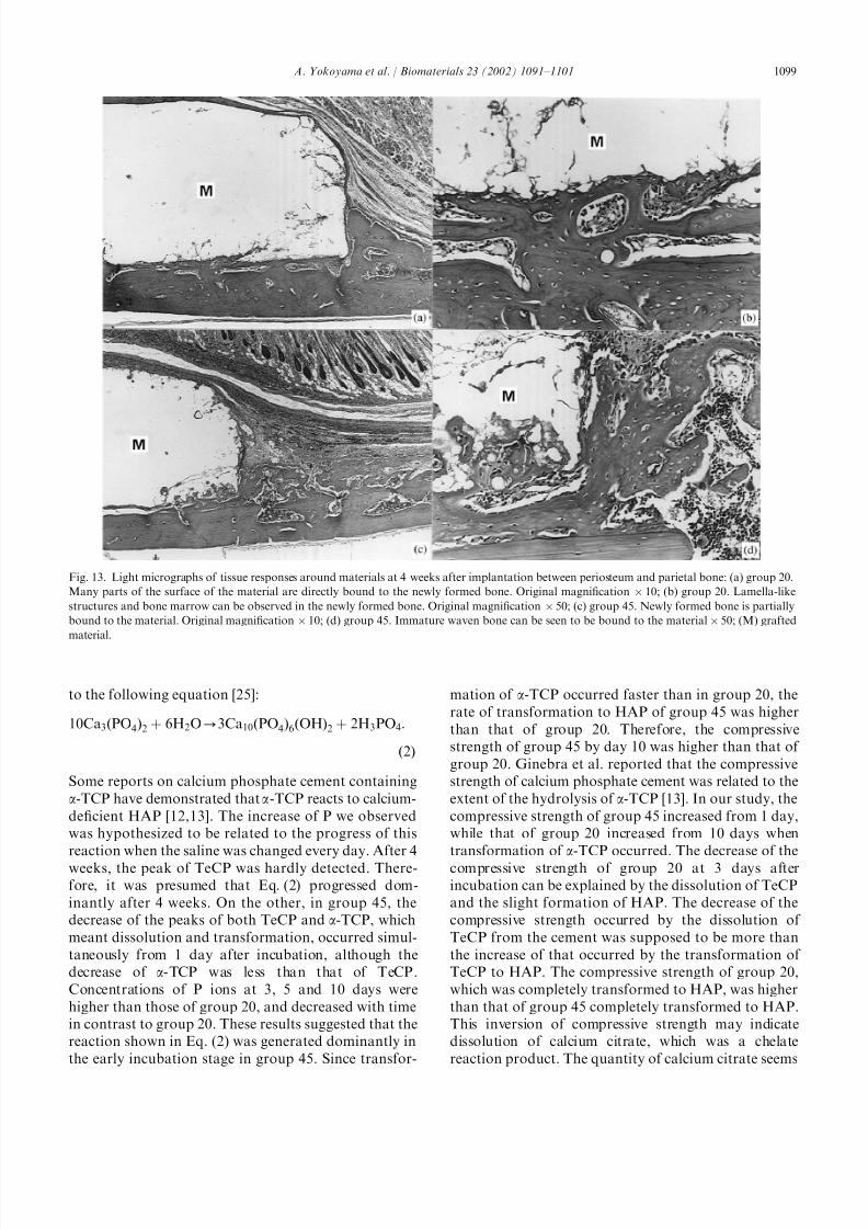
was seen to be partially bound to the cement. At 4 weeks
after surgery, the inflammatory response disappeared in
group 45 and newly formed bone from the parietal bone
was seen to be partially bound to the cement (Figs. 13c
Fig. 8. (A). X-ray diffraction patterns of group 20: (a) powdercomponent; (b) at 1 day after incubation in physiological saline; (c)
at 3 days after incubation in physiological saline; (d) at 5 days after
incubation in physiological saline; (e) at 10 days after incubation in
physiological saline; (f) at 4 weeks after incubation in physiological
saline; (g) at 6 weeks after incubation in physiological saline; (h) at 2
weeks after implantation in subcutaneous tissue; and (i) at 4 weeks
after implantation in subcutaneous tissue. (B): X-ray diffraction
patterns of group 45: (a) powder component; (b) at 1 day after
incubation in physiological saline; (c) at 3 days after incubation in
physiological saline; (d) at 5 days after incubation in physiological
saline; (e) at 10 days after incubation in physiological saline; (f) at 3
weeks after incubation in physiological saline; (g) at 2 weeks after
implantation in subcutaneous tissue; and (h) at 4 weeks after
implantation in subcutaneous tissue.
Fig. 7. (A) Concentrations of Ca ions in the physiological saline
solutions in which the samples were incubated; (B) concentrations of P
ions in the physiological saline in which the samples were incubated.
A. Yokoyama et al. / Biomaterials 23 (2002) 1091–11011096

8/10/2019 Development of Calcium Phosphate Cement Using Chitosan and Citric
http://slidepdf.com/reader/full/development-of-calcium-phosphate-cement-using-chitosan-and-citric 7/11
and d). Many parts of the surface of the grafted material
in group 20 that was bound to the newly formed bone
showed lamella-like structures and bone marrow space
formation (Figs. 13a and b).
4. Discussion
Although sintered HA ceramics have good biocom-
patibility and osteoconductivity, they lack sufficient
plasticity for easy molding into the desired shape. The
newly developed cement reported in this study has
excellent handling properties and moldability. Unlikethe calcium phosphate cements that have been reported
previously, this material had chewing-gum-like consis-
tency after mixing, as shown in Fig. 2. Thus, this
material could be molded to fit the shape of even the
most complicated bone defect and would be useful in
oral and plastic surgery. This chewing-gum-like con-
sistency is thought to be due to the addition of chitosan
to the liquid component. Chitosan is a polysaccharide, a
partially deacetylated chitin (98% deacetylated chitin
was used in this study), which has high viscosity in
solution [18], and which has recently been widely used
as a biomaterial because of its biocompatibility [19,20].
Cherng reported that the incorporation of chitosanacetate and chitosan lactate could improve the handling
Fig. 10. Light micrographs of tissue responses around materials at 1 week after implantation in subcutaneous tissue: (a) group 20; and (b) group 45;
(M) grafted material. Inflammatory cell infiltration is more prominent in group 45 than in group 20. Original magnification 100.
Fig. 11. Light micrographs of tissue responses around materials at 4 weeks after implantation in subcutaneous tissue: (a) group 20; and (b) group 45;
(M) grafted material. The grafted materials are covered by dense collagen fibers in both groups. Original magnification 100.
Fig. 9. Rate of transformation to HAP.
A. Yokoyama et al. / Biomaterials 23 (2002) 1091–1101 1097

8/10/2019 Development of Calcium Phosphate Cement Using Chitosan and Citric
http://slidepdf.com/reader/full/development-of-calcium-phosphate-cement-using-chitosan-and-citric 8/11
properties of calcium phosphate cement [21]. Takechi
that reported anti-washout calcium phosphate cement
containing chitosan showed excellent biocompa-
tibility [22].
The results from the present study demonstrated that
the concentration of citric acid influenced kinetics,
mechanical properties and biocompatibility of the
cements. The setting reactions of the cements could be
divided into two stages. The initial hardening was a
chelate reaction between citric acid in the liquid
component and calcium in the powder component and
the second reaction was transformation of the compo-
nents of the cements to HAP. It has been reported that
calcium phosphate cement containing carboxylic acid
hardens rapidly, with a poor soluble matrix by chelation
[22,23]. In our experiment, it appeared that the
compressive strength at the early stage was affected by
the degree of chelation. Therefore, the difference
between the compressive strengths of groups 20 and 45
during the initial 10 days after incubation can be
explained as a difference in the degree of chelation due
to variations in the concentration of citric acid. The
transformation of the components of the cements to
HAP also appeared to be influenced by the concentra-
tion of citric acid. The XRD analysis of group 20
showed that the peak of TeCP decreased, and the peak
of a-TCP did not decrease during the initial 5 days after
incubation in saline. These results demonstrated that
only TeCP was dissolved and transformed to HAP.
TeCP was transformed according to the following
equation [24].
3Ca4ðPO4Þ2O þ 3H2O-Ca10ðPO4Þ6ðOHÞ2 þ 2CaðOHÞ2:
ð1Þ
The increase of pH by 5 days after incubation in spite of
the change of the saline solution every day was due to
the dissolution and transformation of TeCP. The lack of
detection of P ions at 3 day suggested that P ions
released by dissolution of TeCP were used for the
formation of HAP. The decrease of the peak of a-TCP
from day 10 suggested that a-TCP was dissolved and
transformed to HAP. a-TCP was transformed according
Fig. 12. Light micrographs of tissue responses around materials at 1 week after implantation between periosteum and parietal bone: (a) group 20.
Thickened periosteum with slight inflammatory cell infiltration can be seen. Original magnification 10; (b) group 20. Newly formed bone from the
parietal bone can be seen. Original magnification 50; (c) group 45. Prominent hypertrophic periosteum can be seen. Original magnification 10; (d)
group 45. Moderate inflammatory cell infiltration can be seen around the grafted material. No new bone formation can be seen 50; (M) grafted
material.
A. Yokoyama et al. / Biomaterials 23 (2002) 1091–11011098
8/10/2019 Development of Calcium Phosphate Cement Using Chitosan and Citric
http://slidepdf.com/reader/full/development-of-calcium-phosphate-cement-using-chitosan-and-citric 9/11
to the following equation [25]:
10Ca3ðPO4Þ2 þ 6H2O-3Ca10ðPO4Þ6ðOHÞ2 þ 2H3PO4:
ð2Þ
Some reports on calcium phosphate cement containing
a-TCP have demonstrated that a-TCP reacts to calcium-
deficient HAP [12,13]. The increase of P we observed
was hypothesized to be related to the progress of this
reaction when the saline was changed every day. After 4
weeks, the peak of TeCP was hardly detected. There-
fore, it was presumed that Eq. (2) progressed dom-
inantly after 4 weeks. On the other, in group 45, the
decrease of the peaks of both TeCP and a-TCP, which
meant dissolution and transformation, occurred simul-
taneously from 1 day after incubation, although the
decrease of a-TCP was less than that of TeCP.
Concentrations of P ions at 3, 5 and 10 days were
higher than those of group 20, and decreased with time
in contrast to group 20. These results suggested that the
reaction shown in Eq. (2) was generated dominantly in
the early incubation stage in group 45. Since transfor-
mation of a-TCP occurred faster than in group 20, the
rate of transformation to HAP of group 45 was higher
than that of group 20. Therefore, the compressive
strength of group 45 by day 10 was higher than that of
group 20. Ginebra et al. reported that the compressive
strength of calcium phosphate cement was related to the
extent of the hydrolysis of a-TCP [13]. In our study, the
compressive strength of group 45 increased from 1 day,
while that of group 20 increased from 10 days when
transformation of a-TCP occurred. The decrease of the
compressive strength of group 20 at 3 days after
incubation can be explained by the dissolution of TeCP
and the slight formation of HAP. The decrease of the
compressive strength occurred by the dissolution of
TeCP from the cement was supposed to be more than
the increase of that occurred by the transformation of
TeCP to HAP. The compressive strength of group 20,
which was completely transformed to HAP, was higher
than that of group 45 completely transformed to HAP.
This inversion of compressive strength may indicate
dissolution of calcium citrate, which was a chelate
reaction product. The quantity of calcium citrate seems
Fig. 13. Light micrographs of tissue responses around materials at 4 weeks after implantation between periosteum and parietal bone: (a) group 20.
Many parts of the surface of the material are directly bound to the newly formed bone. Original magnification 10; (b) group 20. Lamella-like
structures and bone marrow can be observed in the newly formed bone. Original magnification 50; (c) group 45. Newly formed bone is partially
bound to the material. Original magnification 10; (d) group 45. Immature waven bone can be seen to be bound to the material 50; (M) grafted
material.
A. Yokoyama et al. / Biomaterials 23 (2002) 1091–1101 1099

8/10/2019 Development of Calcium Phosphate Cement Using Chitosan and Citric
http://slidepdf.com/reader/full/development-of-calcium-phosphate-cement-using-chitosan-and-citric 10/11
to be related to the difference of kinetics between group
20 and group 45. Monma reported that carboxylic ions
accelerate hydration of a-TCP [23]. Therefore, transfor-
mation to HAP of group 45 appeared to be faster than
that of group 20. The compressive strength at 100%
humidity was higher than that after incubation in
physiological saline. It is thought that interlocking of HAP crystals was inhibited in physiological saline, as
reported by Ishikawa et al. [11]. The setting time was
shorter in group 20 than in the group 45 in spite of its
higher concentration of citric acid. This was due to the
effect of glucose in the liquid component. Tables 1 and 2
show that glucose influenced the setting times and
compressive strengths of the cements. In particular,
group 45, which contained a high concentration of citric
acid, was more strongly affected by glucose than group
20, so glucose was hypothesized to inhibit the effect of
citric acid.
Analysis by XRD was carried out on the cements
implanted in subcutaneous tissue. The degree of transformation to HAP in vivo at 4 weeks after
implantation into rat subcutaneous tissue was lower
than that in vitro at 4 weeks after incubation in
physiological saline, as shown in Figs. 8A and B. The
material implanted in the subcutaneous tissue was
surrounded by fibrous tissue in the organization process;
thus, this material had less chance to come into contact
with body fluids such as blood.
When using an organic acid in liquid component of
the cement, it is necessary to consider the inflammatory
response induced by the acid in the early implantation
stage. The pH value was lower in group 45 than in group20. The presence of surplus acid would cause an
inflammatory response in vivo. Kurashina et al. re-
ported that a-TCP hardened by the addition of acid was
surrounded by fibrous tissue and did not bind directly to
bone in rabbits [26]. They speculated that the acid
caused the formation of the fibrous capsules. In the
present study, a moderate inflammatory response was
seen in the subcutaneous tissue and around the bone
soon after the implantation of samples from group 45.
However, only a slight inflammatory response was seen
in the subcutaneous tissue and around the bone after the
implantation of samples from group 20. Thus, the
concentration of citric acid affected the biocompatibility
of the cement in soft tissue and bone. At 4 weeks after
implantation, there was only a slight inflammatory
response in the subcutaneous tissue and the cement had
bound directly to the bone even in group 45. These
results are similar to those reported by Kurashina [27]
for implants on the parietal bone in rabbits and
Miyamoto [28] for implants in the tibia in rats. For
cement samples implanted between the periosteum and
bone, the surplus acid is buffered by bleeding. Thus, the
inflammatory response induced by the acid is less than
that for samples implanted in subcutaneous tissue.
Furthermore, although the samples were implanted
immediately after setting, there were no cases of the
cement being broken, dissolved, or washed out into
the tissue. For this reason, it is thought that an in-
soluble matrix was formed through chelation. No
resorption of either cement was observed in the
subcutaneous tissue or between the periosteum andparietal bone within 4 weeks. Several studies have
reported the resorption and replacement of calcium
phosphate cement [26–30]. In our study, the duration of
the experiment was too short to investigate the resorp-
tion of the cement. Thus, further experiments are
necessary to investigate it.
5. Conclusion
We developed a new calcium phosphate cement that
consists of chitosan, glucose and citric acid solution asthe liquid component, and a-TCP and TeCP as the
powder components. This cement could be molded to
desired shape because of its chewing-gum-like consis-
tency after mixing, and it demonstrated good biocom-
patibility in both soft and hard tissues. In this study,
liquid components of 20% and 45% citric acid were
used to investigate the influence of acid, and the results
indicated that the concentration of citric acid in the
liquid component influences both the mechanical
properties and biocompatibility of the cement.
References
[1] Kent JN, Finger IM, Quinn JH, Guerra LR. Hydroxyapatite
alveolar ridge reconstraction: clinical experience, complications
and technical modifications. J Oral Maxilofac Surg 1986;
44:37–49.
[2] Kent JN, Block MS, Finger IM, Guerra L, Larsen H, Misiek DJ.
Biointegrated hydroxyapatite coated dental implants: 5-year
clinical observations. J Am Dent Assoc 1990;121:138–44.
[3] Wittkampf ARM. Augmentation of the maxillary alveolar ridge
with hydroxylapatite and fibrin glue. J Oral Maxillofac Surg
1988;46:1019–21.
[4] Hupp JR, Mckenna S. Use of porous hydroxylapatite blocks for
augmentation of atrophic mandibles. J Oral Maxillofac Surg1988;46:538–45.
[5] Monma H, Kanazawa T. The hydration of a-tricalcium phos-
phate. Yogyo-Kyokai-shi 1976;84:209–13.
[6] Brown WE, Chow LC. Combinations of sparingly soluble
calcium phosphates in slurries and paste as mineralizers and
cements. US Patent 4,612,053, 1986.
[7] Hong YC, Wang JT, Hong CY, Brown WE, Chow LC. The
periapical tissue reactions to a calcium phosphate cement in the
teeth of monkeys. J Biomed Mater Res 1991;25:485–98.
[8] Sugawara A, Nishiyama M, Kusama K, Moro I, Nishinura S,
Kudo I, Chow LC, Takagi S. Histolpathological reactions of
calcium phosphate cement. Dent Mater J 1992;11:11–6.
[9] Bermudez O, Botlong MG, Driessens FCM, Planell JA.
Development of some calcium phosphate cements from
A. Yokoyama et al. / Biomaterials 23 (2002) 1091–11011100

8/10/2019 Development of Calcium Phosphate Cement Using Chitosan and Citric
http://slidepdf.com/reader/full/development-of-calcium-phosphate-cement-using-chitosan-and-citric 11/11
combinations a-TCP, MCPM and CaO. J Mater Sci: Mater Med
1994;5:160–3.
[10] Ishikawa K, Takagi S, Chow LC, Ishikawa Y. Properties and
mechanisms of fast-setting calcium phosphate cements. J Mater
Sci: Mater Med 1995;6:528–33.
[11] Ishikawa K, Miyamoto Y, Kon M, Nagayama M, Asaoka K.
Non-decay type fast-setting calcium phosphate cement: composite
with sodium alginate. Biomaterials 1995;16:527–32.
[12] Fernandez E, Ginebra MP, Boltong MG, Driessens FCM,
Ginebra J, De Maeyer EAP, Verbeeck RMH, Planell JA. Kinetic
study of the setting reaction of a calcium phosphate bone cement.
J Biomed Mater Res 1996;32:367–74.
[13] Ginebra MP, Fernandez E, De Maeyer EAP, Verbeeck RMH,
Boltong MG, Ginebra J, Driessens FCM, Planell JA. Setting
reaction and hardening of an apatitic calcium phosphate cement.
J Dent Res 1997;76:905–12.
[14] Sumita M. Development of chitosan/calcium phosphate compo-
site material for bone substitute materials. Kino Zairyo 1989;
9:26–32 (in Japanese).
[15] Sumita M. Composition for forming calcium phosphate type
setting material and process for producing setting materials US
Patent, 5,281,404, 1994.
[16] Mori K, Ohno K, Kudo M, Michi K, Shigeno K, Hirayama Y.
Histological study of self-setting apatite bone substituteF
comparative study of hardening materials and non hardening
materials. J Jpn Stomatol Soc 1993;42:695–703.
[17] Fukase Y, Eanes ED, Takagi S, Chow LC, Brown WE. Setting
reactions and compressive strengths of calcium phosphate
cements. J Dent Res 1990;69:1852–6.
[18] Mita Y. Properties of chitosan and use for material of cosmetics.
In: Food Chemical. I. Tokyo: Shokuhin-Kagaku-Shinbunsha,
1987; 70–78 (in Japanese).
[19] Muzzarelli R, Baldassarre V, Conti F, Ferrara P, Biagini G,
Gazzanelli G, Vasi V. Biological activity of chitosan: ultra-
structural study. Biomaterials 1988;9:247–52.
[20] Muzzarelli RA, Biagini G, Bellardini L, Simonelli L, Castaldini C,
Fratto G. Osteoconduction exerted by methylpyrrolidinone
chitosan used in dental surgery. Biomaterials 1993;14:39–43.
[21] Cherng A, Takagi S, Chow LC. Effects of hydroxypropyl
methyllcellulose and other gelling agents on the handling proper-
ties of calcium phosphate cement. J Biomed Mater Res
1997;35:273–7.
[22] Takechi M, Miyamoto Y, Ishikawa K, Toh T, Yuasa T,
Nagayama M, Suzuki K. Initial histological evaluation of
anti-washout type fast-setting calcium phosphate cement
following subcutaneous implantation. Biomaterials 1998;
19:2057–63.
[23] Monma H. Materials chemistry of calcium phosphate cement. J
Jpn Soc Biomater 1997;15:24–30.
[24] Ishikawa K, Kuwayama N, Tagagi S, Chow LC. The develop-
ment of fast-setting calcium phosphate cement. Jpn J Dent Mater
1993;12:222–3.
[25] Monma H, Goto M, Kohmura T. Effect of additives on hydration
and hardening of tricalcium phosphate. Gypsum Lime 1984;
188:11–6.
[26] Kurashina K, Ogiso A, Kotani A, Takeuchi H, Hirano M.
Histological and microradiographic evaluation of hydrated and
hardened a-tricalcium phosphate/calcium phosphate dibasic
mixtures. Biomaterials 1994;15:429–32.
[27] Kurashina K, Kurita H, Kotani A, Kobayashi S, Kyoshima K,
Hirano M. Experimental cranioplasty and skeltal augmentation
using an a-tricalcium phosphate/dicalcium phosphate dibasic/
tetracalcium phosphate monoxide cement: preliminary short-term
experiment in rabbits. Biomaterials 1998;19:701–6.
[28] Miyamoto Y, Ishikawa K, Takeuchi M, Toh T, Yoshida Y,
Nagayama M, Kon M, Asaoka K. Tissue response to fast-setting
calcium phosphate cement in bone. J Biomed Mater Res
1997;37:457–64.
[29] Friedman CD, Costantino PD, Jones K, Chow LC, Pelzer HJ,
Sisson GA. Hydroxyapatite cement. II. Obliteration and recon-
struction of the cat frontal sinus. Arch Otolaryngo Head Neck
Surg 1991;117:385–9.
[30] Shindo ML, Costantino PD, Friedman CD, Chow LC. Facial
skeletal augmentation using hydroxyapatite cement. Arch
Otolaryngo Head Neck Surg 1993;119:185–90.
A. Yokoyama et al. / Biomaterials 23 (2002) 1091–1101 1101