development of a cyclometalated iridium complex...
TRANSCRIPT
DaltonTransactions
PAPER
Cite this: Dalton Trans., 2014, 43,17463
Received 20th March 2014,Accepted 19th September 2014
DOI: 10.1039/c4dt00845f
www.rsc.org/dalton
Development of a cyclometalated iridium complexwith specific intramolecular hydrogen-bondingthat acts as a fluorescent marker for theendoplasmic reticulum and causes photoinducedcell death†
Soumik Mandal,‡a Dipak K. Poria,‡b Ritabrata Ghosh,b Partho Sarothi Ray*b andParna Gupta*a
Cyclometalated iridium complexes have important applications as phosphorescent probes for cellular
imaging due to their photophysical properties. Moreover, these properties also make them potential can-
didates as photosensitizers for photodynamic therapy (PDT) of tumors and skin diseases. Treatment of
MCF7 breast carcinoma cells with a heteroleptic phosphorescent cyclometalated iridium(III) complex C2
followed by confocal imaging indicates that the complex selectively localizes and exhibits high
fluorescence in the endoplasmic reticulum. In an unprecedented approach, systematic alteration of func-
tional groups or the metal core in C2 to synthesize a series of iridium(III) complexes (C1–C10) and an
organometallic rhenium complex C11 with an imidazolyl modified phenanthroline ligand has indicated
the functional groups and their interactions that are responsible for this selective localization. Remarkably,
the exposure of the cells treated with C2 to irradiation at 405 nm for one hour led to membrane blebbing
and cell death, demonstrating a photosensitizing property of the compound.
IntroductionThe cyclometalated heteroleptic iridium(III) complexes of poly-pyridyl ligands are highly efficient in imaging of cells andcellular organelles, and are important tools for studyingcellular function. The efficacy of the cyclometalated iridium(III)complexes in this field has been demonstrated with a range
of substituted phenyl pyridine and polypyridyl units.1,2 Pre-sently, small-molecular dyes, fluorophore-conjugated anti-bodies or fluorescent proteins are used commercially asimaging agents for cellular organelles such as nuclei, mito-chondria, endoplasmic reticulum (ER), Golgi complex andlysosomes.3 The cyclometalated iridium(III) complexes providea number of important advantages over these currently avail-able imaging agents. These include enhanced chemical andphotochemical stability within the cellular environment, per-meability, minimization of self-quenching due to the signifi-cant Stokes’ shift, long-lived triplet excited state (τ ∼ μs)compared to organic dyes (τ ∼ ns) that helps to eliminate theshort-lived autofluorescence of biological samples, and micro-environment-sensitive emission characteristics which are lessdamaging to cellular components.4,5 Kinetic inertness of thelow-spin octahedral d6 complexes contributes to both reducingthe cytotoxicity of the heavy metal ions and enhancing thestability of the complexes. The high photostability of thesecomplexes allows the continuous monitoring of biologicalevents by fluorescence spectroscopy and microscopy. Highfluorescence lifetimes and tunable emission properties haveresulted in their exploitation in sensors,6 OLEDs,7 OLECs,8 cat-alysts for water splitting9 and dye-sensitized solar cells(DSSCs),10 together with successful usage as cellular imagingagents.11
†Electronic supplementary information (ESI) available: Detailed synthesis;photophysical data (Table S1); pH dependent phosphorescence lifetime ofcomplex C2 (Table S2); crystallographic parameters of C2; selected bond dis-tances and angles of C2 (Table S3); cyclic voltammetric data of complexes C1–
C11 (Table S4); 1H NMR spectra of ligands and complexes (Fig. S1 and S3);ESI-MS spectra of ligands and complexes (Fig. S2 and S4); fluorescence spectraof the complexes in acetonitrile and at pH 4, 7 and 9, exponential decay curve ofC2 (Fig. S5); pH dependent fluorescence spectrum of complexes C1–C11(Fig. S6); DIC and confocal fluorescence images of live MCF7 cells not treatedwith C2 but exposed to photoirradiation at 405 nm for 30 min; the cells weretreated with DCFDA and fluorescence images were obtained at 529 nm after exci-tation at 495 nm (Fig. S7). ESI videos 1 and 2. CCDC 967841. For ESI and crystal-lographic data in CIF or other electronic format see DOI: 10.1039/c4dt00845f‡Authors contributed equally.
aDepartment of Chemical Sciences, Indian Institute of Science Education and
Research-Kolkata, Mohanpur Campus, Mohanpur, Nadia, West Bengal 741252,
India. E-mail: [email protected]; Fax: +91 3473279131; Tel: +91 3473279130bDepartment of Biological Sciences, Indian Institute of Science Education and
Research-Kolkata, Mohanpur Campus, Mohanpur, Nadia, 741252, India.
E-mail: [email protected]
This journal is © The Royal Society of Chemistry 2014 Dalton Trans., 2014, 43, 17463–17474 | 17463
Ope
n A
cces
s A
rtic
le. P
ublis
hed
on 2
3 O
ctob
er 2
014.
Dow
nloa
ded
on 2
6/05
/201
5 16
:06:
12.
Thi
s ar
ticle
is li
cens
ed u
nder
a C
reat
ive
Com
mon
s A
ttrib
utio
n-N
onC
omm
erci
al 3
.0 U
npor
ted
Lic
ence
.
View Article OnlineView Journal | View Issue
A major challenge in the field of cellular imaging is theselective staining of specific cell organelles or sub-cellularcompartments. The factors that influence the cellular uptakeand distribution within the microenvironment include per-meability through the cell membrane, diffusion in the cellularcytoplasm, the charge-interaction associated with the organel-lar membranes during the uptake and interaction with orga-nellar components through non-covalent forces, e.g. hydrogenbonds, ionic bonds, van der Waals forces and hydrophobicinteractions, which results in specific localization. Within themicro- or nano-environment of the cell, specific localization ismostly regulated by pH,12 dielectric constant, hydrophobic andhydrophilic interactions and redox potential.13–16 Cyclometa-lated iridium complexes have been reported as cellularimaging probes for visualization of different organelles likenucleoli,17,18 Golgi complex,19 cytoplasm,1,2,20 mitochondria,21
lysosomes and endosomes22 and endoplasmic reticulum23 aswell as intracellular metal-containing molecules21,24 and theyhave also been reported to act as efficient photosensitizingagents in cells.25 Therefore they have the potential of beingused for photodynamic therapy (PDT) which involves thedestruction of tumor cells by photoirradiation.25,26 PDT usesphotosensitizing agents in a less invasive and more targetedform of cancer treatment. In many cases, the photosensitizingagents such as porphyrins, phthalocyanines or methyleneblue27 have been shown to generate reactive singlet oxygenwhich causes cell death.25,28 The iridium(III) complexesalso have great potential in the field due to their high phos-phorescence lifetime and cell permeability.29
In order to rationally design cyclometalated iridium(III)complexes with specific subcellular localization properties, wehave initially synthesized three simple imidazolyl-phenanthro-line ligands and treated them with a cyclometalated iridium(III)precursor to form phosphorescent iridium(III) complexes.The complexes exhibit significant fluorescence intensitybetween pH 3 and 8 in a pH-dependent manner in vitro. Cellu-lar imaging with these complexes showed differential fluore-scence intensity in cells, and remarkable selectivity for the ERby one of the complexes. Alteration of the ancillary ligand in asystematic manner and synthesis of eight more complexesenabled us to locate the precise functional moieties andtheir interactions responsible for the ER localization andfluorescence emission. A detailed analysis of the complexesrevealed that presence of strong intramolecular hydrogenbonding interactions and cationic nature of the metal complexwill contribute significantly to the specific localization andfluorescence intensity. Finally, the ER localized complex wasalso found to have significant photosensitizing property,efficiently causing cell death induced by photoirradiation at405 nm.
ResultsSynthesis and characterization of complexes C1–C11
The primary criteria for designing a cell-organelle specificimaging agent involve the provision of interactions between
the sub-cellular microenvironment and the potential imagingagent, resulting in specific localization20 and efficient fluore-scence emission characteristics. Taking these criteria into con-sideration we selected the 2-phenyl-(1H-imidazo[4,5-f ][1,10]-phenanthroline backbone as the polypyridyl ligand with sub-stitutions at different positions of the appended phenyl ring(Scheme 1). We initially synthesized three complexes (C1–C3)followed by eight other complexes (C4–C11). Complexes C1–C10 have been synthesized by refluxing [Ir(C^N)2Cl]2 indichloromethane–acetonitrile solution at 60 °C with ligand1–9, where systematic substitution of various functionalgroups have been done. C11 was obtained by the reaction of[Re(CO)5Cl] with ligand 2 in toluene at 110 °C (Chart 1). Thesynthesized complexes were purified and characterized by 1HNMR (Fig. S1 and S2†), high-resolution mass spectrometry(Fig. S3 and S4†), IR and elemental analysis. The photo-physical studies showed that the complexes have absorptionmaxima at around 466–469 nm, 390–410 nm, 344–348 nm,321–334 nm and 272–276 nm in organic solvents. The absorp-tion bands with very high extinction coefficient at ca.270–350 nm have been assigned to spin-allowed ligand-centered 1IL transitions (1π → π*), from both the ppy andphen ligands. The low-energy absorption shoulders at ca.380–410 nm have been assigned to an admixture of spin-allowed metal-to-ligand charge-transfer (metal-to-ligand(1MLCT)(dπ–π* phen) and ligand-to-ligand (1LLCT)(π(ppy)–π*-phen)) processes. The very low intensity band around466–469 nm is assigned to spin-forbidden 3MLCT and 3LLCTtransitions.30 C1–C11, upon excitation at 390 nm in aceto-
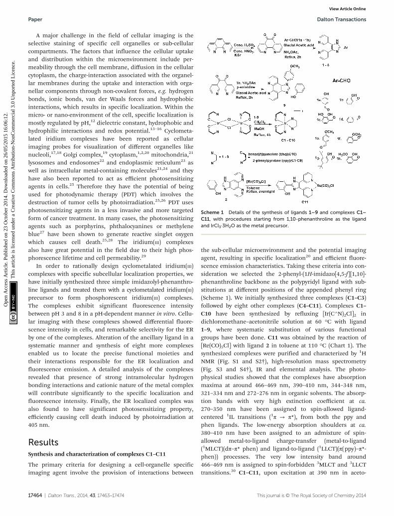
Scheme 1 Details of the synthesis of ligands 1–9 and complexes C1–C11, with procedures starting from 1,10-phenanthroline as the ligandand IrCl3·3H2O as the metal precursor.
Paper Dalton Transactions
17464 | Dalton Trans., 2014, 43, 17463–17474 This journal is © The Royal Society of Chemistry 2014
Ope
n A
cces
s A
rtic
le. P
ublis
hed
on 2
3 O
ctob
er 2
014.
Dow
nloa
ded
on 2
6/05
/201
5 16
:06:
12.
Thi
s ar
ticle
is li
cens
ed u
nder
a C
reat
ive
Com
mon
s A
ttrib
utio
n-N
onC
omm
erci
al 3
.0 U
npor
ted
Lic
ence
.View Article Online
nitrile at 298 K, phosphoresces with λmax ∼ 570–600 nm withphosphorescence quantum yield (ΦPL) and emission lifetime(τe) in the range of 5.40–41.21% and 8.60–17.72 μs respectively(Table S1 and Fig. S5†). These systems possess a large ligand-field stabilization of iridium owing to its high oxidation state,the presence of 5d orbitals and, most importantly, a highligand-field splitting energy due to the presence of two C^Nligands. These enable the iridium to allow intersystem cross-ing from the first singlet excited state to the triplet stateefficiently. Thus a long-lived triplet excited state (τ ∼ μs) andstrong spin–orbit coupling (ζIr = 3909 cm−1) are characteristicsof these complexes.31–33 The photophysical parameters of thecomplexes were quite satisfactory to explore their applicationas cellular imaging agents.
C2 specifically localizes in the endoplasmic reticulum of cells
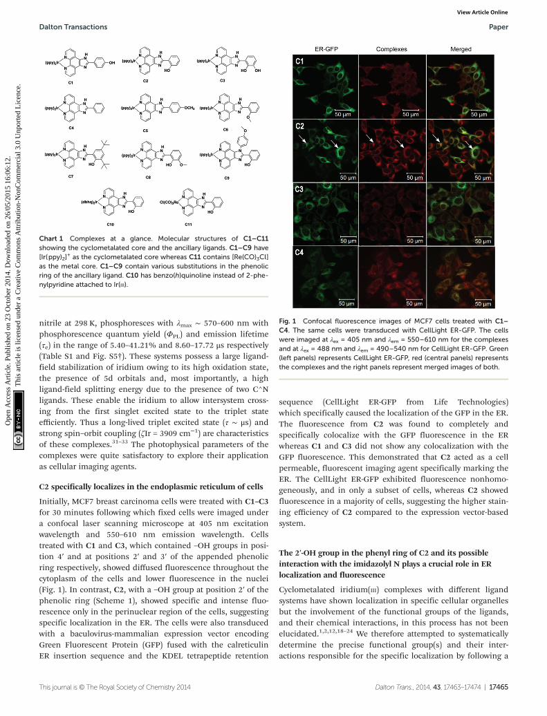
Initially, MCF7 breast carcinoma cells were treated with C1–C3for 30 minutes following which fixed cells were imaged undera confocal laser scanning microscope at 405 nm excitationwavelength and 550–610 nm emission wavelength. Cellstreated with C1 and C3, which contained –OH groups in posi-tion 4′ and at positions 2′ and 3′ of the appended phenolicring respectively, showed diffused fluorescence throughout thecytoplasm of the cells and lower fluorescence in the nuclei(Fig. 1). In contrast, C2, with a –OH group at position 2′ of thephenolic ring (Scheme 1), showed specific and intense fluo-rescence only in the perinuclear region of the cells, suggestingspecific localization in the ER. The cells were also transducedwith a baculovirus-mammalian expression vector encodingGreen Fluorescent Protein (GFP) fused with the calreticulinER insertion sequence and the KDEL tetrapeptide retention
sequence (CellLight ER-GFP from Life Technologies)which specifically caused the localization of the GFP in the ER.The fluorescence from C2 was found to completely andspecifically colocalize with the GFP fluorescence in the ERwhereas C1 and C3 did not show any colocalization with theGFP fluorescence. This demonstrated that C2 acted as a cellpermeable, fluorescent imaging agent specifically marking theER. The CellLight ER-GFP exhibited fluorescence nonhomo-geneously, and in only a subset of cells, whereas C2 showedfluorescence in a majority of cells, suggesting the higher stain-ing efficiency of C2 compared to the expression vector-basedsystem.
The 2′-OH group in the phenyl ring of C2 and its possibleinteraction with the imidazolyl N plays a crucial role in ERlocalization and fluorescence
Cyclometalated iridium(III) complexes with different ligandsystems have shown localization in specific cellular organellesbut the involvement of the functional groups of the ligands,and their chemical interactions, in this process has not beenelucidated.1,2,12,18–24 We therefore attempted to systematicallydetermine the precise functional group(s) and their inter-actions responsible for the specific localization by following a
Chart 1 Complexes at a glance. Molecular structures of C1–C11showing the cyclometalated core and the ancillary ligands. C1–C9 have[Ir(ppy)2]
+ as the cyclometalated core whereas C11 contains [Re(CO)3Cl]as the metal core. C1–C9 contain various substitutions in the phenolicring of the ancillary ligand. C10 has benzo(h)quinoline instead of 2-phe-nylpyridine attached to Ir(III).
Fig. 1 Confocal fluorescence images of MCF7 cells treated with C1–C4. The same cells were transduced with CellLight ER-GFP. The cellswere imaged at λex = 405 nm and λem = 550–610 nm for the complexesand at λex = 488 nm and λem = 490–540 nm for CellLight ER-GFP. Green(left panels) represents CellLight ER-GFP, red (central panels) representsthe complexes and the right panels represent merged images of both.
Dalton Transactions Paper
This journal is © The Royal Society of Chemistry 2014 Dalton Trans., 2014, 43, 17463–17474 | 17465
Ope
n A
cces
s A
rtic
le. P
ublis
hed
on 2
3 O
ctob
er 2
014.
Dow
nloa
ded
on 2
6/05
/201
5 16
:06:
12.
Thi
s ar
ticle
is li
cens
ed u
nder
a C
reat
ive
Com
mon
s A
ttrib
utio
n-N
onC
omm
erci
al 3
.0 U
npor
ted
Lic
ence
.View Article Online
“mutational approach” of systematically altering the variousfunctional groups on the phenyl ring.
Firstly, in order to investigate the role of the 2′-OH group,cells were treated with cyclometalated iridium(III) complex C4containing the 2-phenyl-(1H-imidazo[4,5-f ][1,10]phenanthro-line) as an ancillary ligand, i.e. without any –OH groups. Thiscomplex (C4) showed high intensity fluorescence (quantumyield = 41.21%, τ = 14.57 μs) and considerable ER localization,but the localization was less specific compared to C2, withfluorescence exhibited in the nucleus and to some extent inthe cytoplasm (Fig. 1). This suggests that the specific localiz-ation in the ER requires the presence of the –OH group at 2′position of the appended phenyl ring. However, the othergroups in the complex also play a role in conferring the fluo-rescence property. High fluorescence of C4 might be due tothe absence of fluorescence quenching by the –OH moiety.
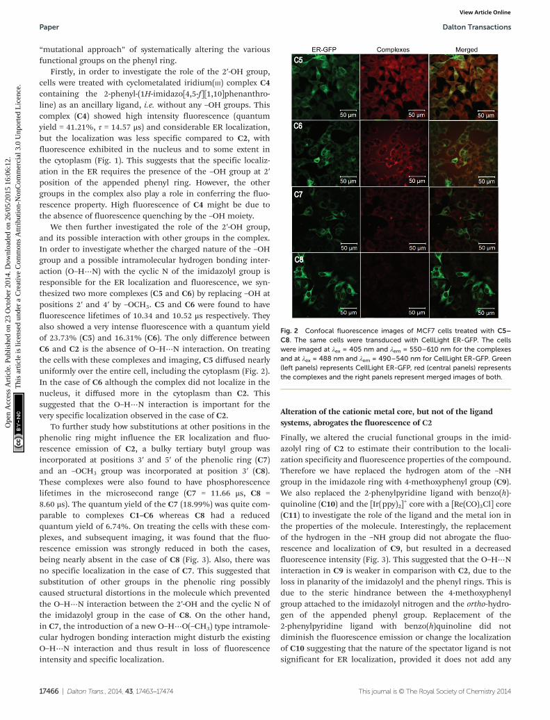
We then further investigated the role of the 2′-OH group,and its possible interaction with other groups in the complex.In order to investigate whether the charged nature of the –OHgroup and a possible intramolecular hydrogen bonding inter-action (O–H⋯N) with the cyclic N of the imidazolyl group isresponsible for the ER localization and fluorescence, we syn-thesized two more complexes (C5 and C6) by replacing –OH atpositions 2′ and 4′ by –OCH3. C5 and C6 were found to havefluorescence lifetimes of 10.34 and 10.52 μs respectively. Theyalso showed a very intense fluorescence with a quantum yieldof 23.73% (C5) and 16.31% (C6). The only difference betweenC6 and C2 is the absence of O–H⋯N interaction. On treatingthe cells with these complexes and imaging, C5 diffused nearlyuniformly over the entire cell, including the cytoplasm (Fig. 2).In the case of C6 although the complex did not localize in thenucleus, it diffused more in the cytoplasm than C2. Thissuggested that the O–H⋯N interaction is important for thevery specific localization observed in the case of C2.
To further study how substitutions at other positions in thephenolic ring might influence the ER localization and fluo-rescence emission of C2, a bulky tertiary butyl group wasincorporated at positions 3′ and 5′ of the phenolic ring (C7)and an –OCH3 group was incorporated at position 3′ (C8).These complexes were also found to have phosphorescencelifetimes in the microsecond range (C7 = 11.66 μs, C8 =8.60 μs). The quantum yield of the C7 (18.99%) was quite com-parable to complexes C1–C6 whereas C8 had a reducedquantum yield of 6.74%. On treating the cells with these com-plexes, and subsequent imaging, it was found that the fluo-rescence emission was strongly reduced in both the cases,being nearly absent in the case of C8 (Fig. 3). Also, there wasno specific localization in the case of C7. This suggested thatsubstitution of other groups in the phenolic ring possiblycaused structural distortions in the molecule which preventedthe O–H⋯N interaction between the 2′-OH and the cyclic N ofthe imidazolyl group in the case of C8. On the other hand,in C7, the introduction of a new O–H⋯O(–CH3) type intramole-cular hydrogen bonding interaction might disturb the existingO–H⋯N interaction and thus result in loss of fluorescenceintensity and specific localization.
Alteration of the cationic metal core, but not of the ligandsystems, abrogates the fluorescence of C2
Finally, we altered the crucial functional groups in the imid-azolyl ring of C2 to estimate their contribution to the locali-zation specificity and fluorescence properties of the compound.Therefore we have replaced the hydrogen atom of the –NHgroup in the imidazole ring with 4-methoxyphenyl group (C9).We also replaced the 2-phenylpyridine ligand with benzo(h)-quinoline (C10) and the [Ir(ppy)2]
+ core with a [Re(CO)3Cl] core(C11) to investigate the role of the ligand and the metal ion inthe properties of the molecule. Interestingly, the replacementof the hydrogen in the –NH group did not abrogate the fluo-rescence and localization of C9, but resulted in a decreasedfluorescence intensity (Fig. 3). This suggested that the O–H⋯Ninteraction in C9 is weaker in comparison with C2, due to theloss in planarity of the imidazolyl and the phenyl rings. This isdue to the steric hindrance between the 4-methoxyphenylgroup attached to the imidazolyl nitrogen and the ortho-hydro-gen of the appended phenyl group. Replacement of the2-phenylpyridine ligand with benzo(h)quinoline did notdiminish the fluorescence emission or change the localizationof C10 suggesting that the nature of the spectator ligand is notsignificant for ER localization, provided it does not add any
Fig. 2 Confocal fluorescence images of MCF7 cells treated with C5–C8. The same cells were transduced with CellLight ER-GFP. The cellswere imaged at λex = 405 nm and λem = 550–610 nm for the complexesand at λex = 488 nm and λem = 490–540 nm for CellLight ER-GFP. Green(left panels) represents CellLight ER-GFP, red (central panels) representsthe complexes and the right panels represent merged images of both.
Paper Dalton Transactions
17466 | Dalton Trans., 2014, 43, 17463–17474 This journal is © The Royal Society of Chemistry 2014
Ope
n A
cces
s A
rtic
le. P
ublis
hed
on 2
3 O
ctob
er 2
014.
Dow
nloa
ded
on 2
6/05
/201
5 16
:06:
12.
Thi
s ar
ticle
is li
cens
ed u
nder
a C
reat
ive
Com
mon
s A
ttrib
utio
n-N
onC
omm
erci
al 3
.0 U
npor
ted
Lic
ence
.View Article Online
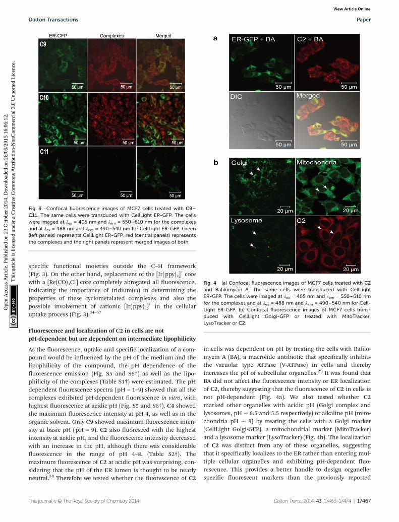
specific functional moieties outside the C–H framework(Fig. 3). On the other hand, replacement of the [Ir(ppy)2]
+ corewith a [Re(CO)3Cl] core completely abrogated all fluorescence,indicating the importance of iridium(III) in determining theproperties of these cyclometalated complexes and also thepossible involvement of cationic [Ir(ppy)2]
+ in the cellularuptake process (Fig. 3).34–37
Fluorescence and localization of C2 in cells are notpH-dependent but are dependent on intermediate lipophilicity
As the fluorescence, uptake and specific localization of a com-pound would be influenced by the pH of the medium and thelipophilicity of the compound, the pH dependence of thefluorescence emission (Fig. S5 and S6†) as well as the lipo-philicity of the complexes (Table S1†) were estimated. The pHdependent fluorescence spectra (pH = 1–9) showed that all thecomplexes exhibited pH-dependent fluorescence in vitro, withhighest fluorescence at acidic pH (Fig. S5 and S6†). C4 showedthe maximum fluorescence intensity at pH 4, as well as in theorganic solvent. Only C9 showed maximum fluorescence inten-sity at basic pH (pH = 9). C2 also fluoresced with the highestintensity at acidic pH, and the fluorescence intensity decreasedwith an increase in the pH, although there was considerablefluorescence in the range of pH 4–8. (Table S2†). Themaximum fluorescence of C2 at acidic pH was surprising, con-sidering that the pH of the ER lumen is thought to be nearlyneutral.38 Therefore we tested whether the fluorescence of C2
in cells was dependent on pH by treating the cells with Bafilo-mycin A (BA), a macrolide antibiotic that specifically inhibitsthe vacuolar type ATPase (V-ATPase) in cells and therebyincreases the pH of subcellular organelles.29 It was found thatBA did not affect the fluorescence intensity or ER localizationof C2, thereby suggesting that the fluorescence of C2 in cells isnot pH-dependent (Fig. 4a). We also tested whether C2marked other organelles with acidic pH (Golgi complex andlysosomes, pH ∼ 6.5 and 5.5 respectively) or alkaline pH (mito-chondria pH ∼ 8) by treating the cells with a Golgi marker(CellLight Golgi-GFP), a mitochondrial marker (MitoTracker)and a lysosome marker (LysoTracker) (Fig. 4b). The localizationof C2 was distinct from any of these organelles, suggestingthat it specifically localizes to the ER rather than entering mul-tiple cellular organelles and exhibiting pH-dependent fluo-rescence. This provides a better handle to design organelle-specific fluorescent markers than the previously reported
Fig. 3 Confocal fluorescence images of MCF7 cells treated with C9–C11. The same cells were transduced with CellLight ER-GFP. The cellswere imaged at λex = 405 nm and λem = 550–610 nm for the complexesand at λex = 488 nm and λem = 490–540 nm for CellLight ER-GFP. Green(left panels) represents CellLight ER-GFP, red (central panels) representsthe complexes and the right panels represent merged images of both.
Fig. 4 (a) Confocal fluorescence images of MCF7 cells treated with C2and Bafilomycin A. The same cells were transduced with CellLightER-GFP. The cells were imaged at λex = 405 nm and λem = 550–610 nmfor the complexes and at λex = 488 nm and λem = 490–540 nm for Cell-Light ER-GFP. (b) Confocal fluorescence images of MCF7 cells trans-duced with CellLight Golgi-GFP or treated with MitoTracker,LysoTracker or C2.
Dalton Transactions Paper
This journal is © The Royal Society of Chemistry 2014 Dalton Trans., 2014, 43, 17463–17474 | 17467
Ope
n A
cces
s A
rtic
le. P
ublis
hed
on 2
3 O
ctob
er 2
014.
Dow
nloa
ded
on 2
6/05
/201
5 16
:06:
12.
Thi
s ar
ticle
is li
cens
ed u
nder
a C
reat
ive
Com
mon
s A
ttrib
utio
n-N
onC
omm
erci
al 3
.0 U
npor
ted
Lic
ence
.View Article Online
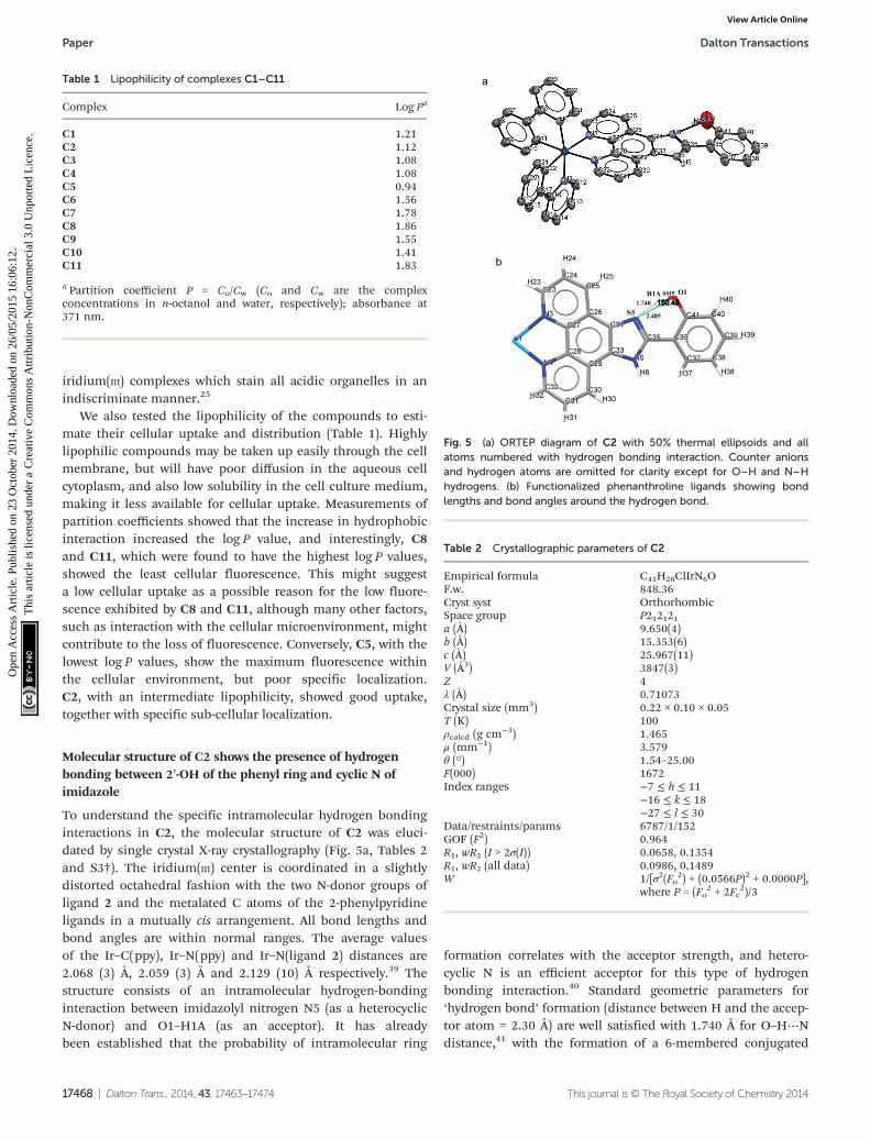
iridium(III) complexes which stain all acidic organelles in anindiscriminate manner.25
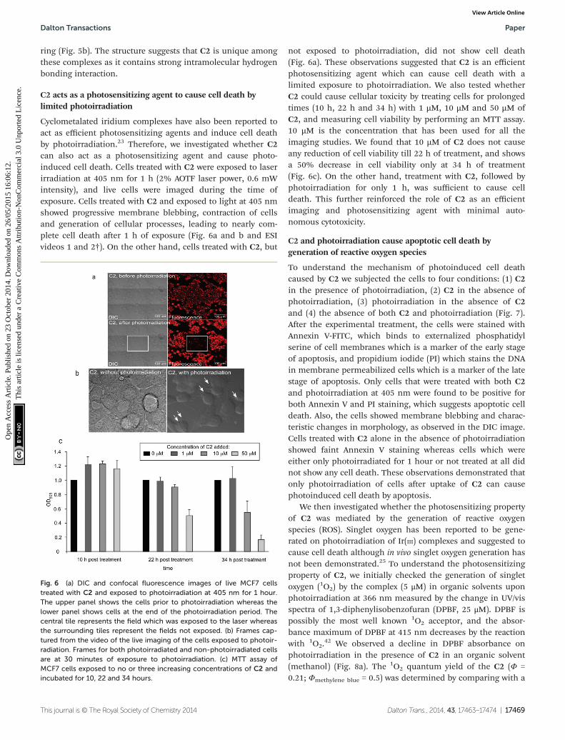
We also tested the lipophilicity of the compounds to esti-mate their cellular uptake and distribution (Table 1). Highlylipophilic compounds may be taken up easily through the cellmembrane, but will have poor diffusion in the aqueous cellcytoplasm, and also low solubility in the cell culture medium,making it less available for cellular uptake. Measurements ofpartition coefficients showed that the increase in hydrophobicinteraction increased the log P value, and interestingly, C8and C11, which were found to have the highest log P values,showed the least cellular fluorescence. This might suggesta low cellular uptake as a possible reason for the low fluore-scence exhibited by C8 and C11, although many other factors,such as interaction with the cellular microenvironment, mightcontribute to the loss of fluorescence. Conversely, C5, with thelowest log P values, show the maximum fluorescence withinthe cellular environment, but poor specific localization.C2, with an intermediate lipophilicity, showed good uptake,together with specific sub-cellular localization.
Molecular structure of C2 shows the presence of hydrogenbonding between 2′-OH of the phenyl ring and cyclic N ofimidazole
To understand the specific intramolecular hydrogen bondinginteractions in C2, the molecular structure of C2 was eluci-dated by single crystal X-ray crystallography (Fig. 5a, Tables 2and S3†). The iridium(III) center is coordinated in a slightlydistorted octahedral fashion with the two N-donor groups ofligand 2 and the metalated C atoms of the 2-phenylpyridineligands in a mutually cis arrangement. All bond lengths andbond angles are within normal ranges. The average valuesof the Ir–C(ppy), Ir–N(ppy) and Ir–N(ligand 2) distances are2.068 (3) Å, 2.059 (3) Å and 2.129 (10) Å respectively.39 Thestructure consists of an intramolecular hydrogen-bondinginteraction between imidazolyl nitrogen N5 (as a heterocyclicN-donor) and O1–H1A (as an acceptor). It has alreadybeen established that the probability of intramolecular ring
formation correlates with the acceptor strength, and hetero-cyclic N is an efficient acceptor for this type of hydrogenbonding interaction.40 Standard geometric parameters for‘hydrogen bond’ formation (distance between H and the accep-tor atom = 2.30 Å) are well satisfied with 1.740 Å for O–H⋯Ndistance,41 with the formation of a 6-membered conjugated
Fig. 5 (a) ORTEP diagram of C2 with 50% thermal ellipsoids and allatoms numbered with hydrogen bonding interaction. Counter anionsand hydrogen atoms are omitted for clarity except for O–H and N–Hhydrogens. (b) Functionalized phenanthroline ligands showing bondlengths and bond angles around the hydrogen bond.
Table 1 Lipophilicity of complexes C1–C11
Complex Log Pa
C1 1.21C2 1.12C3 1.08C4 1.08C5 0.94C6 1.56C7 1.78C8 1.86C9 1.55C10 1.41C11 1.83
a Partition coefficient P = Co/Cw (Co and Cw are the complexconcentrations in n-octanol and water, respectively); absorbance at371 nm.
Table 2 Crystallographic parameters of C2
Empirical formula C41H28ClIrN6OF.w. 848.36Cryst syst OrthorhombicSpace group P212121a (Å) 9.650(4)b (Å) 15.353(6)c (Å) 25.967(11)V (Å3) 3847(3)Z 4λ (Å) 0.71073Crystal size (mm3) 0.22 × 0.10 × 0.05T (K) 100ρcalcd (g cm−3) 1.465μ (mm−1) 3.579θ (°) 1.54–25.00F(000) 1672Index ranges −7 ≤ h ≤ 11
−16 ≤ k ≤ 18−27 ≤ l ≤ 30
Data/restraints/params 6787/1/152GOF (F2) 0.964R1, wR2 (I > 2σ(I)) 0.0658, 0.1354R1, wR2 (all data) 0.0986, 0.1489W 1/[σ2(Fo
2) + (0.0566P)2 + 0.0000P],where P = (Fo
2 + 2Fc2)/3
Paper Dalton Transactions
17468 | Dalton Trans., 2014, 43, 17463–17474 This journal is © The Royal Society of Chemistry 2014
Ope
n A
cces
s A
rtic
le. P
ublis
hed
on 2
3 O
ctob
er 2
014.
Dow
nloa
ded
on 2
6/05
/201
5 16
:06:
12.
Thi
s ar
ticle
is li
cens
ed u
nder
a C
reat
ive
Com
mon
s A
ttrib
utio
n-N
onC
omm
erci
al 3
.0 U
npor
ted
Lic
ence
.View Article Online
ring (Fig. 5b). The structure suggests that C2 is unique amongthese complexes as it contains strong intramolecular hydrogenbonding interaction.
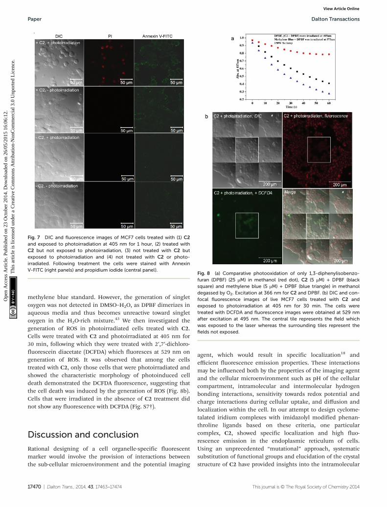
C2 acts as a photosensitizing agent to cause cell death bylimited photoirradiation
Cyclometalated iridium complexes have also been reported toact as efficient photosensitizing agents and induce cell deathby photoirradiation.23 Therefore, we investigated whether C2can also act as a photosensitizing agent and cause photo-induced cell death. Cells treated with C2 were exposed to laserirradiation at 405 nm for 1 h (2% AOTF laser power, 0.6 mWintensity), and live cells were imaged during the time ofexposure. Cells treated with C2 and exposed to light at 405 nmshowed progressive membrane blebbing, contraction of cellsand generation of cellular processes, leading to nearly com-plete cell death after 1 h of exposure (Fig. 6a and b and ESIvideos 1 and 2†). On the other hand, cells treated with C2, but
not exposed to photoirradiation, did not show cell death(Fig. 6a). These observations suggested that C2 is an efficientphotosensitizing agent which can cause cell death with alimited exposure to photoirradiation. We also tested whetherC2 could cause cellular toxicity by treating cells for prolongedtimes (10 h, 22 h and 34 h) with 1 µM, 10 µM and 50 µM ofC2, and measuring cell viability by performing an MTT assay.10 µM is the concentration that has been used for all theimaging studies. We found that 10 µM of C2 does not causeany reduction of cell viability till 22 h of treatment, and showsa 50% decrease in cell viability only at 34 h of treatment(Fig. 6c). On the other hand, treatment with C2, followed byphotoirradiation for only 1 h, was sufficient to cause celldeath. This further reinforced the role of C2 as an efficientimaging and photosensitizing agent with minimal auto-nomous cytotoxicity.
C2 and photoirradiation cause apoptotic cell death bygeneration of reactive oxygen species
To understand the mechanism of photoinduced cell deathcaused by C2 we subjected the cells to four conditions: (1) C2in the presence of photoirradiation, (2) C2 in the absence ofphotoirradiation, (3) photoirradiation in the absence of C2and (4) the absence of both C2 and photoirradiation (Fig. 7).After the experimental treatment, the cells were stained withAnnexin V-FITC, which binds to externalized phosphatidylserine of cell membranes which is a marker of the early stageof apoptosis, and propidium iodide (PI) which stains the DNAin membrane permeabilized cells which is a marker of the latestage of apoptosis. Only cells that were treated with both C2and photoirradiation at 405 nm were found to be positive forboth Annexin V and PI staining, which suggests apoptotic celldeath. Also, the cells showed membrane blebbing and charac-teristic changes in morphology, as observed in the DIC image.Cells treated with C2 alone in the absence of photoirradiationshowed faint Annexin V staining whereas cells which wereeither only photoirradiated for 1 hour or not treated at all didnot show any cell death. These observations demonstrated thatonly photoirradiation of cells after uptake of C2 can causephotoinduced cell death by apoptosis.
We then investigated whether the photosensitizing propertyof C2 was mediated by the generation of reactive oxygenspecies (ROS). Singlet oxygen has been reported to be gene-rated on photoirradiation of Ir(III) complexes and suggested tocause cell death although in vivo singlet oxygen generation hasnot been demonstrated.25 To understand the photosensitizingproperty of C2, we initially checked the generation of singletoxygen (1O2) by the complex (5 μM) in organic solvents uponphotoirradiation at 366 nm measured by the change in UV/visspectra of 1,3-diphenylisobenzofuran (DPBF, 25 μM). DPBF ispossibly the most well known 1O2 acceptor, and the absor-bance maximum of DPBF at 415 nm decreases by the reactionwith 1O2.
42 We observed a decline in DPBF absorbance onphotoirradiation in the presence of C2 in an organic solvent(methanol) (Fig. 8a). The 1O2 quantum yield of the C2 (Φ =0.21; Φmethylene blue = 0.5) was determined by comparing with a
Fig. 6 (a) DIC and confocal fluorescence images of live MCF7 cellstreated with C2 and exposed to photoirradiation at 405 nm for 1 hour.The upper panel shows the cells prior to photoirradiation whereas thelower panel shows cells at the end of the photoirradiation period. Thecentral tile represents the field which was exposed to the laser whereasthe surrounding tiles represent the fields not exposed. (b) Frames cap-tured from the video of the live imaging of the cells exposed to photoir-radiation. Frames for both photoirradiated and non-photoirradiated cellsare at 30 minutes of exposure to photoirradiation. (c) MTT assay ofMCF7 cells exposed to no or three increasing concentrations of C2 andincubated for 10, 22 and 34 hours.
Dalton Transactions Paper
This journal is © The Royal Society of Chemistry 2014 Dalton Trans., 2014, 43, 17463–17474 | 17469
Ope
n A
cces
s A
rtic
le. P
ublis
hed
on 2
3 O
ctob
er 2
014.
Dow
nloa
ded
on 2
6/05
/201
5 16
:06:
12.
Thi
s ar
ticle
is li
cens
ed u
nder
a C
reat
ive
Com
mon
s A
ttrib
utio
n-N
onC
omm
erci
al 3
.0 U
npor
ted
Lic
ence
.View Article Online
methylene blue standard. However, the generation of singletoxygen was not detected in DMSO–H2O, as DPBF dimerizes inaqueous media and thus becomes unreactive toward singletoxygen in the H2O-rich mixture.43 We then investigated thegeneration of ROS in photoirradiated cells treated with C2.Cells were treated with C2 and photoirradiated at 405 nm for30 min, following which they were treated with 2′,7′-dichloro-fluorescein diacetate (DCFDA) which fluoresces at 529 nm ongeneration of ROS. It was observed that among the cellstreated with C2, only those cells that were photoirradiated andshowed the characteristic morphology of photoinduced celldeath demonstrated the DCFDA fluorescence, suggesting thatthe cell death was induced by the generation of ROS (Fig. 8b).Cells that were irradiated in the absence of C2 treatment didnot show any fluorescence with DCFDA (Fig. S7†).
Discussion and conclusion
Rational designing of a cell organelle-specific fluorescentmarker would involve the provision of interactions betweenthe sub-cellular microenvironment and the potential imaging
agent, which would result in specific localization18 andefficient fluorescence emission properties. These interactionsmay be influenced both by the properties of the imaging agentand the cellular microenvironment such as pH of the cellularcompartment, intramolecular and intermolecular hydrogenbonding interactions, sensitivity towards redox potential andcharge interactions during cellular uptake, and diffusion andlocalization within the cell. In our attempt to design cyclome-talated iridium complexes with imidazolyl modified phenan-throline ligands based on these criteria, one particularcomplex, C2, showed specific localization and high fluo-rescence emission in the endoplasmic reticulum of cells.Using an unprecedented “mutational” approach, systematicsubstitution of functional groups and elucidation of the crystalstructure of C2 have provided insights into the intramolecular
Fig. 8 (a) Comparative photooxidation of only 1,3-diphenylisobenzo-furan (DPBF) (25 μM) in methanol (red dot), C2 (5 μM) + DPBF (blacksquare) and methylene blue (5 μM) + DPBF (blue triangle) in methanoldegassed by O2. Excitation at 366 nm for C2 and DPBF. (b) DIC and con-focal fluorescence images of live MCF7 cells treated with C2 andexposed to photoirradiation at 405 nm for 30 min. The cells weretreated with DCFDA and fluorescence images were obtained at 529 nmafter excitation at 495 nm. The central tile represents the field whichwas exposed to the laser whereas the surrounding tiles represent thefields not exposed.
Fig. 7 DIC and fluorescence images of MCF7 cells treated with (1) C2and exposed to photoirradiation at 405 nm for 1 hour, (2) treated withC2 but not exposed to photoirradiation, (3) not treated with C2 butexposed to photoirradiation and (4) not treated with C2 or photo-irradiated. Following treatment the cells were stained with AnnexinV-FITC (right panels) and propidium iodide (central panel).
Paper Dalton Transactions
17470 | Dalton Trans., 2014, 43, 17463–17474 This journal is © The Royal Society of Chemistry 2014
Ope
n A
cces
s A
rtic
le. P
ublis
hed
on 2
3 O
ctob
er 2
014.
Dow
nloa
ded
on 2
6/05
/201
5 16
:06:
12.
Thi
s ar
ticle
is li
cens
ed u
nder
a C
reat
ive
Com
mon
s A
ttrib
utio
n-N
onC
omm
erci
al 3
.0 U
npor
ted
Lic
ence
.View Article Online
chemical interactions responsible for its photophysical pro-perties and specific subcellular localization. A specific hydro-gen bonding interaction between the 2′-OH group in theappended phenyl ring and the –NH group attached to theimidazole ring was found to confer localization specificity to thecomplex. Also, the highly stable resonance-assisted hydrogenbonding resulted in enhanced π-delocalization within thecomplex C2, and resulted in improved fluorescence due toexcited state intramolecular proton transfer (ESIPT).44 On theother hand, replacement of the –NH group with N-anisidine inC9, which is expected to maintain the hydrogen bonding inter-action, did not alter the ER specific localization, but reducedthe fluorescence intensity substantially. This is possibly due tothe distortion of planarity between the imidazolyl group andthe appended phenyl group, increasing the proportion of theopen form of the molecule relative to the closed form contribu-ted by the intramolecular hydrogen bonding.
While changes in the functional groups in the phenyland imidazolyl rings only altered subcellular localizationor reduced fluorescence emission, the replacement of the[Ir(ppy)2]
+ with [Re(CO)3Cl] in C11 completely abrogated allfluorescence, suggesting that the cationic nature of Ir(III) isplaying a crucial role in cellular uptake. On the other hand,replacement of 2-phenylpyridine ligand with benzo(h)quino-line did not substantially alter ER localization or fluorescenceemission suggesting that a change in the C–H frameworkof the spectator ligand does not have a profound effect on sub-cellular localization.
The fluorescence emission of cyclometalated iridium(III)complexes is known to be pH dependent based on the ancil-lary ligand system.45 In our study, in vitro fluorescence of thecomplexes was found to be pH-dependent, with maximumfluorescence of most of the complexes being in the acidic pHrange. However, the high fluorescence of C2 in the ER, whichis reported to have neutral pH, and the absence of a decreasein fluorescence intensity under Bafilomycin A treatmentshowed that the fluorescence of C2 in cells was not pH depen-dent. This is unlike the properties of cyclometalated Ir(III) com-plexes reported previously, which specifically stained acidicorganelles.25 This provides a better tool to design organelle-specific fluorescent markers which might be targeted tospecific sub-cellular compartments or organelles by substitut-ing different functional groups. Also, the intermediate lipo-philicity of the complex appears to have contributed to theefficient uptake and subcellular localization, suggesting thatefficient diffusion through the cytoplasm might be beneficialfor targeting to specific organelles.
Furthermore, the complex has been found to be an efficientphotosensitizing agent, causing extensive photoinduced celldeath upon exposure to light at 405 nm for one hour. Ir(III)complex functionalized nanoparticles have been reported tohave applications in PDT and a recent study has shown aniridium(III) complex with the N,N-diethylamino group in theancillary ligand21,23–25 to cause photoinduced necrosis-like celldeath. However, C2 was found to cause photoinduced apopto-tic cell death and very little cytotoxicity in the absence of
photoirradiation. These results suggest that the complex hasenormous potential for application as a cellular imaging agentin cell biology as well as a photosensitizer for PDT in medi-cine. Further studies need to be directed at elucidating themechanism by which C2 causes apoptotic cell death as a resultof photoirradiation.
Experimental section
The starting materials IrCl3·3H2O, 2-phenylpyridine, 1,10-phenanthroline, aldehydes, and n-octanol were purchasedfrom Sigma-Aldrich and used without further purification. Allthe solvents were dried by usual methods prior to use. Thecyclometalated iridium(III) chloro bridged dimer [Ir(ppy)2Cl]2was prepared according to the methods described in theliterature.46
Synthesis of ligands
The ligands 1–8 47 and 9 (–N-Ph has been replaced by N-p-anisidine)48 were synthesized with a similar procedure to thatreported earlier. Ligands 3, 5, 6, and 8 were not reportedearlier. See the ESI† for details of the synthesis and Fig. S1and S3† for NMR and ESI-MS data.
Synthesis of complexes
Complexes C2 and C4 were synthesized with a similar pro-cedure to that reported earlier.40 Complexes C1, C3, C5, C6,C7, C8, C9, C10 and C11 were not reported earlier. See the ESIfor details of the synthesis and Fig. S2 and S4† for NMR andESI-MS data.
Physical measurements
IR spectra were obtained on a Perkin-Elmer Spectrum RXIspectrophotometer with samples prepared as KBr pellets.Elemental analyses were performed on a Perkin-Elmer 2400series II CHN series. Electronic spectra were recorded on aU-4100, HITACHI spectrometer. 1H NMR spectra were obtainedon a Jeol 400 MHz NMR spectrometer using TMS as theinternal standard. Electrochemical measurements were madeusing a PAR model 273 potentiostat. A platinum disk workingelectrode, a platinum wire auxiliary electrode and an aqueousAg/Ag+ reference electrode were used in a three electrode con-figuration. Electrochemical measurements were made under adinitrogen atmosphere. All electrochemical data were collectedat 298 K and are uncorrected for junction potentials. Massspectra were recorded on a Q-Tof Micromass spectrometer bypositive-ion mode electrospray ionization. Fluorescencespectra were taken on a HORIBA JOBINYVON spectrofluorom-eter Fluoromax 4. Quantum yield data reported here weremeasured relative to quinine sulfate in 0.1 M H2SO4 (λex =350 nm, Φ = 0.577). The integration of the emission spectrawas obtained from the Fluoromax-4 instrument directly. Thephotoluminescence spectra were measured on a Horiba JobinYvon spectrometer equipped with a 150 W Xe lamp. The life-time measurements were done by exciting the sample using a
Dalton Transactions Paper
This journal is © The Royal Society of Chemistry 2014 Dalton Trans., 2014, 43, 17463–17474 | 17471
Ope
n A
cces
s A
rtic
le. P
ublis
hed
on 2
3 O
ctob
er 2
014.
Dow
nloa
ded
on 2
6/05
/201
5 16
:06:
12.
Thi
s ar
ticle
is li
cens
ed u
nder
a C
reat
ive
Com
mon
s A
ttrib
utio
n-N
onC
omm
erci
al 3
.0 U
npor
ted
Lic
ence
.View Article Online
pulsed Xe lamp. Lifetime measurements were performed withdry deaerated dichloromethane solution (conc. 10−5 M). Thefirst order exponential decay curve was fitted to obtain thephosphorescence lifetime.
X-ray crystallography
Crystal data were collected on a Bruker SMART APEXII CCDarea-detector diffractometer using graphite monochromatedMo Kα radiation (λ = 0.71073 Å). For the crystal, X-ray datareduction was carried out using the Bruker SAINT program.The structures were solved by direct methods using theSHELXS-97 program and refinement was done using theSHELXL-97 program. Selected crystal data and data collectionparameters for all the complexes are given in Table 1. X-raydata reduction, structure solution and refinement were doneusing the SHELXL-97 program package.49
Partition coefficient determination50
The lipophilicity of complexes C1–C11 was determined bythe “shake flask” method using a pH 7.4 phosphate buffer(0.129 M NaCl) and n-octanol as solvents. Each compound wasdissolved in the phase in which it is most soluble, resulting intypical concentrations of 50–350 μM. Duplicate determinationsusing three different solvent ratios were performed for eachcomplex. Following the mixing and phase separation, eachphase was analyzed for solute content, and the concentrationwas determined using spectrophotometric methods. All then-octanol–water partition coefficients were determined byUV/vis spectroscopy. Octanol and buffer solutions were presa-turated. Fifty rotations were performed by hand, followedby 1 h of settling time. Equilibration and absorption measure-ments were made at 20 °C.
Study of pH-dependent fluorescence
Tris buffer solution (500 ml, 0.1 M) was prepared and a seriesof buffer solutions having pH in the range 2–9 was prepared byadding 1 M HCl/NaOH aqueous solution to the Tris solution.The pH of the resulting solutions was recorded using a pHmeter. 31 × 10−5 M stock solutions of all the complexes wereprepared in dry DMSO, and in the fluorescence cuvette, 3.0 mlbuffer solution and 0.1 ml complex solution in DMSO weremixed and fluorescence emission was recorded at 25 °C.
Singlet oxygen (1O2) quantum yield
Quantum yields for singlet oxygen generation in CH3OH weredetermined by monitoring the photooxidation of 1,3-dipheny-lisobenzofuran (DPBF) sensitized by the Ir complexes. Valuesof the triplet photosensitizers were calculated according to amodified literature method.51 The DPBF and samples withDPBF were irradiated at 405 nm and methylene blue was irra-diated at 575 nm.25 The light source of a spectrofluorometerwas used as an irradiation light source (150 W Xe source) toobtain constant monochromatic light. The absorbance ofDPBF was adjusted to around 1.0 at 415 nm in oxygen satu-rated CH3OH, and the absorbance of the sensitizers wasadjusted to 0.2–0.3 at the irradiation wavelength. The photo-
oxidation of DPBF was monitored at intervals of 5 s. Thequantum yields of singlet oxygen generation (ΦΔ) were calcu-lated by a relative method using methylene blue (in methanol)(ΦΔ = 0.50)52 as the reference. The following equation wasused:
ΦΔðC2Þ ¼ ΦΔðstdÞðmC2 �FstdÞ=ðmstd �FC2Þ
where subscripts ‘C2’ and ‘std’ designate the iridium complexand methylene blue, respectively, m is the slope of a plot ofdifferences in changes in absorbance of DPBF (at 415 nm)with the irradiation time and F is the absorption correctionfactor, which is given by F = 1–10−A (absorbance at theirradiation wavelength).
Cellular imaging
MCF7 human breast carcinoma cells were maintained inDMEM with 10% FBS. Cells were treated with 10 µM of thecomplexes in 0.1% DMSO in DMEM medium. Cells weretreated for 30 minutes, following which they were washed withPBS (pH 7.4) and fixed with 4% paraformaldehyde. Fixed cellswere imaged in a Zeiss LSM710 confocal laser scanning micro-scope at 405 nm excitation wavelength and 550 nm–610 nmemission wavelengths. For identifying the ER, cells were alsotransduced with a baculovirus-mammalian expression vectorencoding Green Fluorescent Protein (GFP) fused with the calre-ticulin ER insertion sequence and the KDEL tetrapeptideretention sequence (CellLight ER-GFP from Life Technologies)according to the manufacturer’s instructions. A similar treat-ment was also performed for identifying the Golgi complex(CellLight ER-GFP from Life Technologies). Mitochondria andlysosomes were stained with MitoTracker and LysoTracker dyes(Life Technologies) respectively according to the manufac-turer’s instructions. For the inhibition of V-type ATPases, cellswere treated with 0.5 µM Bafilomycin A1 (Sigma Life Sciences)for 1 hour prior to treatment with the complex. For live cellimaging, cells were treated with the complex 10 minutes priorto the initiation of live cell imaging. Live imaging at 405 nmwas done for 1 hour. For checking apoptotic cell death, cellswere stained with Annexin-V FITC and propidium iodide (PI)after treatment with C2 and photoirradiation. For checkingROS generation, cells were treated with 2′,7′-dichlorofluore-scein diacetate (DCFDA), a fluorogenic dye that fluoresces inthe presence of ROS with excitation and emission maxima at495 nm and 529 nm respectively.
Cell viability assay
MCF7 cells were treated with 1 µM, 10 µM and 50 µM of thecomplex and kept for 10 hours, 22 hours and 34 hours. At theend of these incubation times, cells were washed with PBS and0.5 mg ml−1 of 3-[4,5-dimethylthiazol-2-yl]-2,5-diphenyl tetra-zolium bromide (MTT from Sigma Life Sciences) was added tothe medium. Cells were incubated for 3 hours following whichcells were lysed with 0.1 N HCl in isopropanol and absorptionreadings were taken at 595 nm.
Paper Dalton Transactions
17472 | Dalton Trans., 2014, 43, 17463–17474 This journal is © The Royal Society of Chemistry 2014
Ope
n A
cces
s A
rtic
le. P
ublis
hed
on 2
3 O
ctob
er 2
014.
Dow
nloa
ded
on 2
6/05
/201
5 16
:06:
12.
Thi
s ar
ticle
is li
cens
ed u
nder
a C
reat
ive
Com
mon
s A
ttrib
utio
n-N
onC
omm
erci
al 3
.0 U
npor
ted
Lic
ence
.View Article Online

Acknowledgements
Soumik Mandal and Dipak K. Poria are thankful to CSIR, NewDelhi for Senior Research Fellowship. This work is supportedby DST, New Delhi through research grants ST/FT/CS-057/2009awarded to Parna Gupta and Wellcome Trust-DBT IndiaAlliance Intermediate Fellowship WT500139/Z/09/Z awarded toPartho Sarothi Ray. We thank Dr. G. Subba Rao, MCB, IISc.Bangalore for helpful discussions.
Notes and references
1 K. K.-W. Lo, A. W.-T. Choi and W. H.-T. Law, Dalton Trans.,2012, 41, 6021.
2 Q. Zhao, C. Huanga and F. Li, Chem. Soc. Rev., 2011, 40,2508.
3 S. A. Hilderbrand, Methods Mol. Biol., 2010, 591, 17.4 C. L. Ho, K.-L. Wong, H.-K. Kong, Y.-M. Ho, C. T.-L. Chan,
W.-M. Kwok, K. S.-Y. Leung, H.-L. Tam, M. H.-W. Lam,X.-F. Ren, A.-M. Ren, J.-K. Feng and W.-Y. Wong, Chem.Commun., 2012, 48, 2525.
5 H. Sun, L. Yang, H. Yang, S. Liu, W. Xu, X. Liu, Z. Tu,H. Su, Q. Zhao and W. Huang, RSC Adv., 2013, 3, 8766.
6 V. W.-W. Yam and K. M.-C. Wo, Chem. Commun., 2011, 47,11579.
7 C.-H. Chang, Z.-J. Wu, C.-H. Chiu, Y.-H. Liang, Y.-S. Tsai,J.-L. Liao, Y. Chi, H.-Y. Hsieh, T.-Y. Kuo, G.-H. Lee,H.-A. Pan, P.-T. Chou, J.-S. Lin and M.-R. Tseng, ACS Appl.Mater. Interfaces, 2013, 5, 7341.
8 A. C. Brooks, K. Basore and S. Bernhard, Inorg. Chem.,2013, 52, 5794.
9 P. N. Curtin, L. L. Tinker, C. M. Burgess, E. D. Cline andS. Bernhard, Inorg. Chem., 2009, 48, 10498.
10 E. Baranoff, J.-H. Yum, I. Jung, R. Vulcano, M. Grtzel andM. K. Nazeeruddin, Chem. – Asian J., 2010, 5, 496.
11 Y. You and W. Nam, Chem. Soc. Rev., 2012, 41, 7061.12 J. Han and K. Burgess, Chem. Rev., 2010, 110, 2709.13 M. Patra, G. Gasser and N. Metzler-Nolte, Dalton Trans.,
2012, 41, 6350.14 C. G. Hartinger, N. Metzler-Nolte and P. J. Dyson, Organo-
metallics, 2012, 31, 5677.15 I. Romero-Canelón and P. J. Sadler, Inorg. Chem., 2013, 52,
12276.16 Z. Liu and P. J. Sadler, Acc. Chem. Res., 2014, 47,
1174.17 C. Li, M. Yu, Y. Sun, Y. Wu, C. Huang and F. A. Li, J. Am.
Chem. Soc., 2011, 133, 11231.18 K. Y. Zhang, S. Po-Yam Li, N. Zhu, I. Wai-Shan Or, M. Shau-
Ha Cheung, Y.-W. Lam and K. K.-W. Lo, Inorg. Chem., 2010,49, 2530.
19 K. Y. Zhang, H.-W. Liu, T. T.-H. Fong, X.-G. Chen andK. K.-W. Lo, Inorg. Chem., 2010, 49, 5432.
20 H. Wu, T. Yang, Q. Zhao, J. Zhou, C. Li and F. A. Li, DaltonTrans., 2011, 40, 1969.
21 S. P.-Y. Li, C. T.-S. Lau, M.-W. Louie, Y.-W. Lam,S. H. Cheng and K. K.-W. Lo, Biomaterials, 2013, 34,7519.
22 P. Steunenberg, A. Ruggi, N. S. van den Berg, T. Buckle,J. Kuil, F. W. B. van Leeuwen and A. H. Velders, Inorg.Chem., 2012, 51, 2105.
23 R. Cao, J. Jia, X. Ma, M. Zhou and H. Fei, J. Med. Chem.,2013, 56, 3636.
24 Y. You, S. Cho and W. Nam, Inorg. Chem., 2013, 53, 1804.25 S. Moromizato, Y. Hisamatsu, T. Suzuki, Y. Matsuo, R. Abe
and S. Aoki, Inorg. Chem., 2012, 51, 12697.26 J. P. Celli, B. Q. Spring, I. Rizvi, C. L. Evans, K. S. Samkoe,
S. Verma, B. W. Pogue and T. Hasan, Chem. Rev., 2010, 110,2795.
27 H. Ali and J. E. van Lier, Chem. Rev., 1999, 99, 2379.28 T. Yogo, Y. Urano, A. Mizushima, H. Sunahara, T. Inoue,
K. Hirose, M. Iino, K. Kikuchi and T. Nagano, Proc. Natl.Acad. Sci. U. S. A., 2008, 105, 28.
29 C.-W. Lai, Y.-H. Wang, C.-H. Lai, M.-J. Yang, C.-Y. Chen,P.-T. Chou, C.-S. Chan, Y. Chi, Y.-C. Chen and J.-K. Hsiao,Small, 2008, 4, 218.
30 S. Ladouceur and E. Zysman-Colman, Eur. J. Inorg. Chem.,2013, 2985.
31 M. S. Lowry and S. Bernhard,Chem. – Eur. J., 2006, 12, 7970.32 S. Campagna, M. Cavazzini, M. Cusumano, M. L. Di Pietro,
A. Giannetto, F. Puntoriero and S. Quici, Inorg. Chem.,2011, 50, 10667.
33 P-K. Lee, W. H.-T. Law, H.-W. Liu and K. K.-W. Lo, Inorg.Chem., 2011, 50, 8570.
34 Z.-B. Zheng, Y.-Q. Wu, K.-Z. Wang and F. Li, Dalton Trans.,2014, 53, 3273.
35 E. E. Langdon-Jones, N. O. Symonds, S. E. Yates,A. J. Hayes, D. Lloyd, R. Williams, S. J. Coles, P. N. Hortonand S. J. A. Pope, Inorg. Chem., 2014, 53, 3788.
36 R. G. Balasingham, M. P. Coogan and F. L. Thorp-Green-wood, Dalton Trans., 2011, 40, 11663.
37 L. Raszeja, A. Maghnouj, S. Hahn and N. Metzler-Nolte,ChemBioChem, 2011, 12, 371.
38 J. H. Kim, L. Johannes, B. Goud, C. Antony,C. A. Lingwood, R. Daneman and S. Grinstein, Proc. Natl.Acad. Sci. U. S. A., 1998, 95, 2997.
39 Q. Zhao, S. Liu, M. Shi, F. Li, H. Jing, T. Yi and C. Huang,Organometallics, 2007, 26, 5922.
40 C. Laurence, K. A. Brameld, J. Graton, J.-Y. Le Questel andE. Renault, J. Med. Chem., 2009, 52, 4073.
41 C. Bilton, F. H. Allen, G. P. Shields and J. A. K. Howard,Acta Crystallogr., Sect. B: Struct. Sci., 2000, 56, 849.
42 F. Wilkinson, W. P. Helman and A. B. Ross, J. Phys. Chem.Ref. Data, 1993, 22, 113.
43 I. Kraljic, Biochimie, 1986, 68, 807.44 A. I. Ciuciu, K. Skonieczny, D. Koszelewski, D. T. Gryko and
L. Flamigni, J. Phys. Chem. C, 2013, 117, 791.45 Y. Himeda, N. Onozawa-Komatsuzaki, S. Miyazawa,
H. Sugihara, T. Hirose and K. Kasuga, Chem. – Eur. J., 2008,14, 11076.
46 M. Nonoyama, Bull. Chem. Soc. Jpn., 1974, 47, 767.
Dalton Transactions Paper
This journal is © The Royal Society of Chemistry 2014 Dalton Trans., 2014, 43, 17463–17474 | 17473
Ope
n A
cces
s A
rtic
le. P
ublis
hed
on 2
3 O
ctob
er 2
014.
Dow
nloa
ded
on 2
6/05
/201
5 16
:06:
12.
Thi
s ar
ticle
is li
cens
ed u
nder
a C
reat
ive
Com
mon
s A
ttrib
utio
n-N
onC
omm
erci
al 3
.0 U
npor
ted
Lic
ence
.View Article Online

47 B. Coban, U. Yildiz and A. Sengul, J. Biol. Inorg. Chem.,2013, 18, 461.
48 K. Skonieczny, A. I. Ciuciu, E. M. Nichols, V. Hugues,M. Blanchard-Desce, L. Flamigni and D. T. Gryko, J. Mater.Chem., 2012, 22, 20649.
49 G. M. Sheldrick, Acta Crystallogr., Sect. A: Fundam. Crystal-logr., 2008, 64, 112.
50 A. M. Angeles-Boza, H. T. Chifotides, J. D. Aguirre,A. Chouai, P. K. L. Fu, K. R. Dunbar and C. Turro, J. Med.Chem., 2006, 49, 6841.
51 N. Adarsh, R. R. Avirah and D. Ramaiah, Org. Lett., 2010,12, 5720.
52 M. C. DeRosa and R. J. Crutchley, Coord. Chem. Rev., 2002,233–234, 351.
Paper Dalton Transactions
17474 | Dalton Trans., 2014, 43, 17463–17474 This journal is © The Royal Society of Chemistry 2014
Ope
n A
cces
s A
rtic
le. P
ublis
hed
on 2
3 O
ctob
er 2
014.
Dow
nloa
ded
on 2
6/05
/201
5 16
:06:
12.
Thi
s ar
ticle
is li
cens
ed u
nder
a C
reat
ive
Com
mon
s A
ttrib
utio
n-N
onC
omm
erci
al 3
.0 U
npor
ted
Lic
ence
.View Article Online