development, disease and repair - escardio.org · development, disease and repair. leiden...
TRANSCRIPT

Cardiac stem cellsdevelopment, disease and repair
Leiden University Medical CenterAnatomy and Embryology
Robert [email protected]
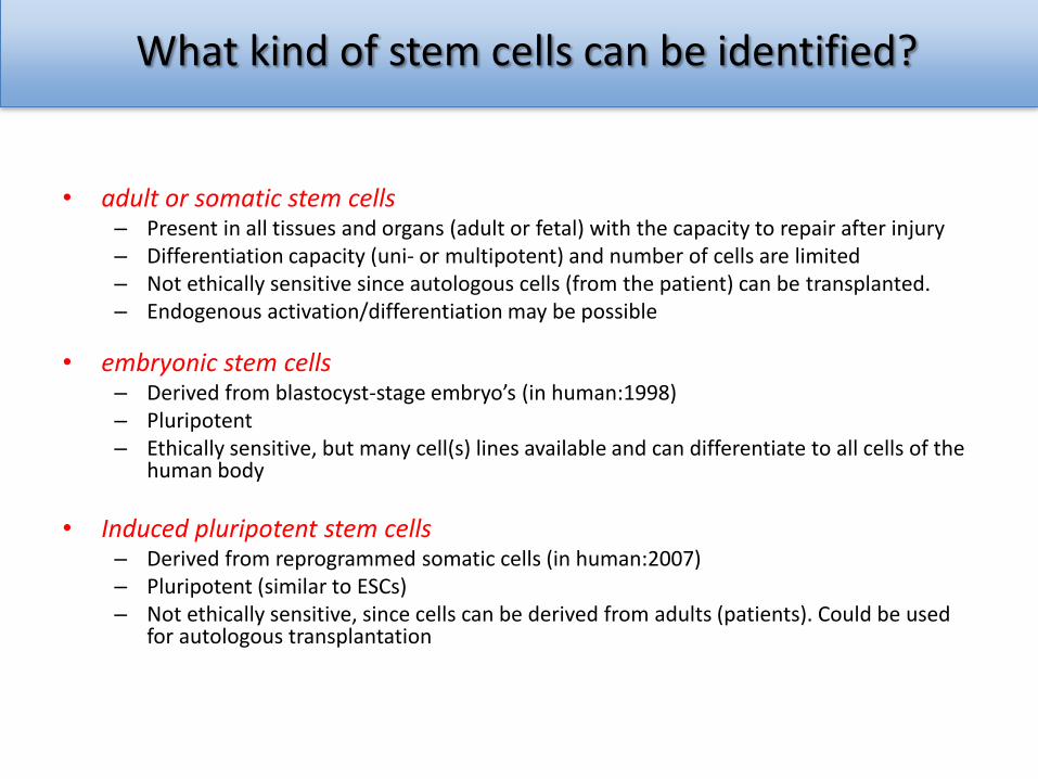
What kind of stem cells can be identified?
• adult or somatic stem cells – Present in all tissues and organs (adult or fetal) with the capacity to repair after injury – Differentiation capacity (uni- or multipotent) and number of cells are limited– Not ethically sensitive since autologous cells (from the patient) can be transplanted.– Endogenous activation/differentiation may be possible
• embryonic stem cells– Derived from blastocyst-stage embryo’s (in human:1998)– Pluripotent– Ethically sensitive, but many cell(s) lines available and can differentiate to all cells of the
human body
• Induced pluripotent stem cells– Derived from reprogrammed somatic cells (in human:2007)– Pluripotent (similar to ESCs)– Not ethically sensitive, since cells can be derived from adults (patients). Could be used
for autologous transplantation
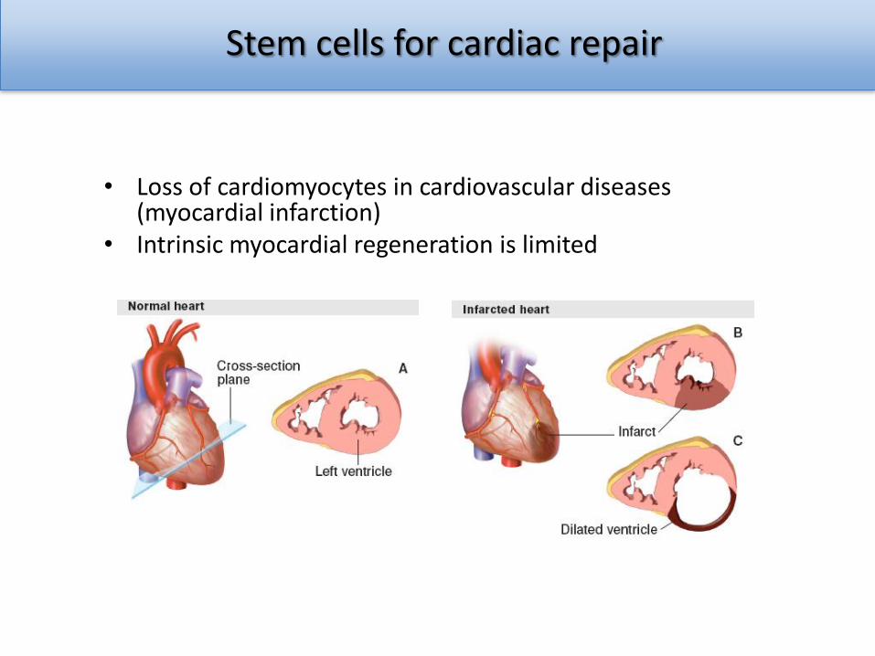
Stem cells for cardiac repair
• Loss of cardiomyocytes in cardiovascular diseases (myocardial infarction)
• Intrinsic myocardial regeneration is limited
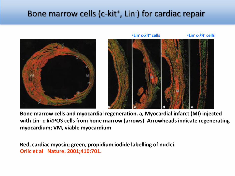
Bone marrow cells (c-kit+, Lin-) for cardiac repair
•Lin- c-kit+ cells •Lin- c-kit- cells
Bone marrow cells and myocardial regeneration. a, Myocardial infarct (MI) injected with Lin- c-kitPOS cells from bone marrow (arrows). Arrowheads indicate regenerating myocardium; VM, viable myocardium
Red, cardiac myosin; green, propidium iodide labelling of nuclei. Orlic et al Nature. 2001;410:701.
Adult stem cells for cardiac repair
• Clinical trials with bone marrow cells with limited success– Slight improvement of heart function– No cardiac regeneration
• Mesenchymal stem cells (MSCs)– Stromal cells obtained from the bone marrow, but also from many other tissues
• Adipose tissue, umbilical cord blood, placenta, pericvascular tissues, etc.
– Self-renewal capacity– Multipotent differentiation capacity
• chondrocytes, osteoblasts, adipocytes, cardiomyocytes)
• Role of MSCs in cardiac repair– Improved heart function
• Migration to injury site• Immunosuppressive properties• Increased vascularization• Release of growth factors (VEGF, IGF-1)• Cardiac differentiation from MSCs is limited
cardiac stem cells
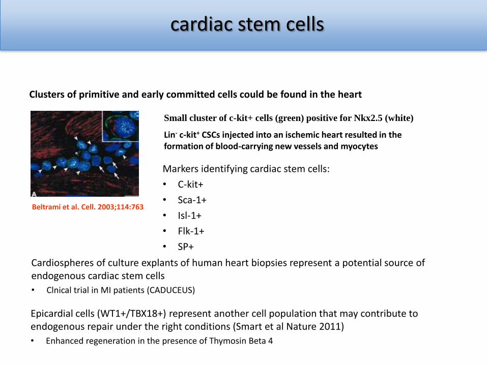
Clusters of primitive and early committed cells could be found in the heart
Small cluster of c-kit+ cells (green) positive for Nkx2.5 (white)
Lin- c-kit+ CSCs injected into an ischemic heart resulted in the formation of blood-carrying new vessels and myocytes
Beltrami et al. Cell. 2003;114:763
Markers identifying cardiac stem cells:
• C-kit+
• Sca-1+
• Isl-1+
• Flk-1+
• SP+
Cardiospheres of culture explants of human heart biopsies represent a potential source of endogenous cardiac stem cells
• Clnical trial in MI patients (CADUCEUS)
Epicardial cells (WT1+/TBX18+) represent another cell population that may contribute to endogenous repair under the right conditions (Smart et al Nature 2011)
• Enhanced regeneration in the presence of Thymosin Beta 4
Human Pluripotent Stem Cells
generation of cardiac progenitor cells and cardiomyocytes
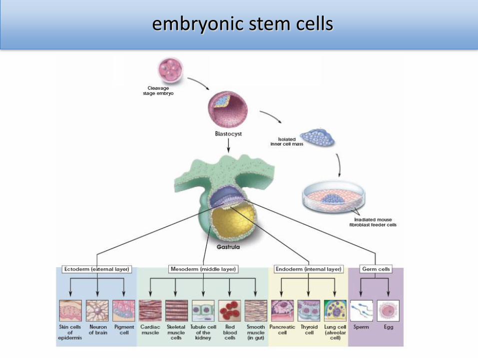
embryonic stem cells
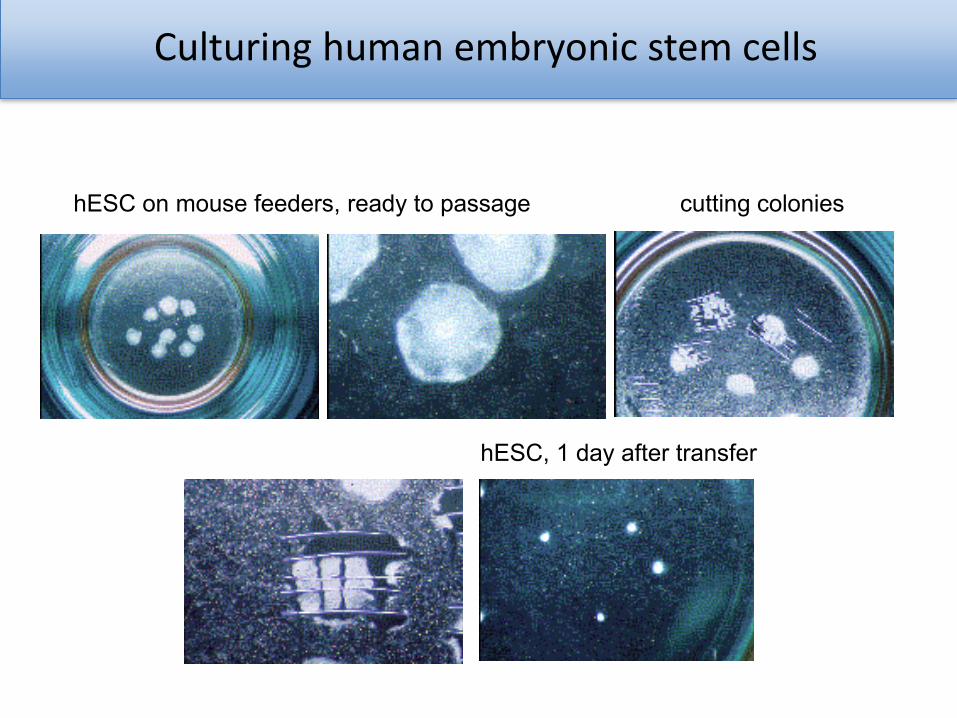
Culturing human embryonic stem cells
hESC on mouse feeders, ready to passage cutting colonies
hESC, 1 day after transfer
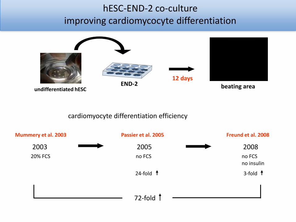
undifferentiated hESC beating areaEND-212 days
24-fold 3-fold
72-fold
Mummery et al. 2003 Passier et al. 2005 Freund et al. 2008
2003 2005 200820% FCS no FCSno FCS
cardiomyocyte differentiation efficiency
no insulin
hESC-END-2 co-cultureimproving cardiomycocyte differentiation

hESC differentiating to cardiomyocytes
hESCs
gastrulation
ectoderm
endoderm
mesoderm cardiac progenitors
CM
UH 1d 3d 6d 9d 12ddifferentiation
OCT-4TNCPLNNKX2-5BraT
SOX17
CL1A+B CL2A+B CL3 CL4A+B
NANOG
epiblast
ISL-1
MESP1
Beqqali et Stem Cells 2006
Differentiating hESC-CM follow “waves” of expression comparable to in vivo cardiac development!
hESC-END2 coculture
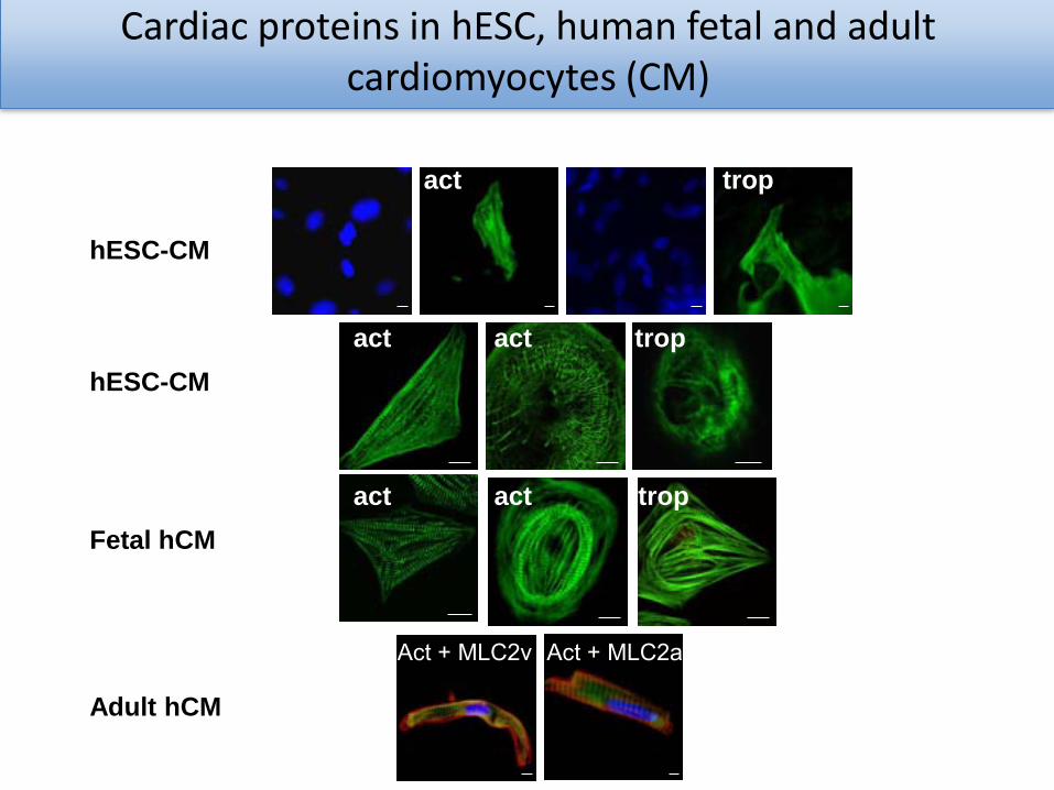
Cardiac proteins in hESC, human fetal and adult cardiomyocytes (CM)
hESC-CM
act trop
hESC-CM
actact trop
Fetal hCM
actact trop
Adult hCM
Act + MLC2v Act + MLC2a
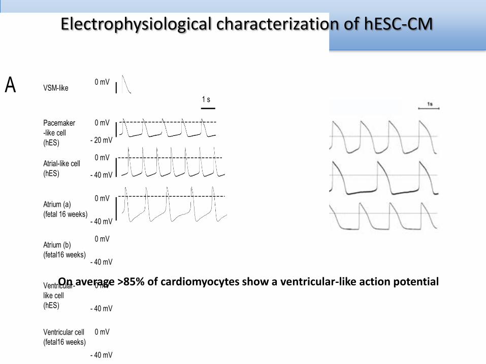
VSM-like(hES) 1 s
0 mV
- 30 mV
Ventricular cell(fetal16 weeks)
0 mV
- 40 mV
Ventricular-like cell(hES)
0 mV
- 40 mV
Atrial-like cell(hES)
0 mV
- 40 mV
Pacemaker-like cell(hES)
0 mV
- 20 mV
A
Atrium (a)(fetal 16 weeks)
0 mV
- 40 mV
Atrium (b)(fetal16 weeks)
0 mV
- 40 mV
Phen
yleph
rine 10
-4 M
Phen
yleph
rine 10
-4 M
rest
Carba
chol
10-4
M
Isopren
aline
10-6
M
be
ats
/min
0
80
40
120
rest
Carba
chol
10-4 M
Isopren
aline
10-6
M
hfetalhES
hES
Humanfetalatrium
Humanfetal ventricular
± 5 minutes5 M Verapamil
5 M Verapamil ± 5 minutes
0
-60
0
-60
5 M Verapamil ± 5 minutes
0
-60
5 seconds
B
C
On average >85% of cardiomyocytes show a ventricular-like action potential
Electrophysiological characterization of hESC-CM
Human pluripotent stem cell-derived cardiomyocytes
cell transplantation for cardiac repair?

Effect on cardiac function?
Model of acute myocardial infarctionMale SCID mice (n=13-15 per group)• MI (LAD ligation) +
– 1 million GFP-HES3 from beating areas END-2 co-culture (20% CM)
– 1 million non-CM differentiated from GFP-HES3
• MRI (9.4 T) after 2 days, 4 weeks, 12 weeks
4 Chamber view
Cardiac function improvement at 4 weeks not sustained!
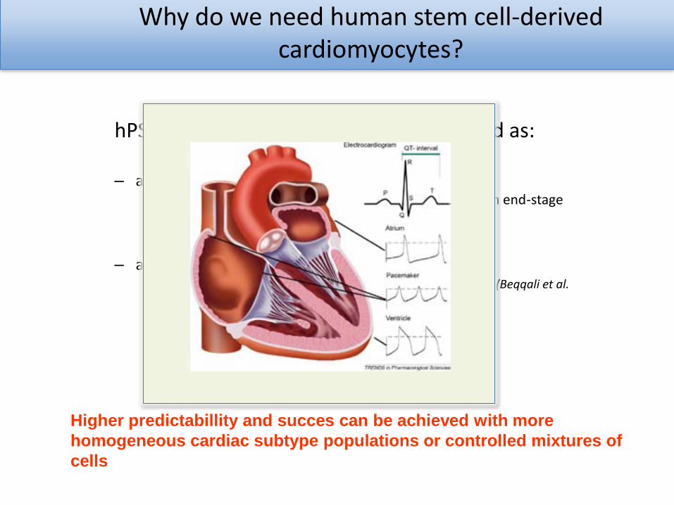
Why do we need human stem cell-derived cardiomyocytes?
hPSC-derived cardiomyocytes may be used as:
– a possible source for cell replacement therapy• As alternative for heart transplantation in patients with end-stage
heart failure (van Laake et al. Stem Cell Research 2007)
• Biological pacemaker
– a human in vitro model for:• cardiomyocyte differentiation or cardiac development (Beqqali et al.
Stem Cells 2006)
• drug toxicity (Braam et al. Stem Cell Research 2010)
• cardiac disease– target and drug discovery
Higher predictabillity and succes can be achieved with more
homogeneous cardiac subtype populations or controlled mixtures of
cells
Maandag, 7 maart 2011Afdelingsbezoek RvB 17
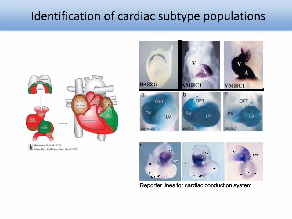
Identification of cardiac subtype populations
NKX2.5
Reporter lines for cardiac conduction system
Maandag, 7 maart 2011Afdelingsbezoek RvB 18
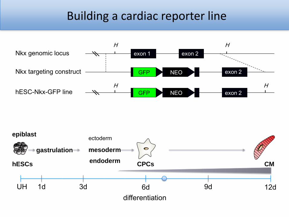
Building a cardiac reporter line
hESCs
gastrulation
ectoderm
endoderm
mesoderm
CPCs CM
UH 1d 3d 6d 9d 12ddifferentiation
epiblast
GFP NEO
GFP NEO
exon 1 exon 2
exon 2
exon 2
H H
H H
Nkx genomic locus
Nkx targeting construct
hESC-Nkx-GFP line
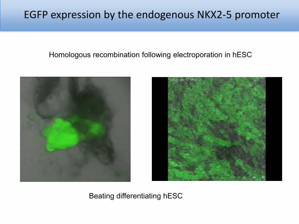
EGFP expression by the endogenous NKX2-5 promoter
Homologous recombination following electroporation in hESC
Beating differentiating hESC
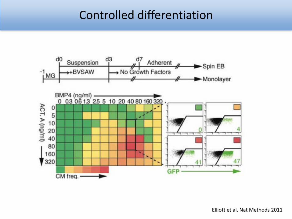
Controlled differentiation
Elliott et al. Nat Methods 2011
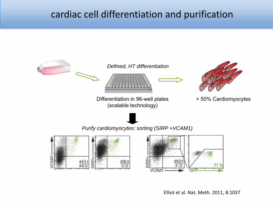
cardiac cell differentiation and purification
> 50% Cardiomyocytes
Defined, HT differentiation
Differentiation in 96-well plates (scalable technology)
Purify cardiomyocytes: sorting (SIRP +VCAM1)
Elliot et al. Nat. Meth. 2011, 8:1037
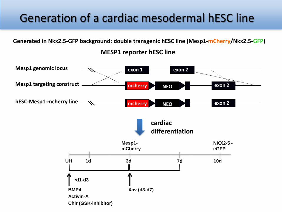
Generation of a cardiac mesodermal hESC line
Mesp1 genomic locus
Mesp1 targeting construct
hESC-Mesp1-mcherry line
mcherry NEO
NEO
exon 1 exon 2
exon 2
exon 2mcherry
MESP1 reporter hESC line
Generated in Nkx2.5-GFP background: double transgenic hESC line (Mesp1-mCherry/Nkx2.5-GFP)
Mesp1-
mCherry
UH 1d 3d 7d 10d
•d1-d3
BMP4
Activin-A
Chir (GSK-inhibitor)
Xav (d3-d7)
NKX2-5 -
eGFP
cardiac differentiation
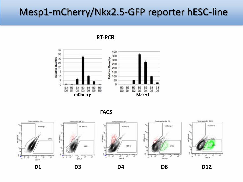
Mesp1-mCherry/Nkx2.5-GFP reporter hESC-line
mCherry Mesp1
RT-PCR
FACS
D1 D3 D4 D8 D12
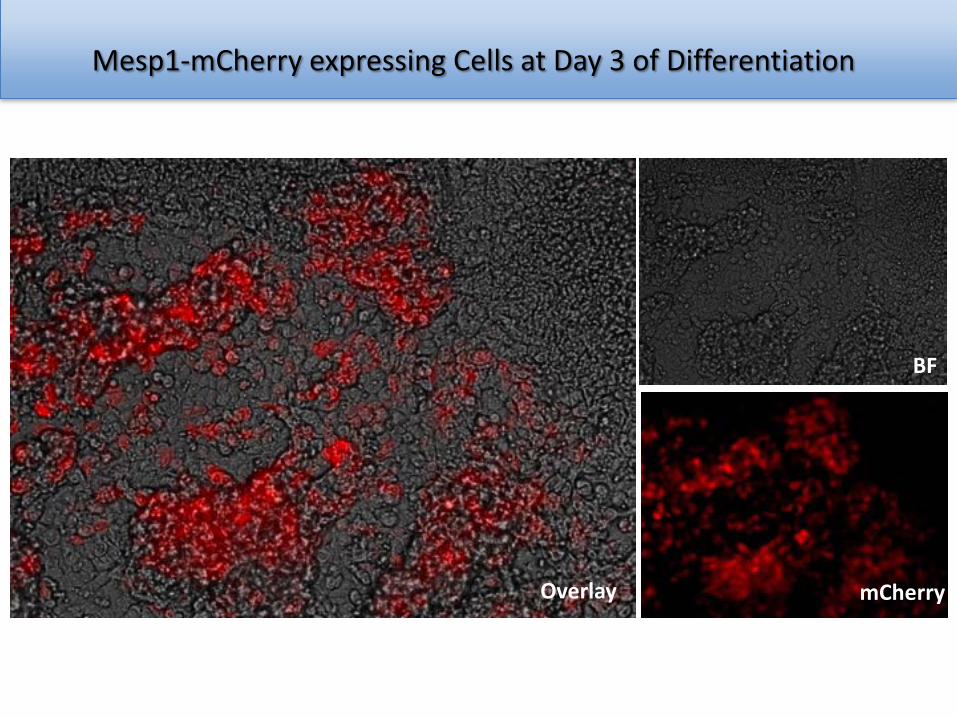
Mesp1-mCherry expressing Cells at Day 3 of Differentiation
Overlay
BF
mCherry
How do we get cardiac subtype populations?(atrial, ventricular, pacemaker cells)
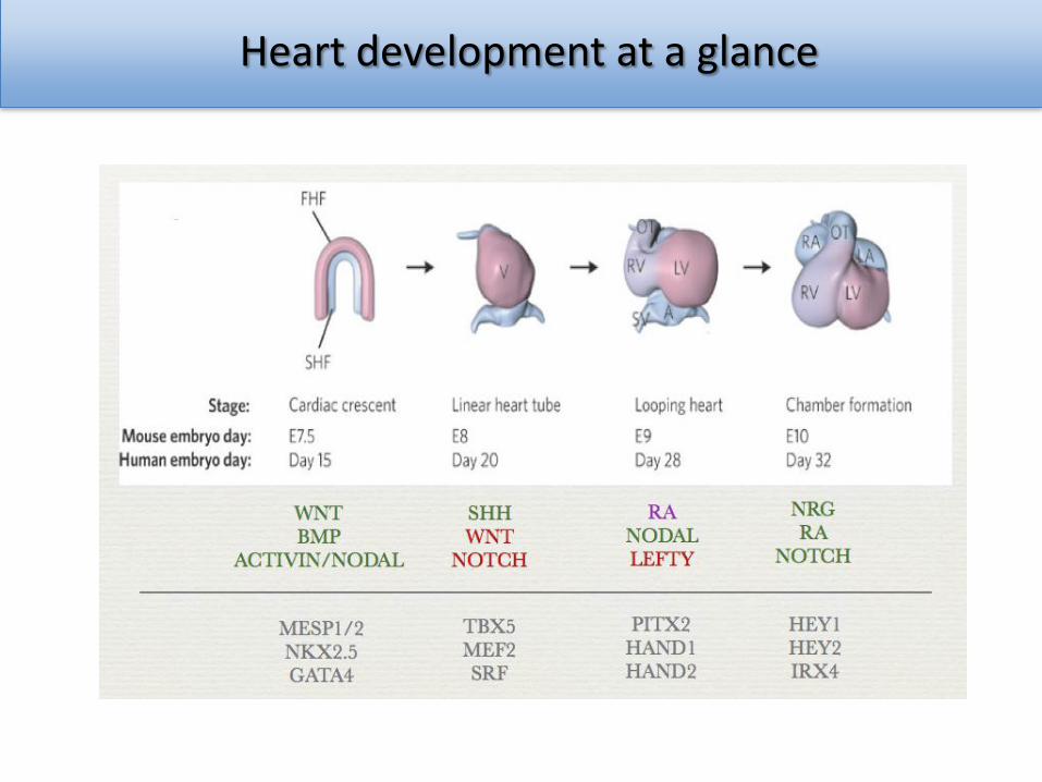
Heart development at a glance
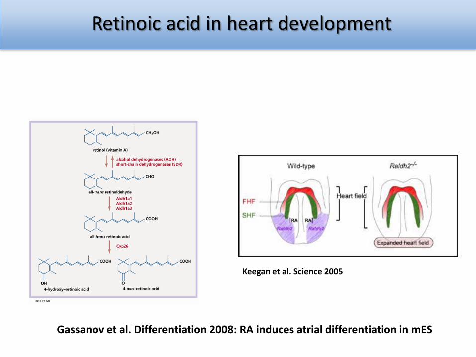
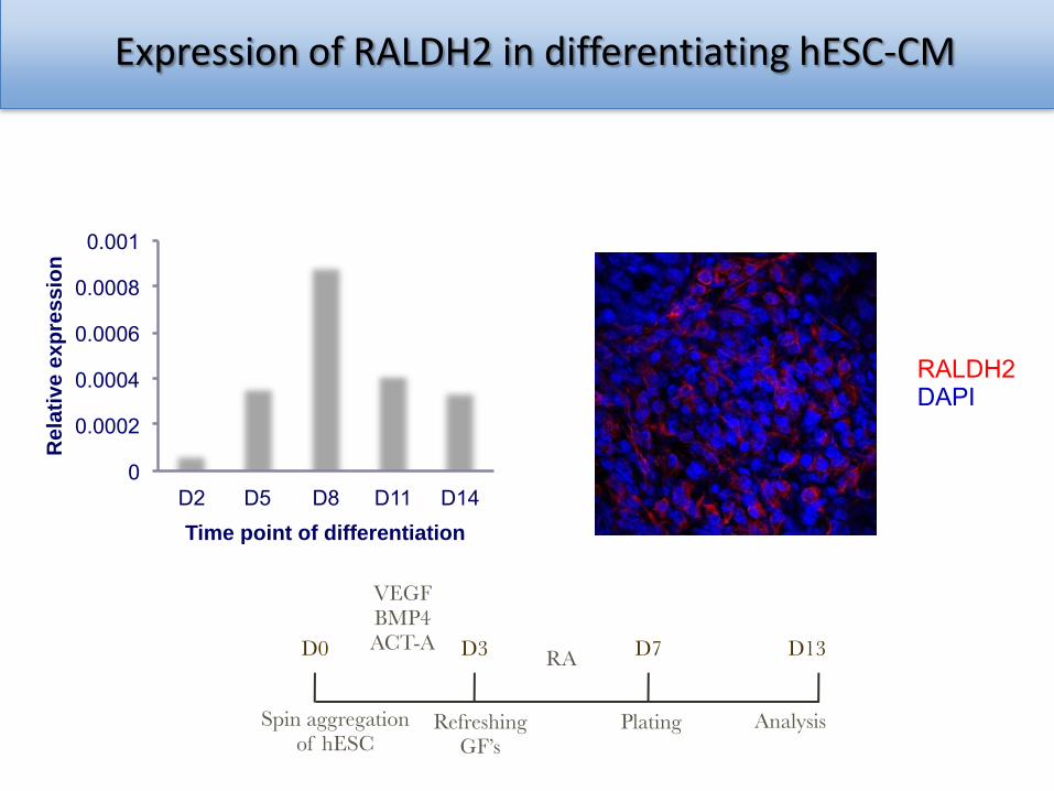
Retinoic acid in heart development
Gassanov et al. Differentiation 2008: RA induces atrial differentiation in mES
Keegan et al. Science 2005
0
0.0002
0.0004
0.0006
0.0008
0.001
D2 D5 D8 D11 D14
Re
lati
ve
ex
pre
ssio
n
Time point of differentiation
RALDH2 DAPI
Expression of RALDH2 in differentiating hESC-CM
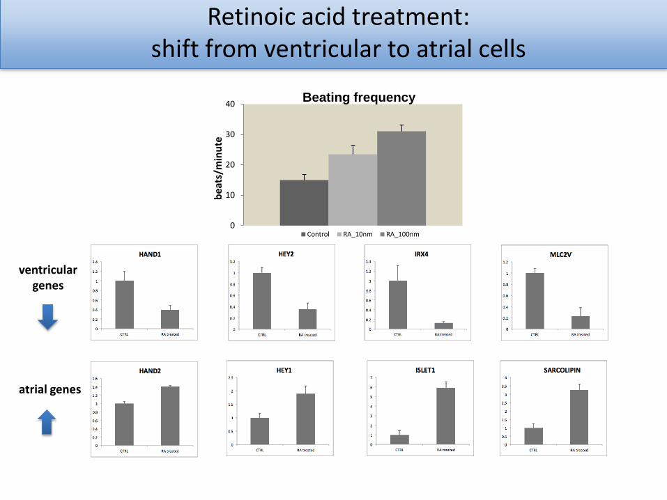
Retinoic acid treatment: shift from ventricular to atrial cells
0
10
20
30
40
bea
ts/m
inu
te
Beating frequency
Control RA_10nm RA_100nm
ventricular genes
atrial genes
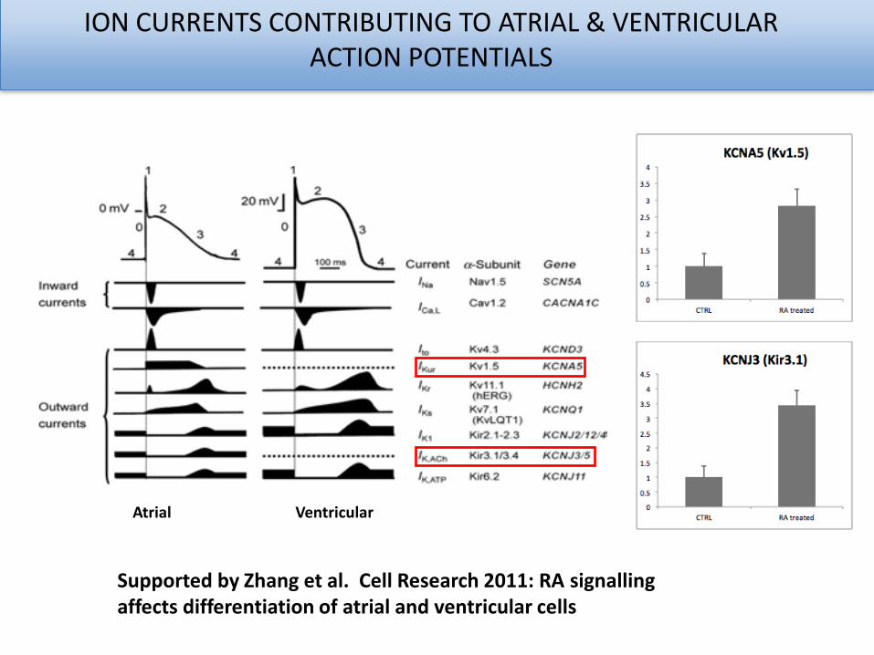
ION CURRENTS CONTRIBUTING TO ATRIAL & VENTRICULAR ACTION POTENTIALS
Atrial Ventricular
Supported by Zhang et al. Cell Research 2011: RA signallingaffects differentiation of atrial and ventricular cells
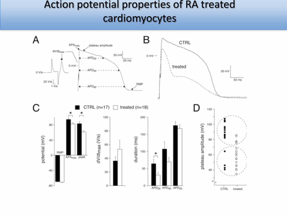
Action potential properties of RA treated cardiomyocytes
Conclusions
• Mesenchymal stem cells and cardiac stem cells are promising cell sources forthe treatment of cardiac disease.
– Transplantation, tissue engineering, endogenous activation
• Human pluripotent stem cells for transplantation: tissue engineering usingmixtures of cardiac cells from defined differentiation cultures will be the nextstep
• Genetic cardiac reporter lines faithfully recapitulate the “in vivo” lineage– Molecular mechanisms for expansion and differentiation can be studied
• Refined protocols enable cardiac subtype specification (retionic acid atrialCMs)
– Advantageous for tissue engineering, drug screening, disease modeling
Acknowledgements
Harsha Deepti DevallaMarcelo RibeiroSabine Den HartoghVerena RönzJantine MonshouwerChantal SchreursMarie-Christine WellerYann DeckerJuan Antonio Guadix
Christine MummeryRichard DavisDorien Ward
Arie VerkerkDavid ElliotAndrew ElefantyEd Stanley