development and validation of an indirect enzyme immunoassay for detection of ovine antibody to...
TRANSCRIPT
veterinary microbiology
Veterinary Microbiology 54 (1997) 357-368 ELSEVIER
Short communication
Development and validation of an indirect enzyme immunoassay for detection of ovine antibody to
Brucella ovis
Ana M. Vigliocco a3 * , Patricia S. Silva Paulo a, Joaquin Mestre a, Gabriel C. Briones a, Graciela Draghi b, Mirta Tossi a,
Klaus Nielsen ’
a National Commission of Atomic Energy Au. de1 Libertador 8250, 1429 Buenos Aires, Argentina
b E.E.A.-INTA, Mercedes, Province of Corrientes, Argentina
’ Immunology Section, ADRI. Box 11300, Station H. Nepean, Ont. K2H 8P9, Canada
Received 29 November 1995; accepted 20 September 1996
Abstract
An indirect enzyme-linked immunosorbent assay (ELISA) for the serodiagnosis of Brucella
ovis infection was developed. The assay uses a mouse monoclonal antibody to bovine IgG, horseradish peroxidase (HRPO) conjugate that cross-reacts with immunoglobulin from sheep and a purified antigen from Brucellu oris. The ELISA data were read and analyzed according to a targeting procedure. The ELISA results were compared with a cold complement fixation test (CFT). Sera from 675 rams from three uninfected flocks were used to determine the ELISA
cut-off value (O.D. 405 nm: 0.095) and the diagnostic specificity of the ELISA (100%) and the CFT (99.69% f 0.42). The ELISA cut-off value was corroborated by receiver operating character- istic (ROC) analysis. Six hundred and forty semen and serum samples from 419 rams from two naturally infected flocks were collected before and after mating-time during two consecutive years. All semen samples were cultured and Brucellu ouis was isolated from 28 samples. Sera from the 28 rams with positive semen were used to determine the diagnostic sensitivity of the ELISA (96.43% f 6.8) and of the CFT (including suspected positive samples with titers of 1:5; 88.89% k 11.85). Considering the CFT suspicious and the anti-complementary reactions as positive resulted in a diagnostic sensitivity value of 89.28% f 11.46. Six hundred and ten serum samples from the 640 sera were used to determine relative sensitivity (excluding sera with 1:5) at: ELISA/CFI 97.26% _t 3.74 and CFT/ELISA was 71.72% f 8.87. The percent agreement, beyond chance measured by the Kappa index was 79.7. Relative sensitivity ELISA/CFI
* Corresponding author. Fax: +54-l-3798540
0378.1135/97/$17.00 Copyright 0 1997 Elsevier Science B.V. All rights reserved PfI SO378-1 135(96)01285-O

358 A.M. Vigliocco et al./ Veterinary Microbiology 54 11997) 357-368
(including I:5 titers in the CFT as positive) was 94.9% + 4.83 and CFT/ELISA was 72.84% _t 8.59. The Kappa index was 79.4.
Ke_words: Brucella ouis; Sheep-bacteria; ELBA: Diagnosis-bacteria
1. Introduction
Brucella ovis is one of the most important causes of epididymitis in rams. The rams infected with Brucda ouis have reduced fertility and, in advanced cases, are infertile (McGowan and Schultz, 1956).
Eradication of this disease is dependant upon effective methods of diagnosis and
control. Clinical and bacteriological methods are not adequate for the detection of the disease
in large numbers of sheep, because both methods fail to detect all infected animals
(Jones et al., 1975; Worthington et al., 1985). Several serological methods have been used to detect antibodies to Brucella ouis
including agar gel diffusion, complement fixation test (CFT), agglutination, indirect hemagglutination, indirect immunofluorescence and enzyme linked immunosorbent as- say (ELISA). The CFT, used alone or together with genital palpation and the bacterio- logical examination of semen, has been reported to be a successful diagnostic test for
eradication of sheep brucellosis (Hughes and Claxton, 1968; Ris, 1974). However, the CFT applied to routine diagnostic work has found limited application because of
numerous disadvantages such as the occurrence of anti-complementary and hemolyzed sera, prozone phenomena and the need to use a highly labile reagent (complement). In addition, culture positive rams with negative or fluctuacting CFT titre are not uncommon and the prevalence of CFT negative infected rams is high among chronically infected
animals (Worthington et al., 1985). More recently, the use of the ELISA to improve serological diagnosis of Brucelh
ouis infection in rams has been investigated (Chin, 1983; Rahaley et al., 1983; Afzal et al., 1984; Ris et al., 1984; Spencer and Burgess, 1984; Worthington et al., 1984; Walker
et al., 1985; Cho and Niilo, 1987). This paper describes an indirect ELISA procedure developed and validated for the
serological diagnosis of Brucellu ouis infection in mature Corriedale rams reared under field conditions (Mercedes, Province of Corrientes, Argentina). The ELISA cut-off value was determined and the performance of the test in terms of its diagnostic specificity and sensitivity was determined. The ELISA sensitivity relative to the CFT was calculated
and the Kappa index was determined (Fleiss, 1981).
2. Materials and methods
2.1. Biological materials
2.1.1. Positive reference serum Two rams were experimentally infected with 1 ml of Brucella ouis (Strain 11) live
suspension (dose lo9 CFU ml- ‘) by intratesticular inoculation. Brucella ovis, strain 11

A.M. Vigliocco et al. / Veterinary Microbiology 54 f 19971357-368 359
(isolated by Centro Panamericano de Zoonosis, Argentina) was grown in Brucella agar
(Difco Laboratories, Detroit), plus 10% bovine serum in 10% CO, at 36.5”C. The strain was subsequently recovered from the semen of both rams. The rams were bled and the sera were used as positive controls in the ELISA and the CIT.
2.1.2. Animals Six hundred and seventy five mature rams from three uninfected flocks with no
clinical and epidemiological evidence of brucellosis were used to determine ELISA cut-off value and the diagnostic specificity of the ELISA and CFT.
Four hundred and nineteen rams from two naturally infected flocks (detected by gel
diffusion and clinical examination 5 years ago) were sampled before and after mating- time during two consecutive years. The 640 semen and serum samples obtained were used to calculate the ELISA sensitivity relative to the CFT and vice versa.
The sera from culture positive rams were used to calculate the diagnostic sensitivity of the ELISA and CFT.
2.1.3. Sample collection The blood sampling was carried out in field conditions. Blood was collected without
anticoagulant by jugular venopuncture. The blood was maintained at room temperature for 24 h followed by 1 h at 4°C. Sera were separated and stored at - 20°C until the ELISA or CFT for detection of Brucella ouis antibodies were carried out.
The semen samples were collected by electro-ejaculation (Bailey ejaculator No.
21150, Western Instrument Company, Denver). Each semen sample was taken in sterile absorbent cotton swab which was inserted and packed in semi-solid media (Transport
medium, Amies, with charcoal) (Difco Manual, 1984) and cooled immediately. The semen samples were maintained at 4°C and transported to the laboratory for processing within 36 h of collection. Samples were cultured on Thayer-Martin medium (Thayer and Martin, 1964) modified by Brown (Brown et al., 1971) increasing nistatina
concentration ten times (Marin, Clara Maria, personal communication 1994), incubated in 10% CO, at 365°C and examined daily for seven to ten days. The colonies suspected
of being Brucella were sub-cultured on Brucella serum medium under the same conditions.
Suspect strains were identified by macroscopical appearance, acriflavin testing, oxidase and urease reactions, biotyped using standard procedures (Alton et al., 1988) and phagetyped with the Brucella phage strains R/C as described by Corbel (Corbel and Thomas. 1985).
2.2. Methods
2.2.1. Complement fiation test A standard direct cold CFI (Laboratory Branch Task Force, 1965) (tube method 18 h
fixation at 4°C) was used for detection of antibody to Brucella ovis. Briefly, the test was based on addition of five 50% hemolytic units of guinea pig complement, followed after 18 h by addition of sensitized erythrocytes and visually read against a color standard. The antigen was obtained according to Myers (Myers et al., 1972) using Brucella ovis

360 A.M. Vigliocco et al. / Veterinary Microbiology 54 (1997) 357-368
REO 198. The sera were inactivated for 60 min at 60°C and tested at 15, 1:lO and 1:20 dilutions with serum controls at 15 and 1: 10 dilutions. Hemolysis of 50% or less in a given serum dilution was considered significant. For interpretation, a reaction at a 15 serum dilution was considered suspicious while reactions at 1: 10 or higher serum dilution were designated as positive.
Serum samples which gave unclear titers or showed prozone reaction were tested in
doubling dilutions of up to 1: 160.
2.3. ELISA test
2.3.1. Controls
Control sera consisted of a strong positive reactor (TG), a weak positive reactor (Ql),
a negative serum (42) and a buffer control without serum (43). The TG and Ql controls were obtained from two experimentally infected rams. The
sera were pooled and titrated using negative serum to make serial two-fold dilutions of the positive serum pool. Each dilution was tested in the ELISA protocol under the same conditions as a test sample. The TG control was chosen as that dilution which lies on the
linear portion of the titration curve at a point which is approximately 30% below the antibody saturation level (Fig. l).The Ql control was chosen as the dilution which
1.28
0.32
0 0.25 0.5 1 2 4 16 32 64 128 256 512 102420484100
l/DILUTION
Fig. 1. Titration curve of the positive serum reference standard. Reciprocal dilutions of the reference standard
are plotted against optical density at 405 nm (Y-axis). The reference standard dilution used for targeting (TG)
the enzyme immunoassay and that used for the weak positive control (Ql) as well as the cut-off optical density are indicated.

A.M. Vigliocco et al. / Veterinary Microbiology 54 (19971357-368 361
Table 1
Assay control. The control limits for four control sera and final development time of the reaction used in
ELBA
Control
a TG: mean+2SD
a Ql: mean+2SD
a Q2: mean k 3SD
a Q3: mean + 3SD
b TD: mean + 2SD
Optical density
1.037 * 0.094
0.234 k 0.094
0.018+0.020
0.0045+0.018
Minutes
9.292 + 5.02
TG: strong positive control; Ql: weak positive control; Q2: negative control; 43: buffer control; TD:
development time; SD: standard deviation.
a The data are expressed in OD values at 405 nm.
b The data are expressed in minutes.
produced an optical density (OD) approximately greater than the positive/negative
cut-off value (Fig. 1). The Q2 control serum was obtained from a healthy ram. The upper control limit for
this standard did not overlap the positive/negative cut-off (Table 1). All controls were tested in quadruplicate in each plate. The mean OD of the controls
and the time development (TD) of the reactions were calculated. The specifications
limits and the standard deviation (SD) were set for the mean obtained from 58 plates. If any of the results fell outside the determined limits, the assay was repeated. (Table 1).
2.3.2. Serum dilution Mean OD values of positive and negative control serum samples at various dilutions
were determined and a I:200 dilution gave the highest positive-to-negative ratio with the
lowest background reading. This dilution was used with all the test samples and controls
in the ELISA.
2.3.3. Antigen The antigen was obtained from Brucellu ovis (strain 11) according to the procedure
of Myers (Myers et al., 1972) and purified SRA-pp (Vigliocco et al., 1982; Suarez et al., 1988) was used. It was then freeze-dried and in this form was stable for at least 1 year. For the ELISA test, the antigen was reconstituted in double distilled water at 0.62 mg ml-’ protein (Lowry et al., 1951) and was stored at - 20°C. The reconstituted antigen solution retained its activity for at least two years.
Reconstituted antigen was titrated by comparing OD readings of various antigen dilutions using six replicates of TG, Ql and 42 control serum samples at a I:200 dilution. A 1:3000 dilution produced the highest specific reaction (TG) with the lowest
nonspecific reaction (42) and achieved an OD value of 1.000 in approximately 10 min with the TG control. This dilution was selected to be used in subsequent ELISAs.
The antigen showed no deterioration in prepared plates stored at - 20°C for at least
two months.
2.3.4. Conjugate A lyophilized monoclonal antibody to bovine IgG,-horseradish peroxidase (HRPO)
conjugate (Henning and Nielsen, 1987; Henning and Nielsen, 1992) was used in the

362 A.M. Vigliocco et al. / Veterinary Microbiology 54 (1997) 357-368
ELISA test. It was reconstituted with double distilled water and glycerol (1: 1) and stored
at 4°C. For titration, the conjugate was tested at several dilutions, using six replicates of TG,
Ql and 42 controls. The dilution which produced an OD value of approximately 1.000 at 10 min of reaction with TG control was chosen. The average dilution was 1:lOOO.
Antigen, conjugate and serum dilutions for titration were measured at 10 min using a raw-OD protocol (Nielsen et al., 1992).
2.3.5. Procedure The antigen, diluted in 0.06 M sodium carbonate buffer (pH 9.6) was passively
adsorbed to polystyrene plates (Nunc 2-69620, Denmark) at 50 pi/well. For the maximum sensitization, plates were incubated 18 h at 20°C and then washed five times
in 0.01 M phosphate buffered saline containing 0.05% tween 20, pH 7.2 (PBS/T). Control (in quadruplicate) and test (in duplicate) sera were applied at 1:200 in PBS/T, 50 pi/well, for 1 h at 20°C. After five washes in PBS/T, appropiately diluted
anti-immunoglobulin-enzyme conjugate was added, 50 pi/well, and incubated for 1 h at 20°C. After five washes in PBS/T, 100 ~1 of substrate/chromogen (4.0 mM hydrogen peroxide and 1 .O mM 2,2’-azino-bis(3-ethylbenz-thiazoline-6-sulfonic acid) diammonium salt in 0.05 M citrate buffer, pH 4.5) were added. The plate was shaken continuously on an orbital shaker and the OD was measured at 405 nm in a photometer (Titertek Multiskan) interfaced with a computer, by the targeting procedure (Wright et
160- 140 MEAN: 0,018 SD: 0,017
120
5 100
2
= s 80
E 60
40
20
0 ti.015 b.03. - c-i.046 ' b.06' . d.075 . b.06 . 6.108 . 6.1.
OPTICAL DENSITY
Fig. 2. Frequency distribution of indirect ELISA absorbance values obtained with 675 ram sera from three
uninfected flocks. The cut-off value (0.095) was selected to provide an assay specificity value of 100%

A.M. Vigliocco et al. / Veterinary Microbiology 54 (1997) 357-368 363
al., 1985), to minimize inter-plate variability. This is a protocol that uses ELISA
software to automate microtiter plate reading and data reduction for the Brucella ELISA (Nielsen et al., 1992). Briefly, the OD of the TG serum is measured after 4 min of substrate/chromogen interaction. The mean of the four pretarget values is then used to determine the development time (TD) required to produce a target value as near as possible to 1.0 OD units. At this time the plate is re-read.
2.3.6. Test performance The efficacy of each test was considered in terms of the sensitivity and specificity as
defined by Vecchio (Vecchio, 1966). The Kappa index was calculated according to the formula of Fleiss (Fleiss. 1981).
3. Results
3.1. Determination of the ELISA cut-off value and diagnostic specifici of ELISA and CFT
The 675 ram sera collected from three uninfected flocks gave in ELISA test a mean OD value of 0.018 and a SD of 0.017. Fig. 2 shows the frequency distribution of the
sera with a characteristic positive skew.
100
90
92
99.5 100 100.5 101
% specificity
Fig. 3. ROC analysis of % sensitivity (Y-axis) plotted against % specificity (X-axis) for various cut-off values
of indirect ELBA. From the graph, it is evident that a cut-off of 0.095 optical density units provide the
maximum sum of sensitivity and specificity (apex of the curve) and this value was selected for use. The
specificity values were calculated from the results in the 675 ram sera from three uninfected flocks. The
sensitivity values were calculated from the results in the 71 CFf positive ram sera from two infected flocks.

364 A.M. Vigliocco et al. / Veterinary Microbiology 54 (1997) 357-368
Two and sixteen serological false positives were evident when tbe ELISA cut-off value was calculated as the mean f 4 SD (0.086) or the mean f 3 SD (0.069), respectively. Therefore a cut-off value of 0.095 was selected to achieve a specificity
value with the negative sera of 100%. This cut-off was confirmed using both positive and negative serum samples by receiver operating characteristic (ROC) analysis (Metz, 1978) (Fig. 3).
In the CFT, performed on the same samples, 649 sera were negative, 24 anti-comple-
mentary and two were suspicious. The diagnostic specificity of the CFT was 99.69% &- 0.42 (if the anti-complementary sera were considered as negative).
Table 2
Characterization of non-smooth Bmcella isolated from semen and the serological response to ELBA and CFT
ST a Phage Growth On Dyes b URE 0x1 ELISAd CFT
thionin fuchsin safr ’ - -
a b c a b d
1 + + 2 + + 3 + f 4 + f 5 + + 6 + _ 7 + + 8 + - 9 + +
10 + + 11 + + 12 + + 13 + + 14 + + 15 + + 16 + + 17 + + 18 + + 19 + + 20 + _ 21 + + 22 + - 23 + + 24 + + 25 + + 26 + + 27 + + 28 + +
+ + + + + + + + + + + + + + + + + + + + + + + + + + + +
+ + + + + + + + -I-
+
f
+
+
+
+
+
+
+
+
+
-I-
i
+
+
+
+
f
+
+ + + - + + + + + _ + + + + + + + + + + + + + + + + + +
+ + + - + + - + + _ + + + + + + + + + + + + + + + + + +
_ -
f - - - _ - _ _ - - _ _ _ _ - - - - - - - - _ _ _ _ _ _ _ - - - _ _ _ _ - - _ - _ _ _ _ - - _ - - -
L?z - - -
_ 0.070 - 0.126 - 0.155 _ 0.188 - 0.193 - 0.299 _ 0.310 _ 0.358 - 0.441 - 0.456 - 0.496 _ 0.500 _ 0.517 _ 0.538 _ 0.568 _ 0.571 - 0.610 - 0.680 _ 0.679 - 0.734 - 0.866 _ 0.893 - 0.910 - 0.922 - 1.044 - 1.124 - 1.294 - 1.328
- + Se
+
+ -
+
+
+
+
+
+
+
+
AC
+ -
+
+
+
+
+
+
+
+
+
+
+
a Strains.
b Concentration of dyes: a = 10 pg/ml; b = 20 pg/ml; c = 40 pg/ml; d = 100 pg/ml. ’ Safranine.
d Optical density at 405 nm;
e Suspicious 1:5;
URE: urease activity; OXI: oxidase activity, AC: anti-complementary, CFTz complement fixation test.
A.M. Vigliocco et al./ Veterinary Microbiology 54 (1997) 357-368
3.2. Diagnostic sensitivity
365
The 640 semen samples from two infected flocks were cultured and the isolated strains characterized. Twenty eight samples were positive when cultured for Brucella ovis and their characteristics are summarized in Table 2. The ELISA test determined 27 sera as positive and one as negative for a diagnostic sensitivity 96.43% + 6.8.
The CFT was positive with 23 sera, one serum was suspicious (151, three were
negative and one was anti-complementary. The only negative serum in the ELISA was also negative in the CFT (Table 2, serum 1).
The diagnostic sensitivity of the CFT without the anti-complementary serum and
considering the suspicious reactions as positive was 88.89% f 11.85. The diagnostic sensitivity of the CFT considering the suspicious reactions and the anti-complementary serum as positive was 89.28% f. 11.46.
3.3. Relatiue sensitivity of ELJSA / CFT
The CFT performed on 640 sera revealed 22 anti-complementary reactions and 8 sera were hemolyzed. Therefore, 610 sera were used to determine relative sensitivity. The CFT gave 73 positive, 6 suspicious and 531 negative reactions.
140
OPTICAL DENSITY 5 0.8 1.1
23
18.4
4.6
3
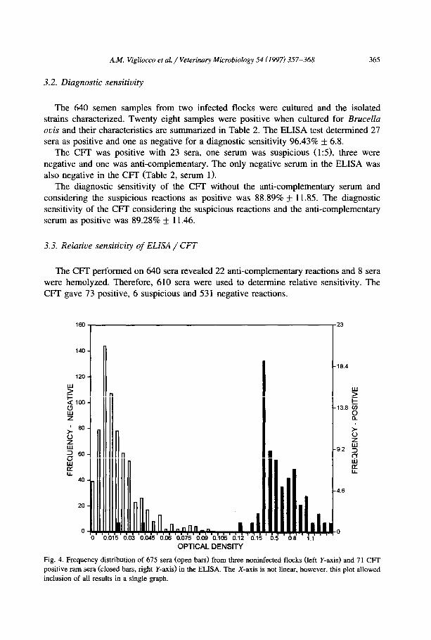
Fig. 4. Frequency distribution of 675 sera (open bars) from three noninfected flocks (left Y-axis) and 71 CFI
positive ram sera (closed bars, right Y-axis) in the ELISA. The X-axis is not linear, however, this plot allowed
inclusion of all results in a single graph.

366 A.M. Vigliocco et al. / Veterinary Microbiology 54 (1997) 357-368
The ELISA test gave 99 positive and 505 negative (without considering the 6 CFT suspicious sera). The ELISA sensitivity relative to the CFT was 97.26% + 3.74 and CFT/ELISA was 71.72% + 8.87 and the Kappa index was 79.7.
Fig. 4 depicts the ELISA frequency distribution of negative and positive sera in the CFT.
Among the 6 sera suspicious in the CFT, 4 were ELISA positive and 2 ELISA
negative. If we consider the suspicious sera as positive to CFT, ELISA test gave 103 positive
and 507 negative results. The CFT gave 79 positive and 531 negative results. The
ELISA relative sensitivity to the CFT was 94.94% f 4.83 and CFT/ELISA was 72.84% f 8.59. The Kappa index was 79.4.
4. Discussion
The ELISA procedure described in this study was a relatively simple test to perform
and may be applicable for widespread use. The application of the protocol from ELISA software program to automate microtiter
plate reading and data reduction for the Brucella ELISA used (Nielsen et al., 1992) permits extensive quality control of the assay.
The data presented in this study clearly indicate that the ELISA test was more
specific and more sensitive than cold CIT. Of the 675 ram serum samples from uninfected flocks ELISA detected no false
positives using a cut-off value of 0.095 while CFI gave 0.29% false suspicious (tine 1:5). Two false suspicious samples in the CFI were ELISA negative. The ELISA detected antibody in more sera from culture positive rams than did the WT. This may be in part due to the failure of the CFT to detect a number of rams shedding Brucella ovis in their semen, reducing the effectiveness of this test (Burgess and Norris, 1982). It
is recognized that the diagnostic sensitivity data of the present paper was calculated
from a small group of animals. Of 32 sera from culture positive rams (kindly supplied by Dr. Ignacio MORIYON,
Navarra, Spain, FICAPAL collection) in addition to our 28 sera, the ELISA gave a total of 57 positive and three negative reactions; the CFI gave 52 positive, 7 negative and one anticomplementary reaction. The three ELISA negative sera were also negative in
the CFI (data not shown). The ELISA sensitivity relative to the CFI was difficult to evaluate because it was not
possible to determine whether an animal was infected with the exception of sera from
the 28 sheep with semen positive cultures. The relatively high rati of sera that were ELISA positive compared to the CFI is not
due to ELISA false positive, reactions (ELISA specificity was 100%) but may be due to CFT false negative reactions: It has been established that the CFf, in early and late infections, may give false negative results, the latter perhaps due to an excess of IgG, isotype of antibody (Burgess et al., 1982).
The ELISA,procedure described follows the format of most indirect ELISAs and should therefore be adaptable to diagnostic use. This, combined with its demonstrated

A.M. Vigliocco et al. / Veterinary Microbiology 54 (1997) 357-368 367
specificity and sensitivity makes it a valuable tool for the eradication of Brucella ouis infection of sheep.
Acknowledgements
The authors wish to acknowledge financial support from the International Atomic Energy Agency (Project ARG/5/003). The authors also wish to acknowledge expert advice from Dr. J. Dargie, Dr. I. Moriyon, Dr. J. Blasco, Dr. D. Gall and Dr. G. Schurig. The R/C phage was kindly provided by Dr. J. Blasco. Serum and semen samples were obtained with assistance from INTA, Mercedes, Corrientes. The field work of J. Benitez is gratefully acknowledged as is the Pilaga Company for making the rams available for testing. The support of and discussions with Dr. H. Figueiras are gratefully acknowl-
edged.
References
Afzal, M., Tengerdy, R.P., Squire, P.G. and Ellis, R.P., 1984. Characterization of Brucella ouis lipopoly-
saccharide and its use for diagnosis of ram epididymitis by enzyme-linked immunosorbent assay. J. Clin.
Microbial., 20: 1159-l 164.
Alton, G.G., Jones, L.M., Angus, R.D. and Verger, J.M., 1988. Techniques for the Brucellosis Laboratory.
INRA, Paris.
Brown, G.M., Ranger, C.R. and Kelley, D.J., 1971. Selective medium for the isolation of Brucella oois. Cornell Vet., 61: 26.5-280.
Burgess, G.W. and Norris, M.J., 1982. Evaluation of the cold complement fixation test for diagnosis of ovine
brucellosis. Aust. Vet. J., 59: 23-25.
Burgess, G.W., MC Donald, J.W. and Norris, M.J., 1982. Epidemiological studies on ovine brncellosis in
select ram flocks. Aust. Vet. J., 59: 45-47.
Corbel, M.J. and Thomas, E.L., 1985. Use of phage for the identification of Brucella canis and Brucella oc:is cultures. Res. Vet. Sci., 38: 3.5-40.
Chin, J.C., 1983. Comparison of different antigenic preparations for the detection of ovine serum antibodies
against Brucella ouis by ELISA. Aust. Vet. J., 60: 261-264.
Cho, H.J. and Niilo, L., 1987. Diagnostic sensitivity and specificity of an enzyme-linked immunosorbent assay
for the diagnosis of Brucella ouis infection in rams. Can. J. Vet. Res. 51: 99-103.
Difco Manual, 1984. Difco Laboratories, Detroit, MI 48232. 10th Ed. (Grlfica Letra S.A., Madrid) pp.
1008-1009.
Fleiss. J.L., 1981. Statistical Methods for Rates and Proportions, 2nd Ed. (J. Wiley, New York) ch. 13.
Henning. D.M. and Nielsen, K.H., 1987. Peroxidase labelled monoclonal antibodies for use in enzyme
immunoassay. J. Immunoassay, 8: 297-307.
Henning, D.M. and Nielsen, K.H., 1992. Cross-reactivity of monoclonal antibodies to bovine immuno-
globulins with immunoglobuIins of other species. Vet. Immunol. Immunopathol. 34: 235-243.
Hughes, K.L. and Claxton, P.D., 1968. Brucella oois infection. An evaluation of microbiological, serological
and clinical methods of diagnosis in the ram. Aust. Vet. J. 44: 41-47.
Jones, L.M., Dubray, Y.G. and Marly, J., 1975. Comparison of methods of diagnosis of Brucdlo o~‘is infection of rams. Ann. Rech. V&t., 6: 1 l-22.
Laboratory Branch Task Force, 1965. Standard diagnosis complement fixation. Public Health Monograph (US)
No. 14.
Lowry, O.H., Rosebrough, N.J., Farr, L. and Rendall, R.J. 1951. Protein measurement with the folin phenol
reagent. J. Biol. Chem.. 193: 265-275.

368 A.M. Vigliocco et al./ Veterinary Microbiology 54 (1997) 357-368
McGowan, B. and Schultz, G., 1956. Epididymitis of rams: Clinical description and field aspects. Cornell Vet.,
46: 277-281.
Metz, C.E. 1978. Basic Principles of ROC Analysis. Seminars in Nuclear Medicine. Vol VIII, No. 4 , October,
pp. 283-298.
Myers, D.M., Jones, L.M. and Varela Diaz, V.M., 1972. Studies of antigen for complement fixation and gel
diffusion test in the diagnosis of infections caused by Brucella ouis and other Brucelh Appl. Microbial.,
23: 894-902.
Nielsen, K.H., Gall, D.E., Henning, D. and Garcia, M., 1992. Enzyme Immunoassay: Application to Diagnosis
of Bovine Brucellosis. Monograph, ADRI, pp. 199-225.
Rahaley, R.S., Dennis, M. and Smeltzer, MS., 1983. Comparison of the enzyme linked immunoasorbent assay
and complement fixation test for detecting Brucella ovis antibodies in sheep. Vet. Rec., 112: 467-470.
Ris, D.R., 1974. The complement fixation test for the diagnosis of Brucda ouis infection in sheep. N. Z. Vet.
J., 22: 143-146.
Ris, R.D., Hamel, K.L. and Long, D.L., 1984. Comparison of an enzyme-linked immunospecitic assay
(ELISA) with the cold complement fixation test for the serodiagnosis of Brucella ouis infection. N. 2. Vet.
J., 32: 18-20.
Spencer, T.L. and Burgess, G.W., 1984. Enzyme linked immunosorbent assay for Brucella ouis specific
antibody in ram sera. Res. Vet. Sci., 36: 194-198.
Suarez, C.E., Pacheco, G. and Vigliocco, A.M., 1988. Characterization of Brucella ouis surface antigens. Vet.
Microbial., 18: 349-356.
Thayer J.D. and Martin J.E., 1964. A selective medium for the cultivating of gonorrhea and N. meningitidis.
Public Health Dept. USA, 79: 49.
Vecchio, T.J., 1966 Predictive value of a single diagnostic test in unselected populations. N. Engl. J. Med.
274: 1171-1173.
Vigliocco, A.M., Suarez, C.E. and Pacheco G., 1982. Aislamiento, purificacibn, caracterizacidn y posterior
marcaci6n con lz51 de las distintas fracciones inmunol6gicamente activas de1 antigen0 superficial de
Brucella oh. Rev. Mil. de Vet., 30: 87-99.
Walker, R.L., Lea Master, B.R., Stellflug, J. and Biberstein E.L., 1985. Use of enzyme-linked immunosorbent
assay for detection to Brucella ouis in sheep: Field trial. Am. J. Vet. Res. 46: 1642-1646.
Worthington, R.W., Weddell, W. and Penrose M.E., 1984. A comparison of three serological tests for the
diagnosis of B. ovis infection in rams. N. Z. Vet. J., 32: 58-60.
Worthington, R.W., Stevenson, B.J. and Lisle, G.W., 1985. Serology and semen culture for the diagnosis of
Brucella ouis infection in chronically infected rams. N. Z. Vet. J., 33: 84-86.
Wright, P.F., Kelly, W. and Gall, D.E., 1985. Application of a timing protocol to the reduction of inter-plate
variability in the indirect enzyme immunoassay for detection of anti-Brucella antibody. J. Immunoassay, 6:
189-199.