development and evaluation of mucoadhesive tablets of ... · cinnarizine into mucoadhesive drug...
TRANSCRIPT
https://biointerfaceresearch.com/ 6365
Article
Volume 10, Issue 5, 2020, 6365 - 6376
https://doi.org/10.33263/BRIAC105.63656376
Development and Evaluation of Mucoadhesive Tablets of
Cinnarizine Using Carboxymethylated Guar Gum by
Compression Coating Technique
Pankaj Giri 1 , Inderbir Singh 1,*
1 Chitkara College of Pharmacy, Chitkara University, Punjab, India
* Correspondence: [email protected] (I.S.);
Scopus Author ID 7401774846
Received: 15.04.2020; Revised: 7.05.2020; Accepted: 8.05.2020; Published: 13.05.2020
Abstract:The e study was designed to perform carboxymethylation of guar gum and to develop
mucoadhesive tablets of cinnarizine by compression coating technology. Carboxymethyl guar gum was
synthesized and characterized by FTIR, SEM, XRD and DSC techniques. Inner core tablets of
cinnarizine were compression coated with guar gum and carboxymethylated guar gum. The formulated
mucoadhesive tablets were evaluated for various tablet parametric tests viz. hardness, friability, content
uniformity, thickness. Ex vivo mucoadhesion strength and in vitro drug release studies were also
conducted. Appearance of new FTIR peaks, surface morphology analysis by SEM, reduction in
crystallinity by XRD and appearance of the endothermic peak in DSC thermogram point out towards
successful carboxymethylation of guar gum. Mucoadhesive strength of F1CGG to F4CGG batches of
mucoadhesive tablets prepared with carboxymethyl guar gum found higher than F1GG to F4GG batches
of tablets prepared with guar gum. Carboxymethyl guar gum exhibited sustained release effect on in
vitro release cinnarizine from compression coated mucoadhesive tablets.
The study confirms that carboxymethylation of guar gum improves the mucoadhesive properties of pure
guar gum. Also, the compression coating of carboxymethylated guar gum enhances the gastric retention
time and sustained release of cinnarizine. Carboxymethyl guar gum can be used as mucoadhesive
polymer for developing different mucoadhesive drug delivery systems.
Keywords: Carboxymethylation; Guar gum; Compression coating; Mucoadhesive tablets;
Cinnarizine.
© 2020 by the authors. This article is an open access article distributed under the terms and conditions of the Creative
Commons Attribution (CC BY) license (https://creativecommons.org/licenses/by/4.0/).
1. Introduction
Mucoadhesive drug delivery systems offer several advantages including increased
residence time of drug at the site of application, increased permeability of drug into the
systemic circulation and enhanced bioavailability of the drug. Mucoadhesion phenomenon
could be illustrated in three steps: first step involves wetting and swelling of the polymer;
second, interpenetration and/or entanglement of the polymer and the mucin chains; and finally,
the formation of bond leading to adhesion of the polymer to the mucosal surface. Mucoadhesive
drug delivery systems could be designed for potentially targeting ocular, oral, buccal, nasal,
vaginal and rectal routes for administration of drugs [1,2].
Cinnarizine is an antihistaminic drug used for the control of nausea and vomiting due
to motion sickness. It exhibits pH-dependent dissolution behavior and dissolves readily at pH
1 (1.5 mg/ml), and has a very low solubility at pH values greater than 4. Cinnarizine, being a

https://doi.org/10.33263/BRIAC105.63656376
https://biointerfaceresearch.com/ 6366
weakly basic drug, precipitates in the basic pH of the small intestine. Incorporation of
cinnarizine into mucoadhesive drug delivery system retains the drug in the stomach
environment for a longer duration. This could enhance the bioavailability of cinnarizine [3].
Singh and Rana developed mucoadhesive tablets of cinnarizine using Eudragit RLPO and iron
oxide [4].
Guar gum is a biopolymer obtained from the seeds of Cyamopsis tetragonolobus of
family Leguminosae. It is mainly composed of high molecular weight polysaccharides in which
(1→4)β-D mannopyranosyl units are linked with (1→6)α-D galactopyranosyl residues. It has
a strong hydrogen bond forming capability in water which makes it a useful stabilizer and
thickner. Due to its thickening, gelling, emulsifying, pH stability, binding and film forming
properties, it is widely used in pharmaceutical, food, textile, paper, oil, cosmetic and paint
industries [5,6]. Carboxymethylation of guar gum was successfully performed and was used
for developing matrix tablets of metronidazole [7,8].
Compression coating is a solvent free coating technique in which the inner core is
coated with the outer layer by compression. Materials used in the outer layer could be employed
for modifying mechanical strength, altering drug release pattern and enhancing drug stability.
Mucoadhesive and gastro-floating tablets have already been developed by compression coating
employing conventional polymeric materials [9, 10].
In the present research, carboxymethylation of guar gum was performed and FTIR,
SEM, XRD, DSC techniques were employed for the characterization of the modified gum.
Core tablets of cinnarizine were prepared which were compression coated with pure guar gum
and carboxymethylated guar gum. The formulated compression coated tablets were evaluated
for various tablet parametric tests. Ex vivo mucoadhesion and in vitro drug release studies were
also performed.
2. Materials and Methods
2.1. Materials.
Cinnarizine was received as a gift from Ind-Swift Pharmaceutical, India. Guar gum was
kindly supplied as a gift sample by Hydrocolloid Plantations, New Delhi, India. MCC (Avicel-
112), sodium hydroxide, monochloro-acetic acid, methanol, glacial acetic acid, PVP K30, talc,
magnesium stearate were procured from Loba Chemie, Mumbai, India. All chemicals and
reagents employed in the study were of analytical grade.
2.2. Carboxymethylation of guar gum.
Carboxymethylation reaction of guar gum was performed by modifying the method
described by Gong et al. [11]. Purified gum (10 g) was dissolved in 500 ml of distilled water
followed by dropwise addition of sodium hydroxide over s period of 30 minutes with
continuous stirring. Monochloro acetic acid was added slowly to the above mixture. The
temperature of the reaction mixture was raised to 50 ºC and stirred for 4 hrs. Methanol (85 %)
was added to the mixture for precipitating the carboxymethylated gum. The modified gum was
washed, dried and stored in desiccator. Reaction for the synthesis of carboxymethyl guar gum
is depicted in Figure 1.

https://doi.org/10.33263/BRIAC105.63656376
https://biointerfaceresearch.com/ 6367
Figure 1. Synthesis reaction for the modification of guar gum into carboxymethylated guar gum.
2.3. Characterization of carboxymethyl guar gum
The carboxymethyl guar gum was characterized by FTIR spectroscopy, SEM, XRD,
and DSC techniques.
2.3.1. Fourier transform infrared spectroscopy.
The Fourier transform infrared (FTIR) spectra of sampleswere obtained using
attenuated total reflectance (ATR)-FTIR spectrophotometer (Alpha, Bruker, Japan). The
samples of pure gum and carboxymethylated gum werescanned in the spectral region of 4000
cm-1 to 400 cm-1.
2.3.2. Scanning electron microscopy.
Scanning electron microscopy (SEM) photomicrographs weretaken for studying
surface morphology of pure gum and carboxymethylated gum by scanning electron microscope
(Hitachi S 4300 SE/N) equipped with a secondary electron at an accelerating voltage of 10 kV.
Samples were mounted directly ontothe SEM sample stub using double-sided sticking carbon
tape under reduced pressure (0.001 mm Hg).
2.3.3. X-ray diffraction analysis.
Samples of pure gum and carboxymethylated gum were mounted in the sample cell and
scanned between 2 θ of 0-60º with a counting time of 0.1 seconds step size. X-ray patterns of
the films were obtained with XPERT-PRO equipment (PANalytical, Netherland).
2.3.4. Differential scanning calorimetry.
Samples of pure gum and carboxymethylated gum were sealed hermetically in flat bottom
aluminum cells and were subjected todifferential scanning calorimetry (DSC) analysis
(MettlerToledo Star System, 305, Switzerland) at a heating rate of10 ºC/min under nitrogen
atmosphere.
2.4. Preparation of core tablets of cinnarizine.
Core tablets were prepared as per the formula given inTable 1. All the ingredients were
passed through 60# sieve,followed by mixing for 15 minutes by tumbling. Tablets with
atheoretical weight of 80 mg were obtained using multipunchtableting machine (A K
Industries, Nakodar, India) fitted with6-mm concave round die-punch tooling [12].

https://doi.org/10.33263/BRIAC105.63656376
https://biointerfaceresearch.com/ 6368
Table 1. Composition of core tablet of cinnarizine.
Constituents Quantity (mg)
Cinnarizine 15
Avicel-112 58
PVP K-30 5
Talc 1
Magnesium Stearate 1
Total weight 80
2.5. Compression coating of core tablets.
The formulated core tablets were press coated with anappropriate blend of coating
polymer as shown in Table 2. Avicel-112 was added quantity sufficient for making the total
tablet weight equal to 600 mg. Half the quantityof the coating polymer was filled into the die
cavity (8.5 mmdiameter). The core tablet was placed in the centre of the diecavity, which was
then filled with the remainder of the coatingmaterial. Then, it was compressed around the core
tablets at an applied force of 5000 kg using 8.5 mm concave punchesfitted to multipunch
tableting machine [13].
Table 2. Compression coating composition for mucoadhesive tablet batches of pure guar gum and
carboxymethylated guar gum Batches Guar Gum
(mg)
Carboxymethylated Guar Gum
(mg)
PVP K30
(mg)
Talc
(mg)
F1GG 350 - 20 5
F2GG 400 - 20 5
F3GG 450 - 20 5
F4GG 500 - 20 5
F1CGG - 350 20 5
F2CGG - 400 20 5
F3CGG - 450 20 5
F4CGG - 500 20 5
2.6. Evaluation of core and compression coated tablets.
To ensure the uniformity and mechanical integrity of the prepared tablets, the following
parameters like weight variation, hardness, friability, and drug content were measured using
the parameters calculated as per standard parameters.
2.6.1. Hardness and friability.
Hardness and friability were determined using the validated Monsanto hardness tester
and the Roche friabilator (Campbell Electronics, Mumbai, India), respectively.
2.6.2. Thickness.
The thickness of the tablets was determined using Digital Vernier Caliper (Mitutoyo
Absolute Digimatic Caliper, Japan). Five tablets from each formulation were used and the
average values were calculated.
2.6.3. Drug content.
Five tablets were weighed individually and powdered. The powder equivalent to the
average weight of tablets was weighed and drug was extracted in 0.1N HCL, the drug content

https://doi.org/10.33263/BRIAC105.63656376
https://biointerfaceresearch.com/ 6369
was determined to measure the absorbance at 254 nm after suitable dilution using UV- Vis
double beam spectrophotometer (AU 2701, Systronics, Mumbai, India).
2.6.4. Ex vivo determination of mucoadhesive strength.
Mucoadhesion testing of the prepared compression coated tablets was performed using
texture analyzer (TA.XT plus, Stable MicroSystems, UK). Sample tablet was fixed to the
cylindrical probe with the help of double side adhesive tape. The pig stomach tissue (about
20x20 mm) was equilibrated for 15 min at 37.0 ± 0.5 ºC before placing onto the holder stage.
The probe with the sample tablet attached was immersed in the test medium for a specified
time prior to the test, the hydrated disc was then moved downward to contact with soaked tissue
at a specified force and maintained until a specified time. The probe was withdrawn at a
specified test speed and the maximum detachment force (Fmax) required to separate the
sample tablet fitted probe from the tissue could be obtained directly from Texture Exponent32
software. The preliminary settings of the instrument were: test speed 0.5 mm/s, contact force
1.0 N, contact time 60 s and return distance 15 mm. The probe without a sample tablet was also
tested to check the uniformity of the animal tissue [14].
2.6.5.Invitro drug release.
The in vitro dissolution study on compression coated mucoadhesive tablets of
cinnarizine was carried out in a paddle-type six station dissolution apparatus (DS 8000,
LabIndia, India) with a stirring speed of 50 rpm at 37 ± 0.5C using 900 ml of 0.1 N HCl (pH
1.2) as dissolution medium. At predetermined time intervals, 5 ml samples were withdrawn,
filtered through a 0.45 µm membrane filter, diluted and analyzed at 254 nm using a UV/VIS
double beam spectrophotometer (2202, Systronics, India).Cumulative percentage of drug
release was calculated using an equation obtained from the calibration curve. The dissolution
profile data of all formulated batches of tablets were fitted to various models such as zero-order
(cumulative % drug release vs time), first-order (log cumulative % drug remaining vs time),
Higuchi [15] (cumulative % drug release vs square root of time), Korsmeyer et al. [16] (log
cumulative % drug release vs log time) and Hixon and Crowell [17] (cube root of cumulative
% drug remaining vs time) models to ascertain the kinetic modeling of drug release.
3. Results and Discussion
3.1. Characterization techniques.
For characterizing the synthesized carboxymethylated guar gum, various techniques
were employed and are discussed here under.
3.1.1. FTIR
The FTIR spectra of native guar gum and carboxymethylated guar gum are shown in
Figure 2 and Figure 3. In the FTIR spectra of guar gum, broad band at 3444.89 cm-1 is attributed
to O−H stretching vibration. A symmetrical stretching vibration due to -CH2- group was
observed at 2925.24 cm-1. Band at 1639.18 cm-1 was assigned to O−H bonds of absorbed water
molecules and the bands between 800-1000 cm-1 were due to skeletal stretching vibrations of
guar gum.

https://doi.org/10.33263/BRIAC105.63656376
https://biointerfaceresearch.com/ 6370
Figure 2. FTIR spectra of pure guar gum.
The carboxymethylated guar gum shows broad band of around 3438.52 cm−1 is
attributed to O-H stretching vibration. The reduced intensity of absorption band at 3438.52
cm−1 indicates the carboxymethylation of O-H groups of guar gum. Increased intensity and
sharpness peaks at 1078.81 cm−1 (C−O symmetrical and asymmetrical vibrations) and 1374.30
cm−1 (COO− symmetric stretching vibrations) indicate that the hydroxyl groups of guar gum
molecules were carboxymethylated.
Figure 3. FTIR spectra of carboxymethylated guar gum
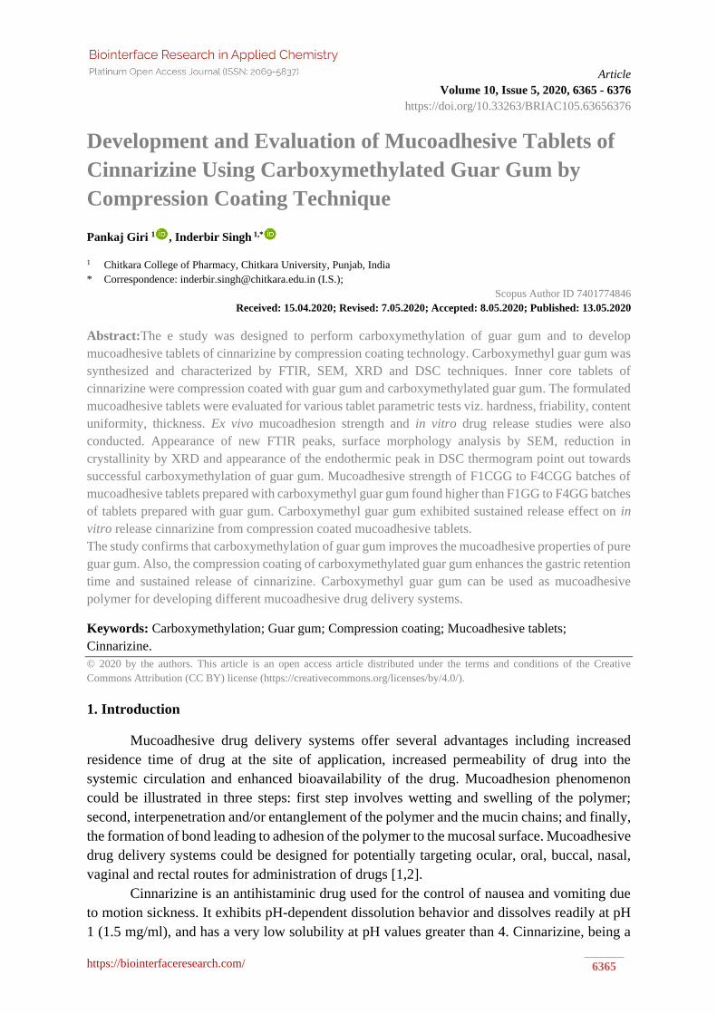
3.1.2. SEM
Scanning electron microscopy was performed for investigating the surface morphology
of the pure guar gum and carboxymethylated guar gum (Figure 4,5). Pure gum exhibited
irregular but smooth surface with round edges. However, carboxymethylated gum depicts
surface roughness with relatively irregular edges. The carboxymethylation process could be
accounted for these structural changes.
Figure 4. SEM micrographs of pure guar gum at different magnifications.
D:\IBS\New Folder\SAMPLE.1 Carboxymethylted Gum Ghati PELLET 08/02/2020
3438.5
2
2975.6
92927.7
0
2856.9
0
2123.0
9
1639.3
4
1559.7
2
1464.3
31417.3
51374.3
0
1299.5
5
1078.8
1
882.4
5
814.0
3774.3
9
647.6
6
500100015002000250030003500
Wavenumber cm-1
20
40
60
80
10
0
Tra
nsm
itta
nce
[%
]
Page 1 of 1
https://doi.org/10.33263/BRIAC105.63656376
https://biointerfaceresearch.com/ 6371
Figure 5. SEM micrographs of carboxymethylated guar gum at different magnifications.
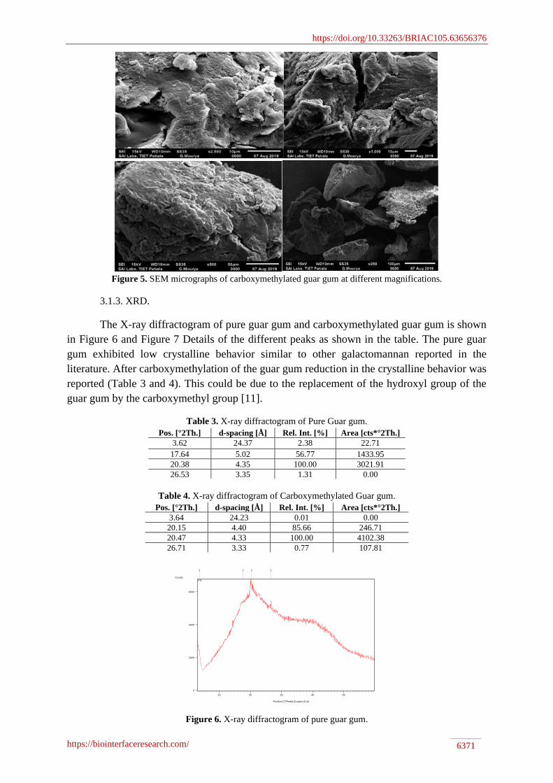
3.1.3. XRD.
The X-ray diffractogram of pure guar gum and carboxymethylated guar gum is shown
in Figure 6 and Figure 7 Details of the different peaks as shown in the table. The pure guar
gum exhibited low crystalline behavior similar to other galactomannan reported in the
literature. After carboxymethylation of the guar gum reduction in the crystalline behavior was
reported (Table 3 and 4). This could be due to the replacement of the hydroxyl group of the
guar gum by the carboxymethyl group [11].
Table 3. X-ray diffractogram of Pure Guar gum.
Pos. [°2Th.] d-spacing [Å] Rel. Int. [%] Area [cts*°2Th.]
3.62 24.37 2.38 22.71
17.64 5.02 56.77 1433.95
20.38 4.35 100.00 3021.91
26.53 3.35 1.31 0.00
Table 4. X-ray diffractogram of Carboxymethylated Guar gum.
Pos. [°2Th.] d-spacing [Å] Rel. Int. [%] Area [cts*°2Th.]
3.64 24.23 0.01 0.00
20.15 4.40 85.66 246.71
20.47 4.33 100.00 4102.38
26.71 3.33 0.77 107.81
Figure 6. X-ray diffractogram of pure guar gum.
Position [°2Theta] (Copper (Cu))
10 20 30 40 50
Counts
0
2000
4000
6000
PG

https://doi.org/10.33263/BRIAC105.63656376
https://biointerfaceresearch.com/ 6372
Figure 7. X-ray diffractogram of carboxymethylated guar gum.
3.1.4. DSC.
Differential scanning calorimetry (DSC) thermograms of pure guar gum and
carboxymethylated guar gum are shown in Figure 8 and 9 respectively. Pure guar gum shows
endothermic peak at 68.96 ºC with -29.76 mJ enthalpy. However, carboxymethylated guar gum
shows endothermic peaks at 62.96 ºC and 267.09 ºC with -12.25 mJ and 0.020 mJ enthalpies
respectively. The endothermic peak at 267.09 ºC in the modified gum could be attributed to the
thermal degradation of chemical compounds inserted on the gum structure after
carboxymethylation.
Figure 8. DSC thermogram of purified Guar gum.
Figure 9. DSC thermogram of carboxymethylated guar gum.
3.2. Evaluation of core tablets.
The results of various evaluation parameters for core tablets are depicted in Table 5.
The weight of the inner core tablets of cinnarizine was 80±5 mg. Hardness and friability of
Position [°2Theta] (Copper (Cu))
10 20 30 40 50
Counts
0
2000
4000
6000
8000
CG

https://doi.org/10.33263/BRIAC105.63656376
https://biointerfaceresearch.com/ 6373
tablets were3.0±0.50 kg/cm2 and 0.80±0.17 % respectively. The thickness of the core tablets
was 1.75 ±0.10 mm.
Table 5. Evaluation parameters of inner core tablets
S.No. Parameters Results
1 Weight Variation 80±5 mg
2 Hardness 3.0±0.50 kg/cm2
3 Friability 0.80±0.17 %
4 Thickness 1.75±0.10 mm
3.3. Evaluation of compression coated tablets.
The results of compression coated tablets prepared using pure gum and
carboxymethylated gum in different proportions for coating the previously prepared inner core
tablets of cinnarizine are depicted in Table 6. The mucoadhesive detachment force of F1GG to
F4GG was found to be ranging between 4.32±0.89 and 8.99±0.75 g. For F1CGG to F4CGG
the value of Fmax was found to be 10.86±1.23 and 18.53±2.08 g. A significant increase in
mucoadhesive property of carboxymethyl guar gum was noticed when compared with pure
guar gum. Moreover, mucoadhesive strength was found to increase with increasing in
concentration of carboxymethyl guar gum as a coating material in the compression coated
mucoadhesive tablets of cinnarizine.
Table 6. Evaluation Parameters of Pure Guar gum and carboxymethylated guar gum used compressed coated
tablet.
Batch No. Zero order First Order Higuchi Korsmeyer-Peppas Hixon-Crowell
r2 K0 r2 K1 r2 KH r2 n KKP r2 K HC
F1GG 0.6716 25.5517 0.9221 -0.8088 0.9738 9.197 0.9684 0.443 6.4395 0.8485 0.1742
F2GG 0.6700 18.5263 0.9572 -0.6396 0.9661 9.2616 0.9941 0.476 6.2728 0.8828 0.1331
F3GG 0.6679 12.4491 0.9682 -0.5173 0.9535 9.3337 0.9994 0.4438 6.0458 0.9138 0.0988
F4GG 0.7278 9.4375 0.9814 -0.3504 0.9315 9.4787 0.9922 0.493 5.6119 0.928 0.0711
F1CGG 0.5038 6.5153 0.8480 -0.3941 0.9552 9.8677 0.9300 0.588 6.267 0.7561 0.0644
F2CGG 0.6627 7.1735 0.9601 -0.3151 0.9356 9.7096 0.9855 0.460 5.7304 0.8967 0.0596
F3CGG 0.7316 7.2908 0.9730 -0.2776 0.9266 9.5731 0.9977 0.515 5.4627 0.9346 0.0557
F4CGG 0.8290 7.7194 0.9233 -0.2629 0.9113 9.6104 0.9926 0.534 4.9473 0.9469 0.0547
3.4. In vitro drug release.
In vitro drug release profiles of core tablet and different batches of compression coated
mucoadhesive tablets prepared with guar gum (F1GG to F4GG) and carboxymethyl guar gum
(F1CGG to F4CGG) are shown in Figure 10 and 11. Carboxymethylated guar gum depicts
sustained release behaviour in the compression coated tablets of cinnarizine.
Figure 10. In vitro drug release from core tablet and different batches of compression coated tablets of guar
gum.

https://doi.org/10.33263/BRIAC105.63656376
https://biointerfaceresearch.com/ 6374
Figure 11. In vitro drug release from core tablet and different batches of compression coated tablets of
carboxymethylated guar gum.
Table 7. In vitro drug release (kinetic modeling) data of the formulated batches.
Batch No. Zero order First Order Higuchi Korsmeyer-Peppas Hixon-Crowell
r2 K0 r2 K1 r2 KH r2 n KKP r2 K HC
F1GG 0.6716 25.5517 0.9221 -0.8088 0.9738 9.197 0.9684 0.443 6.4395 0.8485 0.1742
F2GG 0.6700 18.5263 0.9572 -0.6396 0.9661 9.2616 0.9941 0.476 6.2728 0.8828 0.1331
F3GG 0.6679 12.4491 0.9682 -0.5173 0.9535 9.3337 0.9994 0.4438 6.0458 0.9138 0.0988
F4GG 0.7278 9.4375 0.9814 -0.3504 0.9315 9.4787 0.9922 0.493 5.6119 0.928 0.0711
F1CGG 0.5038 6.5153 0.8480 -0.3941 0.9552 9.8677 0.9300 0.588 6.267 0.7561 0.0644
F2CGG 0.6627 7.1735 0.9601 -0.3151 0.9356 9.7096 0.9855 0.460 5.7304 0.8967 0.0596
F3CGG 0.7316 7.2908 0.9730 -0.2776 0.9266 9.5731 0.9977 0.515 5.4627 0.9346 0.0557
F4CGG 0.8290 7.7194 0.9233 -0.2629 0.9113 9.6104 0.9926 0.534 4.9473 0.9469 0.0547
k0: Zeroorder release rate constant, k1: Firstorder release rate constant, KH: Higuchi release rate constant, KKP:
Korsemeyer–Peppas release rate constant, KHC: Hixson–Crowell release rate constant, r2: Regression line value
The in vitro release data was fitted to different kinetic models such as zero-order, first-
order, Higuchi, Korsmeyer-Peppas and Hixon-Crowell as shown in Table 7. The value of n
(release exponent) 0.45< n < 0.89 indicates non-Fickian drug release transport. Drug release
from the biopolymer coated tablets follows diffusion and erosion of the polymer (anomalous
non-Fickian drug release behavior). When the formulation is in contact with the dissolution
media, the media penetrates the polymer matrix leading to disentanglement and subsequent
dissolution/erosion of polymer chains resulting in the release of the drug molecules from the
dosage form. According to another theory, the glass-rubbery transition of the polymer matrix
leads to an increase in the mobility of polymeric chains allowing the drug molecules to dissolve
and diffuse through the gel layer [18,19].
4. Conclusions
Carboxymethylation of guar gum was performed by simple and efficient chemical
method. FTIR, DSC, XRD and SEM techniques were employed for the characterization of the
modified gum. Compression coating technique was used for coating the core tablets of
cinnarizine for developing sustained release mucoadhesive tablets. Compression coating is an
effective solvent free technique for coating the core tablets. Carboxymethylation is a simple
and economical method for modifying biopolymers. Carboxymethylated biopolymers can be
used for developing different mucoadhesive drug delivery systems for targeting ocular,
pulmonary, buccal, rectal, vaginal and gastrointestinal regions. Regulatory and toxicological
issues of carboxymethylated biopolymer must be adequately handled before exploiting them
for commercial use.
Funding
This research received no external funding.

https://doi.org/10.33263/BRIAC105.63656376
https://biointerfaceresearch.com/ 6375
Acknowledgments
The authors gratefully acknowledge Chitkara College of Pharmacy, Chitkara University,
Punjab, India for support and institutional facilities.
Conflicts of Interest
The authors declare no conflict of interest.
References
1. Andrews, G.P.; Laverty, T.P.; Jones, D.S. Mucoadhesive polymeric platforms for controlled drug delivery.
European Journal of Pharmaceutics and Biopharmaceutics 2009, 71, 505–518,
https://doi.org/10.1016/j.ejpb.2008.09.028.
2. Singh, I.; Rana, V. Enhancement of mucoadhesive property of polymers for drug delivery applications: A
critical review. Reviews on Adhesion and Adhesives 2013, 1, 271–
290,https://doi.org/10.7569/RAA.2013.097307.
3. Sethi, S.; Mangla, B.; Kamboj, S.; Rana, V. A QbD approach for the fabrication of immediate and prolong
buoyant cinnarizine tablet using polyacrylamide-g-corn fibre gum. International Journal of Biological
Macromolecules 2018, 117, 350-361, https://doi.org/10.1016/j.ijbiomac.2018.05.178.
4. Singh, I.; Rana, V. Iron oxide induced enhancement of mucoadhesive potential of Eudragit RLPO:
formulation, evaluation and optimization of mucoadhesive drug delivery system. Expert Opinion on Drug
Delivery 2013, 10, 1179-1191, https://doi.org/10.1517/17425247.2013.790361. 5. George, A.; Shah, P.A.; Shrivastav, P.S. Guar gum: Versatile natural polymer for drug delivery applications.
European Polymer Journal 2019, 112, 722-735, https://doi.org/10.1016/j.eurpolymj.2018.10.042.
6. Sharma, G.; Sharma, S.; Kumar, A.; Al-Muhtaseb, A.H.; Naushad, m.; Ghfar, A.A.; Mola, G.T.; Stadler, F.J.
Guar gum and its composites as potential materials for diverse applications: A review. Carbohydrate
Polymers 2018, 199, 534-545, https://doi.org/10.1016/j.carbpol.2018.07.053.
7. Dodi, G.; Hritcu, D.; Popa, M.I. Carboxymethylation of guar gum: Synthesis and characterization. Cellulose
Chemistry and Technology 2011, 45, 171-176.
8. Singh, R.; Maity, S.; Sa, B. Effect of ionic crosslink on the release of metronidazole from partially
carboxymethylated guar gum tablet. Carbohydrate Polymers 2014, 106, 414-421,
https://doi.org/10.1016/j.carbpol.2014.01.033
9. Sa, Biswanath.; Mukherjee, S.; Roy, S.K. Effect of polymer concentration and solution pH on viscosity
affecting integrity of a polysaccharide coat of compression coated tablets. International Journal of
Biological Macromolecules 2019, 125, 922-930, https://doi.org/10.1016/j.ijbiomac.2018.12.101.
10. Gong, L.; Sun, Y.; Yu, M.; Gao, Y.; Zou, M.; Cheng, G. Development and evaluation of compression coating
gastro-floating tablet of alfuzosin hydrochloride for zero-order controlled release. AAPS PharmSciTech
2018, 19, 3277–3286 https://doi.org/10.1208/s12249-018-1168-z.
11. Gong, H.; Liu, M.; Chen, J.; Han, F.; Gao, C.; Zhang, B. Synthesis and characterization of carboxymethyl
guar gum and rheological properties of its solutions. Carbohydrate Polymers 2012, 88, 1015–1022,
https://doi.org/10.1016/j.carbpol.2012.01.057.
12. Raza, A.; Shen, N.; Li, J.; Chen, Y.; Wang, J.Y. Formulation of zein based compression coated floating
tablets for enhanced gastric retention and tunable drug release. European Journal of Pharmaceutical
Sciences 2019, 132, 163-173, https://doi.org/10.1016/j.ejps.2019.01.025.
13. Malik, D.; Singh, I. Formulation and evaluation of press coated tablets of esomeprazole for colonic delivery.
Asian Journal of Pharmaceutics 2012, 6, 252-258.
14. Koradia, H.; Chaudhari, K. Formulation of unidirectional buccal tablet of mirtazapine: An in vitro and ex
vivo evaluation. Journal of Drug Delivery Science and Technology 2018, 43, 233-242,
https://doi.org/10.1016/j.jddst.2017.10.012.
15. Higuchi, T. Mechanism of sustained-action medication. Theoretical analysis of rate of release of solid drugs
dispersed in solid matrices. Journal of Pharmaceutical Sciences 1963, 52, 1145-1149,
https://doi.org/10.1002/jps.2600521210.
16. Korsmeyer, R.W.; Gurny, R.; Doelker, E.; Buri, P.; Peppas, R.A. Mechanism of solute release from porous
hydrophilic polymers. International Journal of Pharmaceutics 1983, 15, 25-35,
https://doi.org/10.1016/0378-5173(83)90064-9.
17. Hixson, A.W.; Crowell, J.H. Dependence of reaction velocity upon surface and agitation (I) theoretical
consideration. Ind Eng Chem 1931, 23, 923-931, https://doi.org/10.1021/ie50260a018.

https://doi.org/10.33263/BRIAC105.63656376
https://biointerfaceresearch.com/ 6376
18. Barmpalexis, P.; Kachrimanis, K.; Malamataris. S. Statistical moments in modelling of swelling, erosion
and drug release of hydrophilic matrix-tablets. International Journal of Pharmaceutics 2018, 540, 1-10,
https://doi.org/10.1016/j.ijpharm.2018.01.052.
19. Hattori, Y.; Takaku, T.; Otsuka, M. Mechanochemical effect on swelling and drug release of natural polymer
matrix tablets by X-ray computed tomography. International Journal of Pharmaceutics 2018, 539, 31-38,
https://doi.org/10.1016/j.ijpharm.2018.01.020.