development and charecterization of soy lecithin and palm...
TRANSCRIPT

1
DEVELOPMENT AND CHARECTERIZATION OF SOY LECITHIN
AND PALM OIL BASED ORGANOGELS
A THESIS SUBMITTED IN PARTIAL FULFILLMENT
OF THE REQUIREMENTS FOR THE DEGREE OF
BACHELOR OF TECHNOLOGY
IN
BIO-MEDICAL ENGINEERING
SUBMITTED BY
NIROD BARAN
109BM0004
UNDER THE GUIDANCE OF
Dr. KUNAL PAL
DEPARTMENT OF BIOTECHNOLOGY AND MEDICAL ENGINEEIRNG
NATIONAL INSTITUTE OF TECHNOLOGY
ROURKELA-769008

2
DEPARTMENT OF BIOTECHNOLOGY & MEDICAL ENGINEERING,
NATIONAL INSTITUTE OF TECHNOLOGY-ROURKELA
Dated: 7th
May, 2013
CERTIFICATE
This is to certify that the thesis entitled “DEVELOPMENT AND CHARACTERIZATION
OF SOY LECITHIN AND PALM OIL BASED ORGANOGELS” submitted by Mr. Nirod
Baran in partial fulfillment for the requirements for the award of Bachelor of Technology
Degree in Biotechnology at National Institute of Technology, Rourkela is an authentic work
carried out by him under the supervision of the undersigned.
To the best of my knowledge, the matter embodied in the thesis has not been submitted to any
other University / Institute for the award of any Degree or Diploma.
(Dr. KUNAL PAL)
Assistant Professor

3
ACKNOWLEDGEMENTS
I would like to express my deep sense of gratitude and respect to our supervisor, Dr. Kunal Pal,
Assistant Professor, Department of Biotechnology and medical Engineering, National Institute of
Technology Rourkela for his excellent guidance, suggestions and constructive criticism. He has
been very kind, supportive and patient to me while suggesting the outlines of the project and has
also been very helpful in the successful completion of the same. I thank him for his overall
support.
Last but not the least; I would like to extend my heartfelt gratitude to the Ph.D scholar, Mr.
Vinay Kumar Singh, Mr. Sai Sateesh Sagiri and Ms. Beauty Behera, Department of
Biotechnology and Medical Engineering, National Institute of Technology, Rourkela for their
support and guidance. His helping nature and suggestion has helped me to complete this present
work.
Date: Nirod Baran
4
Abbreviations
SL- Soy Lecithin
PO- Palm Oil
DW- Distilled Water
MZ- Metronidazole
CPDR- Cumulative Percent Drug Release
NCIM- National Collection of Industrial Microorganisms
RH- Relative Humidity
BFM- Bright Field Microscopy
RT- Room Temperature
MW- Molecular Weight

5
TABLE OF CONTENTS
Page No.
Abstract 10
Chapter 1 Introduction 11-12
Chapter 2 Literature Review 13-15
Chapter 3 Materials and Methods 16-21
3.1. Materials
3.2. Methods
3.2.1. Preparation of Organogels
3.2.2. Organoleptic Evaluation
3.2.3. Stability Studies
3.2.3.1. Accelerated Stability Test
3.2.3.2. Long Term Stability Test
3.2.4. Microscopic Studies
3.2.5. pH Measurements
3.2.6. FTIR Microscopy
3.2.7. Disintegration Studies
3.2.8. Gel-Sol Transition Test
3.2.9. Heamocomatibilty Studies
3.2.10. In-vitro Drug release
3.2.11. Anti Microbial Assay
Chapter 4 Results and Discussion 22- 35
4.1. Preparation of Organogels
4.2. Organoleptic Evaluation

6
4.3. Stability Studies
4.3.1. Accelerated Stability Test
4.3.2. Long Term Stability Test
4.4. Microscopic Studies
4.5. pH Measurements
4.6. FTIR Microscopy
4.7. Disintegration Studies
4.8. Gel-Sol Transition Test
4.9. Heamocomatibilty Studies
4.10. In-vitro Drug release
4.11. Anti Microbial Assay
Chapter 5 Conclusions 37
Chapter 6 Bibliography 38-42

7
LIST OF TABLES
1. Composition of Organogels used for further Analysis
2. pH value of the selected organogels
3. Disintegration test of the selected gels
4. Gel-Sol Transition of selected organogel
5. Hemocompatibility Test

8
LIST OF FIGURES
1. Mechanism of organogels and emulsion formation
2. Mapping of the compositions forming organogels and emulsions
3. Mapping of the compositions of the formulations after accelerated stability test
4. Representative organogel formulations after the accelerated stability test
5. Bright field micrographs of the organogels showing the dispersed phase structures
6. FTIR Spectroscopy of the OG, MZ loaded and the raw materials
7. CPDR of the developed gels
8. Antimicrobial efficacy of the organogels against E. coli
9. Antimicrobial efficacy of the organogels against B. subtilis

9
ABSTRACT
Preparation and characterization of soy lecithin (SL) and palm oil (PO) based organogels have
been reported in this study. The optimization of the composition of the organogels was carried
out by varying the proportions of SL, PO and water. Microscopic studies suggested the presence
of aqueous phase either as fluid filled fibers or spherical droplets or both, depending on the
composition of the organogels. FTIR study indicated strong intermolecular hydrogen bonding
amongst the organogel components. The pHs of the organogels was found to be ~ 5.75 and were
hemocompatible. The release of metronidazole (MZ; model drug) suggested diffusion mediated
drug release. MZ loaded organogels have shown good antimicrobial property against B. subtilis
and E. coli.

10
CHAPTER 1
INTRODUCTION
11
1. INTRODUCTION
Organogels are semisolid formulations which consist of apolar solvent as the liquid phase. They
are often reported to be thermo-reversible and may be attributed to the physical interactions
amongst the gel components [1-3]. Permanent organogels have also been reported [2].
Organogels have been found to be inherently thermodynamically stable below their gellation
temperature. They possesses viscoelastic properties which is suitable for the development of
topical/ transdermal formulations [1]. The properties of organogels may be tailored by
incorporating water within its structure [4]. Sagiri et. al. (2012) showed that by varying the
composition of organogels, it is possible to alter the drug release profile from the organogels.
Due to the above-mentioned properties, there has been an increased research to device controlled
drug delivery systems based on organogels [5].
Lecithin appears as a combination of zwitter ionic phospholipids (namely, phosphotidyl choline,
phosphotidyl ethanolamine and phosphotidyl inositol), which forms either spherical or
ellipsoidal reverse micellar structures when added to oil [5]. Addition of water into these apolar
solutions results in the many-fold increase in the viscosity [6]. If the composition of the
formulation is correct, the tri-phasic system forms organogels else remain as liquid mixtures [7].
Lecithin organogels have been reported to be thermo-reversible in nature and have a gel-to-sol
transition of ~40°C [8]. The lecithin based organogels have also been reported to be viscoelastic
in nature, an essential entity for the development of topical formulations [8].
Palm oil (PO) is extracted from the mesocarps and kernels of the fruits of the palm tree Elaesis
guineenis and is rich in C16 and C18 fatty acids [9-10]. PO is rich in saturated fatty acids and has
been found to be highly stable against oxidation. Due to this reason, PO has evolved as a suitable
candidate to be used for topical formulations. Sakeena et. al. (2010) has reported the use of PO

12
ester based nanoemulsion of ketoprofen for topical application [11]. Owen et. al. (2000) has
developed contraceptive gel using PO [12].
Taking inspiration from the above, it seems quite justified to develop PO and lecithin based
organogels. Hence, an attempt was made to develop and characterize PO and soy lecithin (SL)
based organogels. The developed organogels were tested as matrices for controlled delivery of
metronidazole (MZ).

13
CHAPTER 2
LITERATURE
REVIEW

14
2. Literature Review
A semi-solid formulation having an external solvent and apolar phase immobilized within a three
dimensional networked structure is known as gel [3]. The organogels are bi-continuous systems
consisting of gelators, apolar and polar solvent[1]. The gelator molecule undergoes physical or
chemical transformations to form fibrous structures which get entangled with each other to form
three-dimensional networked structure. The three-dimensional networked structure, immobilize
the flow of external apolar phase [2].
Lecithin is a zwitter ionic phospholipid with two alkyl tails, when added to oil forms spherical or
ellipsoidal reverse micelles. It is having three phospholipids i.e. phosphotidyl choline (PC),
phosphotidyl ethanolamine (PE) and phosphotidyl inositol (PI) [7]. Different polar solvents such
as glycerol, formamide and ethylene glycol, in addition to water have been studied to check the
ability to induce organogel. The infra red results reveal that molecules of both the gel-forming
solvents and non-gel-forming ones are attached to the lecithin phosphate group through hydrogen
bonds [13].
Lecithin organogel is having a unique property of micelle formation. The micellar aggregates,
much like the polymer molecules, overlap, interpenetrate, entangle, thus forming a temporal
three-dimensional network that brings about viscoelastic properties. For this reason, the micellar
system is in a jelly-like state [14]. It is widely used in everyday life, including human and animal
food, medicine, cosmetics and manifold industrial applications due to the formation of the lipid
matrix which plays a key role in the cellular metabolism[14].
Some organic solvents e.g. ether, linear, branched and cyclic alkanes, esters and amines have
been used with lecithin in order to form organogels [14-15]. But due to toxicity of these apolar
solvent recently oils such as palm oil, sun flower oil, soybean oil, mustard oil have been used to
form organogels [3, 16].

15
Palm oil is made up mostly of glyceridic materials with some non glyceridic materials in small or
trace quantities. Triglycerides and fatty acids composition triglycerides form the major
component and bulk of the glyceridic material present in the palm oil with small amount of
monoglycerides. Fatty acids chains present in the chain and in the structure [9-10]. So thus it
makes palm oil a good apolar solvent in the preparation of organogel and also in the drug
delivery.
The phase behavior of lecithin in n-decane employing water as the polar solvent has been
discussed [14]. At first, with the addition of water, the thickening effect is observed at a certain
specific molar ratio of water to lecithin. After this threshold concentration, further addition of
water leads to a sharp increase in the viscosity and the formation of organogel [5].
The topical administration of drugs cutaneous and percutaneous drug delivery has recently got
high importance as they are having easy noninvasive administration, local enhanced transdermal
delivery, avoidance of local gastrointestinal toxicity and delivery benefits [17]. In a recent
development, phospholipids in addition with some other additives such as water or apolar solvent
have been shown to provide a very promising topical drug delivery medium known as lecithin
organogels [17].
SL organogels have generated considerable interest over the years as a potential topical drug
delivery vehicle [18]. One of the important applications of SL organogel is topical delivery. Due
to existence of aqueous and organic phase, larger interfacial area, well defined structure,
entrapment of solutes within the gel matrix and ability to form micells make it good topical drug
delivery vehicle.

16
CHAPTER 3
MATERIALS
AND
METHODS

17
3. MATERIALS AND METHODS
3.1. MATERIALS
Soy lecithin (SL) was purchased from Otto India Pvt. Ltd., Kokata, India. Edible PO was
purchased from the local market. Trisodium citrate was purchased from Loba Chemie Pvt. Ltd.,
Mumbai, India. Nutrient agar and dialysis tubing (MW cutoff: 60 kDa) were procured from
Himedia, Mumbai, India. Microbial cultures of Bacillus subtilis (NCIM 2699) and Escheratia
coli (NCIM 2563) were obtained from National Collection of Industrial Microorganisms
(NCIM), Pune, India. MZ was received as a gift from Aarti drugs, Mumbai, India. Goat blood
was collected from the local butcher shop. Double distilled water (DW) was used throughout the
studies.
3.2. METHODS
3.2.1. Preparation of organogels
SL and PO based organogels were prepared by fluid fiber mechanism. The optimization of the
organogels formation was carried out by varying the proportions of SL, DW and PO. A ternary
phase diagram was plotted to have an overview of organogel forming area. The development of
organogels was carried out as per the method reported by our group for the development of span
80/ tween 80 and SL based organogels with slight modifications [16, 19]. In short, accurately
weighed SL was taken in culture tube and was dissolved in specified weight of PO (apolar
solvent). The mixture was subsequently incubated at 70 oC for 5 min in a water-bath. Specified
amount of DW, maintained at 70 oC, was added drop-wise to the SL-PO solution with constant
vortexing to obtain a homogeneous mixture. The homogenous mixture was allowed to cool to
room temperature (RT, 25 oC). The formation of gel was confirmed by tube inversion method.

18
3.2.2. Oraganoleptic Evaluation
Freshly prepared organogels were evaluated for organoleptic properties (e.g. odor, color, taste,
appearance and texture) [20].
3.2.3. Stability studies
3.2.3.1. Accelerated stability Test
Freeze-thaw method was used to predict the long-term stability of the organogels. The gels were
incubated alternatively in the water bath (maintained at 70 oC) and temperature controlled
cabinet (maintained at - 4 oC) for 15 min each. The procedure was repeated for five cycles. After
each cycle, gels were inspected for any signs of destabilization. The samples were regarded to be
stable if they survived the harsh conditions for 5 cycles [19].
3.2.3.2. Intermediate stability test
The stability of the pharmaceutical products may be carried out by incubating the samples at a
particular environment for a longer time period. International Conference on Harmonisation
(ICH) guideline of stability of pharmaceutical products indicates the storage of the products at
30°C ± 2°C/65% RH ± 5% RH for 6 months (intermediate stability test) [21].
3.2.4. Microscopic studies
The microstructures of the gels were studied in detail by bright field microscopy (BFM; LEICA-
DM 750 equipped with ICC 50-HD camera), phase contrast microscopy (PCM; Carl Zeiss,
Model HBO 50, Germany) and scanning electron microscopy (SEM; Jeol JSM-6480LV, Japan)
[22-23]. The BFM and PCM were carried out by making thin smears of the organogels. The thin
19
smears were dried in an oven at 45 oC for 12 h and were reanalyzed under PCM. The SEM
studies were conducted by converting the organogels into xerogels as per the reported literature
[24]. The xerogels were sputter-coated with platinum before the SEM studies [25].
3.2.5. pH measurement
The pH of the optimized organogels was measured using a digital ATC pH meter (EI
instruments, model no- 132E) [26].
3.2.6. FTIR spectroscopy
The functional group identification and interaction, if any, amongst the components of the
developed gels were studied by ATR-FTIR spectrophotometer (AlphaE ATR-FTIR, Bruker,
USA). The scanning was done in the range of 4000 cm-1
to 500 cm-1
[27].
3.2.7. Disintegration studies
Tablet disintegration apparatus (Electronics, Model 901, Mumbai, India) was used to study the
disintegration behavior of the organogels. The tests were conducted as per the reported literature
[28]. In short, pellets of organogels (~1 g) were prepared by incubating the organogels at -20 oC
for 30 min. The test was carried out in two disintegration fluids to simulate the gastric (SGF;
0.01 N HCl, pH=2.0) and intestinal (SIF; phosphate buffer saline of pH=6.8) conditions at 37 ±1
ºC. The total time taken for the complete disintegration of the organogels was noted as the
disintegration time [28].

20
3.2.8. Gel Sol Transition Test
Gel-sol transition temperature was found out by incubating the organogels in a water bath, whose
temperature was varied from 27-50 ºC. The temperature, at which the gels started to flow, when
the glass vials were inverted, was noted as the gel-sol transition.
3.2.9. Hemocompatibility studies
The hemocompatibility study was carried out to find out the extent of hemolysis of the blood
cells in the presence of the leachants of the developed organogels. The method has been
described in details elsewhere [29]. The method deals with the incubation of the leachants of the
organogels with the goat blood and analyzing the % hemolysis of the goat blood. The %
hemolysis was calculated as per the formula given below [30-31]:
% 100test Negative
positive Negative
OD ODHemolysis
OD OD
where, ODTest = optical density of test sample, OD Positive = optical density of +ve control and
ODNegative= optical density of -ve control.
3.2.10. In-vitro Drug Release
In-vitro drug release was carried out using a 2-compartment modified Franz’s diffusion cell as
per the previously reported literature [32]. In short, accurately weighed ~1 gm of MZ containing
organogels were taken in the donor chamber. 50 ml of DW was used as the dissolution medium
and was kept in the receptor chamber. The receptor volume was stirred at 100 rpm at constant
temperature 37 ± 1 °C. Previously activated dialysis membrane of MW cut-off - 60 kDa,
obtained from Himedia, Mumbai, was used as the semipermeable membrane. The release study

21
was conducted for a period of 12 h. The whole receptor volume was changed periodically (15
min for first 1hr, 30 min for the next 2 h and 1 h for the next 9 h) with fresh DW. A portion of
the replaced DW was further analyzed under UV- spectrophotometer (UV 3200 double beam,
Labindia) at a wavelength of 321 nm [21]. The drug release studies were carried out in duplicate
[33-34].
3.2.11. Antimicrobial assay
Antimicrobial assay was performed using bore well method. The antimicrobial efficacy of the
MZ loaded organogels were studied against Bacillus subtilis (gram positive bacteria) and
Escherichia coli (gram negative bacteria). Nutrient agar was used as a culture media for the
study. 0.1 g of sample were accommodated in the wells of diameter 5 mm, which was bored
using a sterilized borer.1 ml of nutrient broth cell suspension containing 106 to10
7 cfu/ml of
bacteria was spread on the solid nutrient agar Petri plates using a sterilized spreader. The zone of
inhibition was measured after the plated were incubated for 24 h at 37 ± 1 °C.

22
CHAPTER 4
RESULTS
AND
DISCUSSIONS

23
4. RESULTS AND DISCUSSIONS
4.1. Preparation of Organogels
SL and PO based organogels were developed by varying the proportions of SL, PO and DW. The
organogels were formed by fluid filled fiber mechanism [3]. Depending on the composition of
the mixture of SL, PO and DW, the mixture either formed organogels or remained as liquid
mixtures [35]. It was further confirmed by tube inversion method [35]. Depending on the
composition of the organogels, the color of the developed organogels varied from cream color to
dark brown color. The developed organogels were opaque in nature and were non-birefringent
[3]. Apart from the organogels, emulsions were also formed when the concentration of SL was in
between 10 % and 30 %. Figure 1 shows the compositions of the mixtures of the SL, PO and
DW which resulted in the formation of organogels and emulsions. The organogels formation
was observed when the concentrations of SL, DW and PO were in the range of ~43 % - ~80 %,
~17% - 55% and ~2% to 20%, respectively. Emulsions were formed when the concentrations of
SL, DW and PO were in the range of ~10 % - ~25 %, ~45 % – ~60 % and ~25% to ~35% w/w
(Figure 2).
Figure 1: Mechanism of organogel and emulsion formation

24
Figure 2: Mapping of the compositions forming organogels and emulsions
4.2. Organoleptic Evaluation
The color of the developed organogels was found to be varying from cream to brown in color.
The emulsions were light brown in color. The consistencies of the organogels were improved
with the increase in SL concentration. All the samples were found to be oily to touch and
possessed pleasant odor.
25
4.3. Stability Studies
4.3.1. Accelerated Stability Test
Accelerated stability test was conducted by freeze-thaw method to have an understanding about
the long-term stability of the organogels. The samples were observed for any signs of
destabilization (e.g. phase separation, change in color or odor) during the course of the study. All
the emulsions failed the accelerated stability test and hence were not studied further. Some of the
developed organogels formulations have failed the test (marked in red, Figure 3) The
formulations failed to maintain their structural integrity and started to flow. Rest of the organogel
formulations have passed test. From the triplot it was clear that the organogels which failed the
accelerated stability test were having compositions near to the critical gellation concentration
(figure 3). 5 organogel formulations were selected from the stable formulations as representative
samples for further characterization. 1 % MZ was incorporated within these selected organogels.
The compositions of the representative organogels and their corresponding drug incorporated
organogels have been provided in table 1.

26
Figure 3: Mapping of the compositions of the formulations after accelerated stability test
Figure 4: Representative organogel formulations after the accelerated stability test. (a)
Stable organogel and (b) and Destabilized organogel

27
Table 1: Composition of the organogels used for further analysis
Formulations SL (% w/w) PO (% w/w) DW (% w/w) MZ (% w/w)
PLG1 45 10 45 -
PLG1M 45 10 44 1
PLG2 60 7.5 32.5 -
PLG2M 60 7.5 31.5 1
PLG3 60 15 25 -
PLG3M 60 15 24 1
PLG4 70 5 25 -
PLG4M 70 5 24 1
PLG5 70 10 20 -
PLG5M 70 10 19 1
4.3.2. Long term stability test
All the selected formulations were kept for long-term stability test. The organogels were
incubated at 30 °C ± 2 °C and 75% ±5% RH for 12 months. All the samples passed the test with
no change in the physical appearance i.e. change in color, phase separation or syneresis of the
organogels.
4.4. Microscopic studies
The microstructures of the organogels have been shown in figure 5. When the concentration of
the SL was lowest (~45 %), uniformly distributed spherical-shaped droplets were observed. The
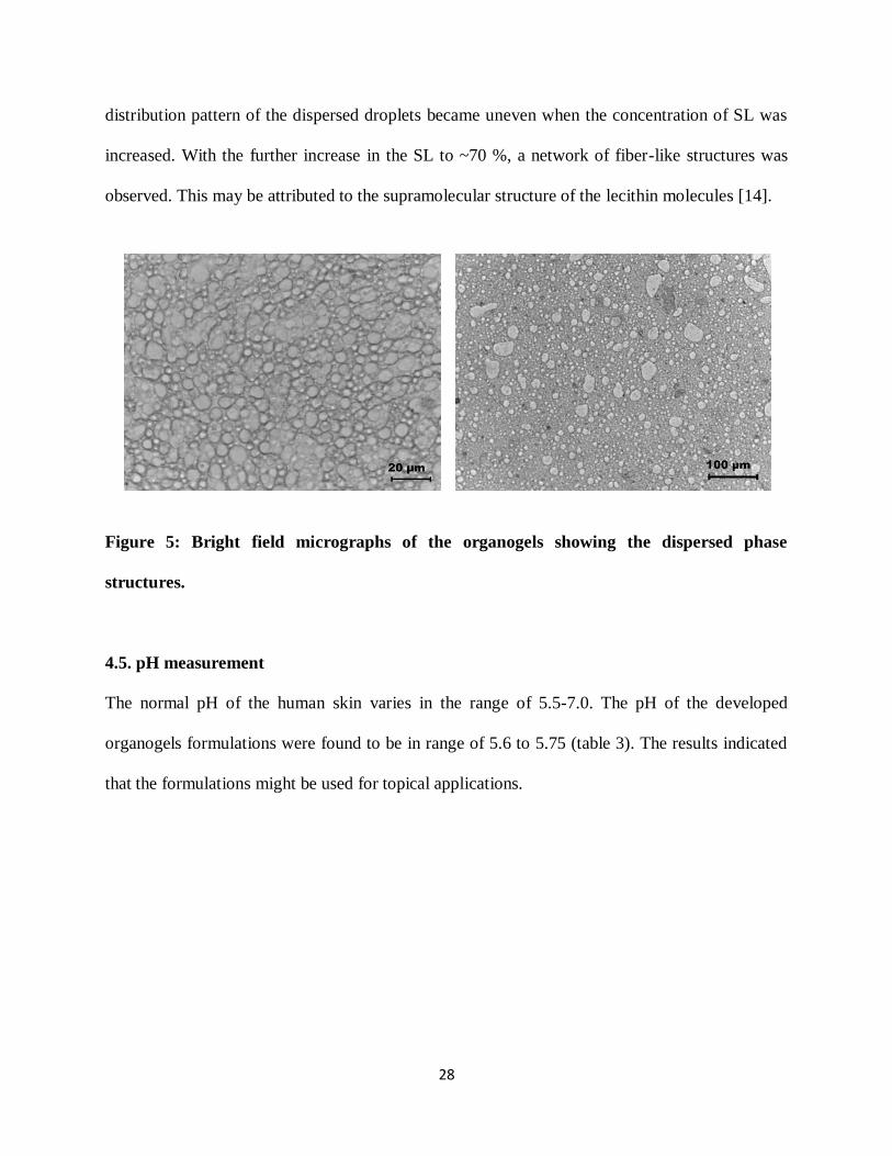
28
distribution pattern of the dispersed droplets became uneven when the concentration of SL was
increased. With the further increase in the SL to ~70 %, a network of fiber-like structures was
observed. This may be attributed to the supramolecular structure of the lecithin molecules [14].
Figure 5: Bright field micrographs of the organogels showing the dispersed phase
structures.
4.5. pH measurement
The normal pH of the human skin varies in the range of 5.5-7.0. The pH of the developed
organogels formulations were found to be in range of 5.6 to 5.75 (table 3). The results indicated
that the formulations might be used for topical applications.

29
Table 2: pH value of the selected organogels
S. No. Sample pH value
1 PLG1 5.61 ± 0.12
2 PLG2 5.63 ± 0.16
3 PLG3 5.71 ± 0.18
4 PLG4 5.75 ± 0.11
5 PLG5 5.73 ± 0.10
4.6. FTIR spectroscopy
4000 3500 3000 2500 2000 1500 1000 500
SL
Ta
ns
mit
tan
ce
(%)
Wave Number (cm-1)
PO
MZ
4000 3500 3000 2500 2000 1500 1000 500
PLG1
Tra
ns
mit
tan
ce
(%
)
Wave Number (cm-1)
PLG1M
PLG2 PLG2M
PLG3
PLG3M
PLG4
PLG4M
PLG5
PLG5M
Figure 6: FTIR study of raw material and their MZ loaded gels

30
FTIR spectrograms of the raw materials (SL and PO) and the organogels (blank and drug loaded)
have been shown in figure 9. SL showed a broad peak at ~3600 cm-1
due to the -OH stretching
vibrations [16]. The absorption peaks at ~ 2848 cm-1
and ~ 1461 cm-1
may be associated to the C-
H stretching and bending vibrations, respectively, of the alkanes. The absorption peak at ~ 1745
cm-1
may be associated to the -CO stretch of the carbonyl groups. The peaks at ~ 721 cm-1
may
be associated to the CH rocking due to the alkanes [14, 16] . PO has shown the absorption peaks
at ~2920 cm−1
and ~2850 cm−1
which was associated to the CH stretching in CH3 and CH2 of
alkanes [36]. The absorption peaks at ~ 1470 cm−1
was due to the CH2 scissoring vibrations [37].
The peak at ~1743 cm−1
may be associated with the stretching vibration of CO group of
triglycerides present in PO [38]. MZ has shown absorption peak at ~3220 cm−1
which may be
associated with the OH stretching. The absorption peak at 3,100 cm−1
may be attributed to C–H
stretching vibration. The absorption peak in the range of 1400–1535 cm−1
may be accounted to
the functional groups present in the imidazole ring [39]. The peak at 1348 cm−1
was attributed to
the NO symmetrical stretch of imidazole. The absorption peak at 1732 cm−1
may be associated
with the C=C double bond [40]. The FTIR spectra of the organogels have shown the presence of
absorption peaks corresponding to the raw materials. A slight shift was observed in few
absorption peaks which may be explained due to the change in the chemical environment around
the components of the organogels. A broad peak was observed at ~ 3350 cm-1
which suggested
the presence of strong intra-molecular/ intermolecular hydrogen bonding amongst the organogel
components. The increase in the intensity as compared to the raw materials indicated an increase
in the hydrogen bonding amongst the gel components [7, 16]. No extra peak was observed in the
MZ loaded organogels. This may due to the fact that MZ was present in very minute
31
concentrations and the peaks associated with MZ were subsided because of the intense peaks of
the other components.
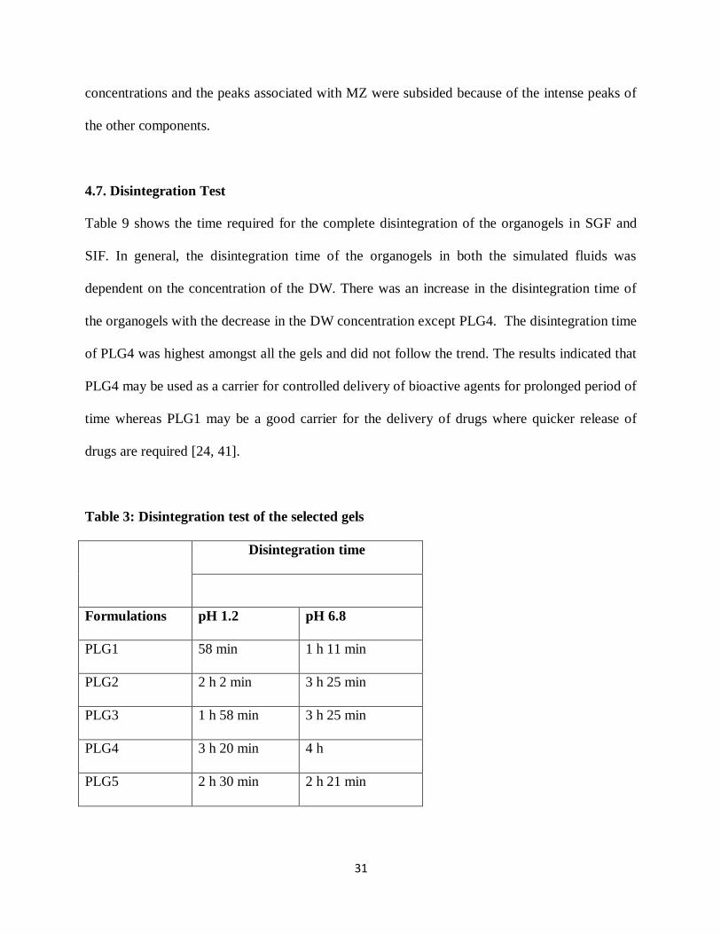
4.7. Disintegration Test
Table 9 shows the time required for the complete disintegration of the organogels in SGF and
SIF. In general, the disintegration time of the organogels in both the simulated fluids was
dependent on the concentration of the DW. There was an increase in the disintegration time of
the organogels with the decrease in the DW concentration except PLG4. The disintegration time
of PLG4 was highest amongst all the gels and did not follow the trend. The results indicated that
PLG4 may be used as a carrier for controlled delivery of bioactive agents for prolonged period of
time whereas PLG1 may be a good carrier for the delivery of drugs where quicker release of
drugs are required [24, 41].
Table 3: Disintegration test of the selected gels
Disintegration time
Formulations pH 1.2 pH 6.8
PLG1 58 min 1 h 11 min
PLG2 2 h 2 min 3 h 25 min
PLG3 1 h 58 min 3 h 25 min
PLG4 3 h 20 min 4 h
PLG5 2 h 30 min 2 h 21 min

32
4.8. Gel Sol Transition:
Table 7: Gel-sol transition of the optimized organogels
Formulations 250C 30
0C 35
0C 40
0C 45
0C 50
0C 55
0C
PLG 1 √ √ √ √ X X X
PLG 2 √ √ √ √ √ √ X
PLG 3 √ √ √ √ √ X X
PLG 4 √ √ √ √ √ √ X
PLG 5 √ √ √ √ √ X X
Gel-sol transition showed the melting point of the developed organogels was in the range of 45
to 550C (table 7). The MP of the gels was found to be increased with SL concentration
suggesting presence of surfactant might have increased the structural integrity. At the same time
it may also be concluded that presence of water also imparted an increase in the structural
organization of the gel samples. PLG 2 has shown higher melting point as compared to PLG 3
which may be associated to the higher DW concentration in PLG2 as compared to PLG3. Both
PLG2 and PLG3 possessed equal SL concentration. So it might be the DW which might have
increased the inter molecular hydrogen bonding.
4.9. Hemocompatibility Test
The results of the hemocompatibility test have been tabulated in the table 12. The results
suggested that the organogels were highly hemocompatible in nature.

33
Table 5: Hemocompatibilty test
Sample Name % Hemolysis
PLG1 3.05 ± 0.08
PLG2 1.42 ± 0.04
PLG3 3.55 ± 0.09
PLG4 0.00 ± 0.00
PLG5 0.00 ± 0.00
4.10. In- vitro drug release
The drug release profiles of MZ from the organogels have been shown in figure 7. The results
suggested that the release of the drug was dependent on the composition of the organogels. The
cumulative percentage drug release (CPDR) of MZ from the organogels was in the order of
PLG1M > PLG2M > PLG3M > PLG4M > PLG5M. This indicated that the CPDR of the drug
was lowered due to the combined effect of the decrease in the concentration of DW and an
increase in the concentration of SL.

34
Figure 7: CPDR of the developed gels
4.11. Antimicrobial assay
The efficiency of the drug incorporated organogels was studied against B. subtilis (a model gram
positive bacterium) and E. coli (a model gram negative bacterium). The zone of inhibition was
measured after 24 h of incubation at 37 ± 1 °C. Metrogyl, the marketed MZ gel, was taken as the
positive control whereas the blank gels were treated as the negative controls. The results
indicated that the developed organogels have shown good antimicrobial efficacy against the
gram positive and gram negative bacterium and the antimicrobial efficiency was comparable to
the marketed formulations (Figure 8-9).

35
Figure 8: Antimicrobial efficacy of the organogels against E. coli
Figure 9: Antimicrobial efficacy of the organogels against B. subtilis

36
CHAPTER 5
CONCLUSION

37
5. CONCLUSION
This study reports the successful development of the SL and PO based organogels. The
preliminary study suggested that the developed systems may be used as potential carriers for
bioactive agents. The organogels were easy to prepare and had shown excellent thermal stability.
The disintegration test suggested the developed systems may be used either as a controlled
release system or as a quick release system, depending on the composition of the organogel. The
developed organogels were found to have good spreadability and viscosity profile desired for the
development of topical formulations. The organogels were found to be highly biocompatible.
The antimicrobial assay of the MZ-loaded organogels has shown comparable antimicrobial as
against Metrogyl®. In short, it may be concluded that the developed organogels may be used as
carriers for antimicrobial bioactive agents for topical application.

38
CHAPTER 6
BIBLIOGRAPHY
39
6. BIBLIOGRAPHY
1. Gupta, S., et al., Organogel: a viable alternative for existing carrier system. system,
2011. 4: p. 5.
2. Vintiloiu, A. and J.-C. Leroux, Organogels and their use in drug delivery—a review.
Journal of Controlled Release, 2008. 125(3): p. 179-192.
3. Sahoo, S., et al., Organogels: Properties and Applications in drug delivery. Designed
Monomers and Polymers, 2011. 14(2): p. 95-108.
4. Behera, B., et al., Span‐60‐based organogels as probable matrices for
transdermal/topical delivery systems. Journal of Applied Polymer Science, 2012. 125(2):
p. 852-863.
5. Kumar, R. and O.P. Katare, Lecithin organogels as a potential phospholipid-structured
system for topical drug delivery: a review. AAPS PharmSciTech, 2005. 6(2): p. 298-310.
6. Scartazzini, R. and P.L. Luisi, Organogels from lecithins. The Journal of Physical
Chemistry, 1988. 92(3): p. 829-833.
7. Pearson, R.H. and I. Pascher, The molecular structure of lecithin dihydrate. 1979.
8. Szuhaj, B.F., Lecithins: sources, manufacture & uses. Vol. 12. 1989: Amer Oil Chemists
Society.
9. Sreekala, M., M. Kumaran, and S. Thomas, Oil palm fibers: Morphology, chemical
composition, surface modification, and mechanical properties. Journal of Applied
Polymer Science, 1998. 66(5): p. 821-835.
10. Sambanthamurthi, R., K. Sundram, and T. YewAi, Chemistry and biochemistry of palm
oil. Progress in Lipid Research, 2000. 39(6): p. 507-558.
11. Sakeena, M., et al., Formulation and in vitro evaluation of ketoprofen in palm oil esters
nanoemulsion for topical delivery. Journal of Oleo Science, 2010. 59(4): p. 223-228.
12. Owen, D.H., J.J. Peters, and D.F. Katz, Rheological properties of contraceptive gels.
Contraception, 2000. 62(6): p. 321-326.
13. Shchipunov, Y.A. and E.V. Shumilina, Lecithin bridging by hydrogen bonds in the
organogel. Materials Science and Engineering: C, 1995. 3(1): p. 43-50.

40
14. Shchipunov, Y.A., Lecithin organogel: a micellar system with unique properties.
Colloids and Surfaces A: Physicochemical and Engineering Aspects, 2001. 183: p. 541-
554.
15. Luisi, P., et al., Organogels from water-in-oil microemulsions. Colloid & Polymer
Science, 1990. 268(4): p. 356-374.
16. Satapathy, D., et al., Sunflower‐oil‐based lecithin organogels as matrices for controlled
drug delivery. Journal of Applied Polymer Science, 2012.
17. Patil, K.D., S. Bakliwal, and S. Pawar, Organogel: topical and transdermal drug delivery
system. Int J Pharm Res Dev, 2011. 3(6): p. 58-66.
18. Lawrence, M.J. and G.D. Rees, Microemulsion-based media as novel drug delivery
systems. Advanced drug delivery reviews, 2000. 45(1): p. 89-121.
19. Sagiri, S.S., et al., Effect of Composition on the Properties of Tween-80–Span-80-Based
Organogels. Designed Monomers and Polymers, 2012. 15(3): p. 253-273.
20. Agrawal, V., et al., Preparation and Evaluation of Tubular Micelles of Pluronic Lecithin
Organogel for Transdermal Delivery of Sumatriptan. AAPS PharmSciTech, 2010. 11(4):
p. 1718-1725.
21. Singh, V.K., et al., Castor oil and sorbitan monopalmitate based organogel as a
probable matrix for controlled drug delivery. Journal of Applied Polymer Science, 2013.
22. Song, J. and R.I. Hollingsworth, Synthesis, conformational analysis, and phase
characterization of a versatile self-assembling monoglucosyl diacylglycerol analog.
Journal of the American Chemical Society, 1999. 121(9): p. 1851-1861.
23. Novales, B., et al., Self-assembly of fatty acids and hydroxyl derivative salts. Langmuir,
2008. 24(1): p. 62-68.
24. Shah, D.K., et al., Development of olive oil based organogels using sorbitan
monopalmitate and sorbitan monostearate: A comparative study. Journal of Applied
Polymer Science, 2012.
25. Rogers, M.A., et al., A novel cryo-SEM technique for imaging vegetable oil based
organogels. Journal of the American Oil Chemists' Society, 2007. 84(10): p. 899-906.
26. Haering, G. and P.L. Luisi, Hydrocarbon gels from water-in-oil microemulsions. The
Journal of Physical Chemistry, 1986. 90(22): p. 5892-5895.
41
27. Belgamwar, V., et al., Pluronic lecithin organogel. Asian Journal of Pharmaceutics,
2008. 2(3): p. 134.
28. Murata, Y., et al., Properties of pectin gel bead modified with pectin hydrolysate and
chitosan. Advances in Polymer Technology, 2005. 24(1): p. 46-50.
29. Behera, B., et al., Span‐60‐based organogels as probable matrices for
transdermal/topical delivery systems. Journal of Applied Polymer Science, 2012.
30. Bhattacharya, C., et al., Development of span 80–tween 80 based fluid-filled organogels
as a matrix for drug delivery. Journal of pharmacy & bioallied sciences, 2012. 4(2): p.
155.
31. Pal, K., A. Banthia, and D. Majumdar, Biomedical evaluation of polyvinyl alcohol–
gelatin esterified hydrogel for wound dressing. Journal of Materials Science: Materials in
Medicine, 2007. 18(9): p. 1889-1894.
32. Willimann, H., et al., Lecithin organogel as matrix for transdermal transport of drugs.
Journal of pharmaceutical sciences, 1992. 81(9): p. 871-874.
33. Tokuyama, H. and Y. Kato, Preparation of thermosensitive polymeric organogels and
their drug release behaviors. European Polymer Journal, 2010. 46(2): p. 277-282.
34. Couffin-Hoarau, A.C., et al., In situ-forming pharmaceutical organogels based on the
self-assembly of L-alanine derivatives. Pharmaceutical research, 2004. 21(3): p. 454-457.
35. Suzuki, M., et al., Supramolecular organogel formation triggered by acid–base
interaction in two-component system consisting of L-lysine derivative and aliphatic acids.
New J. Chem., 2007. 31(9): p. 1654-1660.
36. Ifuku, S., et al., Preparation and characterization of redox cellulose Langmuir–Blodgett
films containing a ferrocene derivative. Journal of Polymer Science Part A: Polymer
Chemistry, 2005. 43(21): p. 5023-5031.
37. Atek, D. and N. Belhaneche-Bensemra, FTIR investigation of the specific migration of
additives from rigid poly (vinyl chloride). European polymer journal, 2005. 41(4): p. 707-
714.
38. Pérez-Mateos, M., P. Montero, and M.C. Gómez-Guillén, Formulation and stability of
biodegradable films made from cod gelatin and sunflower oil blends. Food
Hydrocolloids, 2009. 23(1): p. 53-61.

42
39. Yuan, J. and M. Liu, Chiral molecular assemblies from a novel achiral amphiphilic 2-
(heptadecyl) naphtha [2, 3] imidazole through interfacial coordination. Journal of the
American Chemical Society, 2003. 125(17): p. 5051-5056.
40. Cristescu, R., et al., Functional polyethylene glycol derivatives nanostructured thin films
synthesized by matrix-assisted pulsed laser evaporation. Applied Surface Science, 2009.
255(24): p. 9873-9876.
41. Iwanaga, K., et al., Characterization of organogel as a novel oral controlled release
formulation for lipophilic compounds. International journal of pharmaceutics, 2010.
388(1): p. 123-128.