developing perspectives on molluscan shells, part 1
TRANSCRIPT

CONTENTS
Abstract ........................................................................................................21.1 Introduction .........................................................................................21.2 Insights From Genomics, Transcriptomics, and Proteomics ............131.3 Novelty in Molluscan Biomineralization ..........................................211.4 Conclusions and Open Questions .....................................................24Keywords ...................................................................................................27References ..................................................................................................27
CHAPTER 1
DEVELOPING PERSPECTIVES ON MOLLUSCAN SHELLS, PART 1: INTRODUCTION AND MOLECULAR BIOLOGY
KEVIN M. KOCOT1, CARMEL MCDOUGALL, and BERNARD M. DEGNAN
1Present Address: Department of Biological Sciences and Alabama Museum of Natural History, The University of Alabama, Tuscaloosa, AL 35487, USA; E-mail: [email protected]
School of Biological Sciences, The University of Queensland, St. Lucia, Queensland 4072, Australia

2 Physiology of Molluscs Volume 1: A Collection of Selected Reviews
ABSTRACT
Molluscs (snails, slugs, clams, squid, chitons, etc.) are renowned for their highly complex and robust shells. Shell formation involves the controlled deposition of calcium carbonate within a framework of macromolecules that are secreted by the outer epithelium of a specialized organ called the mantle. Molluscan shells display remarkable morphological diversity, structure, and ornamentation; however, the physiological mechanisms underlying the evolution and formation of the shell are just beginning to be understood. Examination of genes expressed in the mantle and proteins incorporated into the shell suggests that the genetic program underlying shell fabrication is rapidly evolving. This includes lineage-specific integration of conserved, ancient gene families into the mantle gene regulatory network and the evolu-tion of genes encoding proteins with novel repetitive motifs and domain combinations, which results in the expression of markedly different shell matrix protein repertoires in even closely-related molluscs. Here, we review the molecular physiology of shell formation with emphasis on the protein components that are particularly rapidly evolving. Nonprotein components such as chitin, other polysaccharides, and lipids are also reviewed. The high degree of novelty in molluscan biomineralized structures is discussed with emphasis on topics of recent interest including the image-forming aragonitic eye lenses of chiton shells and shell pigments. Finally, unanswered questions including some dealing with basic concepts such as the homology of the nacreous shell layers of gastropods and bivalves are discussed.
1.1 INTRODUCTION
Biomineralization is the process by which living organisms convert ions in solution into solid minerals (Simkiss & Wilbur, 1989). The great success of molluscs can be attributed in part to their ability to secrete calcareous skeletal structures with evidence for molluscan biomineralization extending back to the late Precambrian (Runnegar, 1996). All eight major lineages of Mollusca produce calcified exoskeletons, in the form of shells (such as those produced by bivalves, gastropods, and Nautilus) or sclerites (spines, scales, etc. produced by chitons and aplacophorans). However, secondary reduction or loss of the shell has occurred in several lineages (e.g., Kröger et al., 2011; Wägele & Klussmann-Kolb, 2005). In this chapter, we begin the discussion of molluscan biomineralization physiology with an emphasis on recent insights on the molecular biology of shell formation from studies
Developing Perspectives on Molluscan Shells, Part 1 3
using evolutionary developmental, comparative genomic/transcriptomic, and proteomic approaches. We highlight the importance of comparative studies in understanding the principles of biomineralization and a need for more such studies that include representatives from all lineages of Mollusca.
1.1.1 DIVERSITY AND STRUCTURE OF MOLLUSCAN EXOSKELETONS
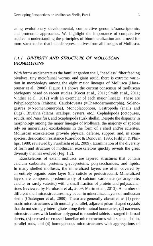
With forms as disparate as the familiar garden snail, “headless” filter feeding bivalves, tiny meiofaunal worms, and giant squid, there is extreme varia-tion in morphology among the eight major lineages of Mollusca (Hasz-prunar et al., 2008). Figure 1.1 shows the current consensus of molluscan phylogeny based on recent studies (Kocot et al., 2011; Smith et al., 2011; Vinther et al., 2012) with an exemplar of each major lineage. These are Polyplacophora (chitons), Caudofoveata (=Chaetodermomorpha), Soleno-gastres (=Neomeniomorpha), Monoplacophora, Gastropoda (snails and slugs), Bivalvia (clams, scallops, oysters, etc.), Cephalopoda (octopuses, squids, and Nautilus), and Scaphopoda (tusk shells). Despite the disparity in morphology among the major lineages of Mollusca, the majority of species rely on mineralized exoskeletons in the form of a shell and/or sclerites. Molluscan exoskeletons provide physical defense, support, and, in some species, desiccation resistance (Carefoot & Donovan, 1995; Fishlyn & Phil-lips, 1980; reviewed by Furuhashi et al., 2009). Examination of the diversity of form and structure of molluscan exoskeletons quickly reveals the great diversity that has evolved (Fig. 1.2).
Exoskeletons of extant molluscs are layered structures that contain calcium carbonate, proteins, glycoproteins, polysaccharides, and lipids. In many shelled molluscs, the mineralized layers are often covered by an entirely organic outer layer (the cuticle or periostracum). Mineralized layers are composed predominantly of calcium carbonate (as aragonite, calcite, or rarely vaterite) with a small fraction of protein and polysaccha-rides (reviewed by Furuhashi et al., 2009; Marin et al., 2013). A number of different shell microstructures may occur in mineralized layers of molluscan shells (Chateigner et al., 2000). These are generally classified as (1) pris-matic microstructures with mutually parallel, adjacent prism-shaped crystals that do not strongly interdigitate along their mutual boundaries, (2) nacreous microstructures with laminar polygonal to rounded tablets arranged in broad sheets, (3) crossed or crossed lamellar microstructures with sheets of thin, parallel rods, and (4) homogeneous microstructures with aggregations of
4 Physiology of Molluscs Volume 1: A Collection of Selected Reviews
irregularly shaped crystallites with a granular appearance (Chateigner et al., 2000; see Bandel, 1990; Carter & Clark, 1985 for detailed discussions of shell microstructure). Of these, the prismatic and nacreous microstructures are the best studied. The prismatic layer is resistant to crack propagation and puncture (Eichhorn et al., 2005; Li & Nardi, 2004; Su et al., 2004), whereas the nacreous layer is best known for being more ductile and fracture resistant (Chateigner et al., 2000; Li et al., 2006). We refer the reader to Chateigner et al. (2000) for high-quality scanning electron micrographs of each of these different microstructure types.
FIGURE 1.1 Current consensus of evolutionary relationships among the major lineages of Mollusca as inferred by Kocot et al. (2011), Smith et al. (2011), and Vinther et al. (2012). Photos are not to scale. Photo of Argopecten (Bivalvia) by Dan Speiser. Photo of Chaetoderma (Caudofoveata) by Christiane Todt. Photo of Laevipilina (Monoplacophora) by Greg Rouse and Nerida Wilson. (Used with permission.)

Developing Perspectives on Molluscan Shells, Part 1 5
FIGURE 1.2 Diversity of mineralized structures fabricated by extant molluscan lineages. A. Micro CT scan of a juvenile specimen of Cryptoplax larvaeformis (Polyplacophora) showing anterior shell valves and sclerites. Specimen is approximately 1-cm wide. Photo by Jeremy Shaw. B. Scanning electron micrograph (SEM) of sclerites of Macellomenia schanderi (Solenogastres). C. Micrograph of sclerites of an undescribed species of Falcidens (Caudofoveata) from New Zealand illuminated with polarized light. Smallest sclerite is approximately 100 µm in length. D. Laterally bisected shell of Nautilus (Cephalopoda). E. Shell of the pearl oyster Pinctada maxima (Bivalvia). F. Shell of the abalone Haliotis asinina (Gastropoda).
6 Physiology of Molluscs Volume 1: A Collection of Selected Reviews
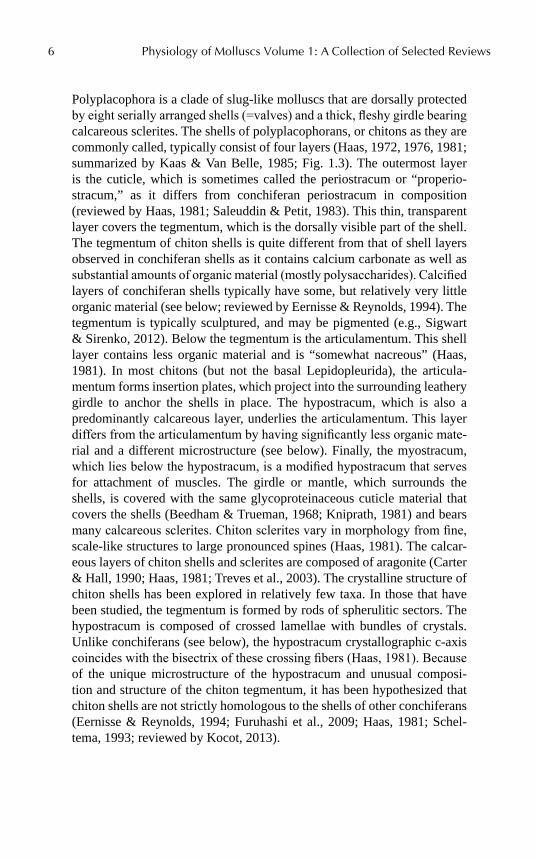
Polyplacophora is a clade of slug-like molluscs that are dorsally protected by eight serially arranged shells (=valves) and a thick, fleshy girdle bearing calcareous sclerites. The shells of polyplacophorans, or chitons as they are commonly called, typically consist of four layers (Haas, 1972, 1976, 1981; summarized by Kaas & Van Belle, 1985; Fig. 1.3). The outermost layer is the cuticle, which is sometimes called the periostracum or “properio-stracum,” as it differs from conchiferan periostracum in composition (reviewed by Haas, 1981; Saleuddin & Petit, 1983). This thin, transparent layer covers the tegmentum, which is the dorsally visible part of the shell. The tegmentum of chiton shells is quite different from that of shell layers observed in conchiferan shells as it contains calcium carbonate as well as substantial amounts of organic material (mostly polysaccharides). Calcified layers of conchiferan shells typically have some, but relatively very little organic material (see below; reviewed by Eernisse & Reynolds, 1994). The tegmentum is typically sculptured, and may be pigmented (e.g., Sigwart & Sirenko, 2012). Below the tegmentum is the articulamentum. This shell layer contains less organic material and is “somewhat nacreous” (Haas, 1981). In most chitons (but not the basal Lepidopleurida), the articula-mentum forms insertion plates, which project into the surrounding leathery girdle to anchor the shells in place. The hypostracum, which is also a predominantly calcareous layer, underlies the articulamentum. This layer differs from the articulamentum by having significantly less organic mate-rial and a different microstructure (see below). Finally, the myostracum, which lies below the hypostracum, is a modified hypostracum that serves for attachment of muscles. The girdle or mantle, which surrounds the shells, is covered with the same glycoproteinaceous cuticle material that covers the shells (Beedham & Trueman, 1968; Kniprath, 1981) and bears many calcareous sclerites. Chiton sclerites vary in morphology from fine, scale-like structures to large pronounced spines (Haas, 1981). The calcar-eous layers of chiton shells and sclerites are composed of aragonite (Carter & Hall, 1990; Haas, 1981; Treves et al., 2003). The crystalline structure of chiton shells has been explored in relatively few taxa. In those that have been studied, the tegmentum is formed by rods of spherulitic sectors. The hypostracum is composed of crossed lamellae with bundles of crystals. Unlike conchiferans (see below), the hypostracum crystallographic c-axis coincides with the bisectrix of these crossing fibers (Haas, 1981). Because of the unique microstructure of the hypostracum and unusual composi-tion and structure of the chiton tegmentum, it has been hypothesized that chiton shells are not strictly homologous to the shells of other conchiferans (Eernisse & Reynolds, 1994; Furuhashi et al., 2009; Haas, 1981; Schel-tema, 1993; reviewed by Kocot, 2013).

Developing Perspectives on Molluscan Shells, Part 1 7
FIGURE 1.3 Structure of a chiton-shell valve. Above: Whole shell valve with cut-out region corresponding to enlargement below. Below: Enlargement showing shell layers. Abbreviations: a, articulamentum; c, crossed lamellar structure of hypostracum; h, hypostracum; m, myostracum; pp, properiostracum (cuticle); t, tegmentum. Modified from Haas (1976).
Caudofoveata (=Chaetodermomorpha) and Solenogastres (=Neome-niomorpha), collectively called Aplacophora, are worm-shaped, shell-less molluscs (reviewed by Todt et al., 2008; Todt, 2013). Although tradition-ally viewed as basal, plesiomorphic molluscs (see Salvini-Plawen & Steiner, 2014 and references therein), recent molecular phylogenetic studies (Kocot et al., 2011; Smith et al., 2011; Vinther et al., 2012) have grouped Apla-cophora + Polyplacophora in a clade called Aculifera (Scheltema, 1993; Fig. 1.1). Examination of fossil paleoloricate “chitons” (Sutton & Sigwart, 2012; Sutton et al., 2012) has led to the interpretation that aplacophorans are derived from chiton-like ancestors that secondarily lost their shells (Sutton

8 Physiology of Molluscs Volume 1: A Collection of Selected Reviews
& Sigwart, 2012; Sutton et al., 2012; Vinther et al., 2012; Vinther, 2014, 2015). Developmental studies have also been cited as evidence for a chiton-like ancestor of Aplacophora (Scheltema & Ivanov, 2002). Although extant aplacophorans lack shells, most of the body surface is covered with a glyco-proteinaceous cuticle and a dense coat of calcareous sclerites. Although the sclerites of the burrowing caudofoveates are relatively uniform, there is great variation in the morphology of solenogaster sclerites. Solenogaster sclerites may be solid or hollow and can exhibit a variety of shapes, such as needles, scales, hooks, and paddles, just to name a few (García-Álvarez & Salvini-Plawen, 2007). Presence of scale-like sclerites in the putatively early branching solenogaster order Pholidoskepia (Salvini-Plawen, 2003) and observation of scale-like sclerites in larvae and early juvenile solenogaster species that later develop hollow needles (Okusu, 2002; Todt & Kocot, 2014) suggests that scale-like sclerites (as also found in Caudofoveata) are plesio-morphic for Aplacophora (Salvini-Plawen, 2003). Aplacophoran spicules are composed of aragonite (Rieger and Sterrer 1975; Scheltema & Ivanov, 2002, 2004), with the long axis of the crystals aligned with the long axis of the spicules (reviewed by Ehrlich, 2010).
Monoplacophora is a small group of around 30 described species of single-shelled molluscs that mostly live in the deep sea (reviewed by Hasz-prunar & Ruthensteiner, 2013; Haszprunar, 2008; Lindberg, 2009). Some authors prefer the more specific name Tryblidia for the extant Monopla-cophora because several extinct “monoplacophorans” are of uncertain phylo-genetic affinity. Most monoplacophorans have a thin outer periostracum, a prismatic shell layer with large quadrangular or hexagonal prisms, and an inner nacreous layer (Erben et al., 1968; Hedegaard & Wenk, 1998; Meen-akshi et al., 1970; Wingstrand, 1985). However, in Veleropilina, Rokopella, and Micropilina, the prismatic layer is apparently absent (see Haszprunar & Ruthensteiner, 2013 for discussion) and the outer shell layer is composed of smooth or granular material with unknown microstructure (presumably homogeneous; Checa et al., 2009; Cruz et al., 2003; Marshall, 2006; Warén & Hain, 1992).
Scaphopods are marine burrowing microcarnivores with a conical shell that is open at both ends. The shell grows from the anterior end and is removed at the posterior end to allow for increased water flow into the mantle cavity as the animal grows (de Paula & Silveira, 2009). Some species produce “tubes” or “pipes” from the posterior mantle margin (Hebert, 1986; Shimek, 1989). The shell may bear longitudinal or, rarely, annular ribs. Generally, scaphopods have a trilayered shell organization similar to that of gastropods and bivalves. The organic periostracum may be thick but
Developing Perspectives on Molluscan Shells, Part 1 9
typically it is very thin or completely eroded in adult animals, probably due to their sand burrowing activity. An outer, very thin crystalline prismatic layer with tightly packed crystals is present in the majority of species of the order Gadilida giving these species a polished appearance. The inner-most shell layer is a complex, crossed-lamellar layer, which may have a regular or irregular structure (Steiner, 1995; Reynolds & Okusu, 1999). The shell is composed of aragonite (Bøggild, 1930).
Cephalopoda includes the extant nautiloids, octopods, vampyropods, and decabrachians (cuttlefish, squid, and Spirula) as well as a rich diversity of fossil forms (reviewed by Kröger et al., 2011; Young et al., 1998). Among the living cephalopods, only members of Nautiloidea have retained an external shell as adults, whereas others have reduced or (more-or-less) completely lost their shell. Cephalopod shell structure and the general mechanisms of shell formation in this group were reviewed by Bandel (1990) and Budel-mann et al. (1991). In Nautiloidea, the most plesiomorphic extant cephalopod lineage, the thick, external shell is aragonitic with prismatic, spherulitic, and nacreous configurations. In Nautilus, internal chambers of the shell are used for buoyancy control; an osmotic gradient is established by active transport of salts to the space between the mantle tissue and the shell. This allows for the extraction of liquid from the hollow chamber and inward diffusion of gas (reviewed in detail by Budelmann et al., 1991). Most of the extant diversity of Cephalopoda is dominated by taxa with internalized and usually highly reduced shells (Birchall & Thomas, 1983; Hunt and El Sherief, 1990; Sousa Reis & Fernandes, 2002). The pelagic cephalopod Spirula has a calcified internal shell similar to that of Nautilus, which is also used for buoyancy control. Cuttlefish (e.g., Sepia) also use their internal shell for this function. Here, the shell is not coiled with relatively few large chambers, but contains small chambers with many flat, subdivided chambers subdivided by serially arranged organic membranes. Most other cephalopods (e.g., octopus and squid) have completely uncalcified, chitinous vestiges of the shell.
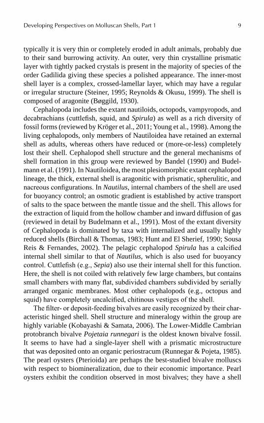
The filter- or deposit-feeding bivalves are easily recognized by their char-acteristic hinged shell. Shell structure and mineralogy within the group are highly variable (Kobayashi & Samata, 2006). The Lower-Middle Cambrian protobranch bivalve Pojetaia runnegari is the oldest known bivalve fossil. It seems to have had a single-layer shell with a prismatic microstructure that was deposited onto an organic periostracum (Runnegar & Pojeta, 1985). The pearl oysters (Pterioida) are perhaps the best-studied bivalve molluscs with respect to biomineralization, due to their economic importance. Pearl oysters exhibit the condition observed in most bivalves; they have a shell
10 Physiology of Molluscs Volume 1: A Collection of Selected Reviews
with an inner nacreous layer, a middle prismatic layer, and an outer protein-aceous layer.
Gastropoda is the most species-rich class of Mollusca. There is a great diversity of shell organization and microstructures within this clade. In the well-studied vetigastropod Haliotis, the shell consists of three layers: an outer organic periostracum (that is often eroded in adults), a prismatic layer made up of needle-shaped crystals enveloped by an organic sheath, and a nacreous layer consisting of aragonitic tablets surrounded and perfused by thin organic matrix (summarized by Marie et al., 2010). Adult patello-gastropods such as Lottia have a shell consisting of five layers (Mann et al., 2012; Marie et al., 2013; Suzuki et al., 2010). The outer-most layer is primarily calcite with a mosaic organization whereas the remaining layers are composed of prismatically arranged crystals of aragonite (Marie et al., 2013; Suzuki et al., 2010). Crossed lamellar shell microstructure is wide-spread in other gastropods (Dauphin & Denis, 2000).
1.1.2 MANTLE TISSUE
Mantle tissue (=pallial tissue; Fig. 1.4) is responsible for the secretion of molluscan shells and sclerites. The mantle forms and isolates a chamber from the external environment (see Simkiss Chapter 2 of this volume) and secretes an organic matrix of polysaccharides (e.g., chitin) and protein, which is presumed to be the site of calcium carbonate crystal nucleation (reviewed by Addadi et al., 2006; Furuhashi et al., 2009; Wilbur & Saleuddin, 1983; Wilbur, 1972). Mantle tissue morphology and the process of shell formation in general are most well-known in bivalves and gastropods. In these taxa, there are conserved cellular and morphogenetic movements that initiate larval shell secretion. Larval shell formation begins at the end of gastrula-tion, with the differentiation and local thickening of a group of ectodermal cells in the post-trochal dorsal region (the shell gland or shell field). These cells elongate and then invaginate transitorily to form the shell gland, which is analogous to the adult mantle and responsible for the secretion of larval shell. The periphery of the shell gland produces an extracellular lamella—the organic periostracum—that will serve as the site of calcium carbonate deposition (Bielefeld & Becker, 1991; Cather, 1967; Hohagen & Jackson, 2013; Kniprath, 1981). Later, the shell gland flattens and grows into the more recognizable adult mantle epithelium (Jackson et al., 2007; Kniprath, 1977, 1980, 1981).

Developing Perspectives on Molluscan Shells, Part 1 11
FIGURE 1.4 Histological sections of molluscan mantle tissues. A. Sclerite secretion in Acanthopleura gemmata (Polyplacophora). B. Various stages of sclerite secretion and lifting through cuticle in Cryptoplax larvaeformis. C. Epidermal papillae, cuticle, and voids from decalcified sclerites in the thick cuticle of Proneomenia custodiens (Solenogastres). D. Mantle tissue of Haliotis asinina (Gastropoda). Specimen prepared by Kathryn Green. Abbreviations: csc, cuticle secreting cells; cu, cuticle; ep, epidermal papillae; inner fold of mantle; mu, muscle; ofd, distal part of outer fold; ofp, proximal part of outer fold; pgb, base of the periostracal groove ; pgl, periostracal groove; pgo, outer fold of the periostracal groove; sc, sclerite; ssc, sclerite secreting cells.
Larval conchiferans (e.g., gastroods, bivalves, scaphopods) typically have a discreet shell gland that secretes the periostracum at its distal edge. The mantle tissue and the periostracum form the crystallization chamber where calcium is deposited adjacent to the periostracum. In contrast to conchiferans, chiton shells are secreted underneath a thin layer of cuticle (the same material that covers the entire dorsum; “properiostracum” sensu Haas, 1981) by a broad “plate field” (Kniprath, 1980; reviewed by Eernisse & Reynolds, 1994). This dramatic difference in shell formation mode has led some workers to question the homology of chiton shells to those of conchif-erans (reviewed by Kocot, 2013; see below).
12 Physiology of Molluscs Volume 1: A Collection of Selected Reviews
A number of studies have examined the anatomy of bivalve (reviewed by Morse and Zardus, 1997; see also Acosta-Salmón & Southgate, 2006; Checa, 2000; Fang et al., 2008) and gastropod (e.g., Fleury et al., 2008; Jackson et al., 2006; Jolly et al., 2004; Kapur & Gibson, 1967; McDou-gall et al., 2011; Sud et al., 2002; Werner et al., 2013; Zylstra et al., 1978) mantle tissue. Bivalve mantle differs from that of gastropods in some key ways. Most notably, the mantle margin, the active site of shell formation, in bivalves has three folds or grooves whereas gastropods generally only have two (Kniprath, 1978; Zylstra et al., 1978). However, this may be an over-generalization as the keyhole limpet Diodora sp. mantle margin has three folds (Budd et al., 2014). In adult bivalves, a ridge between the outer and median fold defines the periostracal groove, which secretes the peri-ostracum. This outer organic shell layer is secreted from basal cells with a greatly infolded apical cell membrane or, in the case of Crassostrea, a specialized “periostracum gland” (Morrison, 1993). The outer epithelium of the mantle (i.e., the surface of the mantle facing the shell) secretes the calcified layers of the shell. Here, different zones of cells secrete different types of layers. In bivalves with a typical three-layered shell consisting of periostracum, prismatic, and nacreous layers, the epithelial cells that secrete the prismatic shell layer are columnar (Carriker, 1992) and distal to the those that secrete nacre, which are cuboidal (Fang et al., 2008; Sudo et al., 1997).
The sclerite-bearing epidermis of chitons (Haas, 1976; Kniprath, 1981) and aplacophorans (Kingsley et al., 2012; Woodland, 1907) contains calcium carbonate-secreting cells, cuticle-secreting cells, and papillae (reviewed by Ehrlich, 2010). In most chitons, an epithelium of columnar cells secretes calcium carbonate portion of the sclerite while marginal cells containing many vesicles secrete the cuticular covering of the sclerite (Haas, 1981, Fig. 1.4A). Sclerite secretion in the chiton Cryptoplax (Fig. 1.4B) is similar but, because this species has a relatively thick cuticle, sclerites must be pushed up through the cuticle. This appears to be achieved by growth of mantle cells (possibly papillae) that subsequently “retreat.” This process is similar to what has been observed in proneomeniid (and other) solenogaster aplacophorans (e.g., Woodland, 1907), which also have a thick cuticle (Fig. 1.4C). Sclerite secretion in the solenogaster aplacophoran Helicoradomenia is similar to that of chitons except just one cell secretes the calcareous portion of the sclerite (as is the case in Proneomenia) and no special cell elongation is needed to push the sclerite through the relatively thin cuticle of this species (Kingsley et al., 2012).
Developing Perspectives on Molluscan Shells, Part 1 13
1.2 INSIGHTS FROM GENOMICS, TRANSCRIPTOMICS, AND PROTEOMICS
At the time of writing this chapter, well-annotated genomes were publicly available from only three molluscs: Lottia gigantea (Simakov et al., 2013), Pinctada fucata (Takeuchi et al., 2012), and Crassostrea gigas (Zhang et al., 2012). However, advances in high-throughput sequencing (reviewed by Metzker, 2010) have made it possible for researchers to deeply sequence the transcriptomes of biological samples as small as a single cell (e.g., Hashim-shony et al., 2012). Studies applying such an approach to the study of molluscan mantle tissue have provided new insight into the genes expressed in mantle and their interactions. Recent phylogenomic studies addressing molluscan evolutionary relationships have also contributed a significant amount of transcriptome data (González et al., 2015; Kocot et al., 2011; Smith et al., 2011; Zapata et al., 2014). Similarly, proteomic tools make it possible to identify the proteins and peptides incorporated into mineralized structures (e.g., Mann & Edsinger-Gonzales, 2014; Mann & Jackson, 2014; Mann et al., 2012). Here, we summarize recent studies that have employed such approaches to improve understanding of the molecular physiology of molluscan biomineralization.
1.2.1 DIFFERENT GENE REPERTOIRES
Several studies have used transcriptomic approaches to identify the biomin-eralization gene repertoires of bivalves including Pinctada (pearl oysters; Fang et al., 2011; Gardner et al., 2011; Huang et al., 2013; Jackson et al., 2010; Jones et al., 2014; Joubert et al., 2010; Kinoshita et al., 2011; McGinty et al., 2012; Shi et al., 2013; Zhao et al., 2012), Mytilus (mussels; Freer et al., 2014; Hüning et al., 2013), Pecten (Artigaud et al., 2014), Hyriopsis (Bai et al., 2010, 2013), and Laternula (Clark et al., 2010; Sleight et al., 2015) and gastropods including Haliotis (abalone; Jackson et al., 2006, 2007, 2010), Patella (Werner et al., 2013) Cepaea (Mann & Jackson, 2014). However, relatively few comparative studies have been performed (Jackson et al., 2010). By directly comparing the transcriptome of nacre-forming cells in a bivalve (Pinctada maxima) and gastropod (Haliotis asinina), Jackson et al. (2010) found tremendous differences in these two mantle transcriptomes, with less than 10% of the genes expressed in the nacre-secreting cells having significant similarity. Of these, most could be identified as being involved in processes other than biomineralization. Notably, P. maxima had high

14 Physiology of Molluscs Volume 1: A Collection of Selected Reviews
representation of genes annotated with lyase activity due to the abundant expression of two alpha carbonic anhydrase (CA) genes. Alpha CAs have previously been shown to be involved in biomineralization in various meta-zoan taxa (Horne et al., 2002; Jackson et al., 2007; Miyamoto et al., 1996; Moya et al., 2008; Wilbur & Saleuddin, 1983).
In order to focus on genes likely involved in the patterning of the nacreous layer of these animals’ shells, Jackson et al. (2010) identified gene prod-ucts that possessed a signal peptide (indicating an extracellular [secreted] protein) from each gene set. From H. asinina they identified 129 sequences and from P. maxima they identified 125 sequences that bear a signal peptide. When these “secretomes” were searched against each other and a variety of databases, the authors found that the majority were unique; 95 (74%) and 71 (57%) of the putative secreted proteins in H. asinina and P. maxima, respectively, shared no similarity with sequences in GenBank’s nonredun-dant protein database or EST databases, or the genome of the patellogas-tropod Lottia gigantea. Of the 54 P. maxima-secreted products that shared similarity with a previously described sequence, 12 of these were previously identified as bivalve-specific biomineralization proteins (McDougall et al., 2013; Yano et al., 2006; Zhang et al., 2006; Aguilera et al., 2014 manuscript in preparation). Interestingly, only six novel H. asinina proteins and one novel P. maxima secreted protein shared similarity with proteins encoded by the Lottia genome, suggesting rapid evolution of lineage-specific biominer-alization gene repertoires.
Proteomic studies have also shed light on differences among molluscan lineages in the molecular physiology of biomineralization (e.g., Joubert et al., 2010; Liao et al., 2015; Mann & Jackson, 2014; Mann et al., 2012; Marie et al., 2011; Marie et al., 2013; Pavat et al., 2012). Marie et al. (2011) observed that the shell protein repertoire of the mussel Mytilus edulis is partly similar to that of other bivalves (i.e., Pinctada), but also shares few similarities with that of the gastropod Haliotis. Also, Marie et al. (2013) examined the proteins incorporated into the shell of the patellogastropod Lottia gigantea. Similar to the results of Jackson et al. (2010), who used a transcriptomic approach, the shell matrix protein (SMP) repertoire of Lottia was found to be more similar to that of the bivalve Pinctada than to that of the vetigastropod Haliotis. Given the fundamental crystallographic differ-ences between the limpet and abalone shells (e.g., presence/absence of nacre and crossed lamellae), these results might suggest that the secretome of the abalone mantle is relatively derived. These works highlight the impor-tance of comparative studies for elucidating the evolution of the molluscan biomineralization toolkit.

Developing Perspectives on Molluscan Shells, Part 1 15
To this end, Mann and Jackson (2014) characterized the transcriptome and shell matrix proteome of another gastropod, the common grove snail Cepaea nemoralis. Interestingly, the shell proteome was dominated by novel proteins with no known protein domains. Specifically, 31 out of the 59 iden-tified shell proteins (52.5%) were completely unknown. Comparison of the C. nemoralis shell proteome to shell proteomes of five molluscan species (Crassostrea gigas, L. gigantea, H. asinina, P. maxima, and P. margaritifera) revealed 28 of 59 C. nemoralis proteins (47.5%) that shared similarity with one or more proteins in shell proteomes of the other species. Interestingly, only one C. nemoralis protein had high similarity to one of the 94 proteins in the shell of H. asinina and only 34 were similar to proteins (631 in total) in the L. gigantea shell proteome. Taken together, these studies indicate that the SMPs directing shell formation in bivalves and gastropods (and even among lineages of gastropods) are markedly different.
1.2.2 COMMON PRINCIPLES
Recent comparative studies have revealed a surprising diversity in the genetic toolkits used in shell secretion by different molluscs. However, there are underlying common principles. All shell- and/or sclerite-forming molluscs use specialized cellular machinery located in the mantle tissue to actively concentrate and secrete calcium carbonate into a closed-off space formed by the mantle and an organic matrix. The shell matrix, which consists of proteins, glycoproteins, chitin, and other polysaccharides, has been shown to be very important in determining the structure of the resulting shell (reviewed by Furuhashi et al., 2009; Marin et al., 2008, 2013).
1.2.2.1 STRUCTURAL PROTEINS
Earlier hypotheses of mollusc shell formation focused on the presence of an extrapallial fluid (e.g., Wilbur & Saleuddin, 1983). However, most contem-porary views of biomineralization refer to a protein–polysaccharide gel rather than a fluid (Addadi et al., 2006; Marin et al., 2013) and view certain SMPs in this gel as the site of nucleation (Evans, 2008). Marin et al. (2008, 2013) and Evans (2008) reviewed the structure, function, and evolution of molluscan shell proteins. Structural proteins are by far the best-known component of the molluscan shell matrix. These proteins appear to function in promoting (Kim et al., 2004, 2006) or inhibiting (Kim et al., 2006; Mann et al., 2007;

16 Physiology of Molluscs Volume 1: A Collection of Selected Reviews
Michenfelder et al., 2003) crystallization of aragonite or calcite and modu-lating the morphology of the structures that are produced (Evans, 2008).
1.2.2.1.1 Acidic Shell Proteins
Highly acidic proteins have been implicated in the biomineralization of many organisms, and molluscs are no exception. The organic matrix of bivalve, gastropod, and polyplacophoran shells contains a high propor-tion of acidic amino acids – particularly aspartate, one of two amino acids that possess a negative charge (the other acidic amino acid, glutamate, is much less common; Hare, 1963; Piez, 1961; Simkiss, 1965). This amino acid bias is reflected in a number of notably acidic characterized SMPs, including MSP1 (pI 3.2; Sarashina & Endo, 2001), Aspein (pI 1.45; Tsuka-moto et al., 2004), Caspartin (Marin et al., 2005), Calprismin (Marin et al., 2005), MPP1 (pI 1.21; Samata et al., 2008), Pif (which is cleaved to produce two acidic peptides with pI’s of 4.99 and 4.65; Suzuki et al., 2013), and the Asprich family (pI 3.1; Gotliv et al., 2005). Additionally, many other SMPs contain short acidic domains, such as N16/Pearlin (Samata et al., 1999), AP7 and AP24 (Michenfelder et al., 2003), some Shematrin proteins (Yano et al., 2006), and Silkmapin (Liu et al., 2015). Recent transcriptomic and proteomic studies have confirmed that the presence of acidic proteins is a common theme in molluscan shells, and have indicated that many more proteins of this nature await characterization (e.g., Jackson et al., 2010; Mann & Jackson, 2014; Marie et al., 2013).
That acidic proteins directly interact with positively charged calcium ions is well-accepted, but their true function within the shell matrix is not completely understood. In the context of in-vitro assays, acidic peptides have been demonstrated to trigger crystal nucleation via the concentration of calcium ions (Hare, 1963), or to control polymorph selection by interacting with and restricting growing crystal step-edges (Michenfelder et al., 2003). The first characterized acidic matrix proteins were isolated from calcitic layers and caused the precipitation of calcite in vitro (Falini et al., 1996; Marin et al., 2005; Takeuchi et al., 2008), prompting speculation that they were involved in the selection of this particular crystal polymorph. Subse-quently, acidic proteins were also identified from aragonitic shell layers (Fu et al., 2005; Suzuki et al., 2009) indicating that the role of these proteins is not restricted to a particular CaCO3 polymorph. Recent research has found that acidic proteins (or mimics thereof) can trigger the formation and stabilization of amorphous calcium carbonate (Politi et al., 2007; Smeets
Developing Perspectives on Molluscan Shells, Part 1 17
et al., 2015), which is thought to be the initial phase of biomineralization in molluscan and other systems (reviewed by Marin et al., 2008; Weiner & Addadi, 2011).
Marie et al. (2007) examined the physical properties of the SMP reper-toire of the (freshwater) unionid bivalve Unio pictorum using trifluoro-methanesulfonic acid-induced deglycosylation. Two-dimensional (2D) gel electrophoresis analysis of the SMPs before and after deglycosylation showed that the SMPs are heavily glycosylated. Glycosylation imparts an acidic pH to SMPs. The sulfated sugar moiety bound to these proteins (Cren-shaw & Ristedt, 1976; Marxen & Becker, 1997; Simkiss, 1965) appears to impart a calcium-binding activity, which is weakened by deglycosylation (Marie et al., 2007). A similar calcium-binding activity has been observed in a vertebrate calcified tissue-associated glycoprotein (Ganss & Hoffman, 1993). Calcium-binding activity imparted by saccharides is also known in echinoderms (Farach-Carson et al., 1989) and has been suspected among mollusc shell components (Samata, 1990) previously.
1.2.2.1.2 Basic Shell Proteins
While the acidic protein fraction has been included in models of biomin-eralization as a major element (e.g., Addadi et al., 2006), the role of basic proteins has generally been overlooked. Basic proteins (or proteins with basic domains) have the potential to interact either directly with carbonate ions, or with other acidic macromolecules within the organic matrix. The existence of basic proteins has been revealed via 2D gel electrophoresis of SMPs from a number of taxa (Furuhashi et al., 2010; Marie et al., 2007; Marie et al., 2009; Pavat et al., 2012), and a growing number of proteins with a predicted basic pI have been characterized, including Lustrin A (Shen et al., 1997), Prisilkin (Kong et al., 2009), PFMG3 (Wang et al., 2011), Perio-stracin (Waite et al., 1979), Perlucin (Weiss et al., 2000), Perlustrin (Weiss et al., 2000), Perlwapin (Treccani et al., 2006), and Perlinhibin (Mann et al., 2007). In pearl oysters, two gene families encoding basic proteins, the lysine (K)-rich mantle proteins (KRMPs; McDougall et al., 2013; Zhang et al., 2006) and Shematrins (McDougall et al., 2013; Yano et al., 2006), are among the most highly expressed genes in the mantle (Jackson et al., 2010; Kinoshita et al., 2011) and are major components of the shell matrix, particu-larly within the prismatic layer (Marie et al., 2012). The level of expression of these proteins indicates that they may function in providing the frame-work of the organic matrix via interactions mediated by basic domains.
18 Physiology of Molluscs Volume 1: A Collection of Selected Reviews
1.2.2.1.3 Silk Proteins and Other Repetitive Low-complexity Domain-containing Proteins
A particularly striking feature of SMPs is the preponderance of repetitive, low-complexity domains found within them. For example, of 39 proteins identified in the Lottia shell matrix identified by Marie et al. (2013), 13 were repetitive low complexity domain-containing (RLCD) proteins; like-wise, 4 out of 14 and 23 out of 83 proteins from abalone shells (Marie et al., 2010) and pearl oyster shells (Marie et al., 2012), respectively, were found to possess RLCDs.
In many cases these RLCD domains contain a high proportion of glycine and alanine residues (e.g., McDougall et al., 2013), explaining why these amino acids were found to be highly abundant in amino acid analyses of shell matrices (Hare, 1963; Piez, 1961; Simkiss, 1965). This particular amino acid composition and the detection of an X-ray diffraction pattern suggestive of a beta-sheet structure drew researchers to liken this component of SMPs to spider silk fibroins, which have similar characteristics (Weiner & Hood, 1975; Weiner & Traub, 1980), and silk-like proteins became a central tenet of the model proposed for molluscan biomineralization (Weiner and Traub, 1984). Subsequent research demonstrated that the beta-sheet diffrac-tion pattern probably originated from chitin within the matrix rather than the silk-like proteins themselves, which are likely to exist in a disordered state and form a hydrogel-like structure (Addadi et al., 2006; Falini et al., 2003; Levi-Kalisman et al., 2001). Interestingly, spider silk fibroins exist in a disordered state within silk glands prior to being extruded in a fibrous form (Hijirida et al., 1996).
Structural disorder of matrix proteins is rapidly becoming a widely recog-nized feature of biomineralized structures in many taxa and, interestingly, is associated with biased amino acid compositions and protein repetitiveness (Kalmar et al., 2012). Therefore, the presence of RLCDs in biomineraliza-tion-associated proteins may reflect their tendency to adopt an intrinsically disordered conformation. A survey of 39 molluscan aragonite-associated proteins revealed that all possessed a disordered region and that many were associated with aggregation motifs (Evans, 2012). Proteins of this type are likely responsible for assembling the framework of shell organic matrices.
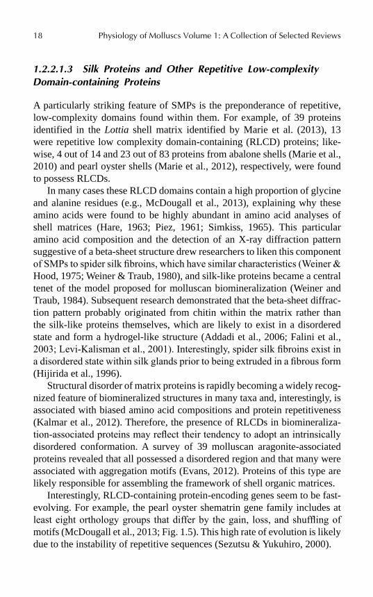
Interestingly, RLCD-containing protein-encoding genes seem to be fast-evolving. For example, the pearl oyster shematrin gene family includes at least eight orthology groups that differ by the gain, loss, and shuffling of motifs (McDougall et al., 2013; Fig. 1.5). This high rate of evolution is likely due to the instability of repetitive sequences (Sezutsu & Yukuhiro, 2000).
Developing Perspectives on Molluscan Shells, Part 1 19
FIGURE 1.5 Schematic representation of sequence motifs in shematrin genes from pearl oysters. Modified from McDougall et al. (2013).
1.2.2.1.4 Modularity
Many SMPs exhibit a modular architecture with each module (i.e., protein domain) having distinct functionality. The most well documented examples of modular SMPs correspond to nacrein and Lustrin A. Nacrein contains a CA domain that is interrupted by the insertion of a RLCD rich in Gly and Asn. CAs have previously been shown to be involved in biomineralization in various metazoans (Horne et al., 2002; Jackson et al., 2007; Miyamoto et al., 1996; Moya et al., 2008). This RLCD region has been proposed to regu-late the activity of the CA domain, acting as an inhibitor of the precipitation of calcium carbonate (Miyamoto et al., 2005). Lustrin A is the most complex multimodular SMP discovered so far and is characterized by numerous proline-, cysteine-, and GS-domains. The C-terminus domain of lustrin A exhibits high similarity with several protease inhibitors (Shen et al., 1997; Gaume et al., 2014). Although most SMPs do not exhibit sequence similarity with known proteins, many proteins contain, in addition to RLCDs, enzy-matic domains such as peroxidase, CA, tyrosinase, or glycosidase domains. For example, Lottia gigantea CA-2 contains Asp- and Glu-rich domains in its C-terminus (Marie et al., 2013).
1.2.2.2 CHITIN AND OTHER POLYSACCHARIDES
Currently, understanding of chitin and other polysaccharides and their func-tion in molluscan shells lag behind that of proteins. Proteins have been found
20 Physiology of Molluscs Volume 1: A Collection of Selected Reviews
in every type of molluscan shell analyzed so far, but whether chitin and/or other polysaccharides are present in all molluscan shells/sclerites is unclear. Furuhashi et al. (2009) provided a detailed review on the understanding of chitin and its role in molluscan shells. The few analyses of the polysac-charides in molluscan shells performed so far suggest that molluscs exhibit different sugar signatures (Marie et al., 2007 2009; Pavat et al., 2012). Chitin has been reliably identified in the shells of at least some bivalves, gastropods, and cephalopods but details on the structure and polymorphism (α- vs. β-chitin) are wanting. A number of different approaches have been used to detect chitin in molluscan shells, but these tests may also produce false positives or provide inaccurate pictures of chitin structure in the pres-ence of other molecules. For example, Calcofluor White binds to chitin as well as certain acidic proteins (Albani et al., 1999, 2000). Inferences with respect to chitin network structure may be inaccurate due to nonspecific binding of such stains to molecules other than chitin. Infrared spectroscopy has also been used to test for chitin presence but insoluble proteins may confound results from this approach. Furuhashi et al. (2009) advocate the use of fluorescence probes with chitin-binding proteins (e.g., GFP-tagged chitin binding protein) and infrared spectroscopy before and after treatment with chitinase more specific tools for detection of chitin than stains such as Calcofluor White. Using the latter approach, they demonstrated the pres-ence of both neutral polysaccharides and chitin in the cuticle of an unidenti-fied solenogaster, the shell plates and sclerites of the chiton Acanthopleura japonica, the shells of the bivalves Pinctada fucata and Atrina japonica, the gastropod Haliotis discus, and the cephalopod Nautilus sp.
Much of our knowledge on chitin in mollusc shells is thanks to transcrip-tomic and proteomic approaches. In an attempt to understand the molecular basis underlying shell formation, Aguilera (2014) analyzed the mantle tran-scriptome of eight bivalve and three gastropod species. This study found over-representation of proteins with polysaccharide-binding domains within the mantle transcriptomes. These include chitin-binding Periotrophin-A, chitin-binding domain, chitinases II, chitinase-insertion domain, polysaccha-ride deacetylase, and galactose-binding domain-like, among others. In addi-tion, Mann and Jackson (2014) described several C. nemoralis shell proteins that have high similarity with other molluscan shell-forming proteins. These include two chitin-binding domain-containing proteins. Further, they also found a protein with a chitin-binding Periotrophin-A domain and a chitinase in most of the sampled gastropods and bivalves. This emphasizes the impor-tance of chitin in shell formation in at least these taxa (Falini and Fermani, 2004; Weiss et al., 2006).
Developing Perspectives on Molluscan Shells, Part 1 21
1.2.2.3 LIPIDS
Lipids have long been known to be a minor constituent of the organic mole-cules found in mollusc shells (Wilbur & Simkiss, 1968). Cobabe and Pratt (1995) investigated the lipid content of the shells of Arca zebra, a hetero-trophic bivalve, Codakia orbicularis, a bivalve that hosts chemoautotrophic bacteria, as well as several fossil bivalves (1.4 myo). They found that lipids comprise between 300 and 700 ppm of the total shell weight and did not vary with trophic strategy. This shell–lipid suite is dominated by cholesterol, fatty acids (recovered as fatty acid methyl esters), ketones, phytadienes, and, in some cases, alkanes. Samata and Ogura (1997) showed that lipids are present in the nacreous layer of Pinctada fucata and Rousseau et al. (2006) showed that lipids are present in the nacreous layer of Pinctada margaritifera. More recently, Farre and Dauphin (2009) examined in detail the lipid composition of the pteriomorph bivalves Pinctada margaritifera and Pinna nobilis. The shells of these bivalves contain polar lipids (phospholipids), sterols (choles-terol), triglycerides (triolein), fatty acids (oleic acid), steroids (stearyl oleate), and waxes. In the nacreous layer, the most abundant lipid components are apolar waxes, free fatty acids, and very polar lipids. Steroids and sterols are represented in lesser amounts and there are only traces of triglycerides. The situation is similar in the prismatic layer except fatty acids are lacking whereas triolein is more abundant in the prismatic layer than the nacreous layer. The physiological function of lipids in molluscan biomineralization is unclear. Extracted phospholipids have the ability to bind calcium ions (Isa & Okazaki, 1987), so they may be involved in calcification.
1.3 NOVELTY IN MOLLUSCAN BIOMINERALIZATION
Perhaps the most fascinating aspect of molluscan biomineralization is the degree of novelty it encompasses at all levels of organization: from the genes and proteins controlling the process, to the diversity of microarchitectures represented, through to the myriad of structures generated. This highly evolvable system is reflected in some astonishing innovations within the molluscan phylum, such as image-forming aragonite lenses found in chiton-shell plates (Speiser et al., 2011, 2014) and the exquisite paper-thin brood chambers of argonauts, which are often mistaken to be true shells but are, in fact, secreted from specialized webs at the tips of the arms of the female and held on to via suckers (Finn, 2013). Novelty can also be generated by the loss or reduction of structures, as seen in many cephalopods and opisthobranchs.

22 Physiology of Molluscs Volume 1: A Collection of Selected Reviews
Structure aside, incredible diversity can also be seen in the coloration incor-porated into molluscan shells and in the minerals from which the structures are composed. Some of these phenomena are explored further below.
1.3.1 REDUCTION OR LOSS OF THE SHELL
Many gastropods, particularly terrestrial and marine slugs, have reduced, internalized, or completely lost the shell. Why would these animals give up the safety afforded to them by the shell? In the terrestrial realm, loss of the shell is likely an evolutionary response to calcium limitation (Solem, 1974, 1978; South, 1992). In the marine realm, this secondary reduction or loss of the shell usually coincides with the sequestration or production of toxic chemical compounds that make these animals noxious or toxic (Derby et al., 2007; Wägele & Klussmann-Kolb, 2005). For example, the shell-less nudi-branch Glossodoris quadricolor feeds on the sponge Latrunculia magnifica and sequesters from it the ichthyotoxic substance latrunculin B. It is thought that this compound then protects it from predation by fish (Mebs, 1985).
Interestingly, in some sea slugs that have secondarily lost shells, subdermal calcareous sclerites are produced (e.g., Brenzinger et al., 2013; Jörger et al., 2010; Schrödl & Neusser, 2010). Subepidermal, calcareous spicules are present in the meiofaunal gastropod taxa Acochlidia, Rhodope-morpha, and potentially Platyhedyle (Saccoglossa). Here, they are consid-ered as an adaptation to the interstitial habitat, probably serving to stabilize certain body parts during movements through the interstices (Jörger et al., 2008). Many larger sea slugs such as nudibranchs also have internalized calcareous spicules. Here, these structures are often spiny and are thought to serve a defensive purpose (Penney, 2006; Thompson, 1960). Whether the production of these spicules is governed by a similar process to that in the Aculifera is unknown.
Further, the shelled deep-sea scaly foot gastropod (Neomphalida) has a foot covered in sclerites, which are noncalcified but contain iron as pyrite and greigite (Chen et al., 2015; Warén et al., 2003). Little is known about the physiology underlying the formation of these structures.
1.3.2 COLORATION OF MOLLUSCAN SHELLS
The natural beauty of seashells never fails to attract the attentions of beach-goers, young and old alike, and has done so since early human history
Developing Perspectives on Molluscan Shells, Part 1 23
(d’Errico et al., 2005). Part of this attraction stems from the stunning array of shapes that molluscan shells exhibit, and part from the often bright or ornately patterned coloration that they possess. The role of coloration in molluscan shells is not well understood; in some cases, the patterning quite effectively camouflages the organism against their habitat; however, in many molluscs this is not the case. Given that many molluscs with colored shells do not have image-forming eyes, reproduce via broadcast spawning, or remain buried in sediment for the extent of the life of the organism, the extravagant patterns are unlikely to serve as a signal to conspecifics (Bauchau, 2001). The fundamental role of coloration has been hypothesized to be as a means to dispose of waste products of metabolism (Comfort, 1951), to increase shell strength (Cain, 1988), or as a means to provide positional information to the mantle (Bauchau, 2001); however, support for all three of these theories is lacking.
The mechanisms underlying the production of color in molluscan shells are diverse (Aguilera et al., 2014; Barnard & De Waal, 2006; Comfort, 1951; Hedegaard et al., 2006). Pigments can be found within the proteinaceous periostracum that covers the outer surface of the shell, and also within the calcified layers themselves (Budd et al., 2014; Needham, 1975). Numerous types of pigments have been identified from molluscan shells, including pyrroles (bilins and porphyrins), polyenes (including carotenoids), and mela-nins (Barnard & De Waal, 2006; Comfort, 1951; Hedegaard et al., 2006). In some cases, these pigments appear closely associated with protein shell components; however, other species do not appear to use protein-associated pigmentation mechanisms (Mann & Jackson, 2014). Some molluscs do not use pigments to create their coloration at all—they have evolved shell micro-structures which produce structural color, that is, color generated through the interference of reflected wavelengths of light from thin films. The most notable example of structural color in molluscs is mother-of-pearl; the archi-tecture of nacre tablets in species such as pearl oysters and abalone results in a stunning display of reflected colors (Rayleigh, 1923; Snow et al., 2004; Webster & Anderson, 1983) that is likely to be the byproduct of an architec-ture that has been optimized for shell strength. However, there are examples of structural color that have clearly evolved to serve a function in their own right, such as the striking iridescent blue lines found on the shell of the limpet Patella pellucida. In this species, the shell ultrastructure maximizes the intensity of blue reflection from the stripes, possibly to mimic the bright blue coloration of toxic nudibranchs found in the same habitat (Li et al., 2015).
Very little is known about how shell coloration and patterning is controlled at the molecular level. A number of studies have demonstrated that

24 Physiology of Molluscs Volume 1: A Collection of Selected Reviews
pigmentation in species displaying intraspecific variation follows Mendelian patterns of inheritance (Evans et al., 2009; Gantsevich et al., 2005; Liu et al., 2009; Luttikhuizen & Drent, 2008), indicating a genetic basis for pigmenta-tion in these molluscs. Evidence for a genetic basis also comes from studies on the juvenile abalone, Haliotis asinina. In this species, the expression of the sometsuke gene maps precisely with areas of red pigmentation on the shell, and the corresponding protein has been isolated from the shell itself (Jackson et al., 2006; Marie et al., 2010). Sometsuke has not been identified in any other molluscan shell proteome, indicating that it may be restricted to abalone.
It appears that the incredible diversity of coloration seen within molluscan shells is reflected in the complexity underlying it. The coloration can be generated by a diversity of pigments (or by no pigment at all!), can fulfill a broad range of functions, and is likely controlled by a number of different molecular processes. The lack of common principles indicates that shell coloration, like many aspects of biomineralization, likely evolved many times independently across the phylum.
1.3.3 CHITON SHELL EYES
In most chitons, the tegmentum is permeated by sensory structures called esthetes, which have a variety of sensory and possibly secretory functions (e.g., Eernisse & Reynolds, 1994; Speiser et al., 2011). In Schizochitonidae and Chitonidae esthetes may be capped with an ocellus that includes a lens (reviewed by Eernisse & Reynolds, 1994). Speiser et al. (2011) recently used electron probe X-ray microanalysis and X-ray diffraction to show that the chiton Acanthopleura granulata has shell eyes with the first aragonite lenses ever discovered. These eyes appear to be used to sense shadows produced by a would-be predator passing over the animal. Further, it appears that the eye structure results in two different refractive indices that are hypothesized to be optimal for function when the animal is submersed in water at high tide and exposed to air at low tide, respectively.
1.4 CONCLUSIONS AND OPEN QUESTIONS
1.4.1 MORE COMPARATIVE STUDIES NEEDED
Numerous recent studies have employed high throughput sequencing and proteomic approaches to improve our understanding of the process of

Developing Perspectives on Molluscan Shells, Part 1 25
biomineralization in molluscs. However, the vast majority of these studies have focused on economically important gastropods and bivalves. Currently, high quality genomes are available only from gastropods and bivalves (Simakov et al., 2013; Takeuchi et al., 2012; Zhang et al., 2012), although comparable data from cephalopods are forthcoming (Albertin et al., 2012). For obvious reasons, high quality genomic resources from other lineages of Mollusca would be highly beneficial toward understanding the evolution of the physiological mechanisms responsible for biomineralization.
Scaphopods are of particular interest because of their apparent close rela-tionship to gastropods and bivalves (Kocot et al., 2011; Smith et al., 2011; Vinther et al., 2012). Because gastropods and bivalves are economically and ecologically important and well-studied with respect to biomineralization, comparative work in Scaphopoda has important bearing on studies in these two groups. In particular, given the apparent differences in biomineraliza-tion between gastropods and bivalves (e.g., Jackson et al., 2010), data from Scaphopoda would help clarify if either gastropods or bivalves are derived with respect to biomineralization or if the process is as highly variable across Mollusca in general (as suspected). Some very detailed studies have addressed scaphopod development (Wanninger & Haszprunar, 2001, 2002, 2003), but little is known about their biomineralization and limited genomic resources are available (Kocot et al., 2011; Smith et al., 2011).
Although relatively hard to obtain (but see Wilson et al., 2009), Mono-placophora would be another very interesting group to study due to its antiquity (Haszprunar, 2008; Haszprunar & Ruthensteiner, 2013; Lindberg, 2009). Genome sequencing of the monoplacophoran Laevipilina antarctica is currently underway (M. Schrödl, personal communication).
Because Aculifera (Aplacophora + Polyplacophora) is sister to all other extant molluscs (Kocot et al., 2011; Smith et al., 2011), studies of this group would provide important evolutionary context for molluscan biomineraliza-tion. Although many aplacophorans live in deep and/or polar habitats, some species are relatively easily accessible and have been successfully spawned in the laboratory (Okusu, 2002; Todt & Wanninger, 2010). In particular, solenogaster aplacophorans produce a phenomenal array of diverse sclerite types (reviewed by García-Álvarez & Salvini-Plawen, 2007). How these structures are achieved is a mystery, but their morphology is likely regulated by the same type of organic matrix found in shelled molluscs. Deeper and wider taxon sampling in comparative studies of biomineralization will help to understand the essential requirements for the production of mineralized structures in the Mollusca.
26 Physiology of Molluscs Volume 1: A Collection of Selected Reviews
1.4.2 ARE SHELLS AND SCLERITES PRODUCED BY DIFFERENT MOLLUSCAN LINEAGES HOMOLOGOUS?
Although aculiferan (chiton + aplacophoran) sclerites, chiton valves, and conchiferan shells are all extracellular calcareous secretions of the mantle, structural, and developmental differences suggest that these features are not strictly homologous (Eernisse & Reynolds, 1994; Furuhashi et al., 2009; Haas, 1981; Scheltema, 1993; reviewed by Kocot, 2013). Specifically, the lack of a true periostracum, periostracal groove, and a differentiated larval shell-secreting epithelium (shell gland) in chitons distinguishes their shell structure and formation from that of the conchiferans. Further, develop-mental studies have shown that chiton shells are secreted by postrochal (2d) cells (Heath, 1899; Henry et al., 2004) during development. These cells (Conklin, 1897; Lillie, 1895), but sometimes also other micromere lineages (2a, 2b, 2c, and sometimes 3c), form the conchiferan shell gland (Damen & Dictus, 1994; Render, 1997). Interestingly, chiton sclerite-secreting cells arise from postrochal (2a, 2c, 3c, and 3d) as well as pretrochal cells (1a and 1d), suggesting that chiton sclerites are not strictly homologous to chiton or conchiferan shells (no cell lineage studies have been conducted in aplacoph-orans). Hence, the gene regulatory networks and physiological mechanisms that produce these structures may differ significantly.
There is also some question regarding the homology of shell layers within the Conchifera. The debate centers on nacre, which is found in bivalve, gastropod, cephalopod, and monoplacophoran lineages (Chateigner et al., 2000). Although generally similar, there are fundamental differences in mineralogy between the taxa; bivalves and monoplacophorans possess “sheet nacre’ (tablets arranged in a brick-like pattern) with alignment of all three axes of the aragonite tablets (bivalves) or a randomly oriented a axis (monoplacophorans), gastropods and cephalopods possess “columnar nacre” (tablets stacked upon each other), with the c-axis of the tablet perpen-dicular to the surface of the shell and the a and b axes aligned within a stack (gastropods) or alignment of all three axes (cephalopods) (Chateigner et al., 2000; Meldrum & Cölfen, 2008). These differences, and the strikingly different nacre building gene sets that underlie them (Jackson et al., 2010), call in to question the assumption of homology of nacre in conchiferan taxa and has bearing on our understanding of the evolution of biomineralization in molluscs. Whether the other shell layers are similarly divergent between molluscan classes remains to be investigated.

Developing Perspectives on Molluscan Shells, Part 1 27
1.4.3 WHAT DOES IT ALL MEAN?
Even with high quality genomic resources spanning the diversity of Mollusca, more data does not mean more understanding. However, genomic resources will continue to provide profound insight into physiological processes such as biomineralization. There is a growing need for implementation of advanced analytical techniques looking at gene family evolution (Aguilera, 2014; De Bie et al., 2006; Domazet-Lošo et al., 2007) and gene networks (Shannon et al., 2003; Smoot et al., 2011). Further, more “traditional” techniques with a much longer history of use in the field of physiology (see Simkiss Chapter 2 of this volume) should not be forgotten in the “-omics” era. Such compara-tive studies will undoubtedly continue to improve understanding of the complex physiological process of molluscan biomineralization.
KEYWORDS
• biomineralization
• shell
• periostracum
• mantle
• silk
• RLCD
REFERENCES
Acosta-Salmón, H.; Southgate, P. C. Wound Healing after Excision of Mantle Tissue from the Akoya Pearl Oyster, Pinctada fucata. Comp. Biochem. Physiol., A: Mol. Integr. Physiol. 2006, 143(2), 264–268.
Addadi, L.; Joester, D.; Nudelman, F.; Weiner, S. Mollusk Shell Formation: A Source of New Concepts for Understanding Biomineralization Processes. Chem.—Eur. J. 2006, 12(4), 980–987.
Aguilera, F.; McDougall, C.; Degnan, B. M. Evolution of the Tyrosinase Gene Family in Bivalve Molluscs: Independent Expansion of the Mantle Gene Repertoire. Acta Biomater. 2014, 10(9), 3855–3865.
Aguilera F. Investigation of Gene Family Evolution and the Molecular Basis of Shell Forma-tion in Molluscs. Ph.D. Thesis, The University of Queensland: Brisbane, Australia, 2014.
28 Physiology of Molluscs Volume 1: A Collection of Selected Reviews
Albani, J. R.; Sillen, A.; Coddeville, B.; Plancke, Y. D.; Engelborghs, Y. Dynamics of Carbo-hydrate Residues of α 1-Acid Glycoprotein (orosomucoid) followed by Red-edge Excita-tion Spectra and Emission Anisotropy Studies of Calcofluor White. Carbohydr. Res. 1999, 322(1), 87–94.
Albani, J. R.; Sillen, A.; Plancke, Y. D.; Coddeville, B.; Engelborghs, Y. Interaction between Carbohydrate Residues of α 1-acid Glycoprotein (Orosomucoid) and Saturating Concentra-tions of Calcofluor White. A Fluorescence Study. Carbohydr. Res. 2000, 327 (3), 333–340.
Albertin, C. B.; Bonnaud, L.; Brown, C. T.; Crookes-Goodson, W. J.; da Fonseca, R. R.; Di Cristo, C.; Dilkes, B. P.; Edsinger-Gonzales, E.; Freeman, Jr., R. M.; Hanlon, R. T. Cepha-lopod Genomics: A Plan of Strategies and Organization. Stand. Genomic Sci. 2012, 7(1), 175.
Artigaud, S.; Thorne, M. A.; Richard, J.; Lavaud, R.; Jean, F.; Flye-Sainte-Marie, J.; Peck, L. S.; Pichereau, V.; Clark, M. S. Deep Sequencing of the Mantle Transcriptome of the Great Scallop Pecten maximus. Mar. Genomics 2014, 15, 3–4.
Bai, Z.; Yin, Y.; Hu, S.; Wang, G.; Zhang, X.; Li, J. Identification of Genes Potentially Involved in Pearl Formation by Expressed Sequence Tag Analysis of Mantle from Fresh-water Pearl Mussel (Hyriopsis cumingii Lea). J. Shellfish Res. 2010, 29(2), 527–534.
Bai, Z.; Zheng, H.; Lin, J.; Wang, G.; Li, J. Comparative Analysis of the Transcriptome in Tissues Secreting Purple and White Nacre in the Pearl Mussel Hyriopsis cumingii. PLoS ONE 2013, 8(1), e53617.
Bandel, K. Cephalopod Shell Structure and General Mechanisms of Shell Formation. In Skelet. Biominer. Patterns Process. Evol. Trends; 1990; pp 97–115. http://onlinelibrary.wiley.com/doi/10.1029/SC005p0097/pdf.
Barnard, W.; De Waal, D. Raman Investigation of Pigmentary Molecules in the Molluscan Biogenic Matrix. J. Raman Spectrosc. 2006, 37, 342–352.
Bauchau, V. Developmental Stability as the Primary Function of the Pigmentation Patterns in Bivalve Shells. Belg. J. Zool. 2001, 131(Suppl. 2), 23–28.
Beedham, G. E.; Trueman, E. R. The Cuticle of the Aplacophora and its Evolutionary Signifi-cance in the Mollusca. J. Zool. 1968, 154(4), 443–451.
Bielefeld, U.; Becker, W. Embryonic Development of the Shell in Biomphalaria glabrata (Say). Int. J. Dev. Biol. 1991, 35, 121–131.
Birchall, J. D.; Thomas, N. L. On the Architecture and Function of Cuttlefish Bone. J. Mater. Sci. 1983, 18(7), 2081–2086.
Bøggild, O. B. The Shell Structure of the Molluscs D. Kgl. Danske Vidensk. Selsk. Skrifter. Naturvidensk. og Math 1930, 9, 230–326.
Brenzinger, B.; Padula, V.; Schrödl, M. Insemination by a Kiss? Interactive 3D-micro-anatomy, Biology and Systematics of the Mesopsammic cephalaspidean Sea Slug Pluscula cuica Marcus, 1953 from Brazil (Gastropoda: Euopisthobranchia: Philinoglossidae). Org. Divers. Evol. 2013, 13(1), 33–54.
Budd, A.; McDougall, C.; Green, K.; Degnan, B. M. Control of Shell Pigmentation by Secre-tory Tubules in the Abalone Mantle. Front. Zool. 2014, 11, 62.
Budelmann, B. U.; Riese, U.; Bleckmann, H. Structure, Function, Biological Significance of the Cuttlefish “lateral lines.” In 1st International Symposium on the Cuttlefish Sepia; Boucaud-Camou, E., Ed.; Centre dePublications de l’Universite de Caen: Caen 1991; pp 201–209.
Cain, A. J. The Scoring of Polymorphic Colour and Pattern Variation and its Genetic Basis in Molluscan Shells. Malacologia 1988, 28(1–2), 1–15.
Developing Perspectives on Molluscan Shells, Part 1 29
Carefoot, T. H.; Donovan, D. A. Functional Significance of Varices in the Muricid Gastropod Ceratostoma foliatum. Biol. Bull. 1995, 189(1), 59–68.
Carriker, M. R. Prismatic Shell Formation in Continuously Isolated (Mytilus edulis) and Periodically Exposed (Crassostrea virginica) extrapallial Spaces: Explicable by the Same Concept. Am. Malacol. Bull. 1992, 9, 193–197.
Carter, J. G.; Hall, R. M. Polyplacophora, Scaphopoda, Archaeogastropoda, and Paragas-tropoda (Mollusca). In Skeletal Biomineralization: Patterns, Processes and Evolutionary Trends; Carter, J. G., Ed.; Van Nostrand Reinhold, New York, 1990; Vol. 2 Atlas and Index, pp 29–31.
Carter, J. G.; Clark, G. R. Classification and Phylogenetic Significance of Molluscan Shell Microstructure. In Molluscs, Notes for a Short Course; Bottjer, D. J.; Hickman, C. S.; Ward, P. D.; Broadhead, T. W., Eds.; University of Tennessee, Department of Geological Sciences Studies in Geology, 1985.
Cather, J. N. Cellular Interactions in the Development of the Shell Gland of the Gastropod, Ilyanassa. J. Exp. Zool. 1967, 166, 205–223.
Chateigner, D.; Hedegaard, C.; Wenk, H. Mollusc Shell Microstructures and Crystallographic Textures. J. Struct. Geol. 2000, 22(11–12), 1723–1735.
Checa, A. A New Model for periostracum and Shell formation in Unionidae (Bivalvia, Mollusca). Tissue Cell 2000, 32(5), 405–416.
Checa, A. G.; Sánchez-Navas, A.; Rodríguez-Navarro, A. Crystal Growth in the Foliated Aragonite of Monoplacophorans (Mollusca). Cryst. Growth Des. 2009, 9(10), 4574–4580.
Chen, C.; Copley, J. T.; Linse, K.; Rogers, A. D.; Sigwart, J. How the Mollusc got its Scales: Convergent Evolution of the Molluscan Scleritome. Biol. J. Linn. Soc. 2015, 114(4), 949–954.
Clark, M.; Thorne, M.; Vieira, F.; Cardoso, J.; Power, D.; Peck, L. Insights into Shell Deposi-tion in the Antarctic Bivalve Laternula elliptica: Gene Discovery in the Mantle Transcrip-tome using 454 Pyrosequencing. BMC Genomics 2010, 11(1), 362.
Cobabe, E. A.; Pratt, L. M. Molecular and Isotopic Compositions of Lipids in Bivalve Shells: A New Prospect for Molecular Paleontology. Geochim. Cosmochim. Acta 1995, 59(1), 87–95.
Comfort, A. The Pigmentation of Molluscan Shells. Biol. Rev. 1951, 26(3), 285–301.Conklin, E. G. The Embryology of Crepidula. J. Morphol. 1897, 13, 1–226.Crenshaw, M. A.; Ristedt, H. The Histochemical Localization of Reactive Groups in Septal
Nacre from Nautilus pompilius L. In The Mechanisms of Mineralization in the Inverte-brates and Plants; 1976; pp 355–367.
Cruz, R.; Weissmüller, G.; Farina, M. Microstructure of Monoplacophora (Mollusca) Shell Examined by Low-voltage Field Emission Scanning Electron and Atomic Force Micros-copy. Scanning 2003, 25(1), 12–18.
Damen, P.; Dictus, W. J. A. G. Cell Lineage of the Prototroch of Patella vulgata (Gastropoda, Mollusca). Dev. Biol. 1994, 162(2), 364–383.
Dauphin, Y.; Denis, A. Structure and Composition of the Aragonitic Crossed Lamellar Layers in Six Species of Bivalvia and Gastropoda. Comp. Biochem. Physiol., A. Mol. Integr. Physiol. 2000, 126(3), 367–377.
De Bie, T.; Cristianini, N.; Demuth, J. P.; Hahn, M. W. CAFE: A Computational Tool for the Study of Fene Family Evolution. Bioinformatics 2006, 22(10), 1269–1271.
d’Errico, F.; Henshilwood, C.; Vanhaeren, M.; van Niekerk, K. Nassarius kraussianus Shell Beads from Blombos Cave: Evidence for Symbolic Behaviour in the Middle Stone Age. J. Hum. Evol. 2005, 48(1), 3–24.
30 Physiology of Molluscs Volume 1: A Collection of Selected Reviews
de Paula, S. M.; Silveira, M. Studies on Molluscan Shells: Contributions from Microscopic and Analytical Methods. Micron 2009, 40(7), 669–690.
Derby, C. D.; Kicklighter, C. E.; Johnson, P. M.; Zhang, X. Chemical Composition of Inks of Diverse Marine Molluscs Suggests Convergent Chemical Defenses. J. Chem. Ecol. 2007, 33(5), 1105–1113.
Domazet-Lošo, T.; Brajković, J.; Tautz, D. A Phylostratigraphy Approach to Uncover the Genomic History of Major Adaptations in Metazoan Lineages. Trends Genet. 2007, 23(11), 533–539.
Eernisse, D. J.; Reynolds, P. D. Polyplacophora. In Microscopic Anatomy of Invertebrates; Harrison, F. W.; Kohn, A. J., Eds.; Wiley-Liss: New York, 1994; Vol. 5, pp 55–110.
Ehrlich, H. Molluscs Spicules. Biol. Mater. Mar. Orig. 2010, 211–242.Ehrlich, H. Chitin and Collagen as Universal and Alternative Templates in Biomineralization.
Int. Geol. Rev. 2010, 52(7–8), 661–699.Eichhorn, S. J.; Scurr, D. J.; Mummery, P. M.; Golshan, M.; Thompson, S. P.; Cernik, R.
J. The Role of Residual Stress in the Fracture Properties of a Natural Ceramic. J. Mater. Chem. 2005, 15(9), 947–952.
Erben, H. K.; Flajs, G.; Siehl, A. Über die Schalenstruktur von Monoplacophoren. Verlag der Akademie der Wissenschaften und der Literatur; in Kommission bei F. Steiner, Wiesbaden, 1968, 1.
Evans, J. S. “Tuning in” to Mollusk Shell Nacre- and Prismatic-associated Protein Terminal Sequences. Implications for Biomineralization and the Construction of High Performance Inorganic−Organic Composites. Chem. Rev. 2008, 108(11), 4455–4462.
Evans, S.; Camara, M.; Langdon, C. Heritability of Shell Pigmentation in the Pacific Oyster, Crassostrea gigas. Aquaculture 2009, 286(3), 211–216.
Evans, J. S. Aragonite-associated Biomineralization Proteins are Disordered and contain Interactive Motifs. Bioinformatics 2012, 28(24), 3182–3185.
Falini, G.; Albeck, S.; Weiner, S.; Addadi, L. Control of Aragonite or Calcite Polymorphism by Mollusk Shell Macromolecules. Science 1996, 271(5245), 67–69.
Falini, G.; Weiner, S.; Addadi, L. Chitin–Silk Fibroin Interactions: Relevance to Calcium Carbonate Formation in Invertebrates. Calcif. Tissue Int. 2003, 72(5), 548–554.
Falini, G.; Fermani, S. Chitin Mineralization. Tissue Eng. 2004, 10(1–2), 1–6.Fang, Z.; Feng, Q.; Chi, Y.; Xie, L.; Zhang, R. Investigation of Cell Proliferation and Differ-
entiation in the Mantle of Pinctada fucata (Bivalve, Mollusca). Mar. Biol. 2008, 153(4), 745–754.
Fang, D.; Xu, G.; Hu, Y.; Pan, C.; Xie, L.; Zhang, R. Identification of Genes Directly Involved in Shell formation and their Functions in Pearl Oyster, Pinctada fucata. PLoS ONE 2011, 6(7), e21860.
Farach-Carson, M. C.; Carson, D. D.; Collier, J. L.; Lennarz, W. J.; Park, H. R.; Wright, G. C. A Calcium-binding, Asparagine-linked Oligosaccharide is Involved in Skeleton formation in the Sea Urchin Embryo. J. Cell Biol. 1989, 109(3), 1289–1299.
Farre, B.; Dauphin, Y. Lipids from the Nacreous and Prismatic Layers of Two Pteriomorpha Mollusc Shells. Comp. Biochem. Physiol., B: Biochem. Mol. Biol. 2009, 152(2), 103–109.
Finn, J. K. Taxonomy and Biology of the Argonauts (Cephalopoda: Argonautidae) with Particular Reference to Australian Material. Molluscan Res. 2013, 33(3), 143–222.
Fishlyn, D. A.; Phillips, D. W. Chemical Camouflaging and Behavioral Defenses against a Predatory Seastar by Three Species of Gastropods from the Surfgrass Phyllospadix Community. Biol. Bull. 1980, 158(1), 34–48.
Developing Perspectives on Molluscan Shells, Part 1 31
Fleury, C.; Marin, F.; Marie, B.; Luquet, G.; Thomas, J.; Josse, C., Serpentini, A.; Lebel, J. M. Shell Repair Process in the Green Ormer Haliotis tuberculata: A Histological and Micro-structural Study. Tissue Cell 2008, 40(3), 207–218.
Freer, A.; Bridgett, S.; Jiang, J.; Cusack, M. Biomineral Proteins from Mytilus edulis Mantle Tissue Transcriptome. Mar. Biotechnol. 2014, 16(1), 34–45.
Fu, G.; Valiyaveettil, S.; Wopenka, B.; Morse, D. E. CaCO3 Biomineralization: Acidic 8-kDa Proteins isolated from Aragonitic Abalone Shell Nacre can Specifically Modify Calcite Crystal Morphology. Biomacromolecules 2005, 6(3), 1289–1298.
Furuhashi, T.; Schwarzinger, C.; Miksik, I.; Smrz, M.; Beran, A. Molluscan Shell Evolution with Review of Shell Calcification Hypothesis. Comp. Biochem. Physiol.: B Biochem. Mol. Biol. 2009, 154(3), 351–371.
Furuhashi, T.; Miksik, I.; Smrz, M.; Germann, B.; Nebija, D.; Lachmann, B.; Noe, C. Comparison of Aragonitic Molluscan Shell Proteins. Comp. Biochem. Phys., B 2010, 155(2), 195–200.
Gantsevich, M.; Tyunnikova, A.; Malakhov, V. The Genetics of Shell Pigmentation of the Mediterranean Mussel Mytilus galloprovincialis Lamarck, 1819 (Bivalvia, Mytilida). Dokl. Biol. Sci. 2005, 404(1), 370–371.
García-Álvarez, O.; Salvini-Plawen, L. Species and Diagnosis of the Families and Genera of Solenogastres (Mollusca). Iberus 2007, 25(2), 73–143.
Ganss, B.; Hoffmann, W. Calcium Binding to Sialic Acids and its Effect on the Conformation of Ependymins. Eur. J. Biochem. 1993, 217(1), 275–280.
Gardner, L.; Mills, D.; Wiegand, A.; Leavesley, D.; Elizur, A. Spatial Analysis of Biominer-alization Associated Gene Expression from the Mantle Organ of the Pearl Oyster Pinctada maxima. BMC Genomics 2011, 12(1), 455.
Gaume B.; Denis, F.; Van Wormhoudt, A.; Huchette, S.; and Jackson, D. J. Characterization and Expression of the Biomineralising Gene Lustrin A During Shell Formation of the Euro-pean Abalone Haliotis tuberculata. Comp. Biochem. Physiol., B 2014, 169, 1–8.
Gotliv, B.-A.; Kessler, N.; Sumerel, J. L.; Morse, D. E.; Tuross, N.; Addadi, L.; Weiner, S. Asprich: A Novel Aspartic Acid-rich Protein Family from the Prismatic Shell Matrix of the Bivalve Atrina rigida. ChemBioChem 2005, 6(2), 304–314.
González, V. L.; Andrade, S. C.; Bieler, R.; Collins, T. M.; Dunn, C. W.; Mikkelsen, P. M.; Taylor, J. D.; Giribet, G. A Phylogenetic Backbone for Bivalvia: An RNA-Seq Approach. Proc. R. Soc. B: Biol. Sci. 2015, 282(1801), 20142332.
Haas, W. Untersuchungen über die Mikro- und Ultrastruktur der Polyplacophorenschale. Biomineralization 1972, 5, 1–52.
Haas, W. Observations on the Shell and Mantle of the Placophora. In The Mechanisms of Mineralization in the Invertebretaes and Plants; University of South Carolina Press: Columbia, 1976; pp 389–402.
Haas, W. Evolution of Calcareous Hardparts in Primitive Molluscs. Malacologia 1981, 21(1–2), 403–418.
Hare, P. E. Amino Acids in the Proteins from Aragonite and Calcite in the Shells of Mytilus californianus. Science 1963, 139(3551), 216–217.
Hashimshony, T.; Wagner, F.; Sher, N.; Yanai, I. CEL-Seq: Single-cell RNA-Seq by Multi-plexed Linear Amplification. Cell. Rep. 2012, 2(3), 666–673.
Haszprunar, G. Monoplacophora (Tryblidia). In Phylogeny and Evolution of the Mollusca; Ponder, W. F.; Lindberg, D. L., Eds.; University of California Press: Berkeley and Los Angeles, 2008; pp 97–104.
32 Physiology of Molluscs Volume 1: A Collection of Selected Reviews
Haszprunar, G.; Schander, C.; Halanych, K. Relationships of Higher Molluscan Taxa. In Phylogeny and Evolution of the Mollusca; Ponder, W. F.; Lindberg, D. L., Eds.; University of California Press: Berkeley and Los Angeles, 2008; pp 19–32.
Haszprunar, G.; Ruthensteiner, B. Monoplacophora (Tryblidia)-some Unanswered Ques-tions. Am. Malacol. Bull. 2013, 31(1), 189–194.
Heath, H. Development of Ischnochiton. Zool. Jahrbuecher Abt. Fuer Anat. Ontog. Tiere 1899, 12, 567–656.
Hebert, A. Reproductive Behavior and Anatomy of Three Central Californian Scaphopods. Master’s Thesis, California State University: Hayward, 1986.
Hedegaard, C.; Wenk, H. Microstructure and Texture Patterns of Mollusc Shells. J. Molluscan Stud. 1998, 64, 133–136.
Hedegaard, C.; Bardeau, J. -F.; Chateigner, D. Molluscan Shell Pigments: An in situ Reso-nance Raman Study. J. Molluscan Stud. 2006, 72(2), 157–162.
Henry, J. Q.; Okusu, A.; Martindale, M. Q. The Cell Lineage of the Polyplacophoran, Chae-topleura apiculata: Variation in the Spiralian Program and Implications for Molluscan Evolution. Dev. Biol. 2004, 272(1), 145–160.
Hijirida, D. H.; Do, K. G.; Michal, C.; Wong, S.; Zax, D.; Jelinski, L. W. 13C NMR of Nephila clavipes Major Ampullate Silk Gland. Biophys. J. 1996, 71(6), 3442–3447.
Hohagen J.; Jackson., D. J. An Ancient Process in a Modern Molluscs: Early Development of the Shell in Lymnaea stagnalis. BMC Dev. Biol. 2013, 13, 27.
Horne, F.; Tarsitano, S.; Lavalli, K. L. Carbonic Anhydrase in Mineralization of the Crayfish Cuticle. Crustaceana 2002, 75(9), 1067–1081.
Huang, X. D.; Zhao, M.; Liu, W. G.; Guan, Y. Y.; Shi, Y.; Wang, Q.; Wu, S. Z.; He, M. X. Gigabase-scale Transcriptome Analysis on Four Species of Pearl Oysters. Mar. Biotechnol. 2013, 15(3), 253–264.
Hüning, A. K.; Melzner, F.; Thomsen, J.; Gutowska, M. A.; Krämer, L.; Frickenhaus, S.; Rosenstiel, P.; Pörtner, H.-O.; Philipp, E. E.; Lucassen, M. Impacts of Seawater Acidifica-tion on Mantle Gene Expression Patterns of the Baltic Sea Blue Mussel: Implications for Shell Formation and Energy Metabolism. Mar. Biol. 2013, 160(8), 1845–1861.
Hunt, S.; El Sherief, A. A Periodic Structure in the “Pen” Chitin of the Squid Loligo vulgaris. Tissue Cell 1990, 22(2), 191–197.
Isa, Y.; Okazaki, M. Some Observations on the Ca2+-binding Phospholipid from Scleractinian Coral Skeletons. Comp. Biochem. Physiol., B: Comp. Biochem. 1987, 87(3), 507–512.
Jackson, D. J.; McDougall, C.; Green, K.; Simpson, F.; Wörheide, G.; Degnan, B. M. A Rapidly Evolving Secretome Builds and Patterns a Sea Shell. BMC Biol. 2006, 4, 40.
Jackson, D. J.; Wörheide, G.; Degnan, B. M. Dynamic Expression of Ancient and Novel Molluscan Shell Genes during Ecological Transitions. BMC Evol. Biol. 2007, 7(1), 160.
Jackson, D. J.; McDougall, C.; Woodcroft, B.; Moase, P.; Rose, R. A.; Kube, M.; Reinhardt, R.; Rokhsar, D. S.; Montagnani, C.; Joubert, C.; Piquemal, D.; Degnan, B. M. Parallel Evolution of Nacre Building Gene Sets in Molluscs. Mol. Biol. Evol. 2010, 27(3), 591–608.
Jörger, K. M.; Neusser, T. P.; Haszprunar, G.; Schrödl, M. Undersized and Underestimated: 3D Visualization of the Mediterranean Interstitial Acochlidian Gastropod Pontohedyle milaschewitchii (Kowalevsky, 1901). Org. Divers. Evol. 2008, 8(3), 194–214.
Jörger, K. M.; Stöger, I.; Kano, Y.; Fukuda, H.; Knebelsberger, T.; Schrödl, M. On the Origin of Acochlidia and Other Enigmatic Euthyneuran Gastropods, with Implications for the Systematics of Heterobranchia. BMC Evol. Biol. 2010, 10(1), 323.
Developing Perspectives on Molluscan Shells, Part 1 33
Jolly, C.; Berland, S.; Milet, C.; Borzeix, S.; Lopez, E.; Doumenc, D. Zona Localization of Shell Matrix Proteins in Mantle of Haliotis tuberculata (Mollusca, Gastropoda). Mar. Biotechnol. 2004, 6(6), 541–551.
Jones, D. B.; Jerry, D. R.; Khatkar, M. S.; Moser, G.; Raadsma, H. W.; Taylor, J. J.; Zenger, K. R. Determining Genetic Contributions to Host Oyster Shell Growth: Quantitative Trait Loci and Genetic Association Analysis for the Silver-lipped Pearl Oyster, Pinctada maxima. Aquaculture 2014, 434, 367–375.
Joubert, C.; Piquemal, D.; Marie, B.; Manchon, L.; Pierrat, F.; Zanella-Cléon, I.; Cochennec-Laureau, N.; Gueguen, Y.; Montagnani, C. Transcriptome and Proteome Analysis of Pinctada margaritifera Calcifying Mantle and Shell: Focus on Biomineralization. BMC Genomics 2010, 11(1), 613.
Kaas, P.; Van Belle, R. A. Monograph of Living Chitons. Vol. 1. Order Neoloricata: Lepido-pleurina, Brill: Leiden, 1985.
Kalmar, L.; Homola, D.; Varga, G.; Tompa, P. Structural Disorder in Proteins Brings Order to Crystal Growth in Biomineralization. Bone 2012, 51(3), 528–534.
Kapur, S. P.; Gibson, M. A. A Histological Study of the Development of the Mantle-edge and Shell in the Freshwater Gastropod, Helisoma duryi eudiscus (Pilsbry). Can. J. Zool. 1967,45(6), 1169–1181.
Kim, I. W.; Morse, D. E.; Evans, J. S. Molecular Characterization of the 30-AA N-terminal Mineral Interaction Domain of the Biomineralization Protein AP7. Langmuir 2004, 20(26), 11664–11673.
Kim, I. W.; Collino, S.; Morse, D. E.; Evans, J. S. A Crystal Modulating Protein from Molluscan Nacre that Limits the growth of Calcite in vitro. Cryst. Growth Des. 2006, 6, 1078.
Kingsley, R.; Froelich, J.; Marks, C.; Spicer, L.; Todt, C. Formation and Morphology of Epidermal Sclerites from a Deep-sea Hydrothermal Vent Solenogaster Helicoradomenia sp. (Solenogastres, Mollusca). Zoomorphology 2012, 132(1), 1–9.
Kinoshita, S.; Wang, N.; Inoue, H.; Maeyama, K.; Okamoto, K.; Nagai, K.; Kondo, H.; Hirono, I.; Asakawa, S.; Watabe, S. Deep Sequencing of ESTs from Nacreous and Pris-matic Layer Producing Tissues and a Screen for Novel Shell Formation-related Genes in the Pearl Oyster. PLoS ONE 2011, 6(6), e21238.
Kniprath, E. Ontogeny of Shell Field in Lymnaea stagnalis. Wilhelm Rouxs Arch. Dev. Biol. 1977, 181(1), 11–30.
Kniprath, E. Growth of the Shell-field in Mytilus (Bivalvia). Zool. Scr. 1978, 7, 119–120.Kniprath, E. Larval Development of the Shell and the Shell Gland in Mytilus (Bivalvia).
Wilhelm Rouxs Arch. Dev. Biol. 1980, 188(3), 201–204.Kniprath, E. Ontogeny of the Molluscan Shell Field: A Review. Zool. Scr. 1981, 10(1), 61–79.Kobayashi, I.; Samata, T. Bivalve Shell Structure and Organic Matrix. Mater. Sci. Eng. C.
2006, 26(4), 692–698.Kocot, K. M.; Cannon, J. T.; Todt, C.; Citarella, M. R.; Kohn, A. B.; Meyer, A.; Santos, S. R.;
Schander, C.; Moroz, L. L.; Lieb, B. Phylogenomics Reveals Deep Molluscan Relation-ships. Nature 2011, 477(7365), 452–456.
Kocot, K. M. Recent Advances and Unanswered Questions in Deep Molluscan Phyloge-netics. Am. Malacol. Bull. 2013, 31(1), 1–14.
Kong, Y.; Jing, G.; Yan, Z.; Li, C.; Gong, N.; Zhu, F.; Li, D.; Zhang, Y.; Zheng, G.; Wang, H.; Xie, L.; Zhang, R. Cloning and Characterization of Prisilkin-39, a Novel Matrix Protein Serving a Dual Role in the Prismatic Layer Formation from the Oyster Pinctada fucata. J. Biol. Chem. 2009, 284(16), 10841–10854.
34 Physiology of Molluscs Volume 1: A Collection of Selected Reviews
Kröger, B.; Vinther, J.; Fuchs, D. Cephalopod Origin and Evolution: A Congruent Picture Emerging from Fossils, Development and Molecules. BioEssays 2011, 8(33), 602–613.
Levi-Kalisman, Y.; Falini, G.; Addadi, L.; Weiner, S. Structure of the Nacreous Organic Matrix of a Bivalve Mollusk Shell Examined in the Hydrated State Using Cryo-TEM. J. Struct. Biol. 2001, 135(1), 8–17.
Li, X.; Nardi, P. Micro/nanomechanical Characterization of a Natural Nanocomposite Mate-rial—the Shell of Pectinidae. Nanotechnology 2004, 15(1), 211.
Li, X.; Xu, Z. H.; Wang, R. In situ Observation of Nanograin Rotation and Deformation in Nacre. Nano Lett. 2006, 6(10), 2301–2304.
Li, L.; Kolle, S.; Weaver, J.; Ortiz, C.; Aizenberg, J.; Kolle, M. A Highly Conspicuous Miner-alized Composite Photonic Architecture in the Translucent Shell of the Blue-rayed Limpet. Nat. Commun. 2015, 6, 6322.
Liao, Z.; Bao, L. F.; Fan, M. H.; Gao, P.; Wang, X. X.; Qin, C. L.; Li, X. M. In-depth Proteomic Analysis of Nacre, Prism, and Myostracum of Mytilus Shell. J. Proteomics 2015, 122, 26–40.
Lillie, F. R. The Development of the Unionidae. J. Morphol. 1895, 10, 1–100.Lindberg, D. R. Monoplacophorans and the Origin and Relationships of Mollusks. Evol.
Educ. Outreach 2009, 2(2), 191–203.Liu, X.; Wu, F.; Zhao, H.; Zhang, G.; Guo, X. A Novel Shell Color Variant of the Pacific
abalone Haliotis discus hannai Ino Subject to Genetic Control and Dietary Influence. J. Shellfish Res. 2009, 28(2), 419–424.
Liu, X.; Dong, S.; Jin, C.; Bai, Z.; Wang, G.; Li, J. Silkmapin of Hyriopsis cumingii, a Novel Silk-like Shell Matrix Protein Involved in Nacre Formation. Gene 2015, 555(2), 217–222.
Luttikhuizen, P.; Drent, J. Inheritance of Predominantly Hidden Shell Colours in Macoma balthica (L.) (Bivalvia: Tellinidae). J. Molluscan Stud. 2008, 74(4), 363–371.
Mann, K.; Siedler, F.; Treccani, L.; Heinemann, F.; Fritz, M. Perlinhibin, a Cysteine-, histi-dine-, and Arginine-rich miniprotein from abalone (Haliotis laevigata) Nacre, Inhibits In Vitro Calcium Carbonate Crystallization. Biophys. J. 2007, 93(4), 1246–1254.
Mann, K.; Edsinger-Gonzales, E.; Mann, M. In-depth Proteomic Analysis of a Mollusc Shell: Acid-soluble and Acid-insoluble Matrix of the Limpet Lottia gigantea. Proteome Sci. 2012, 10(1), 28.
Mann K.; Edsinger-Gonzales, E. The Lottia gigantea Shell Proteome: Re-analysis Including MaxQuant iBAQ Quantitation and Phosphoproteome Analysis. Proteome Sci. 2014, 12, 28.
Mann, K.; Jackson, D. J. Characterization of the Pigmented Shell-forming Proteome of the Common Grove Snail Cepaea nemoralis. BMC Genomics 2014, 15(1), 249.
Marie, B.; Luquet, G.; Pais De Barros, J.-P.; Guichard, N.; Morel, S.; Alcaraz, G.; Bollache, L.; Marin, F. The Shell Matrix of the Freshwater Mussel Unio pictorum (Paleoheterodonta, Unionoida). Involvement of Acidic Polysaccharides from Glycoproteins in Nacre Mineral-ization. FEBS J. 2007, 274(11), 2933–2945.
Marie, B.; Marin, F.; Marie, A.; Bédouet, L.; Dubost, L.; Alcaraz, G.; Milet, C.; Luquet, G. Evolution of Nacre: Biochemistry and Proteomics of the Shell Organic Matrix of the Cephalopod Nautilus macromphalus. ChemBioChem 2009, 10(9), 1495–1506.
Marie, B.; Marie, A.; Jackson, D. J.; Dubost, L.; Degnan, B. M.; Milet, C.; Marin, F. Proteomic Analysis of the Organic Matrix of the Abalone Haliotis asinina Calcified Shell. Proteome Sci. 2010, 8, 54
Marie B.; Le Roy, N.; Zanella-Cleon, I.; Becchi, M.; Marin, F. Molecular Evolution of Mollusc Shell Proteins: Insights from Proteomic Analysis of the Edible Mussel Mytilus. J. Mol. Evol. 2011, 72, 531–546.

Developing Perspectives on Molluscan Shells, Part 1 35
Marie, B.; Joubert, C.; Tayalé, A.; Zanella-Cléon, I.; Belliard, C.; Piquemal, D.; Cochennec-Laureau, N.; Marin, F.; Gueguen, Y.; Montagnani, C. Different Secretory Repertoires Control the Biomineralization Processes of Prism and Nacre Deposition of the Pearl Oyster Shell. Proc. Natl. Acad. Sci. 2012, 109(51), 20986–20991.
Marie, B.; Jackson, D. J.; Ramos-Silva, P.; Zanella-Cléon, I.; Guichard, N.; Marin, F. The Shell-forming Proteome of Lottia gigantea Reveals both Deep Conservations and Lineage-specific Novelties. FEBS J. 2013, 280(1), 214–232.
Marin, F.; Amons, R.; Guichard, N.; Stigter, M.; Hecker, A.; Luquet, G.; Layrolle, P.; Alcaraz, G.; Riondet, C.; Westbroek, P. Caspartin and Calprismin, Two Proteins of the Shell Calcitic Prisms of the Mediterranean Fan Mussel Pinna nobilis. J. Biol. Chem. 2005, 280(40), 33895–33908.
Marin, F.; Luquet, G.; Marie, B.; Medakovic, D. Molluscan Shell Proteins: Primary Structure, Origin, and Evolution. Curr. Top. Dev. Biol. 2008, 80, 209–276.
Marin, F.; Marie, B.; Benhamada, S.; Silva, P.; Le Roy, N.; Guichard, N.; Wolf, S.; Montag-nani, C.; Joubert, C.; Piquemal, D. ‘Shellome’: Proteins Involved in Mollusk Shell Biomin-eralization-diversity, Functions. In Recent Advances in Pearl Research; 2013; pp 149–168. https://www.researchgate.net/profile/Benjamin_Marie2/publication/235752273_'Shellome'_Proteins_Involved_in_Mollusc_Shell_Biomineralization_Diversity_Functions/links/02bfe51320782ecb0c000000.pdf.
Marshall, B. A. Four New Species of Monoplacophora (Mollusca) from the New Zealand Region. Molluscan Res. 2006, 26(2), 61–68.
Marxen, J. C.; Becker, W. The Organic Shell Matrix of the Freshwater Snail Biomphalaria flabrata. Comp. Biochem. Physiol., B: Biochem. Mol. Biol. 1997, 118(1), 23–33.
McDougall, C.; Green, K.; Jackson, D. J.; Degnan, B. M. Ultrastructure of the Mantle of the Gastropod Haliotis asinina and Mechanisms of Shell Regionalization. Cells Tissues Organs 2011, 194(2), 103.
McDougall, C.; Aguilera, F.; Degnan, B. M. Rapid Evolution of Pearl Oyster Shell Matrix Proteins with Repetitive, Low-complexity Domains. J. R. Soc. Interface 2013, 10(82), 20130041.
McGinty, E. L.; Zenger, K. R.; Jones, D. B.; Jerry, D. R. Transcriptome Analysis of Biomin-eralisation-related Genes within the Pearl Sac: Host and Donor Oyster Contribution. Mar. Genomics 2012, 5, 27–33.
Mebs, D. Chemical Defense of a Dorid Nudibranch, Glossodoris quadricolor, From the Red Sea. J. Chem. Ecol. 1985, 11(6), 713–716.
Meldrum, F. C.; Cölfen, H. Controlling Mineral Morphologies and Structures in Biological and Synthetic Systems. Chem. Rev. 2008, 108(11), 4332–4432.
Meenakshi, V. R.; Harpe, P. E.; Watabe, N.; Wilbur, K. M.; Menzies, R. J. Ulstrastructure, Histochemistry and Amino Acid Composition of the Shell of Neopilina. Sci. Rep. Southeast Pac. Exp. 1970, 2, 1–12.
Metzker, M. L. Sequencing Technologies—The Next Generation. Nat. Rev. Genet. 2010, 11(1), 31–46.
Michenfelder, M.; Fu, G.; Lawrence, C.; Weaver, J. C.; Wustman, B. A.; Taranto, L.; Evans, J. S.; Morse, D. E. Characterization of Two Molluscan Crystal-modulating Biomineraliza-tion Proteins and Identification of Putative Mineral Binding Domains. Biopolymers 2003, 70(4), 522–533.
Miyamoto, H.; Miyashita, T.; Okushima, M.; Nakano, S.; Morita, T.; Matsushiro, A. A Carbonic Anhydrase from the Nacreous Layer in Oyster Pearls. Proc. Natl. Acad. Sci. 1996, 93(18), 9657–9660.
36 Physiology of Molluscs Volume 1: A Collection of Selected Reviews
Miyamoto, H.; Miyoshi, F.; Kohno, J. The Carbonic Anhydrase Domain Protein Nacrein is Expressed in the Epithelial Cells of the Mantle and Acts as a Negative Regulator in Calci-fication in the Mollusc Pinctada fucata. Zool. Sci. 2005, 22(3), 311–315.
Morrison, C. M. Histology and Cell Ultrastructure of the Mantle and Mantle Lobes of the Easter Oyster Crassostrea virginica (Gmelin)—A Summary Atlas. Am. Malacol. Bull. 1993, 10(1), 1–24.
Morse, M. P.; Zardus, J. D. Chapter 2: Bivalvia. In The Microscopic Anatomy of Inverte-brates; Harrison, F. W.; Kohn, A. J., Eds.; Wiley-Liss: New York, 1997; Vol. 6A Mollusca II, pp 7–118.
Moya, A.; Tambutté, S.; Bertucci, A.; Tambutté, E.; Lotto, S.; Vullo, D.; Supuran, C. T.; Alle-mand, D.; Zoccola, D. Carbonic Anhydrase in the Scleractinian Coral Stylophora pistil-lata Characterization, Localization, and Role in Biomineralization. J. Biol. Chem. 2008, 283(37), 25475–25484.
Needham, A. The Zoochromes of Helicid Shells. Naturwissenschaften 1975, 62(4), 183–184.Okusu, A. Embryogenesis and Development of Epimenia babai (Mollusca Neomenio-
morpha). Biol. Bull. 2002, 203(1), 87–103.Pavat, C.; Zanella-Cléon, I.; Becchi, M.; Medakovic, D.; Luquet, G.; Guichard, N.; Alcaraz,
G.; Dommergues, J. -L.; Serpentini, A.; Lebel, J. -M.; Marin, F. The Shell Matrix of the Pulmonate Land Snail Helix aspersa maxima. Comp. Biochem. Phys., B: 2012, 161(4), 303–314.
Penney, B. K. Morphology and Biological Roles of Spicule Networks in Cadlina luteomar-ginata (Nudibranchia, Doridina). Invertebr. Biol. 2006, 125(3), 222–232.
Piez, K. A. Amino Acid Composition of Some Calcified Proteins. Science 1961, 134(3482), 841–842.
Politi, Y.; Mahamid, J.; Goldberg, H.; Weiner, S.; Addadi, L. Asprich Mollusk Shell Protein: In Vitro Experiments Aimed at Elucidating Function in CaCO3 Crystallization. CrysEng-Comm 2007, 9(12), 1171–1177.
Rayleigh, L. Studies of Iridescent Colour, and the Structure Producing it—II. Mother-of-pearl. Proc. R. Soc. Lond., A: Mater. 1923, 102(719), 674–677.
Render, J. Cell Fate Maps in the Ilyanassa obsoleta Embryo beyond the Third Division. Dev. Biol. 1997, 189(2), 301–310.
Reynolds, P. D.; Okusu, A. Phylogenetic Relationships among Families of the Scaphopoda (Mollusca). Zool. J. Linn. Soc. 1999, 126(2), 131–154.
Rieger, R. M.; Sterrer, W. New Spicular Skeletons in Turbellaria, and the Occurrence of Spic-ules in Marine Meiofauna. J. Zool. Syst. Evol. Res. 1975, 13(4), 207–278.
Rousseau, M.; Bédouet, L.; Lati, E.; Gasser, P.; Le Ny, K.; Lopez, E. Restoration of Stratum Corneum with Nacre Lipids. Comp. Biochem. Physiol., B: Biochem. Mol. Biol. 2006, 145(1), 1–9.
Runnegar, B.; Pojeta, J. Origin and Diversification of the Mollusca. In The Mollusca; Clarke, M. R.; Wilbur, K. M.; Trueman, E. R., Eds.; 1985; Vol. 10, pp 1–57.
Runnegar, B. Early Evolution of the Mollusca: The Fossil Record. In Origin and Evolutionary Radiation of the Mollusca; Taylor, J. D., Ed.; Oxford University Press: Oxford, 1996; p 77.
Saleuddin, A. S. M.; Petit, H. P. The Mode of Formation and the Structure of the Perios-tracum. In The Mollusca; Academic Press: New York, 1983; Vol. 5, pp 199–234.
Salvini-Plawen, L. On the Phylogenetic Significance of the Aplacophoran Mollusca. Iberus 2003, 21(1), 67–97.
Salvini-Plawen, L. V.; Steiner, G. The Testaria Concept (Polyplacophora + Conchifera) Updated. J. Nat. Hist. 2014, 48(45–48), 2751–2772.
Developing Perspectives on Molluscan Shells, Part 1 37
Samata, T. Ca-binding Glycoproteins in Molluscan Shells with Different Types of Ultrastruc-ture. Veliger 1990, 33(2), 190–201.
Samata, T.; Ogura, M. First Finding of Lipid Component in the Nacreous Layer of Pinctada fucata. J. Foss. Res. 1997, 30, 66.
Samata, T.; Hayashi, N.; Kono, M.; Hasegawa, K.; Horita, C.; Akera, S. A New Matrix Protein Family Related to the Nacreous Layer Formation of Pinctada fucata. FEBS Lett. 1999, 462(1–2), 225–229.
Samata, T.; Ikeda, D.; Kajikawa, A.; Sato, H.; Nogawa, C.; Yamada, D.; Yamazaki, R.; Akiyama, T. A Novel Phosphorylated Glycoprotein in the Shell Matrix of the Oyster Crassostrea nippona. FEBS J. 2008, 275(11), 2977–2989.
Sarashina, I.; Endo, K. The Complete Primary Structure of Molluscan Shell Protein 1 (MSP-1), an Acidic Glycoprotein in the Shell Matrix of the Scallop Patinopecten yessoensis. Mar. Biotechnol. 2001, 3(4), 362–369.
Scheltema, A. H. Aplacophora as Progenetic Aculiferans and the Coelomate Origin of Mollusks as the Sister Taxon of Sipuncula. Biol. Bull. 1993, 184(1), 57–78.
Scheltema, A. H.; Ivanov, D. L. An Aplacophoran Postlarva with Iterated Dorsal Groups of Spicules and Skeletal Similarities to Paleozoic Fossils. Invertebr. Biol. 2002, 121(1), 1–10.
Scheltema, A. H.; Ivanov, D. L. Use of Birefringence to Characterize Aplacophora sclerites. Veliger 2004, 47(2), 153–156.
Schrödl, M.; Neusser, T. P. Towards a Phylogeny and Evolution of Acochlidia (Mollusca: Gastropoda: Opisthobranchia). Zool. J. Linn. Soc. 2010, 158(1), 124–154.
Sezutsu, H.; Yukuhiro, K. Dynamic Rearrangement within the Antheraea pernyi Silk Fibroin Gene is Associated with Four Types of Repetitive Units. J. Mol. Evol. 2000, 51(4), 329–338.
Shannon, P.; Markiel, A.; Ozier, O.; Baliga, N. S.; Wang, J. T.; Ramage, D.; Amin, N.; Schwikowski, B.; Ideker, T. Cytoscape: A Software Environment for Integrated Models of Biomolecular Interaction Networks. Genome Res. 2003, 13(11), 2498–2504.
Shen, X.; Belcher, A. M.; Hansma, P. K.; Stucky, G. D.; Morse, D. E. Molecular Cloning and Characterization of Lustrin A, a Matrix Protein from Shell and Pearl Nacre of Haliotis rufescens. J. Biol. Chem. 1997, 272(51), 32472–32481.
Shi, Y.; Yu, C.; Gu, Z.; Zhan, X.; Wang, Y.; Wang, A. Characterization of the Pearl Oyster (Pinctada martensii) Mantle Transcriptome Unravels Biomineralization Genes. Mar. Biotechnol. 2013, 15(2), 175–187.
Shimek, R. L. Shell Morphometrics and Systematics: A Revision of the Slander, Shallow-water Genus Cadulus of the North-eastern Pacific (Scaphopoda: Gadilida). Veliger 1989, 30, 213–221.
Sigwart, J. D.; Sirenko, B. I. Deep-sea Chitons from Sunken Wood in the West Pacific (Mollusca: Polyplacophora: Lepidopleurida): Taxonomy, Distribution, and Seven New Species. Zootaxa 2012, 3195, 1–38.
Simakov, O.; Marletaz, F.; Cho, S.-J.; Edsinger-Gonzales, E.; Havlak, P.; Hellsten, U.; Kuo, D.-H.; Larsson, T.; Lv, J.; Arendt, D.; et al. Insights into Bilaterian Evolution from Three Spiralian Genomes. Nature 2013, 493(7433), 526–531.
Simkiss, K. The Organic Matrix of the Oyster Shell. Comp. Biochem. Physiol. 1965, 16, 427–435.
Simkiss, K.; Wilbur, K. M. Biomineralization: Cell Biology and Mineral Deposition, Academic Press: San Diego, 1989.

38 Physiology of Molluscs Volume 1: A Collection of Selected Reviews
Sleight, V. A.; Thorne, M. A.; Peck, L. S.; Clark, M. S. Transcriptomic Response to Shell Damage in the Antarctic Clam, Laternula elliptica: Time Scales and Spatial Localisation. Mar. Genomics 2015, 20, 45–55.
Smeets, P. J. M.; Cho, K. R.; Kempen, R. G. E.; Sommerdijk, N. A. J. M.; De Yoreo, J. J. Calcium Carbonate Nucleation Driven by Ion Binding in a Biomimetic Matrix Revealed by in situ Electron Microscopy. Nat. Mater. 2015, 14(4), 394–399.
Smith, S. A.; Wilson, N. G.; Goetz, F. E.; Feehery, C.; Andrade, S. C. S.; Rouse, G. W.; Giribet, G.; Dunn, C. W. Resolving the Evolutionary Relationships of Molluscs with Phylogenomic Tools. Nature 2011, 480(7377), 364–367.
Smoot, M. E.; Ono, K.; Ruscheinski, J.; Wang, P.-L.; Ideker, T. Cytoscape 2.8: New Features for Data Integration and Network Visualization. Bioinformatics 2011, 27(3), 431–432.
Snow, M.; Pring, A.; Self, P.; Losic, D.; Shapter, J. The Origin of the Color of Pearls in Irides-cence from Nano-composite Structures of the Nacre. Am. Miner. 2004, 89(10), 1353–1358.
Solem, A. The Shell Makers. John Wiley & Sons: New York, 1974.Solem, A. Classification of the Land Mollusca, In Pulmonates Fretter, V.; Peake, J., Eds.;
Academic Press: New York, 1978; Vol. 2A, pp 49–97.Sousa Reis, C.; Fernandes, R. Growth Observations on Octopus vulgaris Cuvier, 1797 from
the Portuguese Waters: Growth Lines in the Vestigial Shell as Possible Tools for Age Deter-mination. Bull. Mar. Sci. 2002, 71(2), 1099–1103.
South, A. Terrestrial Slugs: Biology, Ecology, and Control. Chapman and Hall: London, 1992.Speiser, D. I.; Eernisse, D. J.; Johnsen, S. A Chiton Uses Aragonite Lenses to form Images.
Curr. Biol. 2011, 21(8), 665–670.Speiser, D. I.; DeMartini, D. G.; Oakley, T. H. The Shell-eyes of the Chiton Acanthopleura
granulata (Mollusca, Polyplacophora) Use Pheomelanin as a Screening Pigment. J. Nat. Hist. 2014, 48(45–48), 2899–2911.
Steiner, G. Larval and Juvenile Shells of Four North Atlantic Scaphopod Species. Am. Malacol. Bull. 1995, 11, 87–98.
Su, X.-W.; Zhang D.-M.; Heuer, A. H. Tissue Regeneration in the Shell of the Giant Queen Conch, Strombus gigas. Chem. Mater. 2004, 16(4), 581–593.
Sud D.; Poncet, J. M.; Saihi, A.; Lebel, J. M.; Doumenc, D.; Boucaud-Camou, E. A Cyto-logical Study of the Mantle Edge of Haliotis tuberculata L. (Mollusca, Gastropoda) in Relation to Shell Structure. J. Shellfish Res. 2002, 21, 201–210.
Sudo, S.; Fujikawa, T.; Nagakura, T.; Ohkubo, T.; Sakaguchi, K.; Tanaka, M.; Nakashima, K.; Takahashi, T. Structures of Mollusc Shell Framework Proteins. Nature 1997, 387(6633), 563–564.
Sutton, M. D.; Sigwart, J. D. A Chiton Without a Foot. Palaeontology 2012, 55(2), 401–411.Sutton, M. D.; Briggs, D. E. G.; Siveter, D. J.; Siveter, D. J.; Sigwart, J. D. A Silurian Armoured
Aplacophoran and Implications for Molluscan Phylogeny. Nature 2012, 490(7418), 94–97.Suzuki, M.; Kameda, J.; Sasaki, T.; Saruwatari, K.; Nagasawa, H.; Kogure, T. Characteriza-
tion of the Multilayered Shell of a Limpet, Lottia kogamogai (Mollusca: Patellogastropoda), Using SEM-EBSD and FIB-TEM Techniques. J. Struct. Biol. 2010, 171(2), 223–230.
Suzuki, M.; Iwashima, A.; Kimura, M.; Kogure, T.; Nagasawa, H. The Molecular Evolution of the pif Family Proteins in Various Species of Mollusks. Mar. Biotechnol. 2013, 15(2), 145–158.
Suzuki, M.; Saruwatari, K.; Kogure, T.; Yamamoto, Y.; Nishimura, T.; Kato, T.; Nagasawa, H. An Acidic Matrix Protein, pif, is a Key Macromolecule for Nacre Formation. Science 2009, 325(5946), 1388–1390.
Developing Perspectives on Molluscan Shells, Part 1 39
Takeuchi, T.; Sarashina, I.; Iijima, M.; Endo, K. In Vitro Regulation of CaCO(3) Crystal Polymorphism by the Highly Acidic Molluscan Shell Protein Aspein. FEBS Lett. 2008, 582(5), 591–596.
Takeuchi, T.; Kawashima, T.; Koyanagi, R.; Gyoja, F.; Tanaka, M.; Ikuta, T.; Shoguchi, E.; Fujiwara, M.; Shinzato, C.; Hisata, K. Draft Genome of the Pearl Oyster Pinctada fucata: A Platform for Understanding Bivalve Biology. DNA Res. 2012, 19(2), 117–130.
Thompson, T. E. The Development of Neomenia carinata Tullberg (Mollusca Aplacophora). Proc. R. Soc., B: Biol. Sci. 1960, 153, 263–278.
Todt, C.; Okusu, A.; Schander, C.; Schwabe, E. Solenogastres, Caudofoveata, and Polypla-cophora. In Phylogeny and Evolution of the Mollusca; Ponder, W. F.; Lindberg, D. L., Eds.; University of California Press: Berkeley and Los Angeles, 2008; pp 71–96.
Todt, C.; Wanninger, A. Of Tests, Trochs, Shells, and Spicules: Development of the Basal Mollusk Wirenia argentea (Solenogastres) and its Bearing on the Evolution of Trochozoan Larval Key Features. Front. Zool. 2010, 7(6).
Todt, C. Aplacophoran Mollusks—Still Obscure and Difficult? Am. Malacol. Bull. 2013,31(1), 1–7.
Todt, C.; Kocot, K. M. New Records for the Solenogaster Proneomenia sluiteri (Mollusca) from Icelandic Waters and Description of Proneomenia custodiens sp. n. Pol. Polar Res.2014, 35(2), 291–310.
Treccani, L.; Mann, K.; Heinemann, F.; Fritz, M. Perlwapin, an Abalone Nacre Protein with Three Four-disulfide Core (whey acidic protein) Domains, Inhibits the Growth of Calcium Carbonate Crystals. Biophys. J. 2006, 91(7), 2601–2608.
Treves, K.; Traub, W.; Weiner, S.; Addadi, L. Aragonite Formation in the Chiton (Mollusca) Girdle. Helv. Chim. Acta 2003, 86, 1101–1112.
Tsukamoto, D.; Sarashina, I.; Endo, K. Structure and Expression of an Unusually Acidic Matrix Protein of Pearl Oyster Shells. Biochem. Biophys. Res. Co. 2004, 320(4), 1175–1180.
Vinther, J.; Sperling, E. A.; Briggs, D. E.; Peterson, K. J. A Molecular Palaeobiological Hypothesis for the Origin of Aplacophoran Molluscs and their Derivation from Chiton-like Ancestors. Proc. R. Soc., B: Biol. Sci. 2012, 279(1732), 1259–1268.
Vinther, J. A. Molecular Palaeobiological Perspective on Aculiferan Evolution. J. Nat. Hist. 2014, 48(45–48), 2805–2823.
Vinther, J. The Origins of Molluscs. Palaeontology 2015, 58(1), 19–34.Waite, J.; Herbert, A.; Saleuddin, S. M.; Andersen, S. O. Periostracin—A Soluble Precursor
of Sclerotized Periostracum in Mytilus edulis L. J. Comp. Physiol. 1979, 130(4), 301–307.Wang, X.; Liu, S.; Xie, L.; Zhang, R.; Wang, Z. Pinctada fucata Mantle Gene 3 (PFMG3)
Promotes Differentiation in Mouse Osteoblasts (MC3T3-E1). Comp. Biochem. Phys., B 2011, 158(2), 173–180.
Wanninger, A.; Haszprunar, G. The Expression of an Engrailed Protein During Embryonic Shell Formation of the Tusk-shell, Antalis entalis (Mollusca, Scaphopoda). Evol. Dev. 2001, 3(5), 312–321.
Wanninger, A.; Haszprunar, G. Muscle Development in Antalis entalis (Mollusca, Scaph-opoda) and its Significance for Scaphopod Relationships. J. Morphol. 2002, 254(1), 53–64.
Wanninger, A.; Haszprunar, G. The Development of the Serotonergic and FMRF-amidergic Nervous System in Antalis entalis (Mollusca, Scaphopoda). Zoomorphology 2003, 122(2), 77–85.
Warén, A.; Hain, S. Laevipilina antarctica and Micropilina arntzi, Two New Monoplacopho-rans from the Antarctic. Veliger 1992, 35, 165–176.
40 Physiology of Molluscs Volume 1: A Collection of Selected Reviews
Warén, A.; Bengtson, S.; Goffredi, S. K.; Van Dover, C. L. A Hot-vent Gastropod with Iron Sulfide Dermal Sclerites. Science 2000, 302(5647), 1007.
Wägele, H.; Klussmann-Kolb, A. Opisthobranchia (Mollusca, Gastropoda)—More than just Slimy Slugs. Shell Reduction and its Implications on Defence and Foraging. Front. Zool.2005, 2(1), 1–18.
Webster, R.; Anderson, B. W. Gems, Their Sources, Descriptions and Identification. Butter-worths: London, 1983.
Weiner, S.; Addadi, L. Crystallization Pathways in Biomineralization. Annu. Rev. Mater. Res. 2011, 41, 21–40.
Weiner, S.; Hood, L. Soluble Protein of the Organic Matrix of Mollusk Shells: A Potential Template for Shell Formation. Science 1975, 190(4218), 987–989.
Weiner, S.; Traub, W. X-Ray Diffraction Study of the Insoluble Organic Matrix of Mollusk Shells. FEBS Lett. 1980, 111(2), 311–316.
Weiner, S.; Traub, W. Macromolecules in Mollusc Shells and their Functions in Biomineral-ization. Philos. Trans. R. Soc., B 1984, 304(1121), 425.
Weiss, I. M.; Kaufmann, S.; Mann, K.; Fritz, M. Purification and Characterization of Perlucin and Perlustrin, Two New Proteins from the Shell of the Mollusc Haliotis laevigata. Biochem. Biophys. Res. Co. 2000, 267(1), 17–21.
Werner, G. D.; Gemmell, P.; Grosser, S.; Hamer, R.; Shimeld, S. M. Analysis of a Deep Tran-scriptome from the Mantle Tissue of Patella vulgata Linnaeus (Mollusca: Gastropoda: Patel-lidae) Reveals Candidate Biomineralising Genes. Mar. Biotechnol. 2013, 15(2), 230–243.
Wilbur, K. M. Shell Formation in Mollusks. In Chemical Zoology; Florkin, M.; Scheer, B. T., Eds.; Academic Press: New York and London, 1972; Vol. 7, Mollusca, pp 103–145.
Wilbur, K. M.; Saleuddin, A. S. M. Shell Formation. In The Mollusca; Saleuddin, A. S. M.; Wilbur, K. M., Ed.; Academic Press: New York, 1983; Vol. 4; pp 235–287.
Wilbur, K. M.; Simkiss, K. Calcified Shells. Compr. Biochem. 1968, 26, 229–295.Wilson, N. G.; Huang, D.; Goldstein, M. C.; Cha, H.; Giribet, G.; Rouse, G. W. Field Collec-
tion of Laevipilina hyalina McLean, 1979 from Southern California, the Most Accessible Living monoplacophoran. J. Molluscan Stud. 2009, 75(2), 195–197.
Wingstrand, K. G. On the Anatomy and Relationships of Recent Monoplacophora. Galathea Rep. 1985, 16, 7–94.
Woodland, W. Studies in Spicule Formation. Q. J. Microsc. Sci. 1907, 51, 31.Yano, M.; Nagai, K.; Morimoto, K.; Miyamoto, H. Shematrin: A Family of Glycine-rich
Structural Proteins in the Shell of the Pearl Oyster Pinctada fucata. Comp. Biochem. Physiol., B: Biochem. Mol. Biol. 2006, 144(2), 254–262.
Young, R. E.; Vecchione, M.; Donovan, D. T. The Evolution of Coleoid Cephalopods and their Present Biodiversity and Ecology. S. Afr. J. Mar. Sci. 1998, 20(1), 393–420.
Zapata, F.; Wilson, N. G.; Howison, M.; Andrade, S. C.; Jörger, K. M.; Schrödl, M.; Goetz, F. E.; Giribet, G.; Dunn, C. W. Phylogenomic Analyses of Deep Gastropod Relationships Reject Orthogastropoda. Proc. R. Soc., B: Biol. Sci. 2014, 281(1794), 20141739.
Zhang, C.; Xie, L.; Huang, J.; Liu, X.; Zhang, R. A Novel Matrix Protein Family Participating in the Prismatic Layer Framework Formation of Pearl Oyster, Pinctada fucata. Biochem. Biophys. Res. Co. 2006, 344(3), 735–740.
Zhang, G.; Fang, X.; Guo, X.; Li, L.; Luo, R.; Xu, F.; Yang, P.; Zhang, L.; Wang, X.; Qi, H.; et al. The Oyster Genome Reveals Stress Adaptation and Complexity of Shell Formation. Nature 2012, 490(7418), 49–54.

Developing Perspectives on Molluscan Shells, Part 1 41
Zhao, X.; Wang, Q.; Jiao, Y.; Huang, R.; Deng, Y.; Wang, H.; Du, X. Identification of Genes Potentially Related to Biomineralization and Immunity by Transcriptome Analysis of Pearl Sac in Pearl Oyster Pinctada martensii. Mar. Biotechnol. 2012, 14(6), 730–739.
Zylstra, U.; Boer, H. H.; Sminia, T. Ultrastructure, Histology, and Innervation of the Mantle Edge of the Freshwater Pulmonate snails Lymnaea stagnalis and Biomphalaria pfeifferi. Calcif. Tissue Res. 1978, 26(1), 271–282.
