determination of ligament strain during high ankle … · abstract high ankle sprains represent a...
TRANSCRIPT
Abstract High ankle sprains represent a severe injury in sports. External foot rotation is suspected in these
cases, but the mechanism of injury remains unclear. The objective of the current study was to integrate in vitro
and in vivo experiments along with computational models based on rigid bone surfaces and deformable
ligaments of the ankle to investigate the external foot rotation injury mechanism with different shoe constraints
and ankle positioning. Injuries and the highest strains occurred in the anterior deltoid ligament (ADL) when the
foot was held in neutral with athletic tape. Similarly, ADL strains were highest when a football shoe design with
a high rotational stiffness was used to constrain the foot. For a flexible shoe, the anterior tibiofibular ligament
(ATiFL) strain was increased and ATiFL injury occurred due to increased talar eversion. In human subjects
performing a similar movement, the highest strains also occurred in the ATiFL and ADL. The models showed that
ATiFL strain was positively correlated with ankle eversion, but eversion decreased strain in the ADL. Finally, the
consequence of eversion on ATiFL strain was confirmed in the first cadaver study that consistently generated
high ankle sprains in the laboratory.
Keywords ankle joint, external rotation, injury mechanism, ligament strain, syndesmosis sprain.
I. INTRODUCTION
Acute ankle sprain is the most common injury in sports [1] and accounts for 10%‐30% of sports injuries [2].
Injury to the distal tibiofibular syndesmosis ligaments is commonly referred to as a high ankle sprain or
syndesmotic ankle sprain. The distal tibiofibular syndesmosis is comprised of the anterior and posterior
tibiofibular ligaments (ATiFL and PTiFL) and the interosseous ligament (IOL) (Fig. 1). Injury to the anterior deltoid
ligament (ADL), which consists of the tibionavicular ligament (TiNL) and the anterior tibiotalar ligament (ATiTL)
(Fig. 1), is known as a medial ankle sprain. While most ankle sprains are lateral ankle sprains, high and medial
ankle sprains are typically more severe. High ankle sprains result in more time off the field [3], require a longer
recovery period [4] and a different treatment [5], and often cause chronic ankle dysfunction [6]. In a normal
population, high ankle sprains occur in less than 11% of ankle sprains [5], while in professional football the
incidence of this more severe type of ankle sprain is approximately 20% [7]. High ankle sprains have historically
been under‐diagnosed, but more recently a heightened awareness in sports medicine has resulted in more
frequent diagnoses of high ankle sprains [8].
The ATiFL is the main structure of the ankle joint preventing excessive fibular movement and external rotation
of the talus [9]. In the clinical literature, high ankle sprains are mostly attributed to external foot rotation [4].
However, other studies have indicated that the mechanism of injury involves a combination of dorsiflexion,
eversion and external rotation [3],[7]. Although numerous biomechanical studies have been conducted to
investigate ankle injuries, most of them are designed to study ankle fractures rather than sprains. Recently,
there have been injury mechanism studies of lateral ankle sprain in laboratory settings [1],[10] and based on
investigation of game films [11], which have provided insights into the mechanisms of lateral ankle sprains. On
the other hand, few biomechanical studies have been conducted on high and medial ankle sprains.
Excessive rotational traction between the shoe and playing surface has been identified as a risk factor for
lower extremity injuries [12],[13]. Synthetic turfs generally produce higher ankle joint torques than natural
grasses [14], implying an increased risk of injury on artificial turf surfaces. In order to limit or mitigate ankle
injuries, it is important to have a good understanding of the intrinsic (age, sex and anthropometry) and extrinsic
(shoe/surface traction, bracing and environment) risk factors of a particular athlete, as well as an understanding
Eric G. Meyer1Ϯ , Feng Wei2 Ϯ, Keith Button3, John W. Powell4, Roger C. Haut3
1 Experimental Biomechanics Laboratory, Lawrence Technological University 2 Kessler Foundation Research Center
3 Orthopaedic Biomechanics Laboratories, Michigan State University 4 Department of Kinesiology, Michigan State University
Ϯ Both authors contributed equally to this study
Determination of Ligament Strain During High Ankle Sprains due to Excessive External Foot Rotation in Sports
IRC-12-36 IRCOBI Conference 2012
- 277 -
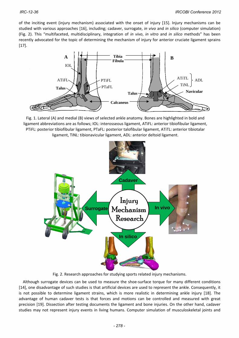
of the inciting event (injury mechanism) associated with the onset of injury [15]. Injury mechanisms can be
studied with various approaches [16], including; cadaver, surrogate, in vivo and in silico (computer simulation)
(Fig. 2). This “multifaceted, multidisciplinary, integration of in vivo, in vitro and in silico methods” has been
recently advocated for the topic of determining the mechanism of injury for anterior cruciate ligament sprains
[17].
Although surrogate devices can be used to measure the shoe‐surface torque for many different conditions
[14], one disadvantage of such studies is that artificial devices are used to represent the ankle. Consequently, it
is not possible to determine ligament strains, which is more realistic in determining ankle injury [18]. The
advantage of human cadaver tests is that forces and motions can be controlled and measured with great
precision [19]. Dissection after testing documents the ligament and bone injuries. On the other hand, cadaver
studies may not represent injury events in living humans. Computer simulation of musculoskeletal joints and
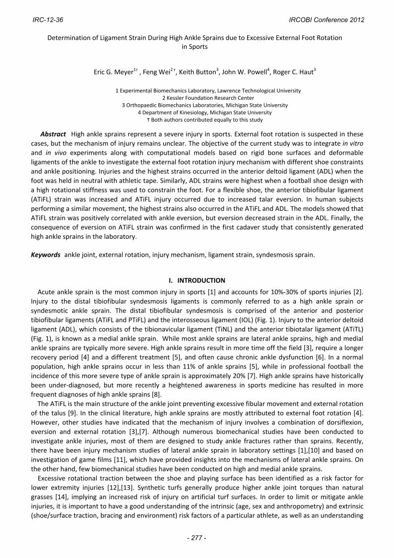
Fig. 1. Lateral (A) and medial (B) views of selected ankle anatomy. Bones are highlighted in bold and
ligament abbreviations are as follows; IOL: interosseous ligament, ATiFL: anterior tibiofibular ligament,
PTiFL: posterior tibiofibular ligament, PTaFL: posterior talofibular ligament, ATiTL: anterior tibiotalar
ligament, TiNL: tibionavicular ligament, ADL: anterior deltoid ligament.
TibiaFibula
Calcaneus
A B
Talus Talus
ATiFL PTiFL
IOL
ATiTL
TiNL ADL
Navicular PTaFL
Fig. 2. Research approaches for studying sports related injury mechanisms.
Cadaver
In vivo Surrogate
In silico
ResearchMechanism
Injury
IRC-12-36 IRCOBI Conference 2012
- 278 -
limbs can also provide useful information about joint mechanics. Validated models can be predictive tools to
understand normal joint function and serve as clinical tools for predicting and helping prevent sports injuries.
While finite element models yield useful information about stresses and strains in bones and ligaments [20],[21]
an advantage of rigid multi‐body models is the ability to rapidly solve for motion‐based mechanics in large
structures [22]. Liacouras and Wayne [23] developed a 3D computational approach to model the lower leg in
order to simulate cadaver ligament sectioning studies of syndesmotic injury and ankle inversion stability. A
recent study [24] followed a similar approach to develop a computational ankle model based on a generic
computed tomography (CT) scan and validated it against two well‐documented cadaver studies for
measurements of ankle ligament strains [25] and ankle joint torques [26]. Simulations of in vivo human
movements using computational models also offer a way to solve for motion‐based mechanics and determine
ligament strains.
The objective of the current study was to use multiple research approaches (Fig. 2) to investigate the
external rotation ankle injury mechanism with different shoe constraints and ankle positioning in order to
achieve a more comprehensive understanding of the relationships between joint biomechanics and the
observed injury mechanism. The hypothesis was that, when real‐world kinematics of the ankle from sports
scenarios were applied to cadaver and human subjects, ligament strains could be predicted using a validated
ankle computer model whose results would correlate with ankle injuries in the form of either high (ATiFL) or
medial (ADL) ankle ligament sprains.
II. METHODS
Experimental Testing
The effect of foot constraint was investigated in cadaver ankles, either by restraining the calcaneus tightly with potting and screws [26] or loosely with athletic tape [27]. Dynamic external rotation experiments were conducted on lower limbs from seven male cadavers (age 40±11 years). The tibia and fibula were sectioned 15 cm distal to the center of the knee and the proximal ends of each shaft were potted in a rectangular aluminum sleeve. The foot was everted to the maximum extent of the range of motion (10‐15°) in all tests. A biaxial testing machine was used to perform the test with the axis of rotation aligned at the ankle center between the medial and lateral malleoli. Compressive preloads of two to three times body weight were applied through the tibia prior to the application of an external torque. A dynamic angular rotation was input with a position controlled waveform. The rotation was increased by increments of five degrees in successive tests until gross ligament injury or bone fracture was observed based on an audible “pop” and corresponding drop in the torque signal. Between successive tests, the ankles were checked for damage and the orientation of the foot was reviewed. After failure, the mode of injury, failure torque and foot rotation were documented for each specimen. Motion analysis of the talus during external rotation was performed on four pairs of ankles using a Vicon motion capture system (OMG plc., Oxford, UK). The effect of shoe rotational stiffness (determined to be the rate at which torque is developed under external
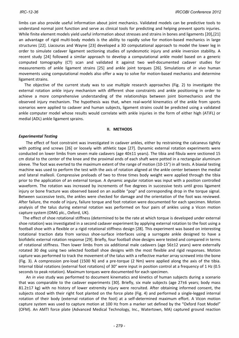
shoe rotation) was investigated in a second cadaver experiment by applying external rotation to the foot using a football shoe with a flexible or a rigid rotational stiffness design [28]. This experiment was based on interesting rotational traction data from various shoe‐surface interfaces using a surrogate ankle designed to have a biofidelic external rotation response [29]. Briefly, four football shoe designs were tested and compared in terms of rotational stiffness. Then lower limbs from six additional male cadavers (age 56±12 years) were externally rotated 30 deg using two selected football shoe designs with the most flexible and rigid responses. Motion capture was performed to track the movement of the talus with a reflective marker array screwed into the bone (Fig. 3). A compression pre‐load (1500 N) and a pre‐torque (2 Nm) were applied along the axis of the tibia. Internal tibial rotations (external foot rotations) of 30° were input in position control at a frequency of 1 Hz (0.5 seconds to peak rotation). Maximum torques were documented for each specimen.
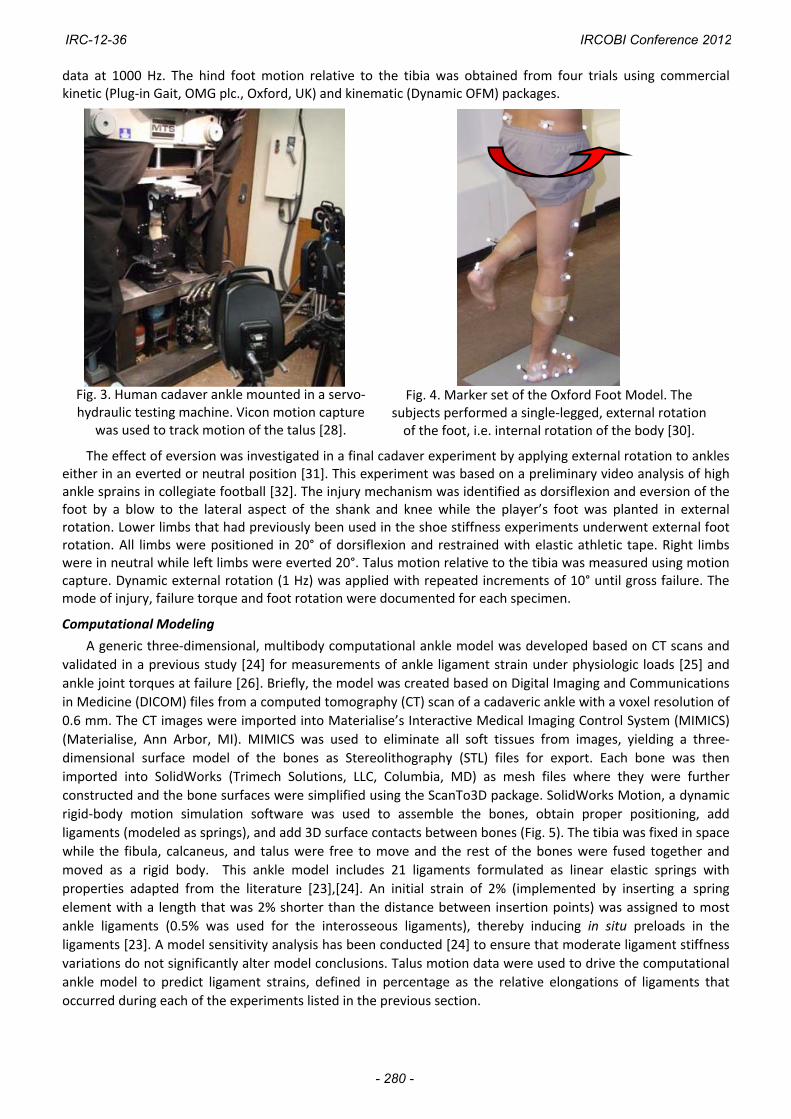
An in vivo study was performed to document kinematics and kinetics of human subjects during a scenario that was comparable to the cadaver experiments [30]. Briefly, six male subjects (age 27±6 years; body mass 81.2±17 kg) with no history of lower extremity injury were recruited. After obtaining informed consent, the subjects stood with their right foot planted on the force plate (Fig. 4) and performed a single‐legged internal rotation of their body (external rotation of the foot) at a self‐determined maximum effort. A Vicon motion capture system was used to capture motion at 100 Hz from a marker set defined by the “Oxford Foot Model” (OFM). An AMTI force plate (Advanced Medical Technology, Inc., Watertown, MA) captured ground reaction
IRC-12-36 IRCOBI Conference 2012
- 279 -
data at 1000 Hz. The hind foot motion relative to the tibia was obtained from four trials using commercial kinetic (Plug‐in Gait, OMG plc., Oxford, UK) and kinematic (Dynamic OFM) packages.
The effect of eversion was investigated in a final cadaver experiment by applying external rotation to ankles either in an everted or neutral position [31]. This experiment was based on a preliminary video analysis of high ankle sprains in collegiate football [32]. The injury mechanism was identified as dorsiflexion and eversion of the foot by a blow to the lateral aspect of the shank and knee while the player’s foot was planted in external rotation. Lower limbs that had previously been used in the shoe stiffness experiments underwent external foot rotation. All limbs were positioned in 20° of dorsiflexion and restrained with elastic athletic tape. Right limbs were in neutral while left limbs were everted 20°. Talus motion relative to the tibia was measured using motion capture. Dynamic external rotation (1 Hz) was applied with repeated increments of 10° until gross failure. The mode of injury, failure torque and foot rotation were documented for each specimen.
Computational Modeling
A generic three‐dimensional, multibody computational ankle model was developed based on CT scans and
validated in a previous study [24] for measurements of ankle ligament strain under physiologic loads [25] and
ankle joint torques at failure [26]. Briefly, the model was created based on Digital Imaging and Communications
in Medicine (DICOM) files from a computed tomography (CT) scan of a cadaveric ankle with a voxel resolution of
0.6 mm. The CT images were imported into Materialise’s Interactive Medical Imaging Control System (MIMICS)
(Materialise, Ann Arbor, MI). MIMICS was used to eliminate all soft tissues from images, yielding a three‐
dimensional surface model of the bones as Stereolithography (STL) files for export. Each bone was then
imported into SolidWorks (Trimech Solutions, LLC, Columbia, MD) as mesh files where they were further
constructed and the bone surfaces were simplified using the ScanTo3D package. SolidWorks Motion, a dynamic
rigid‐body motion simulation software was used to assemble the bones, obtain proper positioning, add
ligaments (modeled as springs), and add 3D surface contacts between bones (Fig. 5). The tibia was fixed in space
while the fibula, calcaneus, and talus were free to move and the rest of the bones were fused together and
moved as a rigid body. This ankle model includes 21 ligaments formulated as linear elastic springs with
properties adapted from the literature [23],[24]. An initial strain of 2% (implemented by inserting a spring
element with a length that was 2% shorter than the distance between insertion points) was assigned to most
ankle ligaments (0.5% was used for the interosseous ligaments), thereby inducing in situ preloads in the
ligaments [23]. A model sensitivity analysis has been conducted [24] to ensure that moderate ligament stiffness
variations do not significantly alter model conclusions. Talus motion data were used to drive the computational
ankle model to predict ligament strains, defined in percentage as the relative elongations of ligaments that
occurred during each of the experiments listed in the previous section.
Fig. 3. Human cadaver ankle mounted in a servo‐hydraulic testing machine. Vicon motion capture
was used to track motion of the talus [28].
Fig. 4. Marker set of the Oxford Foot Model. The subjects performed a single‐legged, external rotation of the foot, i.e. internal rotation of the body [30].
IRC-12-36 IRCOBI Conference 2012
- 280 -
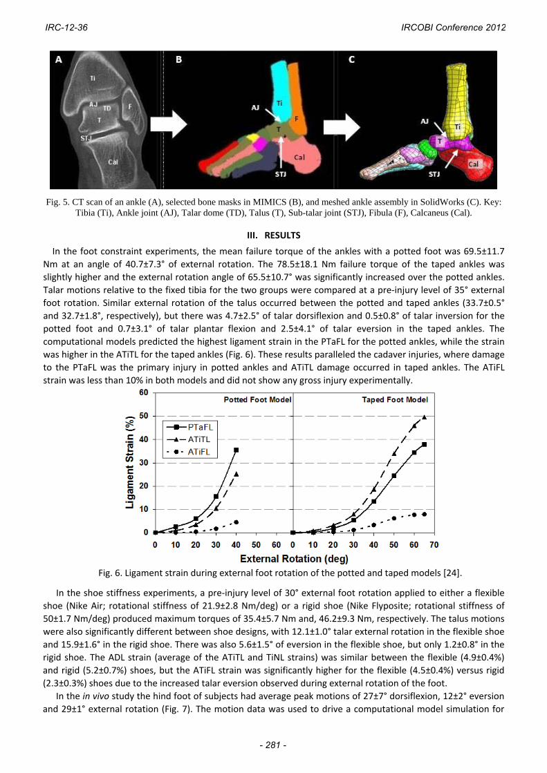
Fig. 5. CT scan of an ankle (A), selected bone masks in MIMICS (B), and meshed ankle assembly in SolidWorks (C). Key: Tibia (Ti), Ankle joint (AJ), Talar dome (TD), Talus (T), Sub-talar joint (STJ), Fibula (F), Calcaneus (Cal).
III. RESULTS
In the foot constraint experiments, the mean failure torque of the ankles with a potted foot was 69.5±11.7
Nm at an angle of 40.7±7.3° of external rotation. The 78.5±18.1 Nm failure torque of the taped ankles was
slightly higher and the external rotation angle of 65.5±10.7° was significantly increased over the potted ankles.
Talar motions relative to the fixed tibia for the two groups were compared at a pre‐injury level of 35° external
foot rotation. Similar external rotation of the talus occurred between the potted and taped ankles (33.7±0.5°
and 32.7±1.8°, respectively), but there was 4.7±2.5° of talar dorsiflexion and 0.5±0.8° of talar inversion for the
potted foot and 0.7±3.1° of talar plantar flexion and 2.5±4.1° of talar eversion in the taped ankles. The
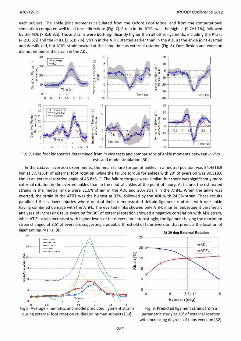
computational models predicted the highest ligament strain in the PTaFL for the potted ankles, while the strain
was higher in the ATiTL for the taped ankles (Fig. 6). These results paralleled the cadaver injuries, where damage
to the PTaFL was the primary injury in potted ankles and ATiTL damage occurred in taped ankles. The ATiFL
strain was less than 10% in both models and did not show any gross injury experimentally.
Fig. 6. Ligament strain during external foot rotation of the potted and taped models [24].
In the shoe stiffness experiments, a pre‐injury level of 30° external foot rotation applied to either a flexible
shoe (Nike Air; rotational stiffness of 21.9±2.8 Nm/deg) or a rigid shoe (Nike Flyposite; rotational stiffness of
50±1.7 Nm/deg) produced maximum torques of 35.4±5.7 Nm and, 46.2±9.3 Nm, respectively. The talus motions
were also significantly different between shoe designs, with 12.1±1.0° talar external rotation in the flexible shoe
and 15.9±1.6° in the rigid shoe. There was also 5.6±1.5° of eversion in the flexible shoe, but only 1.2±0.8° in the
rigid shoe. The ADL strain (average of the ATiTL and TiNL strains) was similar between the flexible (4.9±0.4%)
and rigid (5.2±0.7%) shoes, but the ATiFL strain was significantly higher for the flexible (4.5±0.4%) versus rigid
(2.3±0.3%) shoes due to the increased talar eversion observed during external rotation of the foot.
In the in vivo study the hind foot of subjects had average peak motions of 27±7° dorsiflexion, 12±2° eversion
and 29±1° external rotation (Fig. 7). The motion data was used to drive a computational model simulation for
ATiTL
IRC-12-36 IRCOBI Conference 2012
- 281 -
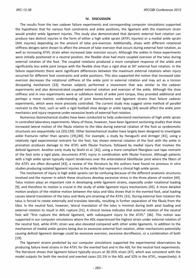
each subject. The ankle joint moments calculated from the Oxford Foot Model and from the computational
simulation compared well in all three directions (Fig. 7). Strain in the ATiFL was the highest (9.2±1.1%), followed
by the ADL (7.8±0.8%). Those strains were both significantly higher than all other ligaments, including the PTaFL
(4.2±0.5%) and the PTiFL (3.6±0.7%). Strain in the ATiFL started earlier than in the ADL as the ankle joint everted
and dorsiflexed, but ATiFL strain peaked at the same time as external rotation (Fig. 8). Dorsiflexion and eversion
did not influence the strain in the ADL.
Fig. 7. Hind foot kinematics determined from in vivo tests and comparisons of ankle moments between in vivo
tests and model simulation [30].
In the cadaver eversion experiments, the mean failure torque of ankles in a neutral position was 84.4±16.9
Nm at 37.7±5.4° of external foot rotation, while the failure torque for ankles with 20° of eversion was 90.3±8.6
Nm at an external rotation angle of 46.8±6.1°. The failure torques were similar, but there was significantly more
external rotation in the everted ankles than in the neutral ankles at the point of injury. At failure, the estimated
strains in the neutral ankle were 31.5% strain in the ADL and 20% strain in the ATiFL. When the ankle was
everted, the strain in the ATiFL was the highest at 32%, followed by the ADL with 24.5% strain. These results
paralleled the cadaver injuries where neutral limbs demonstrated deltoid ligament ruptures with one ankle
having combined damage with the ATiFL. The everted limbs showed only ATiFL injuries. Subsequent parametric
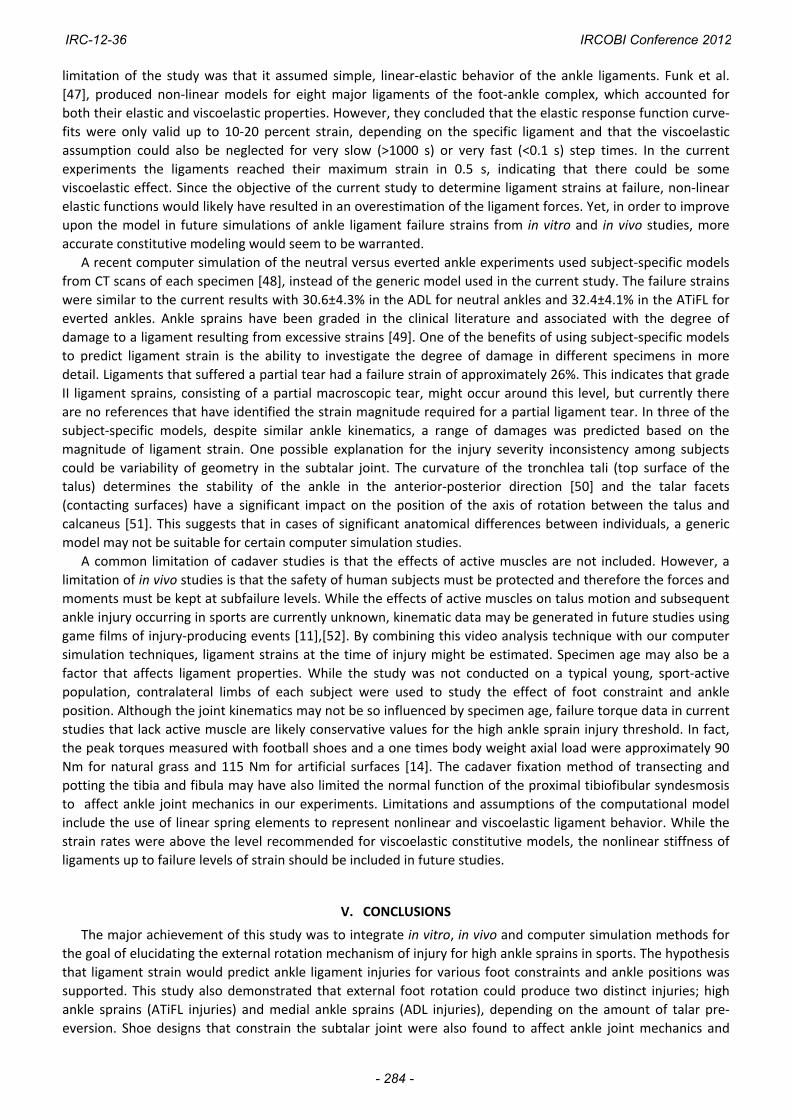
analyses of increasing talus eversion for 30° of external rotation showed a negative correlation with ADL strain,
while ATiFL strain increased with higher levels of talus eversion. Interestingly, the ligament having the maximum
strain changed at 8.5° of eversion, suggesting a possible threshold of talus eversion that predicts the location of
ligament injury (Fig. 9).
Fig 8. Average kinematics and model predicted ligament strains
during external foot rotation studies on human subjects [30].
Fig. 9. Predicted ligament strains from a
parametric study at 30° of external rotation
with increasing degrees of talus eversion [32].
IRC-12-36 IRCOBI Conference 2012
- 282 -
IV. DISCUSSION
The results from the two cadaver failure experiments and corresponding computer simulations supported
the hypothesis that for various foot constraints and ankle positions, the ligament with the maximum strain
would predict ankle ligament injuries. This study also demonstrated that dynamic external foot rotation can
produce two distinct injuries in the form of either a high ankle sprain (ATiFL injuries) or a medial ankle sprain
(ADL injuries) depending on the amount of talar pre‐eversion. Additionally, shoes with different rotational
stiffness designs were shown to affect the amount of talar eversion that occurs during external foot rotation, as
well as increasing ATiFL strain when increased talar eversion occurs. Although the ankles in these experiments
were initially positioned in a neutral position, the flexible shoe had more coupled eversion of the talus during
external rotation of the foot. The coupled rotations produced a more compliant response of the ankle and
significantly less ankle joint torque with the flexible shoe than a rigid shoe at 30° external foot rotation. In the
failure experiments there were significant differences between the maximum external rotations when injury
occurred for different foot constraints and ankle positions. This also supported the notion that increased talar
eversion decreases the rotational stiffness of the ankle joint in external rotation and may act as a torsion
dissipating mechanism [33]. Human subjects performed a movement that was similar to the cadaver
experiments and also demonstrated coupled external rotation and eversion of the ankle. Although the shoe
stiffness and in vivo experiments were at subfailure levels of ankle joint torque, they provided additional and
perhaps a more realistic understanding of joint biomechanics and ligament strains than failure cadaver
experiments, which were more precisely controlled. The current study may suggest some method of parallel
restraint to the foot, such as with a rigid football shoe design or ankle taping [34] would affect the ankle joint
mechanics and injury response under excessive levels of external foot rotation.
Numerous biomechanical studies have been conducted to help understand mechanisms of high ankle sprain
in controlled laboratory experiments. Many of these, however, have been ligament sectioning studies that show
increased lateral motion of the fibula relative to the tibia during external rotation of the foot as syndesmotic
structures are sequentially cut [35]‐[39]. Other biomechanical studies have largely been designed to investigate
ankle fractures rather than sprains [19],[40]. For example, a study by Haraguchi and Armiger [41], using a
relatively rigid experimental restraint for the foot, has shown external rotation of the foot with the ankle in
pronation produces damage to the ATiFL with fibular fracture, followed by medial injury that involves the
deltoid ligament. Another early study by Stiehl et al. [42], using a more compliant fiberglass cast tape restraint
of the foot onto a rigid plate, documents ATiFL injury in combination with fibular fracture. Although patients
with a high ankle sprain typically report tenderness over the anterolateral tibiofibular joint where the fibers of
the ATiFL are often disrupted [43], a review of the literature by the authors have found no previous in vitro
studies producing isolated ligament injuries to the ankle that represent a clinical high ankle sprain.
The mechanism of injury in high ankle sprains can be confusing because of the different anatomic structures
involved and the manner in which these structures develop excessive stress in the three planes of motion [44].
Talus motion plays an important role in developing ankle ligament strains, especially under rotational loading
[9], and therefore its motion is crucial in the study of ankle ligament injury mechanisms [45]. A more detailed
motion analysis of the relative motion between the talus and tibia shows that in the everted foot, axial loading
causes lateral translation of the talus and likely pre‐straining of the ATiFL [31]. During external foot rotation the
talus is forced to rotate externally and translate laterally, resulting in further separation of the fibula from the
tibia. In the neutral foot, however, lateral translation of the talus is minimal during both axial loading and
external rotation to result in ADL rupture [31]. A clinical review indicates that external rotation of the neutral
foot will “first rupture the deltoid ligament, with subsequent injury to the ATiFL” [46]. This notion was
supported in our computer simulations where the ADL experienced the highest strain under external rotation of
the neutral foot, while ATiFL strain was also elevated compared with other ankle ligaments. In addition to the
mechanism of medial ankle sprains being due to excessive external foot rotation, other mechanisms potentially
causing deltoid ligament damage could be excessive eversion, excessive dorsiflexion, or a combination of both
[19].
The ligament strains predicted by our computer simulations supported the experimental observations by
producing failure level strains in the ATiFL for the everted foot and in the ADL for the neutral foot experiments.
The literature shows that ligament failure typically occurs at 30‐35% strain [47], which was consistent with the
model outputs for both the neutral and everted cases (31.5% in the ADL and 32% in the ATiFL, respectively). A
IRC-12-36 IRCOBI Conference 2012
- 283 -
limitation of the study was that it assumed simple, linear‐elastic behavior of the ankle ligaments. Funk et al.
[47], produced non‐linear models for eight major ligaments of the foot‐ankle complex, which accounted for
both their elastic and viscoelastic properties. However, they concluded that the elastic response function curve‐
fits were only valid up to 10‐20 percent strain, depending on the specific ligament and that the viscoelastic
assumption could also be neglected for very slow (>1000 s) or very fast (<0.1 s) step times. In the current
experiments the ligaments reached their maximum strain in 0.5 s, indicating that there could be some
viscoelastic effect. Since the objective of the current study to determine ligament strains at failure, non‐linear
elastic functions would likely have resulted in an overestimation of the ligament forces. Yet, in order to improve
upon the model in future simulations of ankle ligament failure strains from in vitro and in vivo studies, more
accurate constitutive modeling would seem to be warranted.
A recent computer simulation of the neutral versus everted ankle experiments used subject‐specific models
from CT scans of each specimen [48], instead of the generic model used in the current study. The failure strains
were similar to the current results with 30.6±4.3% in the ADL for neutral ankles and 32.4±4.1% in the ATiFL for
everted ankles. Ankle sprains have been graded in the clinical literature and associated with the degree of
damage to a ligament resulting from excessive strains [49]. One of the benefits of using subject‐specific models
to predict ligament strain is the ability to investigate the degree of damage in different specimens in more
detail. Ligaments that suffered a partial tear had a failure strain of approximately 26%. This indicates that grade
II ligament sprains, consisting of a partial macroscopic tear, might occur around this level, but currently there
are no references that have identified the strain magnitude required for a partial ligament tear. In three of the
subject‐specific models, despite similar ankle kinematics, a range of damages was predicted based on the
magnitude of ligament strain. One possible explanation for the injury severity inconsistency among subjects
could be variability of geometry in the subtalar joint. The curvature of the tronchlea tali (top surface of the
talus) determines the stability of the ankle in the anterior‐posterior direction [50] and the talar facets
(contacting surfaces) have a significant impact on the position of the axis of rotation between the talus and
calcaneus [51]. This suggests that in cases of significant anatomical differences between individuals, a generic
model may not be suitable for certain computer simulation studies.
A common limitation of cadaver studies is that the effects of active muscles are not included. However, a
limitation of in vivo studies is that the safety of human subjects must be protected and therefore the forces and
moments must be kept at subfailure levels. While the effects of active muscles on talus motion and subsequent
ankle injury occurring in sports are currently unknown, kinematic data may be generated in future studies using
game films of injury‐producing events [11],[52]. By combining this video analysis technique with our computer
simulation techniques, ligament strains at the time of injury might be estimated. Specimen age may also be a
factor that affects ligament properties. While the study was not conducted on a typical young, sport‐active
population, contralateral limbs of each subject were used to study the effect of foot constraint and ankle
position. Although the joint kinematics may not be so influenced by specimen age, failure torque data in current
studies that lack active muscle are likely conservative values for the high ankle sprain injury threshold. In fact,
the peak torques measured with football shoes and a one times body weight axial load were approximately 90
Nm for natural grass and 115 Nm for artificial surfaces [14]. The cadaver fixation method of transecting and
potting the tibia and fibula may have also limited the normal function of the proximal tibiofibular syndesmosis
to affect ankle joint mechanics in our experiments. Limitations and assumptions of the computational model
include the use of linear spring elements to represent nonlinear and viscoelastic ligament behavior. While the
strain rates were above the level recommended for viscoelastic constitutive models, the nonlinear stiffness of
ligaments up to failure levels of strain should be included in future studies.
V. CONCLUSIONS
The major achievement of this study was to integrate in vitro, in vivo and computer simulation methods for
the goal of elucidating the external rotation mechanism of injury for high ankle sprains in sports. The hypothesis
that ligament strain would predict ankle ligament injuries for various foot constraints and ankle positions was
supported. This study also demonstrated that external foot rotation could produce two distinct injuries; high
ankle sprains (ATiFL injuries) and medial ankle sprains (ADL injuries), depending on the amount of talar pre‐
eversion. Shoe designs that constrain the subtalar joint were also found to affect ankle joint mechanics and
IRC-12-36 IRCOBI Conference 2012
- 284 -
injury response during external rotation of the foot. And finally, this series of experiments was the first to
consistently produce isolated ligament injuries to the ATiFL that represent a clinical high ankle sprain. The study
outcome may provide an avenue for the development of football shoe designs or other protective gear that may
help reduce the incidence and/or severity of medial and high ankle sprains on the athletic field.
VI. ACKNOWLEDGEMENT
This study was funded, in part, by a grant from the National Football League Charities Foundation. The
authors thank Mr. Clifford Beckett, Mr. Jerrod Braman and Mr. Brian Weaver for technical assistance in this
study, Mrs. Jean Atkinson for specimen preparation, Mr. Dean Mueller for the help with procuring the test
specimens and Dr. Seungik Baek for providing the MIMICS software.
VII. REFERENCES
[1] Fong DT, Hong Y, Shima Y, Krosshaug T, Yung PS, Chan KM, Biomechanics of supination ankle sprain: a case
report of an accidental injury event in the laboratory, Am J Sports Med, 37(4): 822‐827, 2009.
[2] Waterman BR, Belmont PJ, Cameron KL, Svoboda SJ, Alitz CJ, Owens BD, Risk factors for syndesmotic and
medial ankle sprain, Am J Sports Med, 39(5): 992‐998, 2011.
[3] Wolfe MW, Uhl TL, Mattacola CG, McCluskey LC, Management of ankle sprains, Am Fam Physician, 63(1):
93‐104, 2001.
[4] Boytim MJ, Fischer DA, Neumann L, Syndesmotic ankle sprains, Am J Sports Med, 19(3): 294‐298, 1991.
[5] Hopkinson WJ, St Pierre P, Ryan JB, Wheeler JH, Syndesmosis sprains of the ankle, Foot Ankle Int, 10(6):325‐
330, 1990.
[6] Gerber JP, Williams GN, Scoville CR, Arciero RA, Taylor DC, Persistent disability associated with ankle
sprains: A perspective examination of an athletic population, Foot Ankle Int, 19(10):653‐660, 1998.
[7] Guise ER, Rotational ligamentous injuries to the ankle in football, Am J Sports Med, 4(1):1‐6, 1976.
[8] Williams GN, Jones MH, Amendola A, Syndesmotic ankle sprains in athletes, Am J Sports Med, 35(7):1197‐
1207, 2007.
[9] Sarsam IM, Hughes SP, The role of the anterior tibio‐fibular ligament in talar rotation: an anatomical study,
Injury, 19(2):62‐64, 1988.
[10] Kristianslund E, Bahr R, Krosshaug T, Kinematics and kinetics of an accidental lateral ankle sprain, J Biomech,
44:2576‐2578, 2011.
[11] Mok KM, Fong DT, Krosshaug T, Engebretsen L, Hung AS, Yung PS, Chan KM, Kinematics analysis of ankle
inversion ligamentous sprain injuries in sports: 2 cases during the 2008 Beijing Olympics, Am J Sports Med,
doi:10.1177/0363546511399384, 2011.
[12] Torg JS, Quedenfeld TC, Landau S, The shoe‐surface interface and its relationship to football knee injuries, J Sports Med, 2(5): 261‐269, 1974.
[13] Lambson RB, Barnhill BS, Higgins RW, Football cleat design and its effect on anterior cruciate ligament
injuries. A three year prospective study, Am J Sports Med, 24(2):155‐159, 1996.
[14] Villwock MR, Meyer EG, Powell JW, Fouty AJ, Haut RC, Football playing surface components and shoe
designs may affect lower extremity injury risk potential: An in situ torsional resistance assessment, Am J
Sports Med, 37(3):518‐525, 2009.
[15] Krosshaug T, Bahr R, Understanding injury mechanisms: a key component of preventing injuries in sport, Br J
IRC-12-36 IRCOBI Conference 2012
- 285 -
Sports Med, 39:324‐329, 2005
[16] Crandall JR, Bose D, Forman J, Untaroiu CD, Arregui‐Dalmases C, Shaw CG, Kerrigan JR, Human surrogates
for injury biomechanics research, Clin Anat, 24:362‐371, 2011.
[17] Quatman CE, Quatman CC, Hewett TE, Prediction and prevention of musculoskeletal injury: a paradigm shift
in methodology, Br J Sports Med, 43:1100‐1107,2009.
[18] Noyes, FR, Mooar LA, Moorman 3rd CT, McGinnis GH, Partial tears of the anterior cruciate ligament.
Progression to complete ligament deficiency, J Bone & Joint Surg, 71:825‐833, 1989.
[19] Funk JR, Ankle injury mechanisms: lessons learned from cadaveric studies, Clin Anat, 24:350‐361, 2011.
[20] Cheung JT, An KN, Zhang M, Consequences of partial and total plantar fascia release: a finite element study,
Foot Ankle Int, 27(2):125‐132, 2006.
[21] Reggiani B, Leardini A, Corazza F, Taylor M, Finite element analysis of a total ankle replacement during the
stance phase of gait, J Biomech, 39(8): 1435‐1443, 2006.
[22] Kwak SD, Blankevoort L, Ateshian GA, A mathematical formulation for 3D quasi‐static multibody models of
diarthrodial joints, Comput Methods Biomech Biomed Eng, 3(1):41‐64, 2000.
[23] Liacouras PC, Wayne JS, Computational modeling to predict mechanical function of joints: application to the
lower leg with simulation of two cadaver studies, J Biomech Eng, 129(6):811‐17, 2007.
[24] Wei F, Hunley SC, Powell JW, Haut RC, Development and validation of a computational model to study the
effect of foot constraint on ankle injury due to external rotation, Ann Biomed Eng, 39(2): 756‐765, 2011.
[25] Colville MR, Marder RA, Boyle JJ, Zarins B, Strain measurement in lateral ankle ligaments, Am J Sports Med,
18(2):196‐200, 1990.
[26] Wei F, Villwock MR, Meyer EG, Powell JW, Haut RC, A biomechanical investigation of ankle injury under
excessive external foot rotation in the human cadaver, J Biomech Eng, 132(9): 2010.
[27] Villwock MR, Meyer EG, Powell JW, Haut RC, External rotation ankle injuries: investigating ligamentous
rupture, Proceedings of ASME Summer Bioengineering Conference, Lake Tahoe, CA, 2009.
[28] Wei F, Meyer EG, Braman JE, Powell JW, Haut RC, Rotational stiffness of football shoes influences talus
motion during external rotation of the foot, J Biomech Eng, DOI:10.1115/1.4005695, 2012.
[29] Villwock MR, Meyer EG, Powell JW, Haut RC, Development and evaluation of a surrogate ankle for use with
a rotational traction measurement apparatus, J Sports Eng Tech, 223:151‐157, 2009.
[30] Wei F, Braman JE, Weaver BT, Haut RC, Determination of dynamic ankle ligament strains from a
computational model driven by motion analysis based kinematic data, J Biomech, 44(15):2636‐2641, 2011.
[31] Wei F, Post JM, Braman, JE, Meyer EG, Powell JW, Haut RC, Eversion during external rotation of the human
cadaver foot produces high ankle sprains, J Orthop Res, DOI:10.1002/jor.22085, 2012.
[32] Wei F, Braman JE, Meyer EG, Powell JW, Haut RC, Mechanism of injury in a high ankle sprain: A simulation
study, Proceedings of the ASME Summer Bioengineering Conference, Farmington, PA, 2011.
[33] Lundberg A, Svensson OK, Bylund C, Goldie I, Selvik G. Kinematics of the ankle/foot complex – Part 2:
pronation and supination, Foot Ankle, 9(5):248‐253, 1989.
[34] Verhagen EA, Van Mechelen W, De Vente W, The Effect of Preventive Measures on the Incidence of Ankle
Sprains, Clin J Sport Med, 10(4):291‐296, 2000.
[35] Johnson EE, Markolf KL, The contribution of the anterior talofibular ligament to ankle laxity, J Bone Joint
Surg Am, 65:81‐88,1983.
IRC-12-36 IRCOBI Conference 2012
- 286 -
[36] Rasmussen O, Tovborg‐Jensen I, Boe S, Distal tibiofibular ligaments. Analysis of function, Acta Orthop Scand,
53: 681‐686, 1982.
[37] Rasmussen O, Stability of the ankle joint. Analysis of the function and traumatology of the ankle ligaments,
Acta Orthop Scand Suppl, 211:1‐75, 1985.
[38] Stormont DM, Morrey BF, An KN, Cass JR, Stability of the loaded ankle. Relation between articular restraint
and primary and secondary static restraints, Am J Sports Med, 13:295‐300, 1985.
[39] Xenos JS, Hopkinson WJ, Mulligan ME, Olson EJ, Popovic NA, The tibiofibular syndesmosis. Evaluation of the
ligamentous structures, methods of fixation, and radiographic assessment, J Bone Joint Surg Am, 77:847‐
856, 1995.
[40] Hirsch C, Lewis J, Experimental ankle‐joint fractures, Acta Orthop Scand, 36:408‐417, 1965.
[41] Haraguchi N, Armiger RS, A new interpretation of the mechanism of ankle fracture, J Bone Joint Surg Am,
91:821‐829, 2009.
[42] Stiehl JB, Skrade DA, Johnson RP, Experimentally produced ankle fractures in autopsy specimens, Clin
Orthop Relat Res, 285:244‐249, 1992.
[43] Smith AH, Bach BR Jr., High ankle sprains: minimizing the frustration of a prolonged recovery, Phys
Sportsmed, 32: 39‐43, 2004.
[44] Lin CF, Gross MT, Weinhold P, Ankle syndesmosis injuries: anatomy, biomechanics, mechanism of injury,
and clinical guidelines for diagnosis and intervention, J Orthop Sports Phys Ther,36:372‐384, 2006.
[45] Hertel J, Denegar CR, Monroe MM, Stokes WL, Talocrural and Subtalar Joint Instability after Lateral Ankle
Sprain, Med Sci Sports Exerc, 31(11):1501‐1508, 1999.
[46] Dattani R, Patnaik S, Kantak A, Srikanth B, Selvan TP, Injuries to the tibiofibular syndesmosis, J Bone Joint
Surg Br, 90:405‐410, 2008.
[47] Funk JR, Hall GW, Crandall JR, Pilkey WD, Linear and quasi‐linear viscoelastic characterization of ankle
ligaments, J Biomech Eng, 122:15‐22, 2000.
[48] Button KD, Wei F, Meyer EG, Fitzsimons K, Haut RC. Determination of in situ ankle ligament strains in cases
of high and medial ankle sprains, Proceedings of the ASME Summer Bioengineeing Conf., Farjardo, Puerto
Rico, 2012.
[49] Renstrom AFH, Lynch SA, Ankle ligament injuries, Rev Bras Med Esporte, 4(3):71‐80, 1998.
[50] Kleippol RP, Blankevoort L, The relation between geometry and function of the ankle joint complex: a
biomechanical review, Knee Surg Sports Traumatol Arthrosc, 18:618‐627, 2010.
[51] Barbaix E, Van Roy P, Clarys JP, Variations of anatomical elements contributing to subtalar joint stability:
intrinsic risk factors for post‐traumatic lateral instability of the ankle? Ergonomics, 43:1718‐1724, 2000.
[52] Kwon JY, Chacko AT, Kadzielski JJ, Appleton PT, Rodriguez EK, A novel methodology for the study of injury
mechanism: ankle fracture analysis using injury videos posted on YouTube.com, J Orthop Trauma, 24:477‐
482, 2010.
IRC-12-36 IRCOBI Conference 2012
- 287 -

IRC-12-36 IRCOBI Conference 2012
- 288 -