determination of free choline in plasma and erythrocyte samples and choline derived from membrane...
TRANSCRIPT
ANALYTICAL BIOCHEMISTRY 152, 178- 182 ( 1986)
Determination of Free Choline in Plasma and Erythrocyte Samples and Choline Derived from Membrane Phosphatidylcholine
by a Chemiluminescence Method
INDRAJIT DAS, JACQUELINE DE BELLEROCHE, CHRISTOPHER J. MOORE, AND FRANK CLIFFORD ROSE
Departments of Biochemistry and Neurology, Charing Cross and Westminster Medical School, Fulham Palace Road, London W6 8RF, England
Received July 16, 1985
A sensitive chemiluminescence method for assay of choline which has been developed for analysis of erythrocyte and plasma levels of choline is reported here. This method includes a charcoal purification step which yields consistent results with plasma and erythrocyte extracts. Further, choline derived from membrane phosphatidylcholine may also be measured by an ex- tension of this method following digestion with phospholipase D. This method has been used to study abnormal levels of erythrocyte choline that occur in cluster headache patients compared to control subjects and migraine patients. In addition, the time course of changes in plasma and erythrocyte choline following a fatty meal have been monitored. Plasma choline levels rise to a maximum between 1 and 3 h after the meal and this is followed by a rise in erythrocyte choline levels which are maximal 3 h after the meal. Q 1986 Academic press, hc.
KEY WORDS: choline; chemiluminescence; plasma; erythrocytes; phosphatidylcholine.
A number of methods have been developed for estimation of choline in plasma and eryth- rocyte samples. Values obtained by the differ- ent methods show considerable variation in control subjects, from the low values (range of mean values 14-37 nmol/ml) obtained from gas chromatography with mass spec- trometry (l-4) and radioenzyme methods (5) to higher values (approx 110 nmol/ml) ob- tained by proton nuclear magnetic resonance and spectrophotometric methods (6). A sen- sitive chemiluminescence method which yields consistent results is reported here. This has been developed from the chemiluminescence method of Israel and Lesbats (78) and includes an additional purification procedure which now makes the method applicable for use with plasma and erythrocyte samples. Further, choline derived from membrane phosphati- dylcholine may also be measured by an exten- sion of this method following digestion with phospholipase D. This approach provides a
simple means of assessing whether abnormal choline levels which occur in conditions such as mania (3,9) and cluster headache (10) reflect an altered membrane phosphatidylcholine level. This approach has been applied by us to the study of cluster headache where not only are levels of serum cholesterol elevated and levels of erythrocyte choline decreased, but re- ceptor activation of adenylate cyclase is de- pressed (1 l), thus indicating a possible link between lipid turnover and coupling mecha- nisms present in the membrane.
MATERIALS AND METHODS
Reagents. All reagents were of analytical grade. Luminol (5-amino-2,3-dihydro- 1,4- phthalazinedione) and choline bromide were obtained from Sigma Chemical Company. Choline oxidase (50 U/ml), horseradish per- oxidase (2500 U/ml), uricase (18 U/ml), and phospholipase D (0.3 U/mg) were obtained from Boehringer Mannheim.
0003-2697186 $3.00 Copyright Q 1986 by Academic Press, Inc. ALI rights of repduction in any form resewed.
178

CHEMILUMINESCENCE ASSAY FOR CHOLINE 179
Patients and control subjects. Blood samples were taken from patients attending the Prin- cess Margaret Migraine Clinic at Charing Cross Hospital. Control subjects were mem- bers of the staff of Charing Cross Hospital and Medical School.
Sample preparation for choline assay. After an overnight fast, blood was drawn from pa- tients and control subjects by venipuncture into plastic tubes containing sodium heparin (0.5 U/ml) as an anticoagulant. Erythrocytes were sedimented at 14OOg for 30 min at 4°C. Plasma was removed, the buffy coat layer was discarded, and a sample of packed erythrocytes was taken. Plasma and erythrocyte samples were deproteinized with ice-cold trichloroace- tic acid (3.8%) and centrifuged, and the su- pernatants were extracted three times with 3 vol of diethyl ether. The aqueous sample was further purified by mixing with an equal vol- ume of charcoal (Norit OL) suspension (40 mg/ml). Charcoal was removed by centrifu- gation and the supematant stored at -20°C. Plasma and erythrocyte extracts were treated in the same way.
Preparation and extraction of erythrocyte membranes. After an overnight fast, blood (10 ml) was collected in a heparinized tube and centrifuged at 1500g for 15 min. The plasma and buffy coat were removed and the eryth- rocytes were resuspended in saline (0.9% so- dium chloride) and centrifuged at 15OOg for 15 min. Membranes were prepared and ex- tracted by the methods of Hanahan and Ek- holm (12) and Rose and Oklander (13) with minor modifications, as detailed below. An al- iquot (2 to 4 ml) of packed erythrocytes was removed and added to 9 vol distilled water with vigorous mixing. After 30 min at room temperature the preparation was centrifuged at 20,OOOg for 40 min at 4°C. The supematant was discarded and the pellet was resuspended in distilled water, mixed, and recentrifuged at 20,OOOg for 40 min. This washing procedure was repeated twice. The resulting pellet was resuspended in distilled water to give a volume corresponding to the original erythrocyte vol- ume and sonicated for 1 min at 4°C. An ali-
quot of sonicate was removed for protein assay by the method of Lowry et al. (14) and the remainder of the sonicated preparation was added with mixing to 11 vol of 2-propanol. After 1 h at room temperature, the preparation was centrifuged for 30 min at 5008. The su- pematant was then removed and evaporated to dryness under vacuum at 30°C. The dried sample was dissolved in 2-propanol and ali- quots were removed for assay of cholesterol, phosphatidylcholine, and phosphorus.
Assay of choline. Choline was measured by the coupling of two enzyme-catalyzed reac- tions, the conversion of choline to betaine by choline oxidase and the oxidation of luminol by peroxidase. The resulting chemilumines- cence was detected and measured by a LKB 1250 luminometer with the intensity of che- miluminescence being recorded as a function of time on a LKB 2210-032 potentiometric chart recorder. A 14C standard photon source was used as a reference.
The following procedure was used for all samples. A cuvette was set up containing 2 ml of 0.2 M Na2HP04, pH 8.6, 10 ~1 of 1 mM
luminol, 5 /*l horseradish peroxidase (2500 U/ ml), and 10 ~1 standard or sample. A baseline level of luminescence was obtained. The re- action was started by the addition of 10 ~1 choline oxidase (5 U/ml). The cuvette was rapidly mixed and replaced in the luminom- eter. The peak height obtained within approx- imately 10 s was proportional to the choline concentration of the sample. Samples were al- ternated with choline standards and a standard curve was constructed each day with fresh choline standard (5- 100 pmol).
A standard curve was constructed each day with fresh choline bromide using four to six different concentrations of choline between 5 and 100 pmol ( 10 ~1 aliquot). The relationship between peak luminometer deflection and amount of added choline standard in the assay system was linear over the range used in this study (5 to 100 pmol). All samples were diluted to give a value lying in this range, although linearity extended well above this range, The lowest concentration of standard (5 pmol)
180 DAS ET AL.
routinely used gave a reasonable peak deflec- tion over the baseline (5% of maximum de- flection at 50-100 mV input voltage). The coefficient of interassay variation was less than 10% for each choline standard used (range 5- 100 pmol).
Recovery of choline was assayed in two ways. First, [3H]choline was added to the packed erythrocyte samples and radioactivity was monitored in the final charcoal-purified extract. Second, a nonradioactive choline standard was treated in the same way as an erythrocyte sample and choline was analyzed in the final extract. The mean recovery of cho- line was 97 1- 5 (3) and 95 f 4 (3), respectively.
Assay ofphosphatidylcholine. Ahquots (0.1 ml) of membrane extract or standard 0.2 mM
phosphatidylcholine (Grade I, purified egg yolk lecithin from Lipid Products Ltd., Red- hill, U. K.) in 2-propanol were incubated with
0.1 ml phospholipase D and 0.4 ml of 50 mM acetate buffer containing 50 mM CaClz, pH 5.6. Phospholipase D was suspended in the same buffer. Samples were incubated at 37°C for 1 h. These conditions were found to yield complete hydrolysis of phosphatidylcholine standards. Choline was purified and assayed as described.
Assay of cholesterol and phosphorus. Cho- lesterol was assayed by the cholesterol oxidase method using a Boehringer Mannheim kit (Boehringer Corporation Ltd.). Phosphorus was assayed by the method of Petitou et al. (15).
RESULTS AND DISCUSSION
Erythrocyte Choline Levels
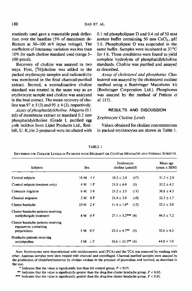
Values obtained for choline concentrations in packed erythrocytes are shown in Table 1.
TABLE 1
ERYTHROCYTE CHOLINE LEVELS IN PATIENTS WITH MIGRAINE OR CLUSTER HEADACHE AND NORMAL SUBJECTS
Subjects Sex
Erythrocyte choline (rmol/l)
Mean age (years t- SEM)
Control subjects
Control subjects (smokers only)
Common migraine
Classical migraine
Cluster headache
Cluster headache patients receiving methylsergide treatment
Cluster headache patients receiving ergotamine containing preparations
Headache patients receiving amitriptyline
16M 1F
4M 1F
6M 5F
2M 8F
10M 2F
4M OF
5M OF
3M IF
18.3 f 2.4 (17)
21.0 + 6.0 (5)
21.2 f 3.5 (11)
21.4 + 3.9 (10)
11.4 + 1.6* (12)
27.1 + 5.2*** (4)
22.4 + 4.7** (5)
56.6 + 13.5** (4)
31.2 f 2.0
33.2 f 4.2
38.8 -t 4.3
32.3 + 1.7
32.1 f 3.0
44.5 + 7.2
32.6 + 4.3
44.0 + 7.0
Note. Erythrocytes were deproteinized with trichloroacetic acid (TCA) and the TCA was removed by washing with ether. Aqueous samples were then treated with charcoal and centrifuged. Charcoal purified samples were assayed by the production of chemiluminescence by choline oxidase in the presence of peroxidase and luminol, as described in the text.
* Indicates that the value is significantly less than the control group, P < 0.05. *+ Indicates that the value is significantly greater than the drug-free cluster headache group, P < 0.02.
*** Indicates that the value is significantly greater than the drug-free cluster headache group, P -e 0.0 1.

CHEMILUMINESCENCE ASSAY FOR CHOLINE 181
These values are higher than those previously reported by us (10) using the chemilumines- cence procedure without a charcoal purifica- tion step. The low values are likely to be due to the presence of an interfering substance present in the extracts which has an inhibitory effect on the reaction (see below). Choline concentrations of normal subjects were com- pared with those of migraine patients (classical migraine, i.e., headache accompanied by an “aura” and common migraine, i.e., headache without associated neurological features) and cluster headache (severe bouts of unilateral head pain lasting 30 min-2 h recurring daily for weeks or months and separated by pain- free intervals). In our previous study carried out without the purification step, a separate group of cluster headache patients was also monitored. In both studies a qualitatively similar result is obtained in which the eryth- rocyte choline levels are significantly reduced to 50% of the control value. Choline concen- trations in both groups of migraine patients (drug-free) were very close to control values (Table 1). The effects of smoking and drug treatments were also examined (separate pa- tients). No significant effect on choline levels in control subjects was seen with smoking. However, amitriptyline significantly enhanced choline levels in headache patients (all groups) and both methysergide and ergotamine con- taining preparations enhanced the levels of choline in cluster headache patients.
Eflect of a Meal on Plasma and Erythrocyte Choline Levels
The effect of a fatty meal was monitored in four normal subjects. Plasma choline levels prior to the meal were 24-38% of the eryth- rocyte values (Fig. 1). Plasma values of choline reached a maximum between 1 and 3 h after a meal and then declined. Erythrocyte values in all four subjects also increased and were maximal 3 h after the meal, presumably arising from plasma choline. It is important therefore when studying migraine and other neurolog- ical conditions to measure erythrocyte choline after fasting to overcome the fluctuations due to dietary choline.
01 I I I 1 II 1 2 3 4
Time after meal (hours)
FIG. 1. Plasma and erythrocyte choline levels at various times after a fatty meal. Blood was taken from four control subjects before and on three further occasions up to 4 h after a fatty meal. Plasma and erythrocyte choline was extracted and analyzed as described under Materials and Methods. Values are for individual samples from each subject and are means of two to three determinations.
Presence of Interfering Substances in Plasma and Erythrocyte Extracts
The possibility that constituents of plasma, e.g., uric acid, peroxide scavengers, e.g., as- corbate or choline derivatives, might interfere with the choline assay was tested. Preincuba- tion with uricase (10 ~1,2 mg/ml) or addition of a physiological concentration of ascorbate (0.1 InM) to the assay mixture had no effect on the analysis of choline standards. Addition of phosphatidylcholine (1 pmol/ml final con- centration) to a choline standard prior to pu- rification did not interfere with the assay at the dilution used in the final assay mixture. CDP-choline is known to be adsorbed by charcoal ( 16) and hence will not interfere with the assay. The possibility that phosphorylcho- line or cytidine diphosphocholine might be degraded during the extraction/assay proce- dure has been excluded by Hise and Mansbach II (17) who found no significant degradation using a spectrophotometric choline oxidase method.
Erythrocyte Membrane Phosphatidylcholine in Control Subjects
Erythrocyte membrane phosphatidylcho- line values related to protein, membrane cho-
182 DAS ET AL.
TABLE 2 ACKNOWLEDGMENTS
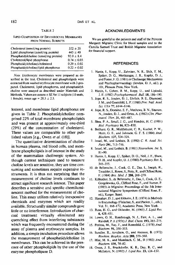
LIPIDCOMPOSITIONOFERYTHROCYTE MEMBRANES FROM NORMALSUBJECTS
Cholesterol (nmol/mg protein) 322 + 20 Lipid phosphorus (nmol/mg protein) 445 f 40 Phosphatidylcholine (nmol/mg protein) 92.0 + 8.4 Cholesterol/lipid phosphorus 0.74 f 0.03 Phosphatidylcholine/cholesterol 0.29 +- 0.02 Phosphatidylcholine/lipid phosphorus 0.22 + 0.02
We are grateful to the patients and staff of the Princess Margaret Migraine Clinic for blood samples and to the Camilla Samuel Trust and British Migraine Association for financial support.
REFERENCES
I. No&. Erythrocyte membranes were prepared as de-
scribed in the text. Cholesterol and phospholipids were extracted from washed erythrocyte membrane with 2-pro- panel. Cholesterol, lipid phosphorus, and phosphatidyl- choline were assayed as described under Materials and Methods. Values are means f SE for 1 I subjects (I 0 male, 1 female), mean age = 28.5 f 2.5.
2.
3.
lesterol, and membrane lipid phosphorus are given in Table 2. Phosphatidylcholine com- prised 22% of total membrane phospholipids and was present at approximately one-third (29%) of the concentration of cholesterol. These values are comparable to other pub- lished values [e.g., Owen et al. (1 S)].
Hanin, I., Kopp, U., Zahniser, N. R., Shih, T. M., Spiker, D. G., Merikangas, J. R., Kupfer, D. J., and Foster, F. G. ( I98 I) in Cholinergic Mechanisms and Psychopharmacology (Jenden, D. J., ed.), p. I8 I, Plenum Press, New York.
Hanin, I., Cohen, B. M., Kopp, U., and Lipinski, J. F. (1982) Psychopharmacol. Bull. 18, 186-190.
Jope, R. S., Jenden, D. J., Ehrlich, B. E., Diamond, J. M., and Gosenfeld, L. F. (1980) Proc. NUB Acad. Sci. USA 77,6 144-6 146.
4.
5.
6.
Jope, R. S., Domino, E. F., Mathews, B. N., Sitaram, N., Jenden, D. J., and Ortez, A. (1982) Clin. Phar- macol. Ther. 31, 483-487.
Shea, P. A., Small, J. G., and Hendrie, H. C. (1981) Biol. Psychiatry 16,825-830.
Beilharz, G. R., Middlehurst, C. R., Kuchel, P. W., Hunt, G. E., and Johnson, G. F. S. (1984) Anal. Biochem. 137,324-329.
The quantitative determination of choline in human plasma, red blood cells, and mem- brane phospholipids is of interest in the study of the mammalian cholinergic system. Al- though current techniques used to measure choline levels are sensitive, they are time con- suming and sometimes require expensive in- struments. It is thus not surprising that the measurement of choline levels continues to attract significant research interest. This paper describes a sensitive and specific chemilumi- nescent method for the measurement of cho- line. This assay utilizes relatively inexpensive chemicals and enzymes which are readily available. Structurally similar compounds gave little or no interference. Introduction of char- coal treatment virtually eliminated any quenching effect from interfering substances which was frequently encountered during the assay of plasma and erythrocyte samples. In addition, a simple incubation procedure allows the measurement of phosphatidylcholine in membranes. This can be achieved in the pres- ence of other phospholipids by the use of the enzyme phospholipase D.
I.
8.
9.
Israel, M., and Lesbats, B. (1980) C. R. Acud. Sci. Paris291,713-716.
Israel, M., and Lesbats, B. (1981) Neurochem. Int. 3, 8 l-90.
Hanin, I., Kopp, U., Spiker, D. G., Neil, J. F., Shaw, D. H., and Kupfer, D. J. (1980) Psychiatry Rex 3, 345-355.
IO.
I I.
de Belleroche, J., Cook, G. E., Das, I., Joseph, R., Tresidder, I., Rouse, S., Petty, R., and Clifford Rose, F. (1984) Brit. Med. J. 288,268-270.
Kilfeather, S., de Belleroche, J., Das, I., Cook, G. E., Gorgolewska, G., Clifford Rose, F., and Turner, P. (1985) in Migraine: Proceedings of the 5th Inter- national Migraine Symposium (Clifford Rose, F., ed.), Karger, Basel.
12.
13.
14.
15.
16.
17.
18.
Hanahan, D. J., and Ekholm, J. E. (1974) in Methods in Enzymology (Fleischer, S., and Packer, L., eds.), Vol. 31, 168-172, Academic Press, New York.
Rose, H. G., and Oklander, M. (I 965) J. Lipid Res. 6,428-43 I.
Lowry, 0. H., Rosebrough, N. J., Farr, A. L., and Randall, P. J. (195 I) J. Biol. Chem. 193,265-275.
Petitou, M., Tuy, F., and Rosenfeld, C. (1978) Anal. Biochem. 91,350-353.
Sundler, R., Arvidson, G., and Akesson, B. (1972) Biochim. Biophys. Ada 280, 559-568.
Hise, M. K., and Mansbach, C. M., II (1983) Anal. Biochem. 135,78-82.
Owen, J. S., Bruckdorfer, K. R., Day, R. C., and McIntyre, N. (I 982) J. Lipid Res. 23, 124-I 32.