determination of a complex crystal structure in the
TRANSCRIPT

warwick.ac.uk/lib-publications
Original citation: Hughes, Colan H., Reddy, G. N. Manjunatha, Masiero, Stefano, Brown, Steven P., Williams, P. Andrew and Harris, Kenneth D. M.. (2017) Determination of a complex crystal structure in the absence of single crystals : analysis of powder X-ray diffraction data, guided by solid-state NMR and periodic DFT calculations, reveals a new 2′-deoxyguanosine structural motif. Chemical Science, 8 (5). pp. 3971-3979. Permanent WRAP URL: http://wrap.warwick.ac.uk/88642 Copyright and reuse: The Warwick Research Archive Portal (WRAP) makes this work of researchers of the University of Warwick available open access under the following conditions. This article is made available under the Creative Commons Attribution 3.0 (CC BY 3.0) license and may be reused according to the conditions of the license. For more details see: http://creativecommons.org/licenses/by/3.0/ A note on versions: The version presented in WRAP is the published version, or, version of record, and may be cited as it appears here. For more information, please contact the WRAP Team at: [email protected]
ChemicalScience
EDGE ARTICLE
Ope
n A
cces
s A
rtic
le. P
ublis
hed
on 1
6 M
arch
201
7. P
urch
ased
by
y.c.
budd
en@
war
wic
k.ac
.uk
on 2
6 M
ay 2
017.
Thi
s ar
ticle
is li
cens
ed u
nder
a C
reat
ive
Com
mon
s A
ttrib
utio
n 3.
0 U
npor
ted
Lic
ence
.
View Article OnlineView Journal | View Issue
Determination of
aSchool of Chemistry, Cardiff University, Pa
[email protected] of Physics, University of WarwcDipartimento di Chimica “G. Ciamician”,
Bologna, via San Giacomo, 11-40126 Bologn
† The experimental datasets for this studfrom the CASTEP calculations are availcatalogue at http://doi.org/10.17035/d.201
‡ Electronic supplementary information (and crystallographic data in CIF or10.1039/c7sc00587c
Cite this: Chem. Sci., 2017, 8, 3971
Received 7th February 2017Accepted 15th March 2017
DOI: 10.1039/c7sc00587c
rsc.li/chemical-science
This journal is © The Royal Society of C
a complex crystal structure in theabsence of single crystals: analysis of powder X-raydiffraction data, guided by solid-state NMR andperiodic DFT calculations, reveals a new20-deoxyguanosine structural motif†‡
Colan E. Hughes,a G. N. Manjunatha Reddy,b Stefano Masiero, c
Steven P. Brown, b P. Andrew Williamsa and Kenneth D. M. Harris *a
Derivatives of guanine exhibit diverse supramolecular chemistry, with a variety of distinct hydrogen-bonding
motifs reported in the solid state, including ribbons and quartets, which resemble the G-quadruplex found in
nucleic acids with sequences rich in guanine. Reflecting this diversity, the solid-state structural properties of
30,50-bis-O-decanoyl-20-deoxyguanosine, reported in this paper, reveal a hydrogen-bonded guanine ribbon
motif that has not been observed previously for 20-deoxyguanosine derivatives. In this case, structure
determination was carried out directly from powder XRD data, representing one of the most challenging
organic molecular structures (a 90-atom molecule) that has been solved to date by this technique. While
specific challenges were encountered in the structure determination process, a successful outcome was
achieved by augmenting the powder XRD analysis with information derived from solid-state NMR data and
with dispersion-corrected periodic DFT calculations for structure optimization. The synergy of
experimental and computational methodologies demonstrated in the present work is likely to be an
essential feature of strategies to further expand the application of powder XRD as a technique for structure
determination of organic molecular materials of even greater complexity in the future.
Introduction
Powder X-ray diffraction (XRD) and solid-state NMR spectros-copy are both rich sources of structural data for polycrystallinematerials. While successful crystal structure determination oforganic molecular solids can now (since the early 1990s) becarried out directly from powder XRD data alone,1–8 the struc-ture determination process is oen enhanced signicantly byconsideration of solid-state NMR data for the same material,allowing specic structural details to be established or vali-dated. In general, solid-state NMR data are used in two ways toassist the process of structure determination from powder XRDdata.9
rk Place, Cardiff, CF10 3AT, UK. E-mail:
ick, Coventry, CV4 7AL, UK
Alma Mater Studiorum – Universita di
a, Italy
y and the magres output (.magres) lesable from the Cardiff University data7.0031643370
ESI) available. CCDC 1535685. For ESIother electronic format see DOI:
hemistry 2017
First, aer completing structure renement (the nal stageof structure determination from diffraction data), periodic DFTcalculations employing the GIPAW (Gauge Including ProjectorAugmented Wave) method10–15 (for example in the CASTEPprogram16) can be used to calculate solid-state NMR data (e.g.,isotropic chemical shis) for the crystal structure, which maythen be compared with the corresponding experimental solid-state NMR data. Clearly, an acceptable level of agreementbetween calculated and experimental solid-state NMR data canprovide strong validation of the crystal structure, augmentingthe validation that is already provided by the rigorous assess-ment17 of the quality of t between experimental and calculatedpowder XRD patterns in the nal Rietveld renement. Thisstrategy is becoming an increasingly popular way of enhancingthe scrutiny and validation of the results obtained in structuredetermination from powder XRD data.18–25
Second, measurements of internuclear couplings from solid-state NMR experiments have the potential to yield informationon specic internuclear distances, molecular conformationsand/or bonding arrangements in the material. For example,measurement of direct (through-space) dipole–dipole interac-tions can be used to determine specic internuclear distancesin the crystal structure. Measurement of indirect (electron-coupled) dipole–dipole interactions (i.e., J-couplings) can also
Chem. Sci., 2017, 8, 3971–3979 | 3971
Chemical Science Edge Article
Ope
n A
cces
s A
rtic
le. P
ublis
hed
on 1
6 M
arch
201
7. P
urch
ased
by
y.c.
budd
en@
war
wic
k.ac
.uk
on 2
6 M
ay 2
017.
Thi
s ar
ticle
is li
cens
ed u
nder
a C
reat
ive
Com
mon
s A
ttrib
utio
n 3.
0 U
npor
ted
Lic
ence
.View Article Online
provide useful structural insights that may be utilized in thestructure determination process. In this regard, J-couplingthrough hydrogen bonds26,27 (e.g., 15N/15N J-coupling inN–H/N hydrogen bonds) can allow the specic functionalgroups engaged in hydrogen-bonding interactions to be iden-tied. Clearly, such knowledge is particularly valuable in thecontext of structure determination from powder XRD data, as itmay allow plausible structural motifs to be identied in trialstructures during the structure solution process or may allowtrial structures containing incorrect motifs to be modied orrejected.
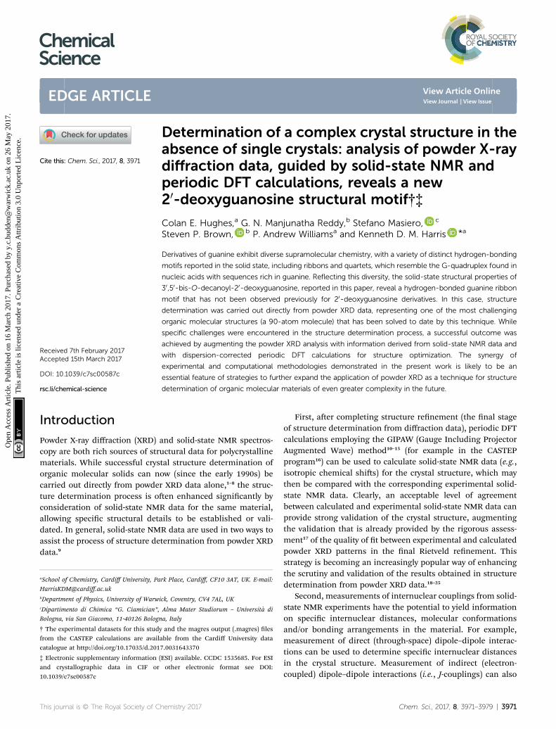
This paper is focused on structure determination directlyfrom powder XRD data in tandem with consideration of solid-state NMR data, specically to elucidate the structure of 30,50-bis-O-decanoyl-20-deoxyguanosine [denoted dG(C10)2; Fig. 1].This material is believed to be polymorphic, as two distinct solidforms have been identied on crystallization from ethanol. Inprevious work, Pham et al.28 referred to these two forms as 2qand 2r. The material studied in the present work corresponds to2q, as the powder XRD data matches the powder XRD data for2q published previously.29 We note that 2q appears to be morereadily obtained, as 2r has only been reported once.28 In order tointroduce a systematic nomenclature, we dene polymorph I ofdG(C10)2 as 2q and we dene polymorph II of dG(C10)2 as 2r.
The dG(C10)2 molecule has found applications in the context ofphotoelectric devices, including photoconductive materials,31–33
biphotonic quantum dots34 and photodetectors with rectifyingproperties.35 It has also been shown36 that dG(C10)2 can reversiblyinterconvert between quartets and ribbons, using a cryptand forcation capture and addition of acid to release the cation. In allthese applications, the hydrogen bonding of the guanine moietiesis a key factor, emphasizing the importance of understanding thepreferred structural properties of dG(C10)2 in the solid state.Among 30,50-bis-O-alkanoyl derivatives of 20-deoxyguanosine,crystal structures have been reported previously only for 30,50-bis-O-acetyl-20-deoxyguanosine [dG(C2)2]37 and 30,50-bis-O-propanoyl-20-deoxyguanosine [dG(C3)2],38 although several 30,50-bis-O-silylderivatives have also been studied39,40 and self-assembly of 20-deoxyguanosine derivatives in solution has been investigated.41–44
Guanine derivatives are known for their rich supramolecularchemistry.47–49 In the solid state, a variety of distinct hydrogen-bonding motifs have been reported, including ribbons and
Fig. 1 Molecular structure of dG(C10)2 showing the atom numberingscheme. The green bracket indicates the Watson–Crick hydrogen-bonding groups. The non-hydrogen atoms of the guanine moiety arelabelled 1 to 10 and the non-hydrogen atoms of the 20-deoxyribosemoiety are labelled 10 to 60 and 100. Note that the atom labelled here asN10 was labelled N2 or NH2 in previous publications28–30 on dG(C10)2.
3972 | Chem. Sci., 2017, 8, 3971–3979
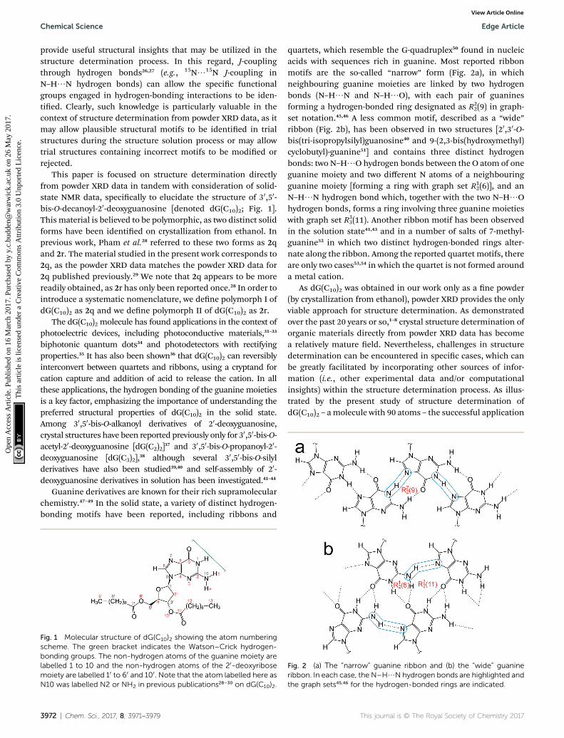
quartets, which resemble the G-quadruplex50 found in nucleicacids with sequences rich in guanine. Most reported ribbonmotifs are the so-called “narrow” form (Fig. 2a), in whichneighbouring guanine moieties are linked by two hydrogenbonds (N–H/N and N–H/O), with each pair of guaninesforming a hydrogen-bonded ring designated as R2
2(9) in graph-set notation.45,46 A less common motif, described as a “wide”ribbon (Fig. 2b), has been observed in two structures [20,30-O-bis(tri-isopropylsilyl)guanosine40 and 9-(2,3-bis(hydroxymethyl)cyclobutyl)-guanine51] and contains three distinct hydrogenbonds: two N–H/O hydrogen bonds between the O atom of oneguanine moiety and two different N atoms of a neighbouringguanine moiety [forming a ring with graph set R1
2(6)], and anN–H/N hydrogen bond which, together with the two N–H/Ohydrogen bonds, forms a ring involving three guanine moietieswith graph set R33(11). Another ribbon motif has been observedin the solution state41,43 and in a number of salts of 7-methyl-guanine52 in which two distinct hydrogen-bonded rings alter-nate along the ribbon. Among the reported quartet motifs, thereare only two cases53,54 in which the quartet is not formed arounda metal cation.
As dG(C10)2 was obtained in our work only as a ne powder(by crystallization from ethanol), powder XRD provides the onlyviable approach for structure determination. As demonstratedover the past 20 years or so,1–8 crystal structure determination oforganic materials directly from powder XRD data has becomea relatively mature eld. Nevertheless, challenges in structuredetermination can be encountered in specic cases, which canbe greatly facilitated by incorporating other sources of infor-mation (i.e., other experimental data and/or computationalinsights) within the structure determination process. As illus-trated by the present study of structure determination ofdG(C10)2 – amolecule with 90 atoms – the successful application
Fig. 2 (a) The “narrow” guanine ribbon and (b) the “wide” guanineribbon. In each case, the N–H/N hydrogen bonds are highlighted andthe graph sets45,46 for the hydrogen-bonded rings are indicated.
This journal is © The Royal Society of Chemistry 2017
Edge Article Chemical Science
Ope
n A
cces
s A
rtic
le. P
ublis
hed
on 1
6 M
arch
201
7. P
urch
ased
by
y.c.
budd
en@
war
wic
k.ac
.uk
on 2
6 M
ay 2
017.
Thi
s ar
ticle
is li
cens
ed u
nder
a C
reat
ive
Com
mon
s A
ttrib
utio
n 3.
0 U
npor
ted
Lic
ence
.View Article Online
of techniques for structure determination from powder XRDdata is not just limited to the case of relatively small molecules.
Methods
The sample of dG(C10)2 was prepared using the methoddescribed previously28 and crystallized from ethanol. PowderXRD data conrmed that the sample was polymorph I, as denedabove. High-quality powder XRD data suitable for structuredetermination were recorded (at 21 �C), for a powder samplecontained in two ame-sealed capillaries, using a Bruker D8Diffractometer (Ge-monochromated CuKa1 radiation) operatingin transmission mode with a Vantec detector covering 3� in 2q.The data were recorded in the 2q range from 3� to 50� (step size0.017�) with a total data collection time of 57 h. A two-dimensional powder XRD pattern was also recorded at ambienttemperature using an Agilent SuperNova Dual Atlas diffractom-eter (CuKa radiation, l ¼ 1.54180 A) in order to assess the extentof “preferred orientation” in the powder sample.55
Periodic DFT calculations for geometry optimization andcalculation of NMR parameters were carried out using theCASTEP program16 (Academic Release version 8.0). Geometryoptimization used ultraso pseudopotentials,56 PBE func-tional,57 semiempirical dispersion corrections (TS correctionscheme58), xed unit cell, preserved space group symmetry andperiodic boundary conditions. Isotropic NMR chemical shiswere calculated using the GIPAW approach,10–14 while J-couplingvalues were calculated at the scalar-relativistic level of theoryusing the ZORA method.59–61 All calculations used a basis setcut-off energy of 700 eV and a Monkhorst–Pack grid62 ofminimum sample spacing 0.05 � 2p A�1. In the rst instance,chemical shis are referenced using the formula
diso(calc) ¼ sref � siso(calc) (1)
where sref is the sum of the mean of the calculated shieldingvalues and the mean of the experimental chemical shis.11 Asecond referencing method uses the formula
diso(calc) ¼ s0 � m siso(calc) (2)
with the values of s0 andm obtained from a least-squares ttingprocedure to optimize the agreement between calculated andexperimental chemical shis.
Structure determination
The powder XRD pattern of polymorph I of dG(C10)2 wasindexed using the DICVOL91 algorithm63 in the program Crys-re,64 giving the following unit cell with monoclinic metricsymmetry: a ¼ 8.33 A, b ¼ 7.82 A, c ¼ 25.79 A, b ¼ 97.5�.However, in the subsequent prole-tting stage of the structuredetermination process, progress was hampered signicantly bytwo specic features of the powder XRD pattern of dG(C10)2,which introduced challenges in achieving an acceptable qualityof prole tting (Fig. 3). First, the powder XRD pattern ofdG(C10)2 contains a very intense peak at low diffraction angle (2q
This journal is © The Royal Society of Chemistry 2017
¼ 3.4�), which is substantially more intense than any of theother peaks. The presence of one peak of dominant intensityinitially raised the possibility that the powder XRD data may bestrongly affected by preferred orientation of the crystallites inthe sample. However, the powder XRD pattern (see Fig. S1 inESI‡) recorded using a two-dimensional detector (for one of thetwo capillaries used to record the one-dimensional powder XRDdata in Fig. 3) exhibited uniform intensity around the Debye–Scherrer rings, indicating that the distribution of crystalliteorientations in the powder sample was essentially random andhence that there was no signicant preferred orientation.
The second challenging aspect concerns the very highbackground in the low-angle region of the powder XRD pattern,arising from a signicant amount of X-ray scattering from air inthe region of the peak at 2q ¼ 3.4�. To achieve a high quality oft in the prole-tting stage, which was carried out using the LeBail technique65 in the GSAS program,66 it was necessary rst tot the baseline of the low-angle region (2q ¼ 3� to 5�) usinga polynomial, which was then subtracted from the experimentaldata. Although this procedure introduced some artefacts to thebaseline, these artefacts were tted successfully by the shiedChebyshev polynomials67 used for baseline correction in the LeBail tting procedure in GSAS. We note that attempts to t theoriginal baseline using this method were not successful.
The modied powder XRD data were then subjected to LeBail tting (Fig. 4a; due to the high intensity of the rst peakrelative to all other peaks, the data between 2q ¼ 6� and 40� areshown separately with an expanded intensity scale in Fig. 4b).The lineshape of the rst peak is rather poorly tted asa consequence of the double baseline tting described above.Nevertheless, the overall quality of t obtained in the Le Bailtting is considered acceptable (Fig. 4a; Rp ¼ 0.93%, Rwp ¼1.23%).
Density considerations suggest that there are two moleculesof dG(C10)2 in the unit cell and, given the fact that the dG(C10)2molecule is chiral, the only plausible space groups are P2 andP21. As these space groups could not be distinguished deni-tively on the basis of systematic absences in the powder XRDdata [although the absence of the (010) peak may point towardsP21], each of these space groups was considered in independentstructure-solution calculations using the direct-space geneticalgorithm technique68–70 in the program EAGER.71–76 Structure-solution calculations for space group P2 did not generate anyplausible trial structures and only space group P21 wasconsidered further.
Previous solid-state NMR studies of dG(C10)2 provide directstructural insights concerning the hydrogen-bonding betweenguanine moieties. Pham et al.28,30 determined the 15N chemicalshis and J-couplings for polymorph I of dG(C10)2 (see Table 1),including a 2hJN7N10 coupling of 5.9 Hz, while Webber et al.29
reported 1H and 13C chemical shis and found evidence forseveral H/H short contacts. The value of 2hJN7N10 providesa strong indication that there is a relatively strong N–H/Nhydrogen bond involving N7 and N10, which provided a robustcriterion for acceptance or rejection of trial structures obtainedin the structure solution from powder XRD data reported here(particularly as a basis for rejecting trial structures that clearly
Chem. Sci., 2017, 8, 3971–3979 | 3973
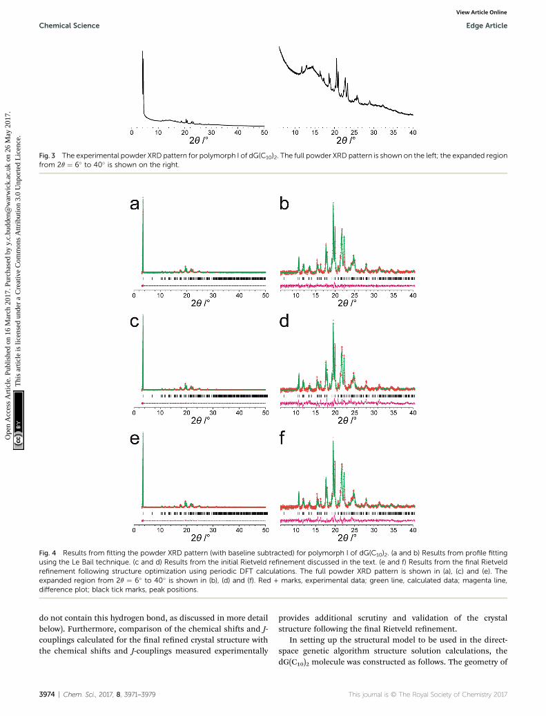
Fig. 3 The experimental powder XRD pattern for polymorph I of dG(C10)2. The full powder XRD pattern is shown on the left; the expanded regionfrom 2q ¼ 6� to 40� is shown on the right.
Fig. 4 Results from fitting the powder XRD pattern (with baseline subtracted) for polymorph I of dG(C10)2. (a and b) Results from profile fittingusing the Le Bail technique. (c and d) Results from the initial Rietveld refinement discussed in the text. (e and f) Results from the final Rietveldrefinement following structure optimization using periodic DFT calculations. The full powder XRD pattern is shown in (a), (c) and (e). Theexpanded region from 2q ¼ 6� to 40� is shown in (b), (d) and (f). Red + marks, experimental data; green line, calculated data; magenta line,difference plot; black tick marks, peak positions.
Chemical Science Edge Article
Ope
n A
cces
s A
rtic
le. P
ublis
hed
on 1
6 M
arch
201
7. P
urch
ased
by
y.c.
budd
en@
war
wic
k.ac
.uk
on 2
6 M
ay 2
017.
Thi
s ar
ticle
is li
cens
ed u
nder
a C
reat
ive
Com
mon
s A
ttrib
utio
n 3.
0 U
npor
ted
Lic
ence
.View Article Online
do not contain this hydrogen bond, as discussed in more detailbelow). Furthermore, comparison of the chemical shis and J-couplings calculated for the nal rened crystal structure withthe chemical shis and J-couplings measured experimentally
3974 | Chem. Sci., 2017, 8, 3971–3979
provides additional scrutiny and validation of the crystalstructure following the nal Rietveld renement.
In setting up the structural model to be used in the direct-space genetic algorithm structure solution calculations, thedG(C10)2 molecule was constructed as follows. The geometry of
This journal is © The Royal Society of Chemistry 2017

Table 1 Calculated and experimental JNN-couplings for polymorph Iof dG(C10)2
Coupling
JNN/Hz
Calculated Experimentala
3JN3N9 4.20 3.5b, 3.8b3JN3N10 5.45 5.32hJN7N10 7.10 5.9
a Taken from Pham et al. (2007).30 b The two values correspond toseparate measurements on each resonance.
Edge Article Chemical Science
Ope
n A
cces
s A
rtic
le. P
ublis
hed
on 1
6 M
arch
201
7. P
urch
ased
by
y.c.
budd
en@
war
wic
k.ac
.uk
on 2
6 M
ay 2
017.
Thi
s ar
ticle
is li
cens
ed u
nder
a C
reat
ive
Com
mon
s A
ttrib
utio
n 3.
0 U
npor
ted
Lic
ence
.View Article Online
the guanine moiety was modelled on the structure of one of themolecules in the reported crystal structure77 of guanosinedihydrate (CCDC ref. code GUANSH10) and the geometry of the20-deoxyribose ring was modelled on that in the reported crystalstructure38 of dG(C3)2 (CCDC ref. code MOFBUE). The two C10
chains were constructed using the average bond lengths andbond angles for similar moieties determined using the programMogul version 1.7.1 (for bonds not involving hydrogen) andfrom Allen et al.78 (for bonds involving hydrogen). The confor-mation of the 20-deoxyribose ring was kept xed during thestructure solution calculation. As the position along the b-axiscan be xed arbitrarily for space group P21, each trial structurewas dened by a total of 27 structural variables (2 positional, 3orientational and 22 torsional variables). The 22 torsionalvariables are specied in Fig. S2.‡
With this model, the structure solution calculations in spacegroup P21 generated trial structures that were consideredplausible, including a geometric relation between N7 and N10consistent with N–H/N hydrogen bonding. In contrast, struc-ture solution calculations using other models for the dG(C10)2molecule (e.g., with the geometry of the 20-deoxyribose ringbased on the average bond lengths and bond angles for similarmoieties) led to trial structures that were considered implau-sible as they did not contain hydrogen bonding between N7 andN10.
The genetic algorithm structure solution calculations inspace group P21 involved the evolution of 32 independentpopulations of 500 structures, with 50 mating operations and250 mutation operations carried out per generation, and a totalof 500 generations in each calculation. In two of the calcula-tions, the trial structure giving the best quality of t betweencalculated and experimental powder XRD data was essentiallythe same structure, and the quality of t was signicantly betterthan the best-t structure obtained in any of the other calcu-lations (see ESI‡ for more details). The trial structure giving thebest quality of t from all the structure-solution calculationswas used as the initial structural model for Rietveld rene-ment,79 which was carried out using the GSAS program.66 In theRietveld renement, restraints were applied to bond lengthsand bond angles based on the initial molecular model (dis-cussed above) and planar restraints were applied to the guaninemoiety and the two carbonyl moieties. These restraints wererelaxed over the course of the renement. A common isotropicatomic displacement parameter was rened for all non-
This journal is © The Royal Society of Chemistry 2017
hydrogen atoms and the value for hydrogen atoms was setequal to 1.2 times the rened value for non-hydrogen atoms. Nocorrections were applied for preferred orientation. The Rietveldrenement at this stage gave a reasonably good t to the powderXRD data (Fig. 4c; Rp ¼ 1.35%, Rwp ¼ 1.86%).
The structure obtained in this Rietveld renement was thensubjected to geometry optimization using the CASTEP program,leading to small shis in atomic positions with an averageatomic displacement of 0.65 A. The most signicant structuralchanges concerned the orientations of the two carbonyl moie-ties in the decanoyl chains. The structure obtained followinggeometry optimization was then used as the starting structuralmodel for a nal Rietveld renement, which gave an improvedt (Fig. 4e; Rp ¼ 1.15%, Rwp ¼ 1.56%) compared to the rstRietveld renement discussed above. The nal rened unit cellparameters were: a ¼ 8.3072(7) A, b ¼ 7.8052(10) A, c ¼25.7246(27) A, b ¼ 97.491(4), V ¼ 1653.73(31) A3 (2q range, 3–50�; 2755 prole points; 289 rened variables). Overall, thecombination of geometry optimization followed by furtherRietveld renement led to an average atomic displacement of0.71 A, with signicant changes in the conformations of thedecanoyl chains (particularly in the region of the carbonylmoieties) and a small shi of the 20-deoxyguanosine moiety,which led to an improvement in geometrical aspects of thehydrogen bonding between guanine moieties in neighbouringmolecules.
Discussion
The crystal structure from the nal Rietveld renement isshown in Fig. 5 and 6. Viewed along the b-axis (Fig. 5), it is clearthat the structure comprises hydrogen-bonded ribbons con-structed from the guanine moieties (the view in Fig. 5 is parallelto the plane of the ribbons). The ribbons run parallel to the b-axis and the guanine moieties within a given ribbon are relatedby the 21 screw axis. The 20-deoxyribose moiety and alkyl chainsoccupy the space between adjacent ribbons. The ribbons involvethree distinct hydrogen bonds: two N–H/O hydrogen bondsinvolving N–H10b and N–H1 of a given molecule as donors andthe C]O group of a neighbouring guanine moiety as theacceptor, and an N–H/N hydrogen bond involving N–H10a asthe donor and the N7 atom of a neighbouring guaninemoiety asthe acceptor (geometric data for these hydrogen bonds are givenin Table S1 in ESI‡). The ribbons in dG(C10)2 are unambiguouslyidentied (compare Fig. 6 and 2b) as the “wide” ribbonmotif,40,51 which has not been observed previously for any 20-deoxyguanosine derivative. A C–H/O hydrogen bond is alsoidentied (see Fig. 6) between C8 of the guanine moiety as theC–H donor and the C]O group containing C110 of a neigh-bouring molecule as the acceptor.
There is also evidence for p/p interactions betweenguanine moieties in adjacent ribbons in the crystal structure ofdG(C10)2, as the distances from the N3, C2 and N10 atoms of oneguanine moiety to the N7, C8 and N9 atoms, respectively, ofa neighbouring molecule are all ca. 3.5 A (Fig. 7). Such p/p
interactions are not observed in the two previously reportedcrystal structures40,51 containing the “wide” ribbon motif.
Chem. Sci., 2017, 8, 3971–3979 | 3975

Fig. 5 Crystal structure of polymorph I of dG(C10)2 viewed along theb-axis (parallel to the direction of the hydrogen-bonded ribbons).
Fig. 6 Crystal structure of polymorph I of dG(C10)2 showing thehydrogen-bonded ribbon of the guanine moieties. In this view, the b-axis is vertical.
Fig. 7 Illustration of p/p interactions between guanine moieties inthe crystal structure of polymorph I of dG(C10)2. The dashed linesrepresent distances of ca. 3.5 A.
Chemical Science Edge Article
Ope
n A
cces
s A
rtic
le. P
ublis
hed
on 1
6 M
arch
201
7. P
urch
ased
by
y.c.
budd
en@
war
wic
k.ac
.uk
on 2
6 M
ay 2
017.
Thi
s ar
ticle
is li
cens
ed u
nder
a C
reat
ive
Com
mon
s A
ttrib
utio
n 3.
0 U
npor
ted
Lic
ence
.View Article Online
The relative arrangement of the 20-deoxyribose and guaninemoieties around the N-glycosidic bond (N9–C10) corresponds tothe syn conformation, with the Watson–Crick hydrogen-bonding groups (see Fig. 1) directed towards the 20-deoxyri-bose ring.80 Signicantly, Webber et al.29 predicted that thecrystal structure of dG(C10)2 should exhibit this structuralfeature, based on the high values of isotropic 13C chemical shifor C8 and C10, which are characteristic of the syn conformation.It is noteworthy that the only guanosine derivative that formsthe “wide” ribbon motif in its crystal structure also has the synconformation.40
The isotropic 1H, 13C and 15N chemical shis calculatedusing the CASTEP program for the crystal structure of poly-morph I of dG(C10)2 determined here are compared with theexperimental values29,30 in Fig. 8 (see also Tables S2–S4‡). Thecalculated (using eqn (1)) and experimental data are in verygood agreement, with RMS deviations of 0.57 ppm, 3.02 ppmand 2.01 ppm for the 1H, 13C and 15N chemical shis, respec-tively. From Fig. 8b, it is evident that the calculated 13C chem-ical shis are higher than the experimental data for theresonances at high ppm and lower than the experimental datafor the resonances at low ppm. This phenomenon is well knownand can be addressed empirically either by establishing thecalculated chemical shis using eqn (2) and the least-squarestting procedure (in which the gradient m may deviate fromunity) described in the Methods section, or by using differentreference shieldings for the high-ppm region and the low-ppm
3976 | Chem. Sci., 2017, 8, 3971–3979
region of the spectrum.11,23,81 As shown in Fig. S3,‡ when thecalculated 13C chemical shis are established using eqn (2), theRMS deviation between calculated and experimental 13Cchemical shis is decreased to 2.51 ppm. Using the sameprocedure (based on eqn (2)) to establish the calculated 1H and15N chemical shis, the RMS deviations between calculated andexperimental data are decreased to 0.39 ppm and 1.99 ppm,respectively.
Hartman et al.82 have reported that GIPAW calculations of13C chemical shis across a range of small organic moleculesgive an RMS deviation of 2.12 ppm when using the procedurebased on eqn (2). Although this deviation is slightly lower thanthat obtained for our results, it is important to note that thedG(C10)2 molecule is signicantly larger and more exible thanany of the molecules considered by Hartman et al. Furthermore,the slightly higher RMS deviation observed for dG(C10)2 may be
This journal is © The Royal Society of Chemistry 2017

Fig. 8 Correlation plots for the calculated and experimental values of the (a) 1H, (b) 13C and (c) 15N isotropic chemical shifts for polymorph I ofdG(C10)2. In each case, the dashed line corresponds to diso(expt) ¼ diso(calc).
Edge Article Chemical Science
Ope
n A
cces
s A
rtic
le. P
ublis
hed
on 1
6 M
arch
201
7. P
urch
ased
by
y.c.
budd
en@
war
wic
k.ac
.uk
on 2
6 M
ay 2
017.
Thi
s ar
ticle
is li
cens
ed u
nder
a C
reat
ive
Com
mon
s A
ttrib
utio
n 3.
0 U
npor
ted
Lic
ence
.View Article Online
caused, in part, by the fact that 13C chemical shis for the CH2
moieties were not included in our analysis as the 13C resonancesfor individual CH2moieties are not resolved in the experimental13C NMR spectrum.
Three 15N/15N J-couplings across N–H/N hydrogen bondsbetween guanine moieties were also calculated (see Table 1),specically the intramolecular couplings 3JN3N10 and
3JN3N9, andthe intermolecular coupling 2hJN7N10. In each case, the calcu-lated J-coupling is higher, to a greater or lesser extent, than theexperimental value, but the calculated values successfullyreect the correct trend.
Concluding remarks
Several aspects of the structure determination of dG(C10)2 frompowder XRD data reported in this paper presented challenges,including the presence of the very intense (001) peak at lowangle in the powder XRD pattern on a very steeply slopingbaseline. This peak represents more than 45% of the totaldiffraction intensity across the 2q range recorded and it wasessential to ensure that the method applied for baselinecorrection did not signicantly distort this peak. This factor,combined with the complexity of the direct-space searchinvolved in the structure-solution calculation (a consequence ofthe large size and exibility of the dG(C10)2 molecule), led toa large number of distinct trial structures giving similar ts tothe data. In order to identify the correct structure solution,
This journal is © The Royal Society of Chemistry 2017
information obtained in previous solid-state NMR studies ofdG(C10)2 proved to be vital, in particular the knowledge thata strong intermolecular N–H/N hydrogen bond exists betweenN7 and N10. Aer initial Rietveld renement, geometry opti-mization using periodic DFT calculations (with xed unit cell)generated a structure which, upon further Rietveld renement,gave an improved t to the experimental powder XRD data,illustrating the utility of introducing geometry optimization asa key step in the overall structure elucidation process. Clearly,the fact that a wide range of solid-state NMR parameterscalculated from the nal rened crystal structure are in goodagreement with the corresponding experimental solid-stateNMR parameters gives additional support to the veracity ofthe structure determined from powder XRD data. The synergy ofexperimental and computational methodologies demonstratedin the present work is likely to be an essential feature of strat-egies to further expand the application of powder XRD asa technique for structure determination of organic molecularmaterials of even greater complexity in the future.
Acknowledgements
We are grateful to Cardiff University for nancial support.CASTEP calculations were carried out on the Cardiff Universitysupercomputer, Raven, run by Advanced Research Computing@ Cardiff. This research was carried out within the frameworkof the EPSRC funded Collaborative Computational Project for
Chem. Sci., 2017, 8, 3971–3979 | 3977

Chemical Science Edge Article
Ope
n A
cces
s A
rtic
le. P
ublis
hed
on 1
6 M
arch
201
7. P
urch
ased
by
y.c.
budd
en@
war
wic
k.ac
.uk
on 2
6 M
ay 2
017.
Thi
s ar
ticle
is li
cens
ed u
nder
a C
reat
ive
Com
mon
s A
ttrib
utio
n 3.
0 U
npor
ted
Lic
ence
.View Article Online
NMR Crystallography CCP-NC (EP/M022501/1). GNMR and SPBacknowledge funding from EPSRC (EP/K003674/1). SMacknowledges the University of Bologna and the EuropeanCommunity (EC FP7 ICT-MOLARNET project 318516).
References
1 P. Lightfoot, M. Tremayne, K. D. M. Harris and P. G. Bruce, J.Chem. Soc., Chem. Commun., 1992, 1012–1013.
2 K. D. M. Harris, M. Tremayne, P. Lightfoot and P. G. Bruce, J.Am. Chem. Soc., 1994, 116, 3543–3547.
3 V. V. Chernyshev, Russ. Chem. Bull., 2001, 50, 2273–2292.4 K. D. M. Harris, M. Tremayne and B. M. Kariuki, Angew.Chem., Int. Ed., 2001, 40, 1626–1651.
5 Structure Determination from Powder Diffraction Data, ed. W.I. F. David, K. Shankland, L. B. McCusker and C.Baerlocher, IUCr/OUP, 2002.
6 H. Tsue, M. Horiguchi, R. Tamura, K. Fujii and H. Uekusa, J.Synth. Org. Chem. Jpn., 2007, 65, 1203–1212.
7 W. I. F. David and K. Shankland, Acta Crystallogr., Sect. A:Found. Crystallogr., 2008, 64, 52–64.
8 K. D. M. Harris, Top. Curr. Chem., 2012, 315, 133–178.9 K. D. M. Harris and M. Xu, in NMR Crystallography, ed. R. K.Harris, R. Wasylishen andM. J. Duer, 2009, ch. 19, pp. 275–287.
10 C. J. Pickard and F. Mauri, Phys. Rev. B: Condens. MatterMater. Phys., 2001, 63, 245101.
11 R. K. Harris, P. Hodgkinson, C. J. Pickard, J. R. Yates andV. Zorin, Magn. Reson. Chem., 2007, 45, S174–S186.
12 J. R. Yates, C. J. Pickard and F. Mauri, Phys. Rev. B: Condens.Matter Mater. Phys., 2007, 76, 024401.
13 T. Charpentier, Solid State Nucl. Magn. Reson., 2011, 40, 1–20.14 C. Bonhomme, C. Gervais, F. Babonneau, C. Coelho,
F. Pourpoint, T. Azaıs, S. E. Ashbrook, J. M. Griffin,J. R. Yates, F. Mauri and C. J. Pickard, Chem. Rev., 2012,112, 5733–5779.
15 S. E. Ashbrook and D. McKay, Chem. Commun., 2016, 52,7186–7204.
16 S. J. Clark, M. D. Segall, C. J. Pickard, P. J. Hasnip,M. J. Probert, K. Refson and M. C. Payne, Z. Kristallogr.,2005, 220, 567–570.
17 Z. Pan, E. Y. Cheung, K. D. M. Harris, E. C. Constable andC. E. Housecro, Cryst. Growth Des., 2005, 5, 2084–2090.
18 M. Tremayne, B. M. Kariuki and K. D. M. Harris, Angew.Chem., Int. Ed. Engl., 1997, 36, 770–772.
19 X. Filip, G. Borodi and C. Filip, Phys. Chem. Chem. Phys.,2011, 13, 17978–17986.
20 D. V. Dudenko, P. A. Williams, C. E. Hughes, O. N. Antzutkin,S. P. Velaga, S. P. Brown and K. D. M. Harris, J. Phys. Chem. C,2013, 117, 12258–12265.
21 P. Li, Y. Chu, L. Wang, R. M.Wenslow Jr, K. Yu, H. Zhang andZ. Deng, CrystEngComm, 2014, 16, 3141–3147.
22 X. Li, A. D. Bond, K. E. Johansson and J. Van de Streek, ActaCrystallogr., Sect. C: Struct. Chem., 2014, 70, 784–789.
23 G. N. M. Reddy, D. S. Cook, D. Iuga, R. I. Walton, A. Marshand S. P. Brown, Solid State Nucl. Magn. Reson., 2015, 65,41–48.
3978 | Chem. Sci., 2017, 8, 3971–3979
24 M. Sardo, S. M. Santos, A. A. Babaryk, C. Lopez, I. Alkorta,J. Elguero, R. M. Claramunt and L. Mafra, Solid State Nucl.Magn. Reson., 2015, 65, 49–63.
25 A. E. Watts, K. Maruyoshi, C. E. Hughes, S. P. Brown andK. D. M. Harris, Cryst. Growth Des., 2016, 16, 1798–1804.
26 A. J. Dingley and S. Grzesiek, J. Am. Chem. Soc., 1998, 120, 54–64.
27 I. G. Shenderovich, S. N. Smirnov, G. S. Denisov,V. A. Gindin, N. S. Golubev, A. Dunger, R. Reibke,S. Kirpekar, O. L. Malkina and H.-H. Limbach, Ber. Bunsen-Ges. Phys. Chem., 1998, 102, 422–428.
28 T. N. Pham, S. Masiero, G. Gottarelli and S. P. Brown, J. Am.Chem. Soc., 2005, 127, 16018–16019.
29 A. L. Webber, S. Masiero, S. Pieraccini, J. C. Burley,A. S. Tatton, D. Iuga, T. N. Pham, G. P. Spada andS. P. Brown, J. Am. Chem. Soc., 2011, 133, 19777–19795.
30 T. N. Pham, J. M. Griffin, S. Masiero, S. Lena, G. Gottarelli,P. Hodgkinson, C. Filipe and S. P. Brown, Phys. Chem.Chem. Phys., 2007, 9, 3416–3423.
31 R. Rinaldi, E. Branca, R. Cingolani, S. Masiero, G. P. Spadaand G. Gottarelli, Appl. Phys. Lett., 2001, 78, 3541–3543.
32 G. Maruccio, P. Visconti, V. Arima, S. D'Amico, A. Biasco,E. D'Amone, R. Cingolani, R. Rinaldi, S. Masiero, T. Giorgiand G. Gottarelli, Nano Lett., 2003, 3, 479–483.
33 R. Rinaldi, G. Maruccio, A. Biasco, V. Arima, R. Cingolani,T. Giorgi, S. Masiero, G. P. Spada and G. Gottarelli,Nanotechnology, 2002, 13, 398–403.
34 A. Neogi, J. Li, P. B. Neogi, A. Sarkar and H. Morkoc, Electron.Lett., 2004, 40, 1605–1606.
35 H. Liddar, J. Li, A. Neogi, P. B. Neogi, A. Sarkar, S. Cho andH. Morkoç, Appl. Phys. Lett., 2008, 92, 013309.
36 S. Pieraccini, S. Masiero, O. Pandoli, P. Samorı andG. P. Spada, Org. Lett., 2006, 8, 3125–3128.
37 L. H. Koole, H. M. Buck, J. A. Kanters and A. Schouten, Can. J.Chem., 1988, 66, 2634–2639.
38 T. Giorgi, F. Grepioni, I. Manet, P. Mariani, S. Masiero,E. Mezzina, S. Pieraccini, L. Saturni, G. P. Spada andG. Gottarelli, Chem.–Eur. J., 2002, 8, 2143–2152.
39 T. Sato, M. Seko, R. Takasawa, I. Yoshikawa and K. Araki, J.Mater. Chem., 2001, 11, 3018–3022.
40 R. Takasawa, I. Yoshikawa and K. Araki, Org. Biomol. Chem.,2004, 2, 1125–1132.
41 G. Gottarelli, S. Masiero, E. Mezzina, G. P. Spada, P. Marianiand M. Recanatini, Helv. Chim. Acta, 1998, 81, 2078–2092.
42 A. L. Marlow, E. Mezzina, G. P. Spada, S. Masiero, J. T. Davisand G. Gottarelli, J. Org. Chem., 1999, 64, 5116–5123.
43 G. Gottarelli, S. Masiero, E. Mezzina, S. Pieraccini, J. P. Rabe,P. Samorı and G. P. Spada, Chem.–Eur. J., 2000, 6, 3242–3248.
44 E. Mezzina, P. Mariani, R. Itri, S. Masiero, S. Pieraccini,G. P. Spada, F. Spinozzi, J. T. Davis and G. Gottarelli,Chem.–Eur. J., 2001, 7, 388–395.
45 M. C. Etter, Acc. Chem. Res., 1990, 23, 120–126.46 M. C. Etter, J. C. MacDonald and J. Bernstein, Acta
Crystallogr., Sect. B: Struct. Sci., 1990, 46, 256–262.47 J. T. Davis, Angew. Chem., Int. Ed., 2004, 43, 668–698.48 J. T. Davis and G. P. Spada, Chem. Soc. Rev., 2007, 36, 296–
313.
This journal is © The Royal Society of Chemistry 2017

Edge Article Chemical Science
Ope
n A
cces
s A
rtic
le. P
ublis
hed
on 1
6 M
arch
201
7. P
urch
ased
by
y.c.
budd
en@
war
wic
k.ac
.uk
on 2
6 M
ay 2
017.
Thi
s ar
ticle
is li
cens
ed u
nder
a C
reat
ive
Com
mon
s A
ttrib
utio
n 3.
0 U
npor
ted
Lic
ence
.View Article Online
49 S. Lena, S. Masiero, S. Pieraccini and G. P. Spada, Chem.–Eur.J., 2009, 15, 7792–7806.
50 M. Gellert, M. N. Lipsett and D. R. Davies, Proc. Natl. Acad.Sci. U. S. A., 1962, 48, 2013–2018.
51 G. S. Bisacchi, J. Singh, J. J. D. Godfrey, T. P. Kissick, T. Mitt,M. F. Malley, J. D. D. Marco, J. Z. Gougoutas, R. H. Muellerand R. Zahler, J. Org. Chem., 1995, 60, 2902–2905.
52 A. Kozma, S. Ibanez, R. Silaghi-Dumitrescu, P. J. S. Miguel,D. Guptaa and B. Lippert, Dalton Trans., 2012, 41, 6094–6103.
53 Y. Inui, M. Shiro, S. Fukuzumi and T. Kojima, Org. Biomol.Chem., 2013, 11, 758–764.
54 J. L. Sessler, M. Sathiosatham, K. Doerr, V. Lynch andK. A. Abboud, Angew. Chem., Int. Ed., 2000, 39, 1300–1303.
55 E. Y. Cheung, K. D. M. Harris and B. M. Foxman, Cryst.Growth Des., 2003, 3, 705–710.
56 D. Vanderbilt, Phys. Rev. B: Condens. Matter, 1990, 41, 7892–7895.
57 J. P. Perdew, K. Burke and M. Ernzerhof, Phys. Rev. Lett.,1996, 71, 3865–3868.
58 A. Tkatchenko and M. Scheffler, Phys. Rev. Lett., 2009, 102,073005.
59 S. A. Joyce, J. R. Yates, C. J. Pickard and F. Mauri, J. Chem.Phys., 2007, 127, 204107.
60 S. A. Joyce, J. R. Yates, C. J. Pickard and S. P. Brown, J. Am.Chem. Soc., 2008, 130, 12663–12670.
61 T. F. G. Green and J. R. Yates, J. Chem. Phys., 2014, 140,234106.
62 H. J. Monkhorst and J. D. Pack, Phys. Rev. B: Condens. MatterMater. Phys., 1976, 13, 5188–5192.
63 A. Boultif and D. Louer, J. Appl. Crystallogr., 1991, 24, 987–993.
64 R. Shirley, The CRYSFIRE System for Automatic PowderIndexing: User's Manual, The Lattice Press, Guildford, U.K.,1999.
65 A. Le Bail, H. Duroy and J. L. Fourquet, Mater. Res. Bull.,1988, 23, 447–452.
This journal is © The Royal Society of Chemistry 2017
66 A. C. Larson and R. B. Von Dreele, Los Alamos NationalLaboratory Report, LAUR, 2004, pp. 86–748.
67 J. C. Mason and D. C. Handscomb, Chebyshev Polynomials,Chapman & Hall/CRC, Boca Raton, Florida, 2003.
68 B. M. Kariuki, H. Serrano-Gonzalez, R. L. Johnston andK. D. M. Harris, Chem. Phys. Lett., 1997, 280, 189–195.
69 K. D. M. Harris, R. L. Johnston and B. M. Kariuki, ActaCrystallogr., Sect. A: Found. Crystallogr., 1998, 54, 632–645.
70 K. D. M. Harris, S. Habershon, E. Y. Cheung andR. L. Johnston, Z. Kristallogr., 2004, 219, 838–846.
71 B. M. Kariuki, P. Calcagno, K. D. M. Harris, D. Philp andR. L. Johnston, Angew. Chem., Int. Ed., 1999, 38, 831–835.
72 B. M. Kariuki, K. Psallidas, K. D. M. Harris, R. L. Johnston,R. W. Lancaster, S. E. Staniforth and S. M. Cooper, Chem.Commun., 1999, 1677–1678.
73 E. Tedesco, G. W. Turner, K. D. M. Harris, R. L. Johnston andB. M. Kariuki, Angew. Chem., Int. Ed., 2000, 39, 4488–4491.
74 D. Albesa-Jove, B. M. Kariuki, S. J. Kitchin, L. Grice,E. Y. Cheung and K. D. M. Harris, ChemPhysChem, 2004, 5,414–418.
75 K. Fujii, A. Lazuen Garay, J. Hill, E. Sbircea, Z. Pan, M. Xu,D. C. Apperley, S. L. James and K. D. M. Harris, Chem.Commun., 2010, 46, 7572–7574.
76 P. A. Williams, C. E. Hughes and K. D. M. Harris, Angew.Chem., Int. Ed., 2015, 54, 3973–3977.
77 U. Thewalt, C. E. Bugg and R. E. Marsh, Acta Crystallogr.,Sect. B: Struct. Crystallogr. Cryst. Chem., 1970, 26, 1089–1101.
78 F. H. Allen, O. Kennard, D. G. Watson, L. Brammer,A. G. Orpen and R. Taylor, J. Chem. Soc., Perkin Trans. 2,1987, S1–S19.
79 H. M. Rietveld, J. Appl. Crystallogr., 1969, 2, 65–71.80 S. Neidle, Principles of Nucleic Acid Structure, Elsevier,
Amsterdam, 2008.81 A. L. Webber, L. Emsley, R. M. Claramunt and S. P. Brown, J.
Phys. Chem. A, 2010, 114, 10435–10442.82 J. D. Hartman, S. Monaco, B. Schatschneider and
G. J. O. Beran, J. Chem. Phys., 2015, 143, 102809.
Chem. Sci., 2017, 8, 3971–3979 | 3979