detection of sulphathiazole in honey samples using a lateral flow immunoassay
TRANSCRIPT
Food Chemistry 129 (2011) 624–629
Contents lists available at ScienceDirect
Food Chemistry
journal homepage: www.elsevier .com/locate / foodchem
Analytical Methods
Detection of sulphathiazole in honey samples using a lateral flow immunoassay
I. Guillén a, J.A. Gabaldón a,⇑, E. Núñez-Delicado a, R. Puchades b, A. Maquieira b, S. Morais b
a Dpto. de Ciencia y Tecnología de Alimentos, Universidad Católica San Antonio de Murcia (UCAM), Avenida de los Jerónimos s/n, 30107 Guadalupe, Murcia, Spainb Centro Universitario de Reconocimiento Molecular y Desarrollo Tecnológico, Universidad Politécnica de Valencia, Camino de Vera s/n, 46071 Valencia, Spain
a r t i c l e i n f o
Article history:Received 25 November 2010Received in revised form 21 March 2011Accepted 25 April 2011Available online 30 April 2011
Keywords:ImmunoassayLFIASulphathiazoleHoney
0308-8146/$ - see front matter � 2011 Elsevier Ltd. Adoi:10.1016/j.foodchem.2011.04.080
⇑ Corresponding author. Tel.: +34 968 278771; fax:E-mail address: [email protected] (J.A. Ga
a b s t r a c t
A lateral flow immunoassay (LFIA) was developed in the competitive reaction format and applied to testsulphathiazole (STZ) residues in honey samples. To prepare the assay test, a hapten conjugate and goatantirabbit antiserum as capture and control reagent, respectively, were dispensed on nitrocellulose mem-brane. Polyclonal antiserum against sulphathiazole was conjugated to colloidal gold nanoparticles andused as the detection reagent. The visual limit of detection (cut-off value) of the sulphathiazole LFIAwas 15 ng/g, reaching qualitative results within 10 min. The assay was evaluated with STZ spiked honeysamples from different geographical origins (n = 25). The results were in good agreement with thoseobtained from liquid chromatography separation and mass spectroscopy detection (LC–MS), indicatingthat the LFIA test might be used as a qualitative method for the determination of sulphathiazole residueswithout expensive equipment. The test was also highly specific, showing no cross-reactivity to otherchemically similar antibiotics. To our knowledge, this is the only work where a development of LFIA testsfor the detection of sulphathiazole residues is performed.
� 2011 Elsevier Ltd. All rights reserved.
1. Introduction
Since the 1990s, there has been an increase in the number ofcases related with contamination of natural honey with residuesof sulphonamides. Honeybees are subject to a number of diseasesthat affect their brood. The American foulbrood (caused byspore-forming Paenibacillus larvae) and the European foulbrood(caused by Melissococcus pluton) are two of the most highly conta-gious and destructive diseases that affect honeybees (Heyndrickxet al., 1996; Shimanuki, 1997). The treatment deals with the useof drugs such as Apicicline that contains 0.4% oxytetracycline and4% sulphathiazole as active compounds. However, the drug doesnot kill the larvae because of the presence of resistant bacteria.In the majority of the developed countries, the use of such antimi-crobials is not approved for the treatment of honey bees. So far,maximum residue limits have not been set for antimicrobial com-pounds in honey by the European Union.
Since the European Union has a total consumption of 0.8 kg/person/year, their production is insufficient to cover demand, sothat about half of the honey consumed is imported. In fact, Europewas in 2008 the main import market, absorbing 47% of global hon-ey imports. Honey sample lots polluted with antibiotic residues is amajor concern to importer food companies.
ll rights reserved.
+34 968 278620.baldón).
Residues of antibacterial drugs in honey are a problematic issuebecause of their toxicological risks, allergenic effects and antibi-otic-resistant microorganisms (Botsoglou & Fletouris, 2001). Nomaximum residue levels (MRLs) for sulphonamide residues in hon-ey are set in the European Union, which indicates that if presentthey must be below the limit of quantitation (LOQ) reached bythe analytical method (Council Regulation EEC No. 2377/90,1990). Since LOQs differ between laboratories, some countrieswithin the European Union have established action limits or toler-ated levels, ranging from 20 to 50 ng/g, referring to the total of allsubstances within the sulphonamide-group or 10 ng/g for one.
The current sulphonamide detection methods are based onchromatography or microbiological growth inhibition (Nouwset al., 1999). Microbial inhibition tests are cheap and easy toperform, but require 2–3 days for microbe growth or may benon-specific or lack the necessary sensitivity for desirable residuemonitoring. Different chromatographic techniques have beenreported for the determination of multiple sulphonamide residuesin honey, including sulphathiazole (Bernal, Nozal, Jimenez, Martín,& Sanz, 2009; Hammel, Mohamed, Gremaud, Le Breton, & Guy,2008; Maudens, Zhang, & Lambert, 2004; Thompson & Noot,2005). Generally, these methods are time-consuming, involvingtedious extraction, concentration, and separation protocols fol-lowed by identification and quantitation using specialised toolsthat made them labour intensive assays performed by qualifiedpersonnel, therefore, they show limited use as first-action tests.
A rapid, sensitive and specific assay would be of great interest todetect sulphonamide residues in a routine analysis as a screening

I. Guillén et al. / Food Chemistry 129 (2011) 624–629 625
method. In this matter, immunoassays are able to detect lowconcentrations in many samples in a short time, and often do notrequire laborious extraction or cleanup steps, making themparticularly suitable for screening purposes (Gabaldon, Maquieira,& Puchades, 1999). ELISA methods are the most widely usedimmunoassays due to high sample throughput. These methodscan dramatically reduce the number of analyses required to detectfood samples for sulphonamide contamination. Therefore, duringthe past decades, a variety of formats have been developed forgeneric multi-sulphonamide screening (Font et al., 2008; Franek,Diblikova, Cernoch, Vass, & Hruska, 2006; Haasnoot, Bienenmann-Ploum, Lamminmäki, Swanenburg, & Rhijn, 2005; Korpimäki,Hagren, Brockmann, & Tuomola, 2004; Pastor-Navarro, Gallego-Iglesias, Maquieira, & Puchades, 2007; Zhang et al., 2007) or specificfor an individual sulphonamide such as sulphathiazole (Lee,Holtzapple, Muldoon, Deshpande, & Stanker, 2001; Pastor-Navarro,García-Bover, Maquieira, & Puchades, 2004).
For simple and rapid qualitative detection, on-site immunoana-lytical techniques are gaining interest in the area of antimicrobialscreening and they are contributing, so far to quality and safetycontrol of the food supply.
In particular, there is a need for cost effective, portable systemsthat can be conducted by users at the point of need. Most of themare basically designed as visual tests that require only low-costinstrumentation, showing high speed that is essential to accept orreject goods on-site (Gabaldon, Maquieira, & Puchades, 2001a,b).
Lateral flow immunoassay is a technology that is currentlywidely applied in several fields (Edwards & Baeumner, 2006;Kalogianni et al., 2007; Koets, Sander, Bogdanovic, Doekes, & vanAmerongen, 2006; Lai, Fung, Xu, Liu, & Xiong, 2009; Nielsenet al., 2008; O’Keeffe et al., 2003; Wang, Quan, Lee, & Kennedy,2006; Wang et al., 2007); however, to our knowledge based onan in-depth survey of recent literature, no LFIA for the rapidscreening of sulphathiazole in honey has been addressed yet.
In the present work, the development and evaluation of aprototype lateral flow immunochromatographic assay (LFIA) foron-site testing of sulphathiazole residues in honey samples wasdescribed. The test kit used colloidal gold nanoparticles as themarker reagent.
2. Material and methods
2.1. Chemicals
Sulphathiazole (STZ) and structurally related sulphonamides,such as sulphadiazine (SDZ), sulphadimethoxine (SDM), sulph-amerazine (SMZ), sulphamethizole (SMT), sulphamethoxazole(SMX), sulphamethoxypyridazine (SMP), sulphapyridine (SPD),sulphisoxazole (SSX) were purchased from Fluka–Sigma–AldrichQuímica (Madrid, Spain). Analytical grade solvents were fromScharlab (Barcelona, Spain). Goat anti-rabbit immunoglobulins(GAR) were purchased from Sigma (Madrid, Spain). All other re-agents used were of analytical grade. Immunoreagents (polyclonalantiserum and hapten conjugates) were developed in a previouswork (Pastor-Navarro et al., 2004). Nitrocellulose membraneCNPC-S3I was from Advanced Microdevices Pvt. Ltd. (AmbalaCantt, India). The sample pad, the conjugate release pad and theabsorbent pad were from Schleicher and Schuell GmbH (Dassel,Germany). Plastic backing was from Estok Plastics (NJ, USA) andthe plastic housing was supplied by Acon Biotech (HangZhou,China).
2.2. Apparatus
The determination of the gold particle size was performed bytransmission electron microscopy (S-3700N model, Hitachi
High-Technologies Europe GmbH, Krefeld, Germany). Opticaldensity of the colloidal solution was obtained using Shimadzumodel UV-1063 spectrophotometer (IZASA, Barcelona, Spain). Thetest and control lines were printed onto a nitrocellulose membraneusing a liquid dispenser (Isoflow, Imagene Technology, Hanover,Germany). The test strips were cut using a CM4000 GuillotineCutting Module (BioDot Inc., Irvine, CA). The centrifuge (Hereausmultifuge 3 S-R) was from VWR International Eurolab S.L. (Madrid,Spain).
2.3. Preparation of colloidal gold nanoparticles
Colloidal gold nanoparticles (AuNP) were prepared according tothe procedure described by Frens (1973). Briefly, 100 mL of 0.01%chlorauric acid solution (in Milli-Q purified water) was heated toboiling and then, 2.0 mL solution of 1% trisodium citrate was addedunder constant stirring. Once the colour of the solution changedfrom blue to dark red, it was boiled again for 15 min. The strengthof the colour shown is closely related to the size and quality of col-loidal gold particles. The size of AuNP was directly dependent onthe amount of trisodium citrate used. The obtained colloidal sus-pension was supplemented with 0.05% (m/v) of sodium azideand stored at 4 �C in a dark-coloured bottle until use. The suspen-sion showed an absorbance peak at 525 nm.
2.4. Labelling antiserum with AuNP
Before conjugation, optimal concentration and pH of antiserumsolution were determined by checkboard titration to obtain thebest sensitivity. Gold nanoparticles, with an average diameter of40 nm, were coated with protein A purified sulphathiazole antise-rum (S3-I). Then, 50 lL of antiserum was added to 10 mL of goldnanoparticles solution containing, per mL, 0.75 absorbance unitsat 520 nm, 0.05% trisodium citrate, 0.20 mM potassium carbonate,and 0.02% sodium azide, this being incubated overnight at roomtemperature. Afterwards, the non-conjugated nanoparticles wereblocked with 1.0 mL of 5% BSA solution for 30 min. The mixturewas centrifuged at 12,000g for 20 min at 4 �C. After that, the pelletwas resuspended in 2 mM borate buffer (pH 7.4) and the mixturewas centrifuged for 15 min at 10,000 rpm. The pellets were resus-pended in 2 mM borate buffer (pH 7.4). The procedure was re-peated twice, and finally pellets were resuspended with 3.0 mLof 10 mM phosphate buffer (pH 7.2), containing 1% BSA, 1% su-crose, and 0.05% sodium azide. The suspension was stored at 4 �Cuntil use. The gold conjugate was sprayed onto a conjugate pad(0.5 lL/cm2, glass fibre membrane) and then dried for 1 h at37 �C. Afterwards, the pad was kept at low humidity (<20% relativehumidity) conditions till use.
2.5. Immobilization of capture reagents
An Isoflow reagent dispenser was used to print two lines on anitrocellulose membrane at a rate of 1.0 lL/cm. After dispensationthe membrane was dried for 12 h at 37 �C and stored under dryconditions at room temperature until use. The LFIA device for thedetection of sulphathiazole was a single-antigen direct immunoas-say. The device consists of a plastic support to which the mem-brane (thickness, 15 ± 1 lm) is mounted. Protein haptenconjugate (OVA-S2) was printed at the ‘‘test line’’ position(0.5 mg/mL), while GAR at ‘‘control line’’ position (1.0 mg/mL).Gold particles conjugated to purified sulphathiazole antiserumwere dispensed onto a conjugate pad. The conjugate pad was thenfixed to the test strip by overlapping the nitrocellulose membraneat its proximal end; the addition of a sample pad completed theassembly by overlapping onto the conjugate pad (Fig. 1).

626 I. Guillén et al. / Food Chemistry 129 (2011) 624–629
2.6. Test procedure
A schematic diagram of the immunochromatography lateral-flow test strip is shown in Fig. 1. The assay was based on the com-petitive reaction format.
Briefly, the sample (100 lL) is dispensed in the sample port ‘‘S’’of the device and it rapidly wet through to the conjugate pad, sol-ubilising the gold-conjugated antiserum. After that, the gold-con-jugate migrates down the nitrocellulose membrane by capillaryaction. At the test ‘‘T’’ line, the gold-conjugate binds to immobi-lized coating conjugate OVA-hapten conjugate), displaying red line.The excess of gold-conjugate antiserum is trapped by goat anti-rabbit immunoglobulins displaying the control line ‘‘C’’ throughthe Fab region (heavy chain of immunoglobulin molecule). Ifsample contains more than 10 ng/g sulphathiazole (STZ), gold-con-jugate-STZ complex competes to bind immobilized OVA-haptenconjugate, obtaining only a red line in C position. On the otherhand, if sample is not contaminated with STZ or the concentrationis lower than 10 ng/g gold conjugate binds to immobilized OVA-hapten conjugate (test line), visualising as a red line at ‘‘T’’ position.The intensity of the test line is inversely proportional to STZ pres-ent in sample. The test is completed in 10 min.
2.7. Screening of honey samples and intralaboratory validation
Honey samples of different geographical origin, i.e. Argentina,China, Mexico, Turkey and Spain, were analysed in this study. Sam-ples were kindly provided by several honey suppliers from ASE-MIEL (Spanish association of honey packers). All samples werestored in a dark and dry place at room temperature until assay.
For the LFIA test, 2 g of honey was first dissolved in 12 mL of1.2 M sodium acetate buffer, pH 5.0. Then, 100 lL of honey samplesolution was placed in the sample port of the LFIA device using aplastic mini Pasteur pipette. The results were determined by thenaked-eye. The samples were also analysed, for confirmatory pur-poses, by HPLC–ESI-MS in an Agilent 1100 series LC/MSD Ion Trap(Agilent Technologies, Waldbronn, Germany), as the referencemethod. To this end, honey samples were extracted as describedby Maudens et al. (2004), with slight modifications. Briefly, an ali-quot of 1.5 g honey was dissolved in 12.5 mL of 1.2 M sodium ace-tate buffer solution, pH 5.0. The mixture was shaken on anultrasonic bath for 15 min and the solution was loaded onto a BondElut SCX (500 mg, 3.0 mL, 40 lm) SPE column (Varian, HarbourCity, CA, USA), conditioned with 3 mL methanol and 3 mL water.The column was washed with 3.0 mL sodium acetate buffer solu-tion. Sulphathiazole was eluted with 3.0 mL acetonitrile and then,the solution was evaporated to dryness at 45 �C under gentlestream of nitrogen. The residue was redissolved in a mobile phaseand an aliquot of 50 lL injected into the chromatographic system.The separation of sulphathiazole was performed on a ZORVAX C18
“T”Test lineOVA-S2
Conjugate padgold S3-I
Sample pad
Flow
“S”Sample port “T”
Test lineOVA-S2
gold S3-I
Flow
“S”
Fig. 1. Schematic diagram of lateral-flo
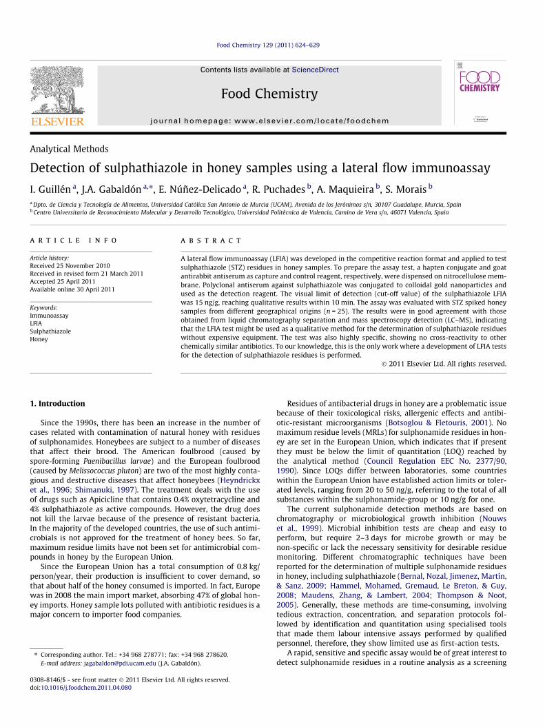
column (50 � 2.1 mm I.D., particle size 3.5 lm) and running alinear gradient from 100% solvent A (0.5% acetic acid/5% methanol,v/v) at 0 min to 50% solvent A and 50% solvent B (methanol) at15 min, at a flow rate of 0.4 mL/min. The nebulizer pressure anddry gas flow (350 �C) were set to 40 psi and 10 L/min, respectively.The STZ was detected using electrospray in the positive ionisationmode. The only molecular-ion species formed in the acidic mobilephase are protonated molecules (Fig. 2). Typical MS settings were:needle voltage 3.5 kV; lens 1: 6.8 V and lens 2: �60 V; capillaryvoltage 110.2, octopole amplitude of 143.8 Vpp, cut-off 69 andamplitude 1.20 V. Two different characteristic fragmentation ionsm/z 108 ([H2NPhO]+) and m/z 156 ([H2NPhSO2]+) were monitoredin the selected reaction monitoring (SRM) mode using a dwell timeof 0.1 s.
3. Results and discussion
The lateral flow immunochromatographic device described inthe present effort yields visual results for the determination of sul-phathiazole residues in honey.
Our previous experiences with colloidal gold based systemshave taught us that a pore size of approximately 15 lm was thebest for both flow rate and reactivity. If larger pore sizes are used,flow rate is usually too fast for reactions to take place at low sen-sitivities, and if smaller pore sizes are used it is difficult to finishthe test in 5–10 min.
As to the colloidal gold particle size, previous experience in goldassays has shown us that a 40 nm particle is the best for strip as-says. These results have also been described in the literature (Shimet al., 2006; Zhou et al., 2004). Besides, smaller particles give lowsignals due to the way the gold scatters light and larger particlestend to migrate slower, generating a purple blue colour, and arealso difficult to work with.
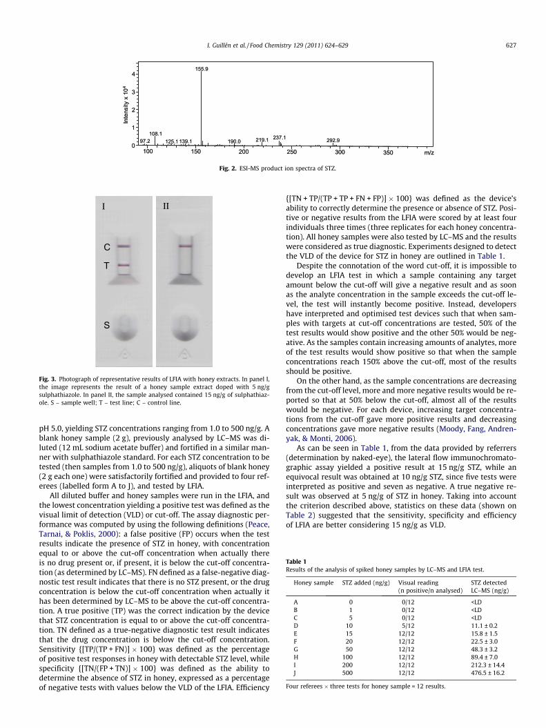
During colloidal gold conjugation, it is important to control thepH of antiserum and that of colloidal gold solution. Both prepara-tions were adjusted to a pH slightly above the isoelectric point ofantiserum before conjugation. Below pKi, antiserum induced floc-culation will occur, whereas above pKi, the adsorption is limiteddue to charge repulsion between the conjugation reagents. A pHof 7.0 was selected as optimum for the stabilisation of the goldsol, since this pH value was the smallest at which flocculation doesnot occur. Control experiments made with buffer or honey withoutsulphathiazole display two red lines – ‘‘C’’ line and the test area(‘‘T’’ line), indicating a negative assay (Fig. 3), whereas honey sam-ples containing sulphathiazole yield a clear red line at the controlarea (‘‘C’’ line) on the device, with no signal – positive test – atthe test line.
For the determination of the visual limit of detection and ana-lytical sensitivity/specificity/efficiency of the LFIA, sulphathiazolestandard (100 mg/g) was diluted with 1.2 M sodium acetate buffer,
PlasticSupport
Nitrocellulosemembrane
“C”Control lineGAR IgG
Absorbent padPlastic
Support
Nitrocellulosemembrane
“C”Control lineGAR IgG
w immunochromatographic assay.
0
1
2
3
4
100 150 200 250 300
97.2108.1
125.1 139.1 190.0 219.1 237.1 292.9
350 m/z
155.9
Inte
nsity
x 1
04
0
1
2
3
4
100 150 200 250 300
97.2108.1
125.1 139.1 190.0 219.1 237.1 292.9
350 m/z
155.9
Inte
nsity
x 1
04
Fig. 2. ESI-MS product ion spectra of STZ.
II
T
C
I
S
Fig. 3. Photograph of representative results of LFIA with honey extracts. In panel I,the image represents the result of a honey sample extract doped with 5 ng/gsulphathiazole. In panel II, the sample analysed contained 15 ng/g of sulphathiaz-ole. S – sample well; T – test line; C – control line.
Table 1Results of the analysis of spiked honey samples by LC–MS and LFIA test.
Honey sample STZ added (ng/g) Visual reading(n positive/n analysed)
STZ detectedLC–MS (ng/g)
A 0 0/12 <LDB 1 0/12 <LDC 5 0/12 <LDD 10 5/12 11.1 ± 0.2E 15 12/12 15.8 ± 1.5F 20 12/12 22.5 ± 3.0G 50 12/12 48.3 ± 3.2H 100 12/12 89.4 ± 7.0I 200 12/12 212.3 ± 14.4J 500 12/12 476.5 ± 16.2
Four referees � three tests for honey sample = 12 results.
I. Guillén et al. / Food Chemistry 129 (2011) 624–629 627
pH 5.0, yielding STZ concentrations ranging from 1.0 to 500 ng/g. Ablank honey sample (2 g), previously analysed by LC–MS was di-luted (12 mL sodium acetate buffer) and fortified in a similar man-ner with sulphathiazole standard. For each STZ concentration to betested (then samples from 1.0 to 500 ng/g), aliquots of blank honey(2 g each one) were satisfactorily fortified and provided to four ref-erees (labelled form A to J), and tested by LFIA.
All diluted buffer and honey samples were run in the LFIA, andthe lowest concentration yielding a positive test was defined as thevisual limit of detection (VLD) or cut-off. The assay diagnostic per-formance was computed by using the following definitions (Peace,Tarnai, & Poklis, 2000): a false positive (FP) occurs when the testresults indicate the presence of STZ in honey, with concentrationequal to or above the cut-off concentration when actually thereis no drug present or, if present, it is below the cut-off concentra-tion (as determined by LC–MS). FN defined as a false-negative diag-nostic test result indicates that there is no STZ present, or the drugconcentration is below the cut-off concentration when actually ithas been determined by LC–MS to be above the cut-off concentra-tion. A true positive (TP) was the correct indication by the devicethat STZ concentration is equal to or above the cut-off concentra-tion. TN defined as a true-negative diagnostic test result indicatesthat the drug concentration is below the cut-off concentration.Sensitivity {[TP/(TP + FN)] � 100} was defined as the percentageof positive test responses in honey with detectable STZ level, whilespecificity {[TN/(FP + TN)] � 100} was defined as the ability todetermine the absence of STZ in honey, expressed as a percentageof negative tests with values below the VLD of the LFIA. Efficiency
{[TN + TP/(TP + TP + FN + FP)] � 100} was defined as the device’sability to correctly determine the presence or absence of STZ. Posi-tive or negative results from the LFIA were scored by at least fourindividuals three times (three replicates for each honey concentra-tion). All honey samples were also tested by LC–MS and the resultswere considered as true diagnostic. Experiments designed to detectthe VLD of the device for STZ in honey are outlined in Table 1.
Despite the connotation of the word cut-off, it is impossible todevelop an LFIA test in which a sample containing any targetamount below the cut-off will give a negative result and as soonas the analyte concentration in the sample exceeds the cut-off le-vel, the test will instantly become positive. Instead, developershave interpreted and optimised test devices such that when sam-ples with targets at cut-off concentrations are tested, 50% of thetest results would show positive and the other 50% would be neg-ative. As the samples contain increasing amounts of analytes, moreof the test results would show positive so that when the sampleconcentrations reach 150% above the cut-off, most of the resultsshould be positive.
On the other hand, as the sample concentrations are decreasingfrom the cut-off level, more and more negative results would be re-ported so that at 50% below the cut-off, almost all of the resultswould be negative. For each device, increasing target concentra-tions from the cut-off gave more positive results and decreasingconcentrations gave more negative results (Moody, Fang, Andren-yak, & Monti, 2006).
As can be seen in Table 1, from the data provided by referrers(determination by naked-eye), the lateral flow immunochromato-graphic assay yielded a positive result at 15 ng/g STZ, while anequivocal result was obtained at 10 ng/g STZ, since five tests wereinterpreted as positive and seven as negative. A true negative re-sult was observed at 5 ng/g of STZ in honey. Taking into accountthe criterion described above, statistics on these data (shown onTable 2) suggested that the sensitivity, specificity and efficiencyof LFIA are better considering 15 ng/g as VLD.

Table 2Statistical analysis of the results in Table 1.
Parameter VLD 10 ng/g VLD 15 ng/g
TP 77 72FN 7 0TN 36 43FP 0 5Sensitivity (%) 91.7 100Specificity (%) 100 89.6Efficiency (%) 94.2 95.8
628 I. Guillén et al. / Food Chemistry 129 (2011) 624–629
On the basis of these findings, VLD was in the range of10–15 ng/g. Even though, we will discriminate all honey sampleshaving an STZ concentration above 15 ng/g.
The specificity of the STZ method was evaluated in comparisonto other analogue compounds: SDZ, SDM, SMZ, SMT, SMX, SMP,SPD and SSX. Stock solutions of each sulphonamide (100 mg/L)and a mix containing all of them (except STZ), at the same concen-tration, were prepared in DMSO and stored a 4 �C and properly di-luted with 1.2 M sodium acetate buffer (pH 5.0), yieldingconcentrations ranging from 0.01 to 50 mg/L. From a blank sample,previously tested by LC–MS, different aliquots (2 g) were fortifiedadding 12 mL acetate buffer of different related compounds.Positive or negative results from the LFIA, three replicates for eachhoney concentration and cross-reactant were analysed.
Two clear bands were observed in the test and control lines oftest, even though these compounds were present at a high level.Each analogue compound was found not cross-reacted when testedat concentrations up to 50 mg/g, only sulphamethoxazole that hasa structure almost identical to STZ has cross reacted at concentra-tions of 10 mg/g. This fact indicates that the polyclonal serum hada high specificity towards sulphathiazole.
Regarding the applicability of the developed prototype, there isa clear difference between laboratory-based techniques and tech-niques for on-site assays. For laboratory use, speed is less impor-tant than throughput while this is the contrary for field assays(Gabaldon et al., 2001a,b). The greatest merits of the immunochro-matographic assay, its simplicity and speed, cannot be demon-strated without the simplest sample preparation. In fortifiedhoney samples, a good agreement was observed when STZ was ex-tracted by both methods (as described in screening of honey sam-ples and intralaboratory validation) and tested by LFIA. Therefore,in all our experiments, samples were directly diluted with acetatebuffer and added to the strip. Since honey is a complex matrix witha large variety in composition, due to different proportions of thepossible sources, nectar and/or honeydew, coming from a greatvariety of plants and origins, the robustness of the STZ-LFIA waschecked on different unifloral and multifloral honey (25) fromArgentina, China, Mexico, Turkey and Spain, free of STZ (checkedby LC–MS). Analysis of 25 blank honey samples yielded negativeresults (no matrix interference or false positive results wereobserved).
In order to calculate the detection capability (CCb) of the assay,the same 25 blank samples were fortified at 15 ng/g with an STZstandard and analysed in triplicate. The assay beta (b) error is zerosince no false negative (false compliant) results were obtained for15 ng/g honey fortified samples. This satisfies Commission Deci-sion 2002/657/EC, (2002), which states that screening techniquesmust have a false compliant rate of <5% (b-error) at the level ofinterest.
This result supports the establishment of 15 ng/g as VLD of theLFIA that will be in compliance with further EU minimum requiredperformance limit (MRPL) of 20 ng/g for STZ in honey, as proposedby the European federation of honey packers and distributors(FEEDM).
Once the prototype was optimised, aliquots of honey samplescontaining 0, 6.7, 14.6 and 33.2 ng/g of STZ, 20 tests properlystored in a sealed bag containing silica, and a protocol assay weredelivered at three honey packers from ASEMIEL to carry out theanalyses in triplicate. All results reported (100%) were in agree-ment with the expected results, two distinct red lines were obser-vable at the ‘‘C’’ line and the test area (‘‘T’’ line) for 0 and 6.7 andonly a clear red line at the control area (‘‘C’’ line) appears for14.6 and 33.2 ng/g of STZ in honey.
On the other hand, there is a commercial ELISA kit (Ridascreensulphonamide; R-biopharm, Darmstadt, Germany) for the mea-surement of nineteen sulphonamides in different matrices suchas milk, meat, fish, egg and shrimps and honey, at the tolerated le-vel (100 ng/g). The kit detects, in more or less extension (crossreaction), all compounds (32% STZ). When it is applied to honeysamples, a previous purification of the whole extract could becarried out using a C18 column. In addition, other multi-residuecompetitive ELISAs to detect seven (Pastor-Navarro et al., 2007;Zhang et al., 2007), 14 (Font et al., 2008), and 19 (Franek et al.,2006) sulphonamides in different matrices such as milk, pig andchicken muscle, fish, egg, honey and hair have been developed.An LFIA for sulphamonomethoxine, sulphamethoxydiazine,sulphadimethoxine and sulphadiazine which has a detectionthreshold of 10 ng/mL, determined with an optical density scanner,in eggs and chicken muscle has also been reported (Wang et al.,2007). By eye measurement, the sensitivity was 20 ng/mL forsulphamonomethoxine, sulphamethoxydiazine, sulphadimethox-ine and 40 ng/mL for sulphadiazine. Three lateral flow strip teststo detect all members of the sulphonamide family of drugs, sulpha-methazine and sulphadimethoxine or only sulphamethazine inmilk, are commercialised by Charm Sciences Inc. (MA, USA) as RosaTests, which show a detection threshold of 10 ng/mL.
However, the current status of the available immunoassays for asingle sulphonamide, such as STZ, is scarce. Currently, two ELISAassays have been described for the detection of STZ. One basedon monoclonal Ab (Lee et al., 2001) shows a sensitivity threshold<100 ng/g when was applied in swine liver tissue, with a relativelyslight cross-reactivity with other 13 sulphonamides; while an-other, that employ the same immunoreagents (Pastor-Navarroet al., 2004) that we use for the development of LFIA, has a mini-mum detectable concentration of 0.03 ng/g, with a sensitivity of3 ng/g in honey samples.
In the present work, we describe an LFIA which has a clear limitof detection at 15 ng/g STZ, which is 10-fold less sensitive than thevalidated ELISA (Pastor-Navarro et al., 2004). The assay can be usedwith small volumes (�100 lL) of diluted honey. The assay wasshown to have 100% diagnostic sensitivity, 89.6% specificity and95.8% efficiency for the detection of STZ in honey. Existing ELISAand other assays for STZ tests are laboratory based, require samplepreparation and are relatively slow compared to the LFIA describedhere, at the first time. In addition, highly trained laboratory person-nel and relatively sophisticated equipment are also necessary forlaboratory-based assays, whereas the LFIA is rapid (�10 min), easyto use, and highly portable.
The STZ lateral flow assay can be performed at the site of honeydelivery such as beehives or beekeepers’ store. It is a qualitativetest using a small quantity of sample and return results within10 min. Compared with centralised laboratory testing, it providesfor rapid buy decision-making by reducing the time spent on trans-porting sampling and retrieving data.
4. Conclusions
A rapid lateral-flow immunochromatographic device with a col-loidal gold-polyclonal probe was developed for the detection of

I. Guillén et al. / Food Chemistry 129 (2011) 624–629 629
sulphathiazole residues in honey samples. The visual limit ofdetection was 15 ng/g, and the test was highly specific towardssulphathiazole, since it only recognises related sulphonamideswhich are present in honey at concentrations above 10 mg/g.
The test showed high diagnostic sensitivity and specificity ratesand resulted very suitable for on site detection of STZ residues inhoney since no sample treatment is required. The results obtainedfor fortified honey samples were in good agreement with those ob-tained by LC–MS.
The LFIA is easy to use, highly portable and the results can beobtained in 10 min without the need for expensive equipment,washing and/or separation steps.
The proposed analytical system, has no equivalence in the mar-ket, and could be used by the honey sector to carry out on-sitescreening for STZ at the beginning of the food chain to improvecommercial trade.
Since the antibody based assays are screening methods in foodanalysis, group specific antibodies for sulphonamides are prefera-ble since positive findings must be confirmed by standard instru-mental method. In this sense, it should be interesting for thedevelopment of a new multianalyte LFIA for sulphonamides andto investigate the potential use of other alternative labels.
Acknowledgements
This work was supported by a grant from Ministry of Educationand Science of Spain, CIT-060000-2007-57.
References
Bernal, J., Nozal, M. J., Jimenez, J. J., Martín, M. T., & Sanz, E. (2009). A new and simplemethod to determine trace levels of sulfonamides in honey by highperformance liquid chromatography with fluorescence detection. Journal ofChromatography A, 1216, 7275–7280.
Botsoglou, N. A., & Fletouris, D. J. (2001). Tetracyclines. In N. A. Botsoglou & D. J.Fletouris (Eds.), Drug Residues in Foods: Pharmacology, Food Safety, and Analysis(pp. 95–100). New York: Marcel Dekker, Inc.
Commission Decision (EEC) No. 2002/657, (2002). Application of 96/23/EUconcerning the performance of analytical methods and interpretation ofresults. Official Journal of the European Communities No. L 221, 8.
Council Regulation (EEC) No. 2377/90, (1990). Community procedure for theestablishment of a maximum residue limits of veterinary medical products infoodstuffs of animal origin. Official Journal of the European Communities No. L224, 1–8 (and amendments).
Edwards, K. A., & Baeumner, A. J. (2006). Optimization of DNA-tagged dye-encapsulating liposomes for lateral-flow assays based on sandwichhybridization. Analytical Bioanalytical Chemistry, 386, 1335–1343.
Font, H., Adrian, J., Galve, R., Estevez, M. C., Castellari, M., Gratacos-Cubarsi, M., et al.(2008). Immunochemical assays for direct sulfonamide antibiotic detection inmilk and hair samples using antibody derivatized magnetic nanoparticles.Journal of Agricultural and Food Chemistry, 56, 736–743.
Franek, M., Diblikova, I., Cernoch, I., Vass, M., & Hruska, K. (2006). Broadspecificityimmunoassays for sulfonamide detection: Immunochemical strategy forgeneric antibodies and competitors. Analytical Chemistry, 78, 1559–1567.
Frens, G. (1973). Controlled nucleation for the regulation of the particle size inmonodisperse gold suspensions. Nature Physical Science, 241, 20–22.
Gabaldon, J. A., Maquieira, A., & Puchades, R. (1999). Current trends inimmunoassay-based kits for pesticide analysis. Critical Reviews in Food Scienceand Nutrition, 39, 519–538.
Gabaldon, J. A., Maquieira, A., & Puchades, R. (2001a). Determination of atrazine invegetable samples using a dipstick immunoassay format. International Journal ofEnvironmental Analytical Chemistry, 82, 145–155.
Gabaldon, J. A., Maquieira, A., & Puchades, R. (2001b). Rapid method for on sitedetermination of atrazine residues in water samples. Assay optimisation.International Journal of Environmental Analytical Chemistry, 82, 133–144.
Haasnoot, W., Bienenmann-Ploum, M., Lamminmäki, U., Swanenburg, M., & Rhijn,H. V. (2005). Application of a multi-sulfonamide biosensor immunoassay for thedetection of sulfadiazine and sulfamethoxazole residues in broiler serum and itsuse as a predictor of the levels in edible tissue. Analytica Chimica Acta, 552,87–95.
Hammel, Y. A., Mohamed, R., Gremaud, E., Le Breton, M. H., & Guy, A. (2008). Multi-screening approach to monitor and quantify 42 antibiotic residues in honey byliquid chromatography–tandem mass spectrometry. Journal of ChromatographyA, 1177, 58–76.
Heyndrickx, M., Vandemeulebroecke, K., Scheldeman, P., Kersters, K., de Vos, P.,Logan, N. A., et al. (1996). A polyphasic reassessment of the genus Paenibacillus,reclassification of Bacillus lautus (Nakamura 1984) as Paenibacillus lautus comb.nov. and of Bacillus peoriae (Montefusco et al. 1993) as Paenibacillus peoriaecomb. nov., and emended descriptions of P. lautus and of P. peoriae. InternationalJournal Systematic Bacteriology, 46, 988–1003.
Kalogianni, D. P., Goura, S., Aletras, A. J., Christopoulos, T. K., Chanos, M. G.,Christofidou, M., et al. (2007). Dry reagent dipstick test combined with 23SrRNA PCR for molecular diagnosis of bacterial infection in arthroplasty.Analytical Biochemistry, 361, 169–175.
Koets, M., Sander, I., Bogdanovic, J., Doekes, G., & van Amerongen, A. (2006). A rapidlateral flow immunoassay for the detection of fungal alpha-amylase at theworkplace. Journal Environmental Monitoring, 8, 942–946.
Korpimäki, T., Hagren, V., Brockmann, E. C., & Tuomola, M. (2004). Genericlanthanide fluoroimmunoassay for the simultaneous screening of 18sulfonamides using an engineered antibody. Analytical Chemistry, 76,3091–3098.
Lai, W., Fung, D., Xu, Y., Liu, R., & Xiong, Y. (2009). Development of a colloidal goldstrip for rapid detection of ochratoxin A with mimotope peptide. Food Control,20, 791–795.
Lee, N., Holtzapple, C. K., Muldoon, M. T., Deshpande, S. S., & Stanker, L. H. (2001).Immunochemical approaches to the detection of sulfathiazole in animal tissues.Food Agricultural Immunology, 13, 5–17.
Maudens, K. E., Zhang, G. F., & Lambert, W. E. (2004). Quantitative analysis of twelvesulfonamides in honey by high-performance liquid chromatography with post-column derivitization and fluorescence detection. Journal of Chromatography A,1047, 85–92.
Moody, D. E., Fang, W. B., Andrenyak, D. M., & Monti, C. (2006). A comparativeevaluation of the instant-view 5-panel test card with OnTrak TestTcup pro 5:comparison with gas chromatography–mas spectrometry. Journal AnalyticalToxicology, 30, 50–56.
Nielsen, K., Yu, W. L., Kelly, L., Bermudez, R., Renteria, T., Dajer, A., et al. (2008).Development of a lateral flow assay for rapid detection of bovine antibody toAnaplasma marginale. Journal Immunoassay Immunochemistry, 29, 10–18.
Nouws, J. F. M., van Egmond, H., Loeffen, G., Schouten, J., Keukens, H.,Smulders, I., et al. (1999). A microbiological assay system for assessment ofraw milk exceeding EU maximum residue levels. International Dairy Journal,9, 85–90.
O’Keeffe, M., Crabbe, P., Salden, M., Wichers, J., van Peteghem, C., Kohen, F., et al.(2003). Preliminary evaluation of a lateral flow immunoassay device forscreening urine samples for the presence of sulphamethazine. JournalImmunological Methods, 278, 117–126.
Pastor-Navarro, N., Gallego-Iglesias, E., Maquieira, A., & Puchades, R. (2007).Development of a group-specific immunoassay for sulfonamides. Applicationto bee honey analysis. Talanta, 71, 923–933.
Pastor-Navarro, N., García-Bover, C., Maquieira, A., & Puchades, R. (2004). Specificpolyclonal-based immunoassays for sulfathiazole. Analytical BioanalyticalChemistry, 379, 1088–1099.
Peace, M., Tarnai, L., & Poklis, A. (2000). Performance evaluation of four on-sitedrug-testing devices for detection of drugs of abuse in urine. Journal AnalyticalToxicology, 24, 589–594.
Shim, W. B., Yang, Z. Y., Kim, Y. Y., Choi, J. G., Je, J. H., Kang, S. J., et al. (2006).Immunochromatography using colloidal gold�antibody probe for the detectionof atrazine in water samples. Journal of Agricultural and Food Chemistry, 54,9728–9734.
Shimanuki, H. (1997). Bacteria. In R. A. Morse & K. Flottum (Eds.), Honey bee pests,predators, and diseases (pp. 35–54). Ohio: AI Root Co. Medina.
Thompson, T. S., & Noot, D. K. (2005). Determination of sulfonamides in honey byliquid chromatography–tandem mass spectrometry. Analytica Chimica Acta, 551,168–176.
Wang, X. L., Li, K., Shi, D., Xiong, N., Jin, X., Yi, J. D., et al. (2007). Development of animmunochromatographic lateral-flow test strip for rapid detection ofsulfonamides in eggs and chicken muscles. Journal of Agricultural and FoodChemistry, 55, 2072–2078.
Wang, S., Quan, Y., Lee, N., & Kennedy, I. R. (2006). Rapid determination offumonisin B1 in food samples by enzyme-linked immunosorbent assay andcolloidal gold immunoassay. Journal of Agricultural and Food Chemistry, 54,2491–2495.
Zhang, H. Y., Wang, L., Zhang, Y., Fang, G. Z., Zheng, W. J., & Wang, S. (2007).Development of an enzyme-linked immunosorbent assay for seven sulfonamideresidues and investigation of matrix effects from different food samples. Journalof Agricultural and Food Chemistry, 55, 2079–2084.
Zhou, P., Lu, Y., Zhu, J., Hong, J., Li, B., Zhou, J., et al. (2004). Nanocolloidal gold-basedimmunoassay for the detection of the N-methylcarbamate pesticide carbofuran.Journal of Agricultural and Food Chemistry, 52, 4355–4359.