detection of colorectal adenomas by routine chromoendoscopy with indigocarmine
TRANSCRIPT

Detection of Colorectal Adenomas by RoutineChromoendoscopy With IndigocarmineJun Haeng Lee, M.D., Jeong Wook Kim, M.D., Yong Kyun Cho, M.D., Chong Il Sohn, M.D.,Woo Kyu Jeon, M.D., Byung Ik Kim, M.D., and Eun Yoon Cho, M.D.Departments of Medicine and Pathology, Sungkyunkwan University School of Medicine, Seoul, Korea
OBJECTIVES: Nonpolypoid adenomas, which can be impor-tant precursors of colorectal cancers, are difficult to findduring routine colonoscopy. The aim of this study was toevaluate the usefulness of routine chromoendoscopy in Ko-rea, where the incidence of colorectal cancer is low com-pared with western countries.
METHODS: Colonoscopy with chromoendoscopy was per-formed in 74 consecutive patients (48 men, 26 women;mean age 53.0 yr). After a careful examination of the wholecolon, a defined segment of the sigmoid colon and rectum(0–30 cm from the anal verge) was stained with 20 ml of0.2% indigocarmine solution with a spraying catheter. Non-polypoid lesions were classified as flat or depressed types.Biopsies were taken from all lesions detected before or afterstaining with indigocarmine.
RESULTS: Indications for colonoscopy included routinecheck-up (21 patients), diarrhea or loose stool (14 patients),abdominal pain (12 patients), constipation (7 patients),bleeding (6 patients), and others (14 patients). Before stain-ing, 58 lesions were found in 30 patients (43.2%). Histologyshowed tubular adenoma in 41 lesions, hyperplastic or in-flammatory changes in 14 lesions, adenocarcinoma in 2lesions, and villous adenoma in 1 lesion. After indigocar-mine staining for normal-looking distal 30 cm colorectalmucosa, 176 lesions were found in 46 patients (62.2%).Histologically, 158 lesions were hyperplastic or inflamma-tory in nature, and 17 lesions (from 11 patients) were tubularadenomas. There was one serrated adenoma. Eighteen ade-nomas seen only after spraying indigocarmine were 2.6 �0.6 mm in diameter, and all of them were classified as flatadenomas. There was no depressed-type adenoma. No ad-enoma with high grade dysplasia, villous histology, or can-cer was found after staining. Presence of macroscopic ad-enomatous lesions or carcinoma before staining could notpredict the existence of adenoma after staining.
CONCLUSIONS: In a large proportion of patients, flat ordepressed adenomas could be found after spraying indigo-carmine for normal-looking colorectal mucosa in Korea.The clinical significance of these diminutive adenomas that
can be found only after spraying contrast agent needs to befurther investigated. (Am J Gastroenterol 2003;98:1284–1288. © 2003 by Am. Coll. of Gastroenterology)
INTRODUCTION
During the last decade, there has been an increasing interestin nonpolypoid colorectal neoplastic lesions (1–11). Suchlesions are difficult to detect and are often overlooked dur-ing routine colonoscopic examinations, and it has beenclaimed that the true incidence of nonpolypoid neoplasticlesions has been underestimated (5–8). Indigocarmine is themost commonly used contrast staining agent for enhance-ment of the superficial structures of the intestinal mucosa(12, 13). Recently, there has been a report from Germanythat chromoendoscopy with indigocarmine may help detectsmall nonpolypoid neoplastic lesions, which are not identi-fied by routine video endoscopy (10). However, the inci-dence of colorectal neoplastic lesions is significantly differ-ent by geographic locations (14), and the role of routinechromoendoscopy in eastern countries has not been deter-mined. The aim of the present study was to evaluate theusefulness of routine chromoendoscopy in Korea, where theincidence of colorectal cancer is low compared with westerncountries (15).
MATERIALS AND METHODS
Colonoscopy with routine chromoendoscopy using indigo-carmine solution (Carmine, Korea United Pharmaceutical,Yoenki, Korea) was prospectively performed in 74 consec-utive patients. Mean age was 53.0 yr (range 30–78, 48 maleand 26 female). Bowel preparation included 4 L PEG elec-trolyte solution in the morning before an afternoon exami-nation. Patients were sedated with midazolam (3–5 mg) ifthere was no contraindication to its use. Electronic videoendoscopes for the lower GI tract (CF-240L and CF-130L,Olympus, Tokyo, Japan) were used during the study.Colonoscopic examinations were performed by three expe-rienced endoscopists (J.H.L., J.W.K., and C.I.S.). Patientswith insufficient bowel preparation, a family history ofpolyposis, GI bleeding within 1 wk, infectious or inflam-
This study was presented as a poster in the 67th annual scientific meeting of theAmerican College of Gastroenterology.
THE AMERICAN JOURNAL OF GASTROENTEROLOGY Vol. 98, No. 6, 2003© 2003 by Am. Coll. of Gastroenterology ISSN 0002-9270/03/$30.00Published by Elsevier Inc. doi:10.1016/S0002-9270(03)00247-8
matory diseases, or total or subtotal strictures had beenexcluded from the study.
The colonoscope was introduced up to the cecum. Resid-ual fecal material and mucus were removed from the mu-cosa with tap water. Subsequently, while retracting theinstrument, biopsy samples were taken for all detectablelesions using a standard cold biopsy forceps (FB-24U-1,Olympus, Tokyo, Japan). After a careful examination of thewhole colorectal mucosa, the endoscope was introducedagain up to 30 cm from the anal verge. During slow with-drawal of the endoscope, approximately 20 ml of indigo-carmine solution (0.2%) were sprayed with a spraying cath-eter (PW-5V-1, Olympus, Tokyo, Japan). Indigo carmine isa blue stain that accentuates the contours of a lesion, pro-viding a detailed view of its border and shape without thenecessity of using a magnifying endoscope. This dye is notabsorbed and has no toxicity when used on the mucosalsurface of the colon. Then, the endoscope was introducedagain up to 30 cm from the anal verge, and the dye-sprayedmucosa of the rectosigmoid colon was examined in detail.All detected lesions were removed for histologic examina-tion by one or two biopsies using the same standard coldbiopsy forceps. Histologically, all biopsy samples were re-viewed by an experienced pathologist (E.Y.C.), and dyspla-sia in adenoma was divided into low and high grade ac-cording to the Vienna classification (16).
Macroscopically, adenomas were classified as polypoidor nonpolypoid (6, 9). Polypoid adenoma was defined as aprotruded adenomatous lesion, and this included both sessileand pedunculated types. Pedunculated polyps were definedas protruding lesions in which a stalk could be demon-strated. Protruding lesions without a stalk were defined assessile polyps. Nonpolypoid adenomas were further classi-fied as flat or depressed. Flat adenoma was defined as anadenoma with either plane or slightly raised areas with adiameter in the axis of the intestinal surface several timesexceeding their height (6). Polyp size was estimated byplacing an opened standard biopsy forceps beside the le-sions.
RESULTS
Indications for colonoscopy included routine check-up (21patients), diarrhea or loose stool (14 patients), abdominalpain (12 patients), constipation (7 patients), bleeding (6patients), and others (14 patients).
Before spraying indigocarmine, 58 lesions were found in30 patients (43.2%). Histology of these lesions showedadenomas in 42 lesions (72.4%), hyperplastic or inflamma-tory changes in 14 lesions (24.1%), and adenocarcinomas in2 lesions (3.4%). Of the 42 adenomas, 36 were tubularadenomas, 3 were tubular adenomas with high grade dys-plasia, and 2 were villous adenomas. Endoscopic features ofthe 42 adenomas were sessile type in 21 (50.0%), pedun-culated type in 6 (14.3%), and nonpolypoid flat lesions in 15
(35.7%). The mean diameter of adenomas was 5.4 � 4.6mm (range 3–30).
After indigocarmine staining for normal-looking distal 30cm colorectal mucosa, 176 lesions were found in 46 patients(62.2%). All of these lesions were smaller than 5 mm inlongest diameter and were removed completely using abiopsy forceps. Histologically, 158 lesions were hyperplas-tic or inflammatory in nature, and 18 lesions (from 12patients) were neoplastic in nature (Table 1, Figs. 1 and 2).Among 18 neoplastic lesions, 17 (94.4%) were tubular ad-enomas, 1 lesion was serrated adenoma, and all of themwere classified as flat adenomas. There was no depressed-type adenoma. The mean size of the 18 adenomas foundafter spraying indigocarmine was 2.6 � 0.6 mm, signifi-cantly smaller than the adenomas found before chromoen-doscopy (p � 0.001, Student t test). No adenoma with highgrade dysplasia, villous histology, or cancer was found afterdye-spraying.
Patients were divided into two groups by the existence ofdetectable neoplastic lesions before dye spraying. In groupA (25 patients, 33.8%), there were adenoma(s) or carcinomathat were detectable before spraying indigocarmine. In thegroup B (49 patients, 66.2%), no neoplastic lesions werefound before spraying indigocarmine. The rates of patientswith small adenomas that were found after spraying indigo-carmine were 24.0% (6/25) in group A and 12.2% (6/49) ingroup B (Table 2). However, this difference was not statis-tically significant (p � 0.317, Fisher exact test).
DISCUSSION
Colonoscopy has been widely accepted as the most effica-cious screening tool for colorectal cancers (17, 18). Inaddition to the detection of colorectal cancers, the mainadvantage of screening colonoscopy is the detection andremoval of preneoplastic and early neoplastic lesions. Dur-ing the last decade, there has been an increasing interest innonpolypoid colorectal neoplastic lesions, which includesthe flat or depressed adenoma, the flat serrated adenoma, andthe de novo carcinoma (1–11). There is a retrospectivereview indicating that 12%–40% of adenomas or earlycolorectal cancers appear flat or as depressions, rather thanas polyps (19). It has also been suggested that flat or de-
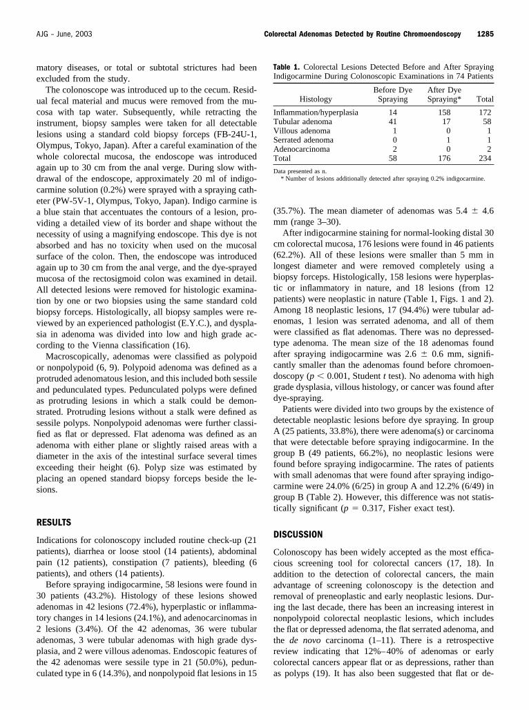
Table 1. Colorectal Lesions Detected Before and After SprayingIndigocarmine During Colonoscopic Examinations in 74 Patients
HistologyBefore Dye
SprayingAfter DyeSpraying* Total
Inflammation/hyperplasia 14 158 172Tubular adenoma 41 17 58Villous adenoma 1 0 1Serrated adenoma 0 1 1Adenocarcinoma 2 0 2Total 58 176 234
Data presented as n.* Number of lesions additionally detected after spraying 0.2% indigocarmine.
1285AJG – June, 2003 Colorectal Adenomas Detected by Routine Chromoendoscopy

pressed adenomas have a propensity for the development ofhigh grade dysplasia at a small size and may be precursorsof flat, ulcerated colorectal carcinomas (1, 2, 5). Therefore,the detection and the removal of such lesions may be veryimportant for the prevention of colorectal cancers. Endo-scopic features of the small nonpolypoid lesions, however,are plaque-like reddish spots or blurring of the vascularpattern (3, 20), so detecting such lesions during colonoscopyis still difficult.
The dye-spraying method has usually been applied onlyto improve visualization of lesions already detected (12, 13,21). Although the diagnostic accuracy may be increased by
the methods with or without a magnifying endoscope (22–24), the detection rate of colorectal lesions itself is notinfluenced. To find more colorectal lesions with small ornonpolypoid features, investigators have tried various meth-ods with routine use of contrast dyes before detecting anylesions. For example, Mitooka et al. (5) administrated anindigocarmine capsule to the patients before the colonicpreparation, and they reported a four- or five-fold increaseddetection rate for nonpolypoid lesions, compared with thosefound without chromoendoscopy. This technique, however,is not suitable for left-sided lesions because the dye rarelyreaches the left-sided colon in sufficient concentration (21).
A B
Figure 1. (A) A small lesion detected after spraying 0.2% indigocarmine onto the normal-looking mucosa of the rectosigmoid colon. (B)Microscopic examination reveals the star-shaped appearance of the glands with an admixture of columnar and goblet cells (hematoxylinand eosin stain, �100).
A B
Figure 2. (A) A small lesion detected after spraying 0.2% indigocarmine onto the normal-looking mucosa of the rectosigmoid colon. (B)Microscopic examination shows a proliferation of adenomatous epithelium with hyperchromatic and stratified nuclei (hematoxylin andeosin stain, �100).
1286 Lee et al. AJG – Vol. 98, No. 6, 2003

In addition, other mucosal lesions, including angiodysplasiaor colitis, may not be recognized when the mucosa is dyedblue. To avoid such limitations, Kiesslich et al. (10) sprayedindigocarmine onto the rectosigmoid mucosa after carefulexamination of the whole colon without spraying dye.Among 48 patients with mucosa of normal appearance be-fore spraying indigocarmine, 27 showed 178 lesions (meansize 0.3 cm) after staining. Histologically, most of theselesions were non-neoplastic, but there were 13 adenomaswith or without high-grade dysplasia. These findings sug-gest that multiple mucosal lesions that are not identified byroutine colonoscopy can be found after spraying contrastdye onto the normal-looking mucosa. However, the clinicalrelevance of this technique is not fully established, becausethe natural history of such minute lesions is not known.
The frequency of colorectal cancer varies remarkablyamong different populations (14). The usefulness of anytechnique to detect cancers and/or precancerous lesions de-pends on the frequency of such lesions in a certain popula-tion. Therefore, we tried to define the role of the routinechromoendoscopic method proposed by Kiesslich et al. (10)in Korea, where the incidence of colorectal cancer is lowerthan in western countries (15). In the present study, wedemonstrated that many small lesions can be found in thenormal-looking colonic mucosa by spraying indigocarmine(Table 1). Approximately 10% of those lesions showedadenomatous histology. These findings are not much differ-ent from the data in Germany (10). However, no lesionsdetected after spraying indigocarmine showed high-gradedysplasia, villous histology, or cancerous changes in thepresent study. We compared the frequency of adenomasdetectable only after spraying contrast agents in a patientwith or without neoplastic lesions before chromoendoscopy(Table 2). When a patient has a neoplastic lesion(s) beforespraying indigocarmine, the rate of additional adenoma ishigher after chromoendoscopy. Twenty-three percent ofsuch patients showed diminutive tubular adenoma(s) in therectosigmoid area after routine chromoendoscopy. This ratewas higher than that of patients without neoplastic lesionsbefore chromoendoscopy, although this difference was notstatistically significant owing to the limited number of pa-tients of the present study. It is well known that colonic
adenomas tend to be multiple in a given patient, and ade-nomas frequently recur after polypectomy (14). Therefore,the influence of the routine chromoendoscopy on the recur-rence rate after removal of colon polyp(s) needs to be furtherstudied.
In the present study, we investigated the role of sprayingindigocarmine on normal-looking mucosa. However, it isnot certain whether this technique can be used for the areaswith chronic inflammation, such as in ulcerative colitis. Therisk of colon cancer is increased in patients with long-standing ulcerative colitis, and mucosal dysplasia (definedas an unequivocal neoplastic alteration of the colonic epi-thelium) is the recognized precursor for developing carci-noma. In western countries, therefore, surveillance colonos-copy with multiple biopsies at 10-cm intervals is commonlyrecommended for patients with extensive colitis for longerthan 8–10 yr (25). Because this approach requires so manybiopsies (more than 30 in most cases), Japanese authoritiesfavor targeted biopsy for suspicious lesions (26). In ouropinion, there is a possibility that a routine dye-sprayingmethod may increase the detection rate of precancerouslesions during surveillance colonoscopy for patients withchronic ulcerative colitis. It also needs to be further studiedin the future.
In conclusion, we found that flat adenomas could befound after spraying indigocarime for normal-lookingrectosigmoid mucosa in a large proportion of patients inKorea. The clinical significance of these diminutive adeno-mas that could be found only after spraying contrast agentneeds to be further investigated.
ACKNOWLEDGMENT
This work was supported by Hyoseok Research Fund.
Reprint requests and correspondence: Byung Ik Kim, M.D.,Kangbuk Samsung Hospital, Department of Medicine, Pyung-dong, Jongro-ku, Seoul 100-634, Korea.
Received Sep. 4, 2002; accepted Nov. 22, 2003.
REFERENCES
1. Muto T, Kamiya J, Sawada T, et al. Small “flat adenoma” ofthe large bowel with special reference to its clinicopathologicfeatures. Dis Colon Rectum 1985;28:847–51.
2. Wolber RA, Owen DA. Flat adenomas of the colon. HumPathol 1991;22:70–4.
3. Adachi M, Muto T, Okinaga K, et al. Clinicopathologic fea-tures of the flat adenoma. Dis Colon Rectum 1991;34:981–6.
4. Kudo S. Endoscopic mucosal resection of flat and depressedtypes of early colorectal cancer. Endoscopy 1993;25:455–61.
5. Mitooka H, Fujimori T, Maeda S, et al. Minute flat depressedneoplastic lesions of the colon detected by contrast chromos-copy using an indigo carmine capsule. Gastrointest Endosc1995;41:453–9.
6. Jaramillo E, Watanabe M, Slezak P, et al. Flat neoplasticlesions of the colon and rectum detected by high-resolution
Table 2. Efficacy of Routine Chromoendoscopy in the Detection ofAdditional Small Adenoma(s) in Patients With or Without Neo-platic Lesions Detected Before Spraying Indogocarmine
Group A(n � 25)
Group B(n � 49)
Neoplastic lesions before dye spraying Yes NoNumber of all additional lesions
detected after dye spraying85 91
Number of adenomas detectedafter dye spraying
6 12
Number of patients with adenoma(s)detected after dye spraying*
6 (24.0%) 6 (12.2%)
* This difference was not statistically significant (p � 0.317, Fisher exact test).
1287AJG – June, 2003 Colorectal Adenomas Detected by Routine Chromoendoscopy

video endoscopy and chromoscopy. Gastrointest Endosc 1995;42:114–22.
7. Rembacken BJ, Fujii T, Cairns A, et al. Flat and depressedcolonic neoplasms: A prospective study of 1000 colonoscopiesin the UK. Lancet 2000;355:1211–4.
8. Saitoh Y, Waxman I, West AB, et al. Prevalence and distinc-tive biologic features of flat colorectal adenomas in a NorthAmerican population. Gastroenterology 2001;120:1657–65.
9. Lambert R, Provenzale D, Ectors N, et al. Early diagnosis andprevention of sporadic colorectal cancer. Endoscopy 2001;33:1042–64.
10. Kiesslich R, von Bergh M, Hahn M, et al. Chromoendoscopywith indigocarmine improves the detection of adenomatousand nonadenomatous lesions in the colon. Endoscopy 2001;33:1001–6.
11. Hayakawa M, Shimokawa K, Kusugami K, et al. Clinicopath-ological features of superficial depressed-type colorectal neo-plastic lesions. Am J Gastroenterol 1999;94:944–9.
12. Acosta MM, Boyce HW, Jr. Chromoendoscopy: Where is ituseful? J Clin Gastroenterol 1998;27:13–20.
13. Shim CS. Staining in gastrointestinal endoscopy: Clinical ap-plications and limitations. Endoscopy 1999;31:487–96.
14. Bresalier RS, Kim YS. Malignant neoplasms of the largeintestine. In: Feldman M, Scharschmidt BF, Sleisenger MH,eds. Gastrointestinal and liver diseases. Philadelphia: W. B.Saunders, 1998:1906–42.
15. Park YJ, Park KJ, Park JG, et al. Prognostic factors in 2230Korean colorectal cancer patients: Analysis of consecutivelyoperated cases. World J Surg 1999;23:721–6.
16. Schlemper RJ, Riddell RH, Kato Y, et al. The Vienna classi-
fication of gastrointestinal epithelial neoplasia. Gut 2000;47:251–5.
17. Rex DK, Lieberman DA. Feasibility of colonoscopy screen-ing: Discussion of issues and recommendations regarding im-plementation. Gastrointest Endosc 2001;54:662–7.
18. Sonnenberg A, Delco F, Inadomi JM. Cost-effectiveness ofcolonoscopy in screening for colorectal cancer. Ann InternMed 2000;133:573–84.
19. Kudo S, Tamura S, Hirota S, et al. The problem of de novocolorectal carcinoma. Eur J Cancer 1995;31A:1118–20.
20. Koba I, Yoshida S, Fujii T, et al. Diagnostic findings inendoscopic screening of superficial colorectal neoplasia: Re-sults from a prospective study. Jpn J Clin Oncol 1998;28:542–5.
21. Fujii T, Hasegawa RT, Saitoh Y, et al. Chromoscopy duringcolonoscopy. Endoscopy 2001;33:1036–41.
22. Eisen GM, Kim CY, Fleischer DE, et al. High-resolutionchromoendoscopy for classifying colonic polyps: A multi-center study. Gastrointest Endosc 2002;55:687–94.
23. Kudo S, Rubio CA, Teixeira CR, et al. Pit pattern in colorectalneoplasia: Endoscopic magnifying view. Endoscopy 2001;33:367–73.
24. Kang NY, Lim CY, Heo JH, et al. The comparison of his-topathologic findings and pit patterns of colorectal tumors.Korean J Gastrointest Endosc 1999;19:904–10.
25. Jewell DP. Ulcerative colitis. In: Feldman M, Friedman LS,Sleisenger MH, eds. Gastrointestinal and liver disease. Phila-delphia: W. B. Saunders, 2002:2039–67.
26. Matsuoka K, Hibi T, Iwao Y, et al. Surveillance for colorectalcancer associated with ulcerative colitis (in Japanese). Stom-ach Intest 2002;37:903–14.
1288 Lee et al. AJG – Vol. 98, No. 6, 2003