detection activity assessment and diagnosis of dental ... address: [email protected] keywords dental...
TRANSCRIPT

Detection ActivityAssessment andDiagnosis of DentalCaries Lesions
Mariana M. Braga, DDS, PhDa, Fausto M. Mendes, DDS, MS, PhDa,Kim R. Ekstrand, DDS, PhDb,*
KEYWORDS
� Dental caries � Caries indices � Caries detection� ICDAS � Radiographic methods � Caries activity
The word diagnosis (plural, diagnoses) is derived from the Greek ‘‘dia’’ meaning‘‘through’’ and ‘‘gnosis’’ meaning ‘‘knowledge’’.1 Thus, ‘‘to diagnose’’ implies that itis only through knowledge about the disease that a diagnosis can be established.Diagnosis can be a complicated process.1,2
Caries disease diagnosis is not the classical hypothetical-deductive process thatdiagnosis often is in the medical world.3 When a patient visits a doctor, the patient tellshis or her symptoms to the doctor. The doctor examines the patient and based on hisdiagnostic hypotheses (knowledge), the doctor chooses the diagnosis that best fitsthe patient’s signs and symptoms and the treatment that offers the best prognosisfor the patient. This systematic approach permits thinking about all the possibilitiesavailable to solve the patient’s problem. Dentists, on the other hand, usually examinetheir patients thinking about how they will treat the patients’ teeth, rather than thinkingabout the condition (diagnoses) of the patients’ teeth. Thus, a great deal of informationrelevant to disease diagnosis can be lost and some treatment options (usually moreconservative alternatives) can be underestimated during the final clinical decisionmaking.3
The examination and evaluation of carious lesions has traditionally been limited tophysical criteria such as size, depth, and presence or absence of cavitation. Theterm for this is caries lesion detection.2,4–6 Caries lesion activity assessment isdifferent from caries lesion detection. The assessment of lesion activity is, togetherwith lesion detection, essential to arrive at the disease diagnosis and the appropriate
a Department of Pediatric Dentistry, Faculdade de Odontologia da Universidade de Sao Paulo,Avenida Professor Lineu Prestes 2227, Sao Paulo, SP 05508-900, Brazilb Department of Cariology and Endodontics, School of Dentistry, Faculty of Health Sciences,University of Copenhagen, 20 Norre alle, DK-2200, Copenhagen N, Denmark* Corresponding author.E-mail address: [email protected]
Dent Clin N Am 54 (2010) 479–493doi:10.1016/j.cden.2010.03.006 dental.theclinics.com0011-8532/10/$ – see front matter ª 2010 Elsevier Inc. All rights reserved.

Braga et al480
clinical treatment decision.2,4–6 In addition to caries lesion detection, lesion or diseaseactivity assessment must also consider etiologic factor evaluations, such as oralhygiene, count of cariogenic micro-organisms in plaque and saliva, use of fluoride,sugar intake, and also some socioeconomic aspects, such as family income andparents’ level of education.2,6,7 It becomes evident that caries disease diagnosis isa difficult task. This article focuses on caries lesion detection including evaluation ofcaries lesion severity. The authors explore conventional and advanced/modernmethods of caries lesion detection, their properties, limitations, and indications.They discuss the interpretation of the results obtained by these methods. Finally,parameters to be used for assessing lesion activity in order to reach a final cariesdisease diagnosis and relate the treatment decision to this diagnosis are discussed.As caries risk assessment is covered in another article (see the article by Youngand Featherstone elsewhere in this issue), the focus of attention in this article is oncaries lesions (signs) and the related treatment decisions.
VISUAL INSPECTION
Visual examination is the most commonly used method for detecting caries lesions,because it is an easy technique that is routinely performed in clinical practice.8 Visualexamination has presented high specificity (proportion of sound sites correctlyidentified), but low sensitivity (proportion of carious sites correctly identified), andlow reproducibility9; the latter because of its subjective nature.
The use of detailed visual indices, however, may improve sensitivity and be animportant factor in minimizing the examiner’s interpretation of the clinical characteris-tics of a lesion, and thus improve reproducibility. Such indices may also describe thecharacteristics of all clinically relevant stages in the caries disease process, makingthem a cost-effective method of recording caries lesions. The use of indices haspermitted early caries signs to be detected and recorded in a reliable and accurateway in visual examination.10–16 However, initial caries lesion stages have generatedmost of the disagreements between examiners in several studies, and their evaluationdemands more training and more time for examination.17
A review found 29 different visual criteria for detecting caries lesions.18 Each systemhas its own particularities and methodology of teeth/surface evaluation.11,18 Only abouthalf of the technologies recommend teeth to be cleaned and/or dried before the exam-ination process (14 criteria), which if not included will increase the risk of missing lesions(Fig. 1). Further, caries lesion activity assessment is not considered by most of theseindices, which is a limitation in clinical practice. In addition, some indices recommendtactile examination to be performed in conjunction with visual examination, and this hasbeen considered questionable. Probing-related surface defects, enlargements, anddamage to dental surfaces have been observed on surfaces with initial carious lesions(see Fig. 1D and E).19,20 Some previous reviews have shown inconclusive results withregard to tactile examination performance, and a lack of information concerning theexaminer’s training and manner of using the explorer (to remove plaque, to gentlyprobe).9,18 The most recent trend is the use of the probe to evaluate enamel surfacetexture (smooth or rough for enamel lesions; hard or soft dentine for dentinal lesions).Another recommendation is evaluation of the presence of discontinuities in enamelor microcavitations by using the WHO probe, which is ball-ended with a sphere pre-senting 0.5 mm in the extremity, allowing this kind of evaluation.
In an attempt to propose an internationally accepted caries detection system, a newindex for caries diagnosis, the International and Caries Detection Assessment System,was created in 2002 by a group of cariologists and epidemiologists, based on visual

Fig. 1. (A–E) Examples showing the benefit of cleaning and drying to detect caries (A–C) andthe harmful effect of probing with a sharp explorer (D, E).
Diagnosis of Dental Caries Lesions 481
examination aided by a WHO probe. The short name of this system is ICDAS.21 Thissystem is a modification of a previous visually ranked caries lesion scoring system thathas been shown to detect occlusal lesions in permanent teeth and to assess theirdepth with acceptable accuracy and reproducibility.13,22
ICDAS is a 2-digit identification system (X-Y). Firstly, the status of the surfaces isrecorded as unrestored, sealed, restored, or crowned. After that, a second code isattributed (Y). This code ranges from measurement of first visual changes in theenamel to extensive cavitation. The description and examples of each code are pre-sented in Fig. 2.23 Before examination, teeth have to be carefully cleaned and exam-inations must be performed with light illumination, an air syringe, plane buccal mirrorand, if necessary, a WHO periodontal probe.
The validity of ICDAS has been tested and expressed in many ways. For example,ICDAS has presented content validity (the system is comprehensible for describingand measuring different degrees of severity of caries lesions). Further, a significantcorrelation with lesion depth in the histologic examination has been shown.15,22,24,25
Criterion validity of ICDAS, which means how well the system is correlated with theactual severity of the caries lesions, was also observed in vitro for permanent andprimary teeth. Its performance has varied from moderate to good. In terms of figures,the sensitivity for occlusal surfaces have varied from 0.63 to 0.82 and specificity from0.63 to 0.94.15,22,24–27
In primary teeth, ICDAS cannot distinguish accurately between lesions related to theouter or inner half of the enamel15; this can be done fairly accurately in permanentteeth. One explanation for this difference in performance is that the enamel in primaryteeth is much thinner compared with permanent enamel.28,29

Fig. 2. Description and clinical examples of each score of ICDAS.
Braga et al482
Few studies have been performed in proximal surfaces using ICDAS. In general, theinterexaminer reproducibility has been similar to that observed for occlusalsurfaces.10,16,27 The system has presented good performance (high sensitivity andspecificity) for in vitro conditions.10,27 However, its sensitivity has been low for prox-imal caries in vivo, whereas the specificity has been high, even when consideringthe noncavitated threshold.16 These properties should encourage the use of ICDASalso in proximal caries detection, although other additional methods should be addedto improve sensitivity on these surfaces.
Initially, ICDAS was devised as a detection system for primary caries. Adjunctcriteria have recently been devised for activity assessment. Thus, the system canbe used for caries lesion activity assessment (LAA) also. The LAA is based on thecombined knowledge of clinical appearance (ICDAS) of the lesion, whether or notthe lesion is in a plaque stagnation area, and the tactile sensation when a ball-endedWHO probe is gently drawn across the surface of the tooth. Such criteria related toactivity receive an individual score (points) based on predictive value in determiningactivity status, and the sum of these points is judged based on a cut-off point

Diagnosis of Dental Caries Lesions 483
(Table 1).24 These individual criteria have presented moderate to good intra- and inter-examiner reproducibility values,15,24 as well as good reproducibility results for thesystem overall.15 This system also presented construct validity (ie, the system isable to reflect theoretical concepts regarding the caries process).24
Two studies have already used the ICDAS1LAA system in caries activity assess-ment.30,31 One of these studies concluded that areas of plaque stagnation were moreassociated with caries lesion activity status than surface texture.30 The other studyused the system successfully to verify an association between caries lesion activityand some biologic parameters.31 Results have suggested that the use of this systemcould overestimate caries lesion activity status for primary teeth, because cavitatedlesions invariably would be considered active, which is not certain in all cases. However,validity parameters could be improved if new cut-off points were adopted. New studiesshould be encouraged to reevaluate the importance attributed to each clinical param-eter or to revise the cut-off point used to classify caries lesion activity in primary teeth.
Using the ICDAS in combination with the LAA criteria described here, it is possible todetect a lesion, estimate its depth or severity, and assess its activity, which are all funda-mental prerequisites for the diagnosis and management of the individual lesion.24,32
Nyvad’s System
Nyvad’s system (Table 2) is another reliable option for activity assessment of nonca-vitated and cavitated caries lesions.14,15 This system has presented construct andpredictive validity (the different status of caries lesions can be predictive of differentoutcomes) concerning caries lesion activity status.14 According to this system, a scorecan be attributed to all observed characteristics of the lesion, eventually classifying thelesion as inactive or active. If a lesion presents at least 1 feature compatible to an activelesion, the examiner should classify the lesion as active. The original system usedplaque as an indicator for caries lesion activity and used standard probes to assessroughness. Some recent studies have performed examinations using Nyvad’s scoring
Table 1Description of the method for lesion activity assessment proposed to be usedafter evaluation with ICDAS
Criterion Description Activity Score
Clinical parameter 1 (Visual appearance: severity score)
ICDAS score 1, 2 (brown lesions) 1ICDAS score 1, 2 (white lesions) 3ICDAS score 3, 4, 5, or 6 4
Clinical parameter 2 (Plaque stagnation)
Plaque stagnation area (PSA) Plaque stagnation area (PSA)along the gingival, below orabove the contact area onproximal surfaces, entranceto the pits and fissures andcavities with irregular borders
3
Non plaque stagnation area (non-PSA) Flat pits and fissures 1
Clinical parameter 3 (Surface texture)
Rough or soft surface on gentle probing 4Smooth or hard surface on gentle probing 2
From Ekstrand KR, Martignon S, Ricketts DJ, et al. Detection and activity assessment of primarycoronal caries lesions: a methodologic study. Oper Dent 2007;32(3):225–35; with permission.

Table 2Description of the scores in the Nyvad system
Score Category Criteria
0 Sound Normal enamel translucency and texture (slight stainingallowed in otherwise sound fissure)
1 Active caries(intact surface)
Surface of enamel is whitish/yellowish, opaque with lossof luster; feels rough when the tip of the probe is movedgently across the surface; generally covered with plaque.No clinically detectable loss of substance. Intact fissuremorphology; lesion extending along the walls of thefissure
2 Active caries(surface discontinuity)
Same criteria as score 1. Localized surface defect(microcavity) in enamel only. No undermined enamelor softened floor detectable with the explorer
Active caries(cavity)
Enamel/dentine cavity easily visible with the naked eye;surface of the cavity feels soft or leathery on gentleprobing. There may or may not be pulpal involvement
4 Inactive caries(intact surface)
Surface of enamel is whitish, brownish, or black. Enamelmay be shiny and feel hard and smooth when the tip ofthe probe is moved gently across the surface. No clinicallydetectable loss of substance. Intact fissure morphology;lesion extending along the walls of the fissure
5 Inactive caries(surface discontinuity)
Same criteria as score 4. Localized surface defect(microcavity) in enamel only. No undermined enamelor softened floor detectable with the explorer
6 Inactive caries(cavity)
Enamel/dentine cavity easily visible with the naked eye;surface of the cavity feels shiny and feels hard on gentleprobing. No pulpal involvement
7 Filling (sound surface)
8 Filling 1 active caries Caries lesion may be cavitated or noncavitated
9 Filling 1 inactive caries Caries lesion may be cavitated or noncavitated
Braga et al484
criteria exactly as published. However, to standardize the methodology used in theexaminations, the Nyvad system was modified in several ways compared with the orig-inal version, adopting inspection after prophylaxis and the use of the WHO probe.15
Although it was not its original purpose, the Nyvad index worked well in assessingthe depth of lesions on primary teeth.15 As observed for ICDAS, microcavities, that is,cavitation limited to enamel (scores 2 and 5 in the Nyvad system, score 3 in ICDAS),usually involve dentin demineralization in primary teeth.15
It is inherent that visual examination must be the main method for caries detection,whereas the methods described below tend to act as adjuncts, depending on thepurpose of the examination. It is worth emphasizing that visual examination is theonly effective method available to assess caries lesion activity.2,14,32,33 The use ofthe indices to assess caries lesion activity described in this article ,should be consid-ered in daily clinical practice.
ADDITIONAL METHODS TO DETECT CARIES LESIONSRadiographic Methods
In permanent teeth, sensitivity values of visual examination obtained in clinical studiesin detecting proximal caries lesions have been around 0.30.34,35 A study by Novaesand colleagues16 obtained similar figures in detecting cavitated lesions in the proximal
Diagnosis of Dental Caries Lesions 485
surfaces of primary molars. Therefore, around 70% of cavitated caries lesions wouldbe missed using visual inspection.
The use of a bitewing radiography as an adjunct to the clinical examination couldpermit more sensitive detection of proximal and occlusal caries lesions in dentinand a better estimation of the lesion depth than the visual inspection performed alone.Moreover, the monitoring of caries lesions could be more reliable and accurate thanusing the conventional clinical examination alone.36
Bitewing projection is the most appropriate radiographic technique for caries detec-tion. This technique requires a film-holder with a wing for the patient to bite. A goodtechnique is necessary to avoid overlapping surfaces, cone cuts, and missingsurfaces. In such conditions, the bitewing radiographs provide valuable informationto complete the clinical diagnosis.36
Some disadvantages, however, are present and must be considered when makingtreatment decisions, mainly on proximal surfaces. First, radiographic images underes-timate the actual lesion depth (measured histologically), and are unable to showaccurately the early stages of enamel caries lesions.36,37 Furthermore, this methodis technique-sensitive and unavoidably exposes the patient to the hazards of ionizingradiation. Another factor is that radiographs do not indicate caries lesion activity andthey are not able to detect the presence of cavitations (cavities), which is an importantpoint in making a decision about treatment, mainly regarding proximal surfaces.36
Nevertheless, the risk-benefit ratio of radiographic examination as an additionalmethod for caries detection justifies its use and is now discussed in more detail.37
The bitewing method can aid the dentist in reaching an appropriate decision in bothocclusal and proximal surfaces. In fact, in occlusal surfaces, if a tooth presents anICDAS score of 0, 1, or 2, a radiograph is not necessary in most cases. If a site is clas-sified as score 5 or 6 by visual inspection, a bitewing radiograph is not necessary to aiddetection (periapical radiography can be indicated to check the proximity of the carieslesion to the pulp and to evaluate the condition of the periapical tissues). However, ifthe clinician scores the teeth as 3 or 4 in an occlusal site, a radiograph could be usedto confirm the detection and to help in clinical decision making (nonsurgical or surgicaltreatment). Detection of caries lesions not detected by clinical examination, mainlyearly dentin caries lesions, favors the use of radiographic examination because themethod has presented high sensitivity in detecting dentinal caries lesions.36 Missedadvanced dentin caries lesions could cause some discomfort for patients until thesubsequent appointment, whereas nondetected enamel caries lesions do not poseany problem for the patient. Thus the radiographic method is beneficial, despitepoor performance in detecting enamel caries lesions.
On occlusal surfaces, the radiographic method has been useful in detecting carieslesions not detected by visual inspection. These lesions have been termed hiddencaries lesions.38 However, it is mostly acknowledged that if a thorough clinical exam-ination on cleaned and dried teeth has been done, ‘‘hidden’’ caries do not exist.2,13,22
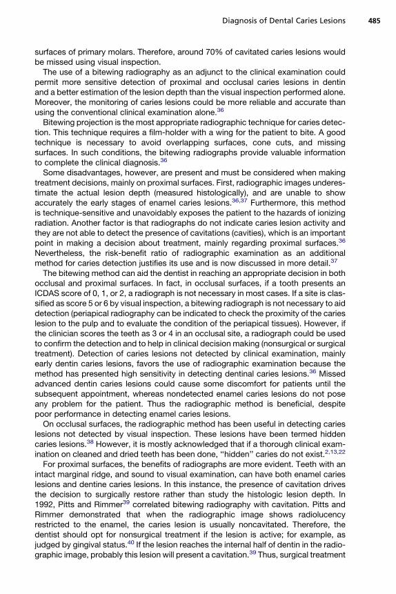
For proximal surfaces, the benefits of radiographs are more evident. Teeth with anintact marginal ridge, and sound to visual examination, can have both enamel carieslesions and dentine caries lesions. In this instance, the presence of cavitation drivesthe decision to surgically restore rather than study the histologic lesion depth. In1992, Pitts and Rimmer39 correlated bitewing radiography with cavitation. Pitts andRimmer demonstrated that when the radiographic image shows radiolucencyrestricted to the enamel, the caries lesion is usually noncavitated. Therefore, thedentist should opt for nonsurgical treatment if the lesion is active; for example, asjudged by gingival status.40 If the lesion reaches the internal half of dentin in the radio-graphic image, probably this lesion will present a cavitation.39 Thus, surgical treatment

Braga et al486
is the best choice. However, when the radiographic image is an initial dentin carieslesion at the proximal surface, some caries lesions will present cavitation; but manyof these surfaces will not have cavitation.10 In these cases, therefore, the cliniciancould provide the temporary separation of the tooth with orthodontic rubbers, tocheck if the surface is cavitated39 and determine the correct treatment decision.More common is the decision to plan for a new examination including a radiographa year later, to monitor the progression and success of nonsurgical interventions.
Another possibility is digital radiography. In this technique, a digital sensor is usedinstead of conventional film and the radiographic image is stored in a computer. Digitalradiographs usually expose patients to lower radiation doses than conventionalmethods. Digital radiographs permit the use of computer facilities, such as thepossibilities of image enhancement and processing of the images, and of sendingthe images to other colleagues.36,41 In general, conventional radiographic methodshave presented sensitivity values around 0.50 to 0.60 and specificity values usuallyhigher than 0.90 in detecting proximal caries lesions in primary and permanentteeth.9,10,16,41
For occlusal surfaces, the sensitivity values of conventional radiography have beenbetween 0.50 and 0.80, and specificities around 0.80.9,13,41,42 Digital methods haveperformed similarly.36
Novel tools, however, have been created for digital radiographic images to improvethe process of caries detection or monitoring. Methods of digital subtraction can beused in 2 radiographic images of the same site recorded in different periods.36,43
This method would allow for monitoring caries lesion progression,43 because the 2radiographs are taken with partly controlled projection angles.36 Computed tomog-raphy techniques have been recommended for caries detection and seems a prom-ising tool for this issue,44 but further studies are necessary to check theperformance and real benefits of the method.
In the use of the radiographic method as an adjunct of clinical examination in detect-ing caries, one can summarize that the conventional bitewing image can aid thedentist in reaching a treatment decision, mainly on proximal surfaces or occlusal sitesscored as 3 or 4 by the ICDAS (see Fig. 2). Digital radiography has not presenteda better performance, but the method exposes the patients to lower doses of ionizingradiation and could offer some assistance to the clinician.
Fiber Optic Transillumination
When a tooth is illuminated by a light, the carious tissue, because of the porosity, scat-ters the light and the enamel shows as a whiter, opaque area in visual inspection. Like-wise, when the dentin is involved in the caries process, a shadow is observed in theunderlying dentin. This phenomenon is observed in lesions classified as score 4 ofthe ICDAS.
The fiber optic transillumination (FOTI) method uses a high-intensity white light toenhance these effects. Thus, carious enamel and dentin appear as shadows withthe use of the FOTI method.45,46 The method is more appropriate for proximal surfacesbut can be used for all surfaces. The device usually is portable and easy to use, but it isnot quantitative and the diagnostic and treatment decisions depend on the dentist’sinterpretation. Therefore, low-reliability values are expected. A digital version of thetechnique, named digital fiber optic transillumination (DIFOTI), has been introduced,whereby the device records the images on a computer. These images can be archivedand assessed in recalls.46 This property could permit monitoring of surface deminer-alizations,41 but this use of DIFOTI has not been tested as yet. One important propertyof DIFOTI is worth mentioning. Because the CCD camera takes an image of the
Diagnosis of Dental Caries Lesions 487
surface only, it shows demineralization and does not correlate with cavitation; there-fore, it should not be interpreted in the same manner as a bitewing radiograph.47
With regard to the performance of the FOTI method in detecting caries lesions,several studies have presented high specificities but low sensitivities for both occlusaland proximal surfaces.9,41,45,46 The method does not use ionizing radiation. However,apart from this advantage, FOTI or DIFOTI methods have not presented more benefitsthan visual or radiographic methods.
Electronic Caries Monitor
The electronic caries monitor (ECM) device uses alternating current and measures thebulk resistance of tooth tissue.46,48 The porosity of caries lesions is filled with fluidswith high concentration of ions from the oral environment, and this more porous tissuedecreases electrical resistance or impedance more than the sound dental tissue.46
ECM is able to detect and quantify this difference.The method presents a probe that is directly applied in an occlusal site, and the
device shows a number that translates the electrical resistance of the site. Highernumbers indicate deeper caries lesions. As the method is quantitative, high-reproduc-ibility values would be expected. Nevertheless, only fair reliability has been reported,probably due to technical problems. With regard to validity, higher sensitivity values(around 0.90) have been obtained compared with conventional methods, in detectingocclusal dentin caries lesions. However, lower specificity values (around 0.80) havebeen observed.9,48 The decision about the activity status of the tooth should not bemade using ECM alone, but the method could be useful as an adjunct to visual inspec-tion. For use in daily clinical practice, however, the method has not presented advan-tages compared with conventional methods. Further, the method cannot yet beapplied to proximal surfaces.
Fluorescence-Based Methods
The knowledge that the presence of a caries lesion provokes changes in the fluores-cence properties of dental tissues has allowed the development of severalfluorescence-based methods for detection and quantification of caries lesions. Thefirst fluorescence-based method introduced onto the market was quantitative light-induced fluorescence (QLF). A QLF device uses a high-intensity halogen lamp, whichemits a blue light (l 5 370 nm) to excite the tooth. When exposed to a light with thiswavelength, dental tissues emit a fluorescence (in green spectrum), which is detectedby the system, and the image is recorded in a computer. Mineral loss in this toothcauses a decrease in the fluorescence. This fluorescence reduction is analyzed bycomputer software and the mineral loss is quantified.46,49
The QLF method permits early detection of enamel demineralization (earlier thanother methods). Furthermore, this method has presented a strong correlation withmineral loss of enamel caries lesions assessed by gold standard analytical methods(microradiography, for instance). Because of this property associated with highreliability values and a video repositioning software, which facilitates the acquisitionof identical images on different occasions,49 the device is an excellent method formonitoring enamel caries lesions to assess whether preventive measures are ableto arrest or remineralize the lesion. Some clinical studies have used the QLF methodto measure the effectiveness of preventive measures for initial caries lesions.50 Never-theless, the device is recommended more for smooth-surface caries lesions restrictedto the enamel. The strong correlation with mineral content of the tooth decreaseswhen the assessments are performed in dentin caries lesions.49

Braga et al488
The method has been also tested in detecting occlusal caries lesions.51,52 However,the results have been disappointing, with low values of specificity (0.11–0.55) andsensitivity (0.64–0.96), according to the different cut-off points used.52 Furthermore,acquisition of images from occlusal surfaces have been time consuming in the clinicalsetting.51
Another fluorescence-based method consists of a diode laser fluorescence device(LF), named DIAGNOdent (KaVo, Charlotte, NC). With the LF method, a diode laseremits a red light (l 5 655 nm), which is absorbed by bacterial by-products such asporphyrins. This light is partially reemitted as near-infrared fluorescence. The devicecaptures this fluorescence and translates it on a numerical scale from 0 to 99: thehigher the number, the deeper the caries lesions.41,53–55
The first version of the LF device (DIAGNOdent) was designed for the detection ofcaries lesions in occlusal and smooth surfaces. For this purpose, the method hasbeen extensively studied and has demonstrated the high reliability of the device indetecting occlusal caries lesions and a moderate correlation with mineral loss56 insmooth-surface caries lesions.41,57–59
With regard to validity, studies have demonstrated good sensitivity and specificityvalues, but the magnitudes of these values have been variable due to different cut-offpoints used in the different studies.58 Another possible explanation for the high vari-ability found in these studies could be due to several possible factors that can alterthe LF readings. The drying time of the site before the LF assessment,55,60 presenceof plaque55,60 or pigmentation,45 and some toothpastes or prophylaxis pastes61 arepossible factors that influence the LF readings.
The sensitivity values for detection of occlusal caries lesions have been higher thanthe specificities, and the values have been usually described between 0.80 and 0.90.62
The specificity values obtained in different studies have been between 0.60 and 0.70.These results, therefore, indicate that the device could be used as an adjunct to visualinspection, and could be an alternative for radiography. However, the older version ofthe device is not designed for proximal caries detection, and thus a radiograph wouldbe necessary to check the proximal surfaces.
Due to this limitation, a new version of the method was designed and intro-duced, named DIAGNOdent pen (LFpen). This new version permits the assessmentof both occlusal and proximal surfaces.63 The device works on the principles of theold version, but the design is different. The tip is rotatable around the axis of itslength, enabling the operator to assess mesial and distal surfaces from both sides(buccal and lingual). The tip designed for proximal surfaces is made of sapphirefiber with a prismatic shape, and the light is directed laterally to the longitudinalaxis of the tip. Another cylindrical tip is recommended for occlusal surfaces, andthe direction of its light is perpendicular to the axis of the length of the tip. Afterexcitation, the tip collects the fluorescence and translates it into a numerical scalefrom 0 to 99.63
The LFpen method has been tested for occlusal caries detection but the focus hereis on the studies of proximal surfaces. Some studies have demonstrated a slight supe-riority of the new device in detecting proximal caries lesions in the in vitro setting.63
Other studies, however, have observed that the device has performed similarly toradiographic methods in detecting cavitated proximal lesions in primary molars. Inthese studies, sensitivities were around 0.60 and the specificities were almost 1.0.10,16
The LFpen device could be an alternative to the radiographic method to aid thedentist in the decision-making process after visual inspection. Nevertheless, theevidence concerning the use of the method in clinical practice is limited, and furtherstudies are necessary to evaluate whether the method could be useful.

Diagnosis of Dental Caries Lesions 489
Summarizing the discussion about ECM and fluorescence-based methods, higherreproducibility values and the possibility of monitoring caries lesions could beexpected due to the quantitative nature of these methods. However, these methodshave shortcomings, for example, lack of LAA, which do not justify use in daily clinicalpractice without including the conventional visual inspection and bitewingradiography.
THE TREATMENT DECISION-MAKING PROCESS
As discussed in the introduction to this article, diagnosis is a fundamental step formaking treatment decisions. The authors suggest the following procedures. First,the dentist should clean and dry the teeth being examined. Then the dentist shouldclassify the teeth according to the severity scores using, for example, the ICDAS.Thereafter, the activity of lesions must be evaluated and a score assigned for eachspecific surface, classifying each surface as an active or inactive caries lesion. A deci-sion-making tree based on this evaluation can help the dentist make decisions (Fig. 3).
If a site is classified as sound (score 0) or with ICDAS scores 1 to 6 with inactivelesion, no treatment is necessary for caries management. Aesthetic or functional reha-bilitation could be considered, but no treatment is necessary to arrest caries progress,because these lesions have already been arrested. When the surface is classified asactive, noncavitated caries lesion (ICDAS scores 1 or 2), nonsurgical treatment isneeded to avoid progression of the lesion to a more severe condition (cavitation).
If the tooth surface is classified as ICDAS score 5 or 6 and the lesion is active,usually surgical treatment is recommended in an attempt to reduce the progressionof the caries lesion and provide conditions for the dentin-pulp reaction.
In active caries lesions classified as ICDAS score 3 or 4, either nonsurgical orsurgical treatment can be considered. A bitewing radiograph can aid the clinician inchoosing between nonsurgical and surgical approaches, because radiography willgive better information about lesion depth and proximity to the pulp. In proximalsurfaces, caries lesions not evident to visual inspection could be better detected usingthe adjunct bitewing radiographic method. If the lesion on the radiographic image isrestricted to the enamel and is active, nonsurgical management is the best choice.If the lesion reaches the middle third or more of the dentin, surgical treatment usuallyis the best option because most of these lesions are cavitated.24,32 On the other hand,
Fig. 3. Decision-making tree for dental caries lesions to be used after examination usingICDAS and lesions activity assessment.

Braga et al490
if the radiographic image indicates an initial dentin caries lesion (the outer third ofdentin), the dentist should check for the presence of cavitation to decide whichnonsurgical or surgical treatment is necessary. Here, temporary separation with ortho-dontic rubbers would be helpful. As an alternative for clinicians, Nyvad’s system canbe used instead of ICDAS.6 Here, the treatment decision is made in a similar way tothat described for ICDAS.
SUMMARY
Traditional thinking about caries management was not about diagnosis but aboutrestoring the caries lesions. Current dental caries management considers cariesdisease to be a dynamic and reversible process.6,63 The caries diagnosis processmust take into account not only caries lesion detection but also the etiologic factorsof dental caries and caries activity with regard to the disease and the lesions. Thus,the detection and assessment of caries lesions discussed in this article is onlya part of the caries disease diagnosis process.
For caries lesion detection and assessment, visual inspection aided by a ball-endedprobe is an essential method and must be performed in all patients. The use of indices,such as the ICDAS, improves the performance of the method mainly in terms of sensi-tivity and reliability. Through visual inspection the presence, severity, and activity oflesions must be assessed.
Other methods can be used as adjuncts to visual inspection. In the clinical practicesetting, the most recommended additional method is radiography, using bitewingprojection. Bitewing radiographs can aid the dentist in reaching a more appropriatetreatment decision, mainly on proximal surfaces or occlusal caries lesions scored 3or 4 on the ICDAS. New technologies have been developed and studied, but nonehas demonstrated significant benefits that justify use in daily clinical practice. Forresearch purposes, however, new technologies could be useful. Further studiesmust be conducted to improve conventional methods, mainly in the assessment ofcaries lesion activity. Studies must also be conducted to find a new technology withbetter performance than conventional methods, or a new method that permitsa more objective assessment of caries lesion activity.
The ICDAS, including the activity assessment system or the Nyvad system, seemsto be the best option for a final caries diagnosis.
REFERENCES
1. Thylstrup A, Fejerskov O. Textbook of clinical cariology. 2nd edition. Copenhagen(Denmark): Munksgaard; 1994.
2. Ekstrand KR, Ricketts DN, Kidd EA. Occlusal caries: pathology, diagnosis andlogical management. Dent Update 2001;28(8):380–7.
3. Bader JD, Shugars DA. What do we know about how dentists make caries-relatedtreatment decisions? Community Dent Oral Epidemiol 1997;25(1):97–103.
4. Nyvad B, Fejerskov O. Assessing the stage of caries lesion activity on the basis ofclinical and microbiological examination. Community Dent Oral Epidemiol 1997;25(1):69–75.
5. Basting RT, Serra MC. Occlusal caries: diagnosis and noninvasive treatments.Quintessence Int 1999;30(3):174–8.
6. Nyvad B. Diagnosis versus detection of caries. Caries Res 2004;38(3):192–8.7. Baelum V, Heidmann J, Nyvad B. Dental caries paradigms in diagnosis and
diagnostic research. Eur J Oral Sci 2006;114(4):263–77.

Diagnosis of Dental Caries Lesions 491
8. Pitts NB. Current methods and criteria for caries diagnosis in Europe. J DentEduc 1993;57(6):409–14.
9. Bader JD, Shugars DA, Bonito AJ. A systematic review of the performance ofmethods for identifying carious lesions. J Public Health Dent 2002;62(4):201–13.
10. Braga MM, Morais CC, Nakama RC, et al. In vitro performance of methods of ap-proximal caries detection in primary molars. Oral Surg Oral Med Oral Pathol OralRadiol Endod 2009;108(4):e35–41.
11. Ekstrand KR. Improving clinical visual detection—potential for caries clinicaltrials. J Dent Res 2004;83(Spec Issue, No C):C67–71.
12. Ekstrand KR, Kuzmina I, Bjorndal L, et al. Relationship between external andhistologic features of progressive stages of caries in the occlusal fossa. CariesRes 1995;29(4):243–50.
13. Ekstrand KR, Ricketts DN, Kidd EA. Reproducibility and accuracy of threemethods for assessment of demineralization depth of the occlusal surface: anin vitro examination. Caries Res 1997;31(3):224–31.
14. Nyvad B, Machiulskiene V, Baelum V. Reliability of a new caries diagnosticsystem differentiating between active and inactive caries lesions. Caries Res1999;33(4):252–60.
15. Braga MM, Mendes FM, Martignon S, et al. In vitro Comparison of Nyvad’s systemand ICDAS-II with lesion activity assessment for evaluation of severity and activityof occlusal caries lesions in primary teeth. Caries Res 2009;43(5):405–12.
16. Novaes TF, Matos R, Braga MM, et al. Performance of a pen-type laser fluores-cence device and conventional methods in detecting approximal caries lesionsin primary teeth—in vivo study. Caries Res 2009;43(1):36–42.
17. Braga MM, Oliveira LB, Bonini GA, et al. Feasibility of the International CariesDetection and Assessment System (ICDAS-II) in epidemiological surveys andcomparability with standard World Health Organization criteria. Caries Res2009;43(4):245–9.
18. Ismail AI. Visual and visuo-tactile detection of dental caries. J Dent Res 2004;83(Spec Issue, No C):C56–66.
19. Ekstrand K, Qvist V, Thylstrup A. Light microscope study of the effect of probingin occlusal surfaces. Caries Res 1987;21(4):368–74.
20. Kuhnisch J, Dietz W, Stosser L, et al. Effects of dental probing on occlusalsurfaces—a scanning electron microscopy evaluation. Caries Res 2007;41(1):43–8.
21. Pitts N. ‘‘ICDAS’’—an international system for caries detection and assessmentbeing developed to facilitate caries epidemiology, research and appropriate clin-ical management. Community Dent Health 2004;21(3):193–8.
22. Ekstrand KR, Ricketts DN, Kidd EA, et al. Detection, diagnosing, monitoring andlogical treatment of occlusal caries in relation to lesion activity and severity: an invivo examination with histological validation. Caries Res 1998;32(4):247–54.
23. Ismail AI, Sohn W, Tellez M, et al. The International Caries Detection and Assess-ment System (ICDAS): an integrated system for measuring dental caries.Community Dent Oral Epidemiol 2007;35(3):170–8.
24. Ekstrand KR, Martignon S, Ricketts DJ, et al. Detection and activity assessmentof primary coronal caries lesions: a methodologic study. Oper Dent 2007;32(3):225–35.
25. Jablonski-Momeni A, Stachniss V, Ricketts DN, et al. Reproducibility and accu-racy of the ICDAS-II for detection of occlusal caries in vitro. Caries Res 2008;42(2):79–87.

Braga et al492
26. Rodrigues JA, Hug I, Diniz MB, et al. Performance of fluorescence methods,radiographic examination and ICDAS II on occlusal surfaces in vitro. CariesRes 2008;42(4):297–304.
27. Shoaib L, Deery C, Ricketts DN, et al. Validity and Reproducibility of ICDAS II inPrimary Teeth. Caries Res 2009;43(6):442–8.
28. Mortimer KV. The relationship of deciduous enamel structure to dental disease.Caries Res 1970;4(3):206–23.
29. Shellis RP. Relationship between human enamel structure and the formation ofcaries-like lesions in vitro. Arch Oral Biol 1984;29(12):975–81.
30. Kuhnisch J, Berger S, Goddon I, et al. Occlusal caries detection in permanentmolars according to WHO basic methods, ICDAS II and laser fluorescencemeasurements. Community Dent Oral Epidemiol 2008;36(6):475–84.
31. Varma S, Banerjee A, Bartlett D. An in vivo investigation of associations betweensaliva properties, caries prevalence and potential lesion activity in an adult UKpopulation. J Dent 2008;36(4):294–9.
32. Ekstrand KR, Zero DT, Martignon S, et al. Lesion activity assessment. MonogrOral Sci 2009;21:63–90.
33. Baelum V, Machiulskiene V, Nyvad B, et al. Application of survival analysis tocarious lesion transitions in intervention trials. Community Dent Oral Epidemiol2003;31(4):252–60.
34. Mialhe FL, Pereira AC, Pardi V, et al. Comparison of three methods for detectionof carious lesions in proximal surfaces versus direct visual examination after toothseparation. J Clin Pediatr Dent 2003;28(1):59–62.
35. Peers A, Hill FJ, Mitropoulos CM, et al. Validity and reproducibility of clinical exam-ination, fibre-optic transillumination, and bite-wing radiology for the diagnosis ofsmall approximal carious lesions: an in vitro study. Caries Res 1993;27(4):307–11.
36. Wenzel A. Bitewing and digital bitewing radiography for detection of carieslesions. J Dent Res 2004;83(Spec Issue, No C):C72–5.
37. Pitts NB. The use of bitewing radiographs in the management of dental caries:scientific and practical considerations. Dentomaxillofac Radiol 1996;25(1):5–16.
38. Weerheijm KL. Occlusal ‘hidden caries’. Dental Update 1997;24(5):182–4.39. Pitts NB, Rimmer PA. An in vivo comparison of radiographic and directly as-
sessed clinical caries status of posterior approximal surfaces in primary andpermanent teeth. Caries Res 1992;26(2):146–52.
40. Ekstrand KR, Bruun G, Bruun M. Plaque and gingival status as indicators forcaries progression on approximal surfaces. Caries Res 1998;32(1):41–5.
41. Yang J, Dutra V. Utility of radiology, laser fluorescence, and transillumination.Dent Clin North Am 2005;49(4):739–52.
42. Mendes FM, Ganzerla E, Nunes AF, et al. Use of high-powered magnification todetect occlusal caries in primary teeth. Am J Dent 2006;19(1):19–22.
43. Ricketts DN, Ekstrand KR, Martignon S, et al. Accuracy and reproducibility ofconventional radiographic assessment and subtraction radiography in detectingdemineralization in occlusal surfaces. Caries Res 2007;41(2):121–8.
44. Tyndall DA, Rathore S. Cone-beam CT diagnostic applications: caries, peri-odontal bone assessment, and endodontic applications. Dent Clin North Am2008;52(4):825–41.
45. Cortes DF, Ellwood RP, Ekstrand KR. An in vitro comparison of a combined FOTI/visual examination of occlusal caries with other caries diagnostic methods andthe effect of stain on their diagnostic performance. Caries Res 2003;37(1):8–16.
46. Pretty IA. Caries detection and diagnosis: novel technologies. J Dent 2006;34(10):727–39.

Diagnosis of Dental Caries Lesions 493
47. Young DA, Featherstone JD. Digital imaging fiber-optic trans-illumination,F-speed radiographic film and depth of approximal lesions. J Am Dent Assoc2005;136(12):1682–7.
48. Longbottom C, Huysmans MC. Electrical measurements for use in caries clinicaltrials. J Dent Res 2004;83(Spec Issue, No C):C76–9.
49. Angmar-Mansson B, ten Bosch JJ. Quantitative light-induced fluorescence(QLF): a method for assessment of incipient caries lesions. DentomaxillofacRadiol 2001;30(6):298–307.
50. Al-Khateeb S, Forsberg CM, de Josselin de Jong E, et al. A longitudinal laserfluorescence study of white spot lesions in orthodontic patients. Am J OrthodDentofacial Orthop 1998;113(6):595–602.
51. Kuhnisch J, Ifland S, Tranaeus S, et al. In vivo detection of non-cavitated carieslesions on occlusal surfaces by visual inspection and quantitative light-inducedfluorescence. Acta Odontol Scand 2007;65(3):183–8.
52. Stookey GK. Quantitative light fluorescence: a technology for early monitoring ofthe caries process. Dent Clin North Am 2005;49(4):753–70.
53. Hibst R, Paulus R, Lussi A. Detection of occlusal caries by laser fluorescence:basic and clinical investigations. Med Laser Appl 2001;16:205–13.
54. Mendes FM, de Oliveira E, Araujo de Faria DL, et al. Ability of laser fluorescencedevice associated with fluorescent dyes in detecting and quantifying earlysmooth surface caries lesions. J Biomed Opt 2006;11(2):24007.
55. Mendes FM, Hissadomi M, Imparato JC. Effects of drying time and the presenceof plaque on the in vitro performance of laser fluorescence in occlusal caries ofprimary teeth. Caries Res 2004;38(2):104–8.
56. Mendes FM, Nicolau J. Utilization of laser fluorescence to monitor caries lesionsdevelopment in primary teeth. J Dent Child 2004;71(2):139–42.
57. Bengtson AL, Gomes AC, Mendes FM, et al. Influence of examiner’s clinicalexperience in detecting occlusal caries lesions in primary teeth. Pediatr Dent2005;27(3):238–43.
58. Braga MM, Mendes FM, Imparato JC, et al. Effect of cut-off points on perfor-mance of laser fluorescence for detecting occlusal caries. J Clin Pediatr Dent2007;32(1):33–6.
59. Lussi A, Megert B, Longbottom C, et al. Clinical performance of a laser fluores-cence device for detection of occlusal caries lesions. Eur J Oral Sci 2001;109(1):14–9.
60. Lussi A, Longbottom C, Gygax M, et al. Influence of professional cleaning anddrying of occlusal surfaces on laser fluorescence in vivo. Caries Res 2005;39(4):284–6.
61. Lussi A, Reich E. The influence of toothpastes and prophylaxis pastes on fluores-cence measurements for caries detection in vitro. Eur J Oral Sci 2005;113(2):141–4.
62. Bader JD, Shugars DA. A systematic review of the performance of a laser fluores-cence device for detecting caries. J Am Dent Assoc 2004;135(10):1413–26.
63. Lussi A, Hack A, Hug I, et al. Detection of approximal caries with a new laser fluo-rescence device. Caries Res 2006;40(2):97–103.