destruction of spirochete borrelia burgdorferi round-body ... · pdf filedestruction of...
TRANSCRIPT
Destruction of spirochete Borrelia burgdorferiround-body propagules (RBs) by theantibiotic TigecyclineØystein Brorsona, Sverre-Henning Brorsonb, John Scythesc, James MacAllisterd, Andrew Wiere,1, and Lynn Margulisd,2
aDepartment of Microbiology, Sentralsykehuset i Vestfold HF, N-3116 Tonsberg, Norway;bDepartment of Pathology, Rikshospitalet, N-0027 Oslo, Norway;cGlad Day Bookshop, Toronto, ON, Canada M4Y 1Z3; dDepartment of Geosciences, University of Massachusetts, Amherst, MA 01003; and eDepartment ofMedical Microbiology, University of Wisconsin, Madison, WI 53706
Contributed by Lynn Margulis, July 31, 2009 (sent for review May 4, 2009)
Persistence of tissue spirochetes of Borrelia burgdorferi as helicesand round bodies (RBs) explains many erythema-Lyme diseasesymptoms. Spirochete RBs (reproductive propagules also calledcoccoid bodies, globular bodies, spherical bodies, granules, cysts,L-forms, sphaeroplasts, or vesicles) are induced by environmentalconditions unfavorable for growth. Viable, they grow, move andreversibly convert into motile helices. Reversible pleiomorphy wasrecorded in at least six spirochete genera (>12 species). Penicillinsolution is one unfavorable condition that induces RBs. This anti-biotic that inhibits bacterial cell wall synthesis cures neither thesecond ‘‘Great Imitator’’ (Lyme borreliosis) nor the first: syphilis.Molecular-microscopic techniques, in principle, can detect in ani-mals (insects, ticks, and mammals, including patients) helices andRBs of live spirochetes. Genome sequences of B. burgdorferi andTreponema pallidum spirochetes show absence of >75% of genesin comparison with their free-living relatives. Irreversible integra-tion of spirochetes at behavioral, metabolic, gene product andgenetic levels into animal tissue has been documented. Irreversibleintegration of spirochetes may severely impair immunologicalresponse such that they persist undetected in tissue. We report invitro inhibition and destruction of B. burgdorferi (helices, RBs �‘‘cysts’’) by the antibiotic Tigecycline (TG; Wyeth), a glycylcyclineprotein-synthesis inhibitor (of both 30S and 70S ribosome sub-units). Studies of the pleiomorphic life history stages in responseto TG of both B. burgdorferi and Treponema pallidum in vivo andin vitro are strongly encouraged.
bacterial resistant stages � doxycycline � medical symbiotics �multiple sclerosis � spirochete cysts
We reexamine evidence and point to mainly Russian studiesignored in English scientific literature that spirochete
round bodies (RBs, also called coccoid bodies, globular bodies,spherical bodies, cysts, granules, L-forms, sphaeroplasts, orvesicles) are fully viable. RBs are spherical, membrane-boundedstructures that appear in pure cultures as they age in proportionto the disappearance of helical forms. They tend to be immotileor less motile than typical helical-shaped spirochetes althoughthey twitch and may move laterally. Analysis by thin sectiontransmission electron microscopy (tsTEM) has revealed thepresence of coiled protoplasmic cylinders and flagella inside RBsthat lead investigators to hypothesize that they are pleiomorphicstages of spirochetes (1) or that they are moribund. Anglophonemedical discussion of spirochetoses (spirochete-associated infir-mities, such as Lyme disease or syphilis) omit mention of ‘‘roundbodies’’ or state that they have no clinical relevance (2). Yetevidence abounds not only that RBs are viable but also that theymay locomote, grow, and reproduce.
Spirochetes threatened by environmental insult form RBs.Unfavorable conditions include changes in solution chemistry:acidity-alkalinity, salts, gas composition; concentrations of an-tibiotics, sugars, or other organic compounds such as aminoacids. Transition from one growth medium to a second of
different viscosity or temperature stimulates the formation ofRBs. Starvation, threat of desiccation, exposure to oxygen gas,total anoxia and/or sulfide may induce RB formation (3–13).RBs revert to the active helical swimmers when favorableconditions that support growth return (3–5).
That RBs reversibly convertible to healthy motile helices isbolstered by the discovery of a new member of the genusSpirochaeta: S. coccoides (14) through 16S ribosomal RNAsequences. Related on phylogenies to Spirochaeta thermophila,Spirochaeta bajacaliforniensis, and Spirochaeta smaragdinae, allof which have helical morphology, S. coccoides does not formspirals or helices; rather, it grows and reproduces in the RB form(14). Clearly, coccoidal spirochete life history stages are viable.
Penicillin, among many other ‘‘unfavorable growth condi-tions,’’ induces RBs (3, 6, 10–13, 15). That penicillin does notcure either of the two ‘‘Great Imitators’’ (Lyme disease andsyphilis) is widely accepted in Russian medical literature. In rabbitsinoculated for study with the venereal spirochete Treponema pal-lidum, borrelias were seen in testicular-tissue sections (16, 17).
RB formation in the test tube is induced by penicillin, espe-cially in the presence of glycine, a protein amino acid andtherefore food. Relevant observations contributed to the PhDdissertation and patent application of Belichenko (18), a studentof medical microbiologist Igor Bazikov, Stavropol, Russia. De-crease in penicillin concentration induced reversion of roundbodies to active helical spirochetes (18). Russian research (19)and ours (5, 20, 21) on spirochetal life history suggests that thecourse of Lyme disease, and probably other spirochetoses suchas syphilis, is altered by penicillin and other antibiotics.
Spirochetes, motile helical Gram-negative eubacteria, areheterotrophs that at optimal temperatures for growth requireabundant moist food. Most ferment sugar in the absence ofoxygen. They form a cohesive taxon, a prokaryotic phylum (22)detectable by DNA sequence analysis that corresponds preciselyto the 16 Svedberg-unit ribosomal RNA (16S rRNA) componentof the small 30S ribosomal subunit. Spirochetes, with theirGram-negative cell walls and internal (i.e., ‘‘periplasmic’’) f la-gella between the inner and outer membranes, are distinctive atthe level of thin section-electron microscopy (23). The plasma(inner) membrane is universal in all bacterial (i.e., prokaryotic)and eukaryotic cells, but the outer lipoprotein membrane thatsurrounds the peptidoglycan layer of the wall characterizes
Author contributions: Ø.B. designed research; Ø.B. and S.-H.B. performed research; S.-H.B.and L.M. contributed new reagents/analytic tools; J.S., J.M., A.W., and L.M. analyzed data;L.M. wrote the paper; and J.S., J.M., A.W., and L.M. provided graphics.
The authors declare no conflict of interest.
1Present address: Department of Biology and Health Sciences, Pace University, 861 BedfordRoad, Pleasantville, NY 10570-2799.
2To whom correspondence should be addressed. E-mail: [email protected] [email protected].
This article contains supporting information online at www.pnas.org/cgi/content/full/0908236106/DCSupplemental.
18656–18661 � PNAS � November 3, 2009 � vol. 106 � no. 44 www.pnas.org�cgi�doi�10.1073�pnas.0908236106
Gram-negative bacteria. Spirochete morphology, definitivelyidentified by EM transverse (cross) section, permits assignmentof the larger spirochetes (�0.5 �m in diameter) to a lower taxon,‘‘species’’ or ‘‘genus.’’ Negative stain images of high quality mayallow assessment of flagella insertions and numbers. Spirochetesdisplay a distinctive flagellar pattern summarized as n:2n:nwhere the fist ‘‘n’’ is the number of flagella at one end, ‘‘2n’’ (orzero for leptospiras) represents overlap of flagella in the middle,and the second ‘‘n’’ is the number of flagella at the oppositeend (24).
Borrelia burgdorferi (4:8:4 or 5:10:5) and other spirochetes innatural habitats [e.g., Treponema pallidum:(1:2:1 or 2:4:2) (25)]are detectable by at least three methods: (i) morphology, espe-cially active motile behavior in live bacteria, (ii) thin sectiontransmission electron micrographs (tsTEM), and (iii) negativestain whole mount EMs. Except for quality high magnificationobservation of spirochetes including their RBs in affected tissueby an experienced microscopist, and to some extent PCR forDNA, no definitive tests for the presence of spirochetes inpatients in late stages exist. Cures of Lyme disease or venerealtreponeme infection (syphilis) lack this level of verification.
Spirochetoses (e.g., leptospiroses, yaws, syphilis, and Lymedisease) are bacterial diseases correlated with continued pres-ence in the body of potentially identifiable spirochetes. Theyobey Koch’s postulates. The little-known history of Lyme bor-reliosis begins with the discovery of spiral bacteria in ticks byDutton before the 1905 publications of Dutton and Todd (26).Spirochetes developed from the large number of ‘‘granules’’ (i.e.,RBs) detected in infected ticks when ambient temperatures roseto 25 °C or higher. The spirochete bacteria were later namedBorrelia duttoni. Hindle (9) injected solutions of ‘‘granularforms’’ (i.e., RBs) in the absence of motile helices (Borreliaduttoni) into mice to show that they caused symptoms ofinfection in the test animals. Borrelia vincentii RBs that hadremained in their ‘‘granular’’ form for 31 months converted tohelical motile spirochetes when transferred to fresh mediumunder conditions favorable for growth (8). However, when otherinvestigators failed to find propagules, dormant spirochetes orpleiomorphic life history stages the RBs were declared to bedying spirochetes. Although terminologcal confusion (cysts,granules, RBs, vesicles, etc.) exacerbated the problem, thedismissal of an entire scientific literature was unjustified (23).
The presence of the eubacterium spirochete Borrelia burgdorf-eri in human tissue correlates with a syndrome of symptoms inpeople known to have experienced ‘‘erythema migrans,’’ amobile circular skin blemish related to a blood meal by immature(young means �1 mm) nymphs that are the most active intransmission of the disease. The acarids are Ixodes scapulariswhereas they are Ixodes pacificus in North America and I. ricinusin Europe. Arthropods in this genus of ticks (class Arachnida,phylum Chelicerata) (22) lack holometabolous development:eggs hatch into small ticks that molt without metamorphosis intonymphs and later adults. Only �50% of those bitten who latertest positive for Lyme borreliosis and present other symptomslike ‘‘Lyme arthritis’’ and neuroborreliosis actually develop thetell-tale �50-cent-size, coin-shaped rash at the site of the bite(27). Symptoms vary greatly in severity, frequency, and persis-tence. When antibodies against tick-borne borreliosis, PCR orcultured borreliosis are detected in patient blood, they arescored ‘‘Lyme disease borreliosis positive,’’ but results arefrequently negative despite infection (28). An estimated 20,000cases of Lyme disease are reported annually in the U.S., but theactual number is estimated to be closer to 200,000 cases per year.In central Europe and Scandinavia, the disease is highly en-demic. Cases of B. burgdorferi in southern Sweden alone rosefrom 164 (in 1992) to 664 (in 2000) per 100,000 population (29).Over 100 strains of Borrelia sp. have been isolated from healthy
ticks and grown in rich liquid medium that contains 6% mam-malian serum cultured at elevated temperatures (30–36 °C).
At least three immunologically distinguishable strains havebeen claimed to transmit the tendency toward symptoms: Bor-relia afzelii, B. burdorferi sensu stricto, and Borrelia garinii.
Persistence of tissue spirochetes of B. burgdorferi has beensuggested since Dutton first reported them in the 19th century.The group of isolates in the B. burdorferii complex sensu latoincludes named ‘‘genospecies:’’ Borrelia valaisiana, Borrelia lu-citania, Borrelia spielmanii, and Borrelia bissettii, with differentorganoheterotrophic optimal in vitro growth requirements. Dif-ferences in strains relative to symptoms is unclear [Fingerle et al.(30)]. Seabirds in the Arctic regions of Norway carry Ixodes uriaeticks from which B. garinii spirochetes were isolated (31). InDanish Ixodes ricinus three different genospecies were simulta-neously present in at least 40% of the ticks (32). Borrelia sensustricto was found in blood plasma from nearly half of theerythema migrans of American patients when large volumes ofblood plasma were used as source of spirochete cultivation (33).Lyme borreliosis, with incidence that has increased in northerncountries, becomes chronic and variable. The percentage ofvirulent strains has greatly increased in recent years (34). Thesite(s) of affected tissue or organ and the intensity, severity,frequency, and duration of symptoms are idiosyncratic. Clinicalfailures to eradicate all traces of symptoms and correlatedimmunological indicators of the presence of the spirochetes havebeen widely reported in mainstream medical literature. Lack ofrelief with no definitive cure of ‘‘Lyme arthritis’’ or ‘‘post-Lymesyndrome’’ and other borreliosis symptoms has led to a ground-swell of health advice, published contradictory claims and othervociferous activity beyond confines of professional medicine. Wehypothesize that Lyme borrelioses is an old chronic bacterialsymbiosis that tends toward necrotrophy in mammals. In healthyimmature ticks, the spirochetes are seasonally transmitted by biteto vertebrates including seabirds, cervids, mice, rabbits, andhumans.
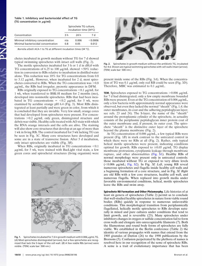
ResultsTigecycline (TG) Incubation. When incubated for 3 h (h) in TGconcentrations �50 �g/mL, spirochete motility was normal inBSK-H medium to which TG had been added. By use ofdarkfield microscopy (DFM) at TG concentrations from 50 to100 �g/mL movement was reduced. When the contents of theincubated tubes were transferred to fresh BSK-H medium, onesingle motile bacterium was observed after incubating for 8 wkat a concentration of 0.4 �g/mL. Therefore, minimal bacterio-cidal concentration (MBC) for 3 h incubation is 0.8 �g/mL.
When motile spirochetes were examined after 24 h incubationin BSK-H medium to which TG was added at 6.25 to 100 �g/mL,the bacterial cells had entirely disintegrated. Those originallyexposed to TG concentrations from 3.12 to 0.4 �g/mL haddegenerated and were not motile. For concentrations from 0.012to 0.2 �g/mL, some spirochetes moved normally whereas othershardly moved at all. They failed to survive and reproduce upontransfer to fresh medium after incubation for times up to 2months. Spirochetes originally exposed to a TG concentration of0.003 �g/mL grew and reproduced to the same level as theantibiotic-free control. Thus, minimal inhibition concentration(MIC) for 24 h incubation is 0.006 �g/mL, and transfer to freshBSK-H medium 1/50 showed MBC to be 0.05 �g/mL.
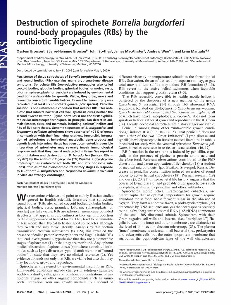
The spirochetes that had been incubated for 7 days (d) withTG/BSK-H were subsequently incubated in fresh BSK-H me-dium for 8 wk at 34 °C, and the MBC turned out to be 0.012�g/mL (Table 1). For TG concentration �0.006 �g/mL, thespirochetes survived and reproduced in fresh BSK-H. Whenexamined by TEM, a few spirochetes lacking the S-layer of thewall (Fig. 1A) and a few RBs with normal ultrastructure werepresent at the concentration of 0.006 �g/mL (Fig. 1B). Spiro-
Brorson et al. PNAS � November 3, 2009 � vol. 106 � no. 44 � 18657
EVO
LUTI
ON
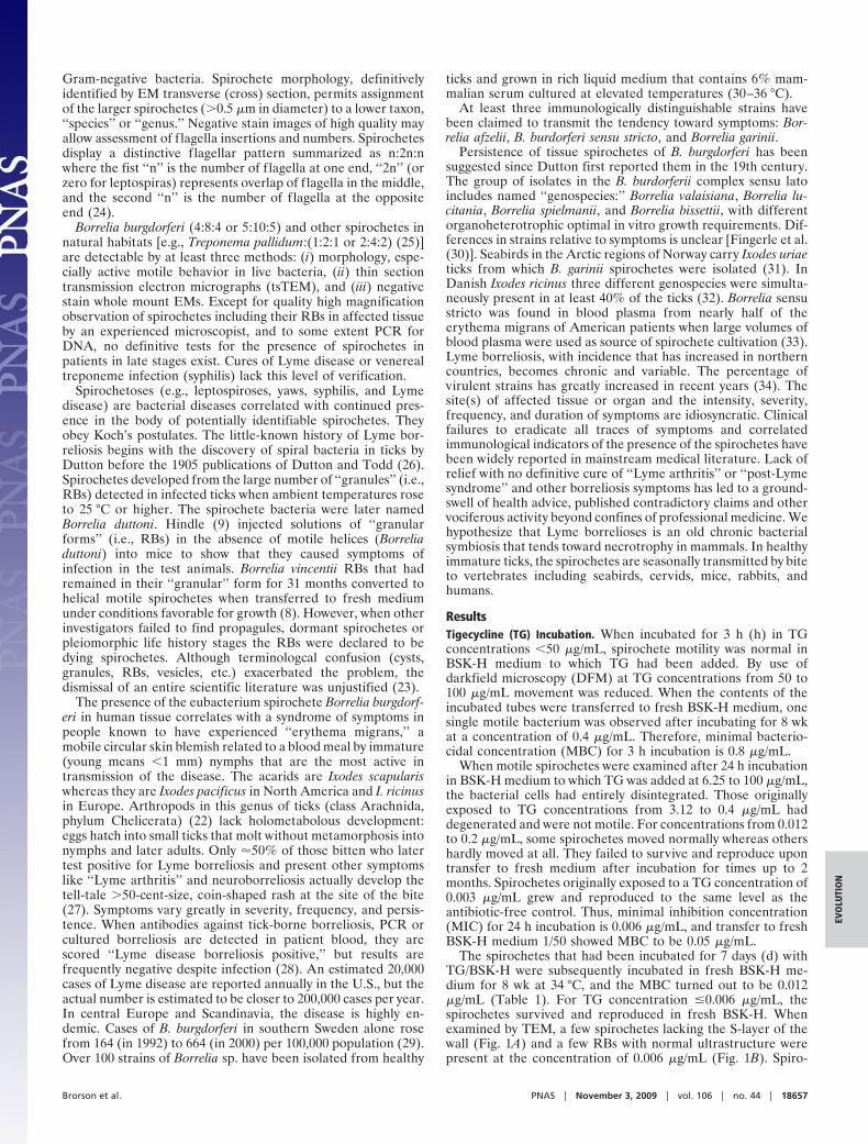
chetes incubated in growth medium without TG for 7 d showedtypical swimming spirochetes with intact cell walls (Fig. 2).
The motile spirochetes incubated for 3 h or 1 d in dH20 withTG concentrations of 6.25 to 100 �g/mL showed �50% reduc-tion in conversion to RBs relative to incubation in distilled wateralone. This reduction was 10% for TG concentrations from 0.8to 3.12 �g/mL. However, when incubated for 2 d, most spiro-chetes converted to RBs. When the TG concentration was �0.8�g/mL, the RBs had irregular, pycnotic appearance in DFM.
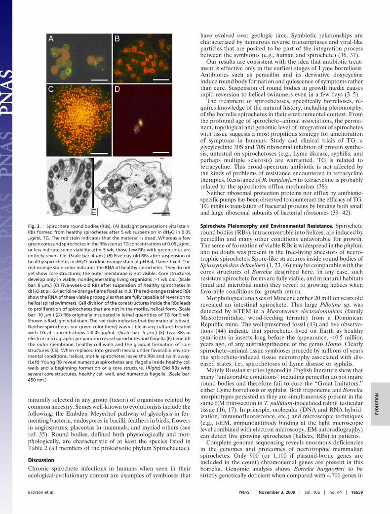
RBs originally exposed to TG concentrations �0.1 �g/mL for1 wk, when transferred to BSK-H medium for 2 months (mo),developed into nonmotile spirochetes. RBs that had been incu-bated in TG concentrations � �0.2 �g/mL for 5 wk, wereexamined by acridine orange pH 6.4 (Fig. 3). Most RBs disin-tegrated at least partially and were green in color, from which itis concluded that they are inviable. Very few small, orange coresthat had developed from spirochetes were present. For concen-trations �0.2 �g/mL only green, disintegrated structure anddebris was visible. Healthy cells treated with AO stain with whichthe RNA orange interacts and the cells are alive. The stainingwill also show core structures that develop at an age of more than1 wk in living RB. The control incubated for 5 wk lacking TG canbe seen in Fig. 3C. Many core structures have developed andsome are in a state of transverse fission. After 5 d incubation,only intact spirochetes are visible (Fig. 3B).
When RBs, originally incubated in TG concentrations �0.1�g/mL for 5 wk, were stained with BacLight vital stain, a fewgreen cores and spirochetal structures (living organism) were
present inside some of the RBs (Fig. 3A). When the concentra-tion of TG was 0.1 �g/mL only red RB could be seen (Fig. 3D).Therefore, MBC was estimated to 0.1 �g/mL.
TEM. Spirochetes exposed to TG concentrations �0.006 �g/mLfor 7 d had disintegrated; only a few empty membrane boundedRBs were present. Even at the TG concentration of 0.006 �g/mL,only a few bacteria with approximately normal appearance wereobserved, but even they lacked the normal ‘‘sheath’’ (Fig. 1 A; theouter membranes, its coat and the adhering peptidoglycan layer;see refs. 23 and 24). The S-layer, the name of the ‘‘sheath’’around the protoplasmic cylinder of the spirochete, in actualityconsists of the periplasmic peptidoglycan inner protein coat ofthe outer membrane and, if present, its outer coat. The spiro-chete ‘‘sheath’’ is the distinctive outer layer of the spirochetebeyond the plasma membrane (Fig. 2).
At TG concentration of 0.006 �g/mL, a few typical RBs werepresent (Fig. 1B) in stark contrast to the untreated control inwhich there were no RBs at all (Fig. 2). Rather, only typicalhelical motile spirochetes were present, indicating conditionsoptimal for growth. RBs exposed to �0.05 �g/mL TG displaymembrane protrusions, cytoplasmic leakage through membranerupture, and other abnormalities (Fig. S1). Healthy RBs ofnormal morphology were present only in untreated controls:those incubated without TG or exposed to very dilute levels(�0.006 �g/mL; Fig. S2). In Fig. 3E Left, young RB revealnumerous spirochetes and flagella inside healthy cell walls anda beginning formation of a core structure, and in Fig. 3E Rightare old RBs with a few core structures, healthy cell wall, andnumerous flagella. When replaced into growth media underfavorable environmental conditions, helical, motile spirochetesleave the RBs and swim away.
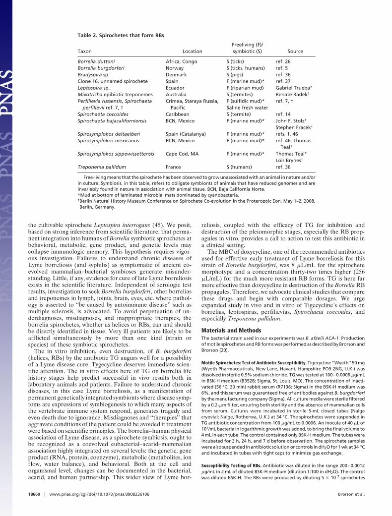
Spirochete RB Formation and Other Pleiomorphy. Life histories of atleast six genera of spirochetes (Table 2) permit us to concludethat well-studied healthy spirochetes in nature form viable roundbodies (RBs) quickly in response to numerous unfavorableconditions. This morphological transition from periplasmicallyf lagellated, helically motile spirochetes to RBs develops natu-rally in mixed and pure cultures under conditions that tend tolimit growth, and is reversible (23). Many spirochetes underinhibitory changes in oxygen or sulfide concentration fail to formcross walls and elongate into unrecognizable filaments (7). Boththe filamentous and round body forms of spirochetes are fullyviable. We established in the Berlin conference (Table 2) theidentity of various propagules with names that extend from the1905 granules of Dutton (26) to the 1998 publications by theBrorsons (5). Confusion perpetrated by the plethora of names isresolved here in our recognition of the seme of spirochete RBs.A seme is a trait of evolutionary importance that has been
Fig. 2. Spirochetes in growth medium without the antibiotic TG, incubatedfor 4 d. Shown are typical swimming spirochetes with cell walls intact (arrow).(TEM; scale bar: 500 nm.)
Table 1. Inhibitory and bacteriocidal effect of TG(TG concentration in �g/ml)
Concentration
Spirochete TG culture,incubation time (34°C)
3 h 24 h 7 d
Minimal inhibitory concentration n/a 0.006 �0.0006Minimal bacteriocidal concentration 0.8 0.05 0.012
Borrelia afzelii ACA-1 to TG at different incubation times (34 °C).
A
B
Fig. 1. Spirochetes incubated for 7 d in growth medium with 0.006 �g/mL TG.(A) Most spirochetes disintegrated (arrows), but a few spirochetes are recog-nized that lack the S-layer of the cell wall. (B) A few viable RBs (arrow) werevisible. (TEM; scale bar: 500 nm.)
18658 � www.pnas.org�cgi�doi�10.1073�pnas.0908236106 Brorson et al.
naturally selected in any group (taxon) of organisms related bycommon ancestry. Semes well-known to evolutionists include thefollowing: the Embden–Meyerhof pathway of glycolysis in fer-menting bacteria, endospores in bacilli, feathers in birds, f lowersin angiosperms, placentas in mammals, and myriad others (seeref. 35). Round bodies, defined both physiologically and mor-phologically, are characteristic of at least the species listed inTable 2 (all members of the prokaryotic phylum Spirochaetae).
DiscussionChronic spirochete infections in humans when seen in theirecological-evolutionary context are examples of symbioses that
have evolved over geologic time. Symbiotic relationships arecharacterized by numerous reverse transcriptases and viral-likeparticles that are posited to be part of the integration processbetween the symbionts (e.g., human and spirochete) (36, 37).
Our results are consistent with the idea that antibiotic treat-ment is effective only in the earliest stages of Lyme borreliosis.Antibiotics such as penicillin and its derivative doxycyclineinduce round body formation and quiescence of symptoms ratherthan cure. Suspension of round bodies in growth media causesrapid reversion to helical swimmers even in a few days (3–5).
The treatment of spirochetoses, specifically borrelioses, re-quires knowledge of the natural history, including pleiomorphy,of the borrelia spirochetes in their environmental context. Fromthe profound age of spirochete–animal associations, the perma-nent, topological and genomic level of integration of spirocheteswith tissue suggests a most propitious strategy for ameliorationof symptoms in humans. Study and clinical trials of TG, aglycylcycline 30S and 70S ribosomal inhibitor of protein synthe-sis, untested on spirochetoses (e.g., Lyme disease, syphilis, andperhaps multiple sclerosis) are warranted. TG is related totetracycline. This broad-spectrum antibiotic is not affected bythe kinds of problems of resistance encountered in tetracyclinetherapies. Resistance of B. burgdorferi to tetracycline is probablyrelated to the spirochetes eff lux mechanism (38).
Neither ribosomal protection proteins nor efflux by antibiotic-specific pumps has been observed to counteract the efficacy of TG.TG inhibits translation of bacterial proteins by binding both smalland large ribosomal subunits of bacterial ribosomes (39–42).
Spirochete Pleiomorphy and Environmental Resistance. Spirocheteround bodies (RBs), intraconvertible into helices, are induced bypenicillin and many other conditions unfavorable for growth.The seme of formation of viable RBs is widespread in the phylumand no doubt was present in the free-living ancestors of necro-trophic spirochetes. Spore-like structures inside round bodies ofSpirosymplokos deltaeiberi (1, 23, 46) may be comparable with thecores structures of Borrelia described here. In any case, suchresistant spirochete forms are fully viable, and in natural habitats(mud and microbial mats) they revert to growing helices whenfavorable conditions for growth return.
Morphological analyses of Miocene amber 20 million years oldrevealed an intestinal spirochete. This large Pillotina sp. wasdetected by tsTEM in a Mastotermes electrodominicus (familyMastotermitidae, wood-feeding termite) from a DominicanRepublic mine. The well-preserved fossil (43) and live observa-tions (44) indicate that spirochetes lived on Earth as healthysymbionts in insects long before the appearance, �0.5 millionyears ago, of any australopithecine of the genus Homo. Clearlyspirochete–animal tissue symbioses precede by millions of yearsthe spirochete-induced tissue necrotrophy associated with dis-eased states, i.e., spirochetoses of Lyme disease or syphilis.
Mainly Russian studies ignored in English literature show thatmany ‘‘unfavorable conditions’’ including penicillin do not injureround bodies and therefore fail to cure the ‘‘Great Imitators,’’either Lyme borreliosis or syphilis. Both treponeme and Borreliamorphotypes persisted as they are simultaneously present in thesame EM thin-section in T. pallidum-inoculated rabbit testiculartissue (16, 17). In principle, molecular (DNA and RNA hybrid-ization, immunofluorescence, etc.) and microscopic techniques(e.g., tsEM, immunoantibody binding at the light microscopiclevel combined with electron microscopy, EM autoradiography)can detect live growing spirochetes (helices, RBs) in patients.
Complete genome sequencing reveals enormous deficienciesin the genomes and proteomes of necrotrophic mammalianspirochetes. Only 900 (or 1,100 if plasmid-borne genes areincluded in the count) chromonemal genes are present in thisborrelia. Genomic analysis shows Borrelia burgdorferi to bestrictly genetically deficient when compared with 4,700 genes in
A B
C D
E
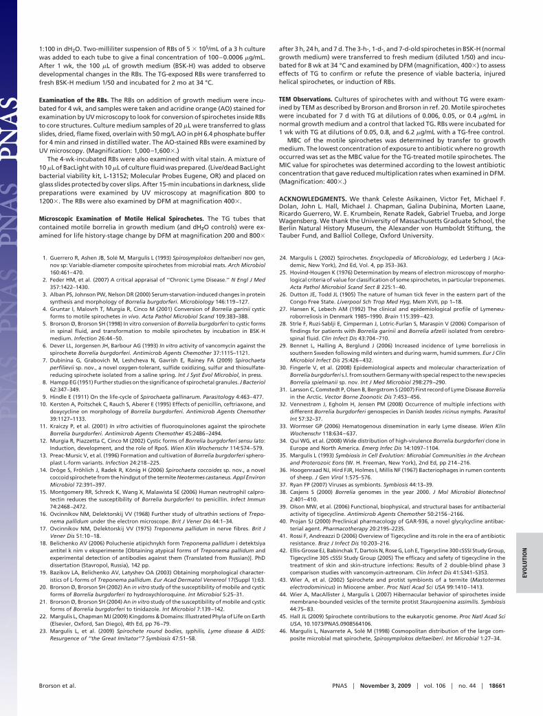
Fig. 3. Spirochete round bodies (RBs). (A) BacLight preparations vital stain.RBs formed from healthy spirochetes after 5 wk suspension in dH2O in 0.05�g/mL TG. The red stain indicates that the material is dead. Whereas a fewgreen cores and spirochetes in the RBs seen at TG concentrations of 0.05 �g/mLor less indicate some viability after 5 wk, those few RBs with green cores areentirely reversible. (Scale bar: 4 �m.) (B) Five-day-old RBs after suspension ofhealthy spirochetes in dH2O-acridine orange stain at pH 6.4, flame-fixed. Thered-orange stain color indicates the RNA of healthy spirochetes. They do notyet show core structures; the outer membrane is not visible. Core structuresdevelop only in viable, nondegenerating living organisms �1 wk old. (Scalebar: 8 �m.) (C) Five-week-old RBs after suspension of healthy spirochetes indH2O at pH 6.4 acridine orange flame fixed as in B. The red-orange stained RBsshow the RNA of these viable propagules that are fully capable of reversion tohelical spiral swimmers. Cell division of the core structures inside the RBs leadsto proliferation of spirochetes that are not in the motile, helical form. (Scalebar: 10 �m.) (D) RBs originally incubated in lethal quantities of TG for 5 wk.Shown is BacLight vital stain. The red stain indicates that the material is dead.Neither spirochetes nor green color (here) was visible in any cultures treatedwith TG at concentrations �0.05 �g/mL. (Scale bar: 5 �m.) (E) Two RBs inelectron micrographic preparation reveal spirochetes and flagella (F) beneaththe outer membrane, healthy cell walls and the gradual formation of corestructures (CS). When replaced into growth media under favorable environ-mental conditions, helical, motile spirochetes leave the RBs and swim away.(Left) Young RB reveal numerous spirochetes and flagella inside healthy cellwalls and a beginning formation of a core structure. (Right) Old RBs withseveral core structures, healthy cell wall, and numerous flagella. (Scale bar:450 nm.)
Brorson et al. PNAS � November 3, 2009 � vol. 106 � no. 44 � 18659
EVO
LUTI
ON
the cultivable spirochete Leptospira interrogans (45). We posit,based on strong inference from scientific literature, that perma-nent integration into humans of Borrelia symbiotic spirochetes atbehavioral, metabolic, gene product, and genetic levels maycollapse immunologic memory. This hypothesis requires vigor-ous investigation. Failures to understand chronic diseases ofLyme borreliosis (and syphilis) as symptomatic of ancient co-evolved mammalian–bacterial symbioses generate misunder-standing. Little, if any, evidence for cure of late Lyme borreliosisexists in the scientific literature. Independent of serologic testresults, investigation to seek Borrelia burgdorferi, other borreliasand treponemes in lymph, joints, brain, eyes, etc. where pathol-ogy is asserted to ‘‘be caused by autoimmune disease’’ such asmultiple sclerosis, is advocated. To avoid perpetuation of un-derdiagnoses, misdiagnoses, and inappropriate therapies, theborrelia spirochetes, whether as helices or RBs, can and shouldbe directly identified in tissue. Very ill patients are likely to beafflicted simultaneously by more than one kind (strain orspecies) of these symbiotic spirochetes.
The in vitro inhibition, even destruction, of B. burgdorferi(helices, RBs) by the antibiotic TG augurs well for a possibilityof a Lyme disease cure. Tygecycline deserves immediate scien-tific attention. The in vitro effects here of TG on borrelia lifehistory stages help predict successful in vivo results both inlaboratory animals and patients. Failure to understand chronicdiseases, in this case Lyme borreliosis, as a manifestation ofpermanent genetically integrated symbionts where disease symp-toms are expressions of symbiogenesis to which many aspects ofthe vertebrate immune system respond, generates tragedy andeven death due to ignorance. Misdiagnoses and ‘‘therapies’’ thataggravate conditions of the patient could be avoided if treatmentwere based on scientific principles. The borrelia–human physicalassociation of Lyme disease, as a spirochete symbiosis, ought tobe recognized as a coevolved eubacterial–acarid–mammalianassociation highly integrated on several levels: the genetic, geneproduct (RNA, protein, coenzyme), metabolic (metabolites, ionflow, water balance), and behavioral. Both at the cell andorganismal level, changes can be documented in the bacterial,acarid, and human partnership. This wider view of Lyme bor-
reliosis, coupled with the efficacy of TG for inhibition anddestruction of the pleiomorphic stages, especially the RB prop-agules in vitro, provides a call to action to test this antibiotic ina clinical setting.
The MBC of doxycycline, one of the recommended antibioticsused for effective early treatment of Lyme borreliosis for thisstrain of Borrelia burgdorferi, was 8 �L/mL for the spirochetemorphotype and a concentration thirty-two times higher (256�L/mL) for the much more resistant RB forms. TG is here farmore effective than doxycycline in destruction of the Borrelia RBpropagules. Therefore, we advocate clinical studies that comparethese drugs and begin with comparable dosages. We urgeexpanded study in vivo and in vitro of Tigecycline’s effects onborrelias, leptospiras, perfilievias, Spirochaeta coccoides, andespecially Treponema pallidum.
Materials and MethodsThe bacterial strain used in our experiments was B. afzelii ACA-1. Productionof motile spirochetes and RB forms was performed as described by Brorson andBrorson (20).
Motile Spirochetes: Test of Antibiotic Susceptibility. Tigecycline ‘‘Wyeth’’ 50 mg(Wyeth Pharmaceuticals, New Lane, Havant, Hampshire PO9 2NG, U.K.) wasdissolved in sterile 0.9% sodium chloride. TG was tested at 100–0.0006 �g/mLin BSK-H medium (B3528; Sigma, St. Louis, MO). The concentration of inacti-vated (56 °C, 30 min) rabbit serum (R7136; Sigma) in the BSK-H medium was6%, and this serum was guaranteed free of antibodies against B. burgdorferiby the manufacturing company (Sigma). All culture media were sterile filteredby a 0.2-�m filter, ensuring both sterility and the absence of mammalian cellsfrom serum. Cultures were incubated in sterile 5-mL closed tubes (Nalgecryovial; Nalge, Rotherwa, U.K.) at 34 °C. The spirochetes were suspended inTG antibiotic concentration from 100 �g/mL to 0.0006. An inocula of 40 �L of107/mL bacteria in logarithmic growth was added, to bring the final volume to4 mL in each tube. The control contained only BSK-H medium. The tubes wereincubated for 3 h, 24 h, and 7 d before observation. The spirochete sampleswere also suspended in antibiotic solution or controls in dH2O for 1 wk at 34 °Cand incubated in tubes with tight caps to minimize gas exchange.
Susceptibility Testing of RBs. Antibiotic was diluted in the range 200–0.0012�g/mL in 2 mL of diluted BSK-H medium (dilution 1:100 in dH2O). The controlwas diluted BSK-H. The RBs were produced by diluting 5 � 10 7 spirochetes
Table 2. Spirochetes that form RBs
Taxon LocationFreeliving (F)/symbiotic (S) Source
Borrelia duttoni Africa, Congo S (ticks) ref. 26Borrelia burgdorferi Norway S (ticks, humans) ref. 5Bradyspira sp. Denmark S (pigs) ref. 36Clone 16, unnamed spirochete Spain F (marine mud)* ref. 37Leptospira sp. Ecuador F (riparian mud) Gabriel Trueba†
Mixotricha epibiotic treponemes Australia S (termites) Renate Radek†
Perfilievia russensis, Spirochaetaperfilievii ref. 7, †
Crimea, Staraya Russia,Pacific
F (sulfidic mud)*Saline fresh water
ref. 7, †
Spirochaeta coccoides Caribbean S (termite) ref. 14Spirochaeta bajacaliforniensis BCN, Mexico F (marine mud)* John F. Stolz†
Stephen Fracek†
Spirosymplokos deltaeiberi Spain (Catalanya) F (marine mud)* refs. 1, 46Spirosymplokos mexicanus BCN, Mexico F (marine mud)* ref. 46, Thomas
Teal†
Spirosymplokos sippewissettensis Cape Cod, MA F (marine mud)* Thomas Teal†
Lois Brynes†
Treponema pallidum France S (humans) ref. 36
Free-living means that the spirochete has been observed to grow unassociated with an animal in nature and/orin culture. Symbiosis, in this table, refers to obligate symbionts of animals that have reduced genomes and areinvariably found in nature in association with animal tissue. BCN, Baja California Norte.*Mud at bottom of laminated microbial mats dominated by cyanobacteria.†Berlin Natural History Museum Conference on Spirochete Co-evolution in the Proterozoic Eon, May 1–2, 2008,Berlin, Germany.
18660 � www.pnas.org�cgi�doi�10.1073�pnas.0908236106 Brorson et al.
1:100 in dH2O. Two-milliliter suspension of RBs of 5 � 105/mL of a 3 h culturewas added to each tube to give a final concentration of 100–0.0006 �g/mL.After 1 wk, the 100 �L of growth medium (BSK-H) was added to observedevelopmental changes in the RBs. The TG-exposed RBs were transferred tofresh BSK-H medium 1/50 and incubated for 2 mo at 34 °C.
Examination of the RBs. The RBs on addition of growth medium were incu-bated for 4 wk, and samples were taken and acridine orange (AO) stained forexamination by UV microscopy to look for conversion of spirochetes inside RBsto core structures. Culture medium samples of 20 �L were transferred to glassslides, dried, flame fixed, overlain with 50 mg/L AO in pH 6.4 phosphate bufferfor 4 min and rinsed in distilled water. The AO-stained RBs were examined byUV microscopy. (Magnification: 1,000–1,600�.)
The 4-wk-incubated RBs were also examined with vital stain. A mixture of10 �L of BacLight with 10 �L of culture fluid was prepared. (Live/dead BacLightbacterial viability kit, L-13152; Molecular Probes Eugene, OR) and placed onglass slides protected by cover slips. After 15-min incubations in darkness, slidepreparations were examined by UV microscopy at magnification 800 to1200�. The RBs were also examined by DFM at magnification 400�.
Microscopic Examination of Motile Helical Spirochetes. The TG tubes thatcontained motile borrelia in growth medium (and dH2O controls) were ex-amined for life history-stage change by DFM at magnification 200 and 800�
after 3 h, 24 h, and 7 d. The 3-h-, 1-d-, and 7-d-old spirochetes in BSK-H (normalgrowth medium) were transferred to fresh medium (diluted 1/50) and incu-bated for 8 wk at 34 °C and examined by DFM (magnification, 400�) to assesseffects of TG to confirm or refute the presence of viable bacteria, injuredhelical spirochetes, or induction of RBs.
TEM Observations. Cultures of spirochetes with and without TG were exam-ined by TEM as described by Brorson and Brorson in ref. 20. Motile spirocheteswere incubated for 7 d with TG at dilutions of 0.006, 0.05, or 0.4 �g/mL innormal growth medium and a control that lacked TG. RBs were incubated for1 wk with TG at dilutions of 0.05, 0.8, and 6.2 �g/mL with a TG-free control.
MBC of the motile spirochetes was determined by transfer to growthmedium. The lowest concentration of exposure to antibiotic where no growthoccurred was set as the MBC value for the TG-treated motile spirochetes. TheMIC value for spirochetes was determined according to the lowest antibioticconcentration that gave reduced multiplication rates when examined in DFM.(Magnification: 400�.)
ACKNOWLEDGMENTS. We thank Celeste Asikainen, Victor Fet, Michael F.Dolan, John L. Hall, Michael J. Chapman, Galina Dubinina, Morten Laane,Ricardo Guerrero, W. E. Krumbein, Renate Radek, Gabriel Trueba, and JorgeWagensberg. We thank the University of Massachusetts Graduate School, theBerlin Natural History Museum, the Alexander von Humboldt Stiftung, theTauber Fund, and Balliol College, Oxford University.
1. Guerrero R, Ashen JB, Sole M, Margulis L (1993) Spirosymplokos deltaeiberi nov gen,nov sp: Variable-diameter composite spirochetes from microbial mats. Arch Microbiol160:461–470.
2. Feder HM, et al. (2007) A critical appraisal of ‘‘Chronic Lyme Disease.’’ N Engl J Med357:1422–1430.
3. Alban PS, Johnson PW, Nelson DR (2000) Serum-starvation-induced changes in proteinsynthesis and morphology of Borrelia burgdorferi. Microbiology 146:119–127.
4. Gruntar I, Malovrh T, Murgia R, Cinco M (2001) Conversion of Borrelia garinii cysticforms to motile spirochetes in vivo. Acta Pathol Microbiol Scand 109:383–388.
5. Brorson Ø, Brorson SH (1998) In vitro conversion of Borrelia burgdorferi to cystic formsin spinal fluid, and transformation to mobile spirochetes by incubation in BSK-Hmedium. Infection 26:44–50.
6. Dever LL, Jorgensen JH, Barbour AG (1993) In vitro activity of vancomycin against thespirochete Borrelia burgdorferi. Antimicrob Agents Chemother 37:1115–1121.
7. Dubinina G, Grabovich M, Leshcheva N, Gavrish E, Rainey FA (2009) Spirochaetaperfilievii sp. nov., a novel oxygen-tolerant, sulfide oxidizing, sulfur and thiosulfate-reducing spirochete isolated from a saline spring. Int J Syst Evol Microbiol, in press.
8. Hampp EG (1951) Further studies on the significance of spirochetal granules. J Bacteriol62:347–349.
9. Hindle E (1911) On the life-cycle of Spirochaeta gallinarum. Parasitology 4:463–477.10. Kersten A, Poitschek C, Rauch S, Aberer E (1995) Effects of penicillin, ceftriaxone, and
doxycycline on morphology of Borrelia burgdorferi. Antimicrob Agents Chemother39:1127–1133.
11. Kraiczy P, et al. (2001) In vitro activities of fluoroquinolones against the spirocheteBorrelia burgdorferi. Antimicrob Agents Chemother 45:2486–2494.
12. Murgia R, Piazzetta C, Cinco M (2002) Cystic forms of Borrelia burgdorferi sensu lato:Induction, development, and the role of RpoS. Wien Klin Wochenschr 114:574–579.
13. Preac-Mursic V, et al. (1996) Formation and cultivation of Borrelia burgdorferi sphero-plast L-form variants. Infection 24:218–225.
14. Droge S, Frohlich J, Radek R, Konig H (2006) Spirochaeta coccoides sp. nov., a novelcoccoid spirochete from the hindgut of the termite Neotermes castaneus. Appl EnvironMicrobiol 72:391–397.
15. Montgomery RR, Schreck K, Wang X, Malawista SE (2006) Human neutrophil calpro-tectin reduces the susceptibility of Borrelia burgdorferi to penicillin. Infect Immun74:2468–2472.
16. Ovcinnikov NM, Delektorskij VV (1968) Further study of ultrathin sections of Trepo-nema pallidum under the electron microscope. Brit J Vener Dis 44:1–34.
17. Ovcinnikov NM, Delektorskij VV (1975) Treponema pallidum in nerve fibres. Brit JVener Dis 51:10–18.
18. Belichenko AV (2006) Poluchenie atipichnykh form Treponema pallidum i detektsiyaantitel k nim v eksperimente [Obtaining atypical forms of Treponema pallidum andexperimental detection of antibodies against them (Translated from Russian)]. PhDdissertation (Stavropol, Russia), 142 pp.
19. Bazikov LA, Belichenko AV, Latyshev OA (2003) Obtaining morphological character-istics of L-forms of Treponema pallidum. Eur Acad Dermatol Venereol 17(Suppl 1):63.
20. Brorson Ø, Brorson SH (2002) An in vitro study of the susceptibility of mobile and cysticforms of Borrelia burgdorferi to hydroxychloroquine. Int Microbiol 5:25–31.
21. Brorson Ø, Brorson SH (2004) An in vitro study of the susceptibility of mobile and cysticforms of Borrelia burgdorferi to tinidazole. Int Microbiol 7:139–142.
22. Margulis L, Chapman MJ (2009) Kingdoms & Domains: Illustrated Phyla of Life on Earth(Elsevier, Oxford, San Diego), 4th Ed, pp 76–79.
23. Margulis L, et al. (2009) Spirochete round bodies, syphilis, Lyme disease & AIDS:Resurgence of ‘‘the Great Imitator’’? Symbiosis 47:51–58.
24. Margulis L (2002) Spirochetes. Encyclopedia of Microbiology, ed Lederberg J (Aca-demic, New York), 2nd Ed, Vol. 4, pp 353–363.
25. Hovind-Hougen K (1976) Determination by means of electron microscopy of morpho-logical criteria of value for classification of some spirochetes, in particular treponemes.Acta Pathol Microbiol Scand Sect B 225:1–40.
26. Dutton JE, Todd JL (1905) The nature of human tick fever in the eastern part of theCongo Free State. Liverpool Sch Trop Med Hyg, Mem XVII, pp 1–18.
27. Hansen K, Lebech AM (1992) The clinical and epidemiological profile of Lymeneu-roborreliosis in Denmark 1985–1990. Brain 115:399–423.
28. Strle F, Ruzi-Sablji E, Cimperman J, Lotric-Furlan S, Maraspin V (2006) Comparison offindings for patients with Borrelia garinii and Borrelia afzelii isolated from cerebro-spinal fluid. Clin Infect Dis 43:704–710.
29. Bennet L, Halling A, Berglund J (2006) Increased incidence of Lyme borreliosis insouthern Sweden following mild winters and during warm, humid summers. Eur J ClinMicrobiol Infect Dis 25:426–432.
30. Fingerle V, et al. (2008) Epidemiological aspects and molecular characterization ofBorrelia burgdorferi s.l. from southern Germany with special respect to the new speciesBorrelia spielmanii sp. nov. Int J Med Microbiol 298:279–290.
31. Larsson C, Comstedt P, Olsen B, Bergstrom S (2007) First record of Lyme Disease Borreliain the Arctic. Vector Borne Zoonotic Dis 7:453–456.
32. Vennestrøm J, Egholm H, Jensen PM (2008) Occurrence of multiple infections withdifferent Borrelia burgdorferi genospecies in Danish Ixodes ricinus nymphs. ParasitolInt 57:32–37.
33. Wormser GP (2006) Hematogenous dissemination in early Lyme disease. Wien KlinWochenschr 118:634–637.
34. Qui WG, et al. (2008) Wide distribution of high-virulence Borrelia burgdorferi clone inEurope and North America. Emerg Infec Dis 14:1097–1104.
35. Margulis L (1993) Symbiosis in Cell Evolution: Microbial Communities in the Archeanand Proterozoic Eons (W. H. Freeman, New York), 2nd Ed, pp 214–216.
36. Hoogenraad NJ, Hird FJR, Holmes I, Millis NF (1967) Bacteriophages in rumen contentsof sheep. J Gen Virol 1:575–576.
37. Ryan FP (2007) Viruses as symbionts. Symbiosis 44:13–39.38. Casjens S (2000) Borrelia genomes in the year 2000. J Mol Microbiol Biotechnol
2:401–410.39. Olson MW, et al. (2006) Functional, biophysical, and structural bases for antibacterial
activity of tigecycline. Antimicrob Agents Chemother 50:2156–2166.40. Projan SJ (2000) Preclinical pharmacology of GAR-936, a novel glycylcycline antibac-
terial agent. Pharmacotherapy 20:219S–223S.41. Rossi F, Andreazzi D (2006) Overview of Tigecycline and its role in the era of antibiotic
resistance. Braz J Infect Dis 10:203–216.42. Ellis-Grosse EJ, Babinchak T, Dartois N, Rose G, Loh E, Tigecycline 300 cSSSI Study Group,
Tigecycline 305 cSSSI Study Group (2005) The efficacy and safety of tigecycline in thetreatment of skin and skin-structure infections: Results of 2 double-blind phase 3comparison studies with vancomycin-aztreonam. Clin Infect Dis 41:S341–S353.
43. Wier A, et al. (2002) Spirochete and protist symbionts of a termite (Mastotermeselectrodominicus) in Miocene amber. Proc Natl Acad Sci USA 99:1410–1413.
44. Wier A, MacAllister J, Margulis L (2007) Hibernacular behavior of spirochetes insidemembrane-bounded vesicles of the termite protist Staurojoenina assimilis. Symbiosis44:75–83.
45. Hall JL (2009) Spirochete contributions to the eukaryotic genome. Proc Natl Acad SciUSA, 10.1073/PNAS.0908564106.
46. Margulis L, Navarrete A, Sole M (1998) Cosmopolitan distribution of the large com-posite microbial mat spirochete, Spirosymplokos deltaeiberi. Int Microbial 1:27–34.
Brorson et al. PNAS � November 3, 2009 � vol. 106 � no. 44 � 18661
EVO
LUTI
ON