design of a safer tracheostomy tube - research |...
TRANSCRIPT
Design of a Safer Tracheostomy Tube1
Arpan SarkarUniversity School of Nashville,
Nashville, TN 37212
Richard J. HendrickDepartment of Mechanical Engineering,
Vanderbilt University,
Nashville, TN 37235
Ray A. LathropDepartment of Mechanical Engineering,
Vanderbilt University,
Nashville, TN 37235
Bret AlvisDepartment of Anesthesiology,
Vanderbilt University,
Nashville, TN 37235
Robert J. Webster, IIIDepartment of Mechanical Engineering,
Vanderbilt University,
Nashville, TN 37235
1 Background
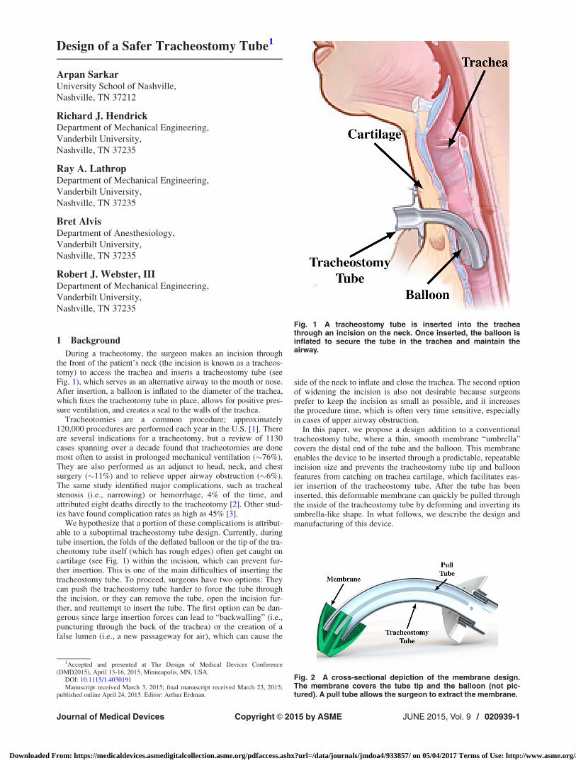
During a tracheotomy, the surgeon makes an incision throughthe front of the patient’s neck (the incision is known as a tracheos-tomy) to access the trachea and inserts a tracheostomy tube (seeFig. 1), which serves as an alternative airway to the mouth or nose.After insertion, a balloon is inflated to the diameter of the trachea,which fixes the tracheotomy tube in place, allows for positive pres-sure ventilation, and creates a seal to the walls of the trachea.
Tracheotomies are a common procedure; approximately120,000 procedures are performed each year in the U.S. [1]. Thereare several indications for a tracheotomy, but a review of 1130cases spanning over a decade found that tracheotomies are donemost often to assist in prolonged mechanical ventilation (�76%).They are also performed as an adjunct to head, neck, and chestsurgery (�11%) and to relieve upper airway obstruction (�6%).The same study identified major complications, such as trachealstenosis (i.e., narrowing) or hemorrhage, 4% of the time, andattributed eight deaths directly to the tracheotomy [2]. Other stud-ies have found complication rates as high as 45% [3].
We hypothesize that a portion of these complications is attribut-able to a suboptimal tracheostomy tube design. Currently, duringtube insertion, the folds of the deflated balloon or the tip of the tra-cheotomy tube itself (which has rough edges) often get caught oncartilage (see Fig. 1) within the incision, which can prevent fur-ther insertion. This is one of the main difficulties of inserting thetracheostomy tube. To proceed, surgeons have two options: Theycan push the tracheostomy tube harder to force the tube throughthe incision, or they can remove the tube, open the incision fur-ther, and reattempt to insert the tube. The first option can be dan-gerous since large insertion forces can lead to “backwalling” (i.e.,puncturing through the back of the trachea) or the creation of afalse lumen (i.e., a new passageway for air), which can cause the
side of the neck to inflate and close the trachea. The second optionof widening the incision is also not desirable because surgeonsprefer to keep the incision as small as possible, and it increasesthe procedure time, which is often very time sensitive, especiallyin cases of upper airway obstruction.
In this paper, we propose a design addition to a conventionaltracheostomy tube, where a thin, smooth membrane “umbrella”covers the distal end of the tube and the balloon. This membraneenables the device to be inserted through a predictable, repeatableincision size and prevents the tracheostomy tube tip and balloonfeatures from catching on trachea cartilage, which facilitates eas-ier insertion of the tracheostomy tube. After the tube has beeninserted, this deformable membrane can quickly be pulled throughthe inside of the tracheostomy tube by deforming and inverting itsumbrella-like shape. In what follows, we describe the design andmanufacturing of this device.
Fig. 1 A tracheostomy tube is inserted into the tracheathrough an incision on the neck. Once inserted, the balloon isinflated to secure the tube in the trachea and maintain theairway.
Fig. 2 A cross-sectional depiction of the membrane design.The membrane covers the tube tip and the balloon (not pic-tured). A pull tube allows the surgeon to extract the membrane.
1Accepted and presented at The Design of Medical Devices Conference(DMD2015), April 13-16, 2015, Minneapolis, MN, USA.
DOI: 10.1115/1.4030191Manuscript received March 3, 2015; final manuscript received March 23, 2015;
published online April 24, 2015. Editor: Arthur Erdman.
Journal of Medical Devices JUNE 2015, Vol. 9 / 020939-1Copyright VC 2015 by ASME
Downloaded From: https://medicaldevices.asmedigitalcollection.asme.org/pdfaccess.ashx?url=/data/journals/jmdoa4/933857/ on 05/04/2017 Terms of Use: http://www.asme.org/about-asme/terms-of-use

2 Methods
We set out to create a membrane attachment (see Fig. 2) thatwould make the tracheostomy tube insertion process smootherand more repeatable. We determined three main design inputs:first, the membrane must be large enough to cover the outer layersof the deflated foam cuff as well as the tip of the tube itself. Sec-ond, the membrane must be capable of being removed withoutdisturbing the tracheostomy tube or balloon. Finally, the mem-brane must be made of a biocompatible material.
These design inputs culminated in the concept of a highlydeformable silicone umbrella membrane. The design intent wasthat this membrane could be placed inside the tracheostomytube immediately prior to insertion, rather than becoming a per-manent fixture to the tube. The umbrella-like membrane isattached to a pull tube that runs the length of the tracheostomytube that the surgeon can grasp to deform the membrane, invert itsumbrella shape, and remove it through the tracheostomy tube (seeFig. 3).
The device was fabricated by filling a 3D-printed mold assem-bly (see Fig. 4) with DragonSkin (a silicone compound, Smooth-On, Inc., Macungie, PA), placing it in a vacuum chamber toremove all bubbles, and allowing it to cure for several hours.While DragonSkin was the available material for our prototype,we intend for the final, biocompatible design to be made from pol-ydimethylsiloxane, a known biocompatible silicone material. Thepull tube was designed as a silicone tube inserted along the axis ofthe membrane and secured with a silicone epoxy (Smooth-On,Inc., Macungie, PA). According to the adhesive specifications forbonding silicone to silicone, the tensile strength of the bond is750 psi, the tear strength is 100 pounds per inch (ppi) of bondingmaterial, and the peel strength is 100 ppi.
3 Results
One concern of this design is the potential for the pull tube toseparate from the membrane during retraction, which could plugthe tube or leave the membrane inside the trachea. To addressthis, we did three identical experiments characterizing the pullforce required to remove the membrane. We tied the pull tube to aspring scale and gradually increased pull force until the membranewas extracted. The pull out force never exceeded 2 lb in theexperiments. Since there is approximately 0.080 in. of membranematerial at the bond and the peel/tear strength of the adhesive is100 ppi, it would require 8 lb of force to peel or tear the membranefrom the pull tube. Therefore, we estimate a factor of safety ofover four for preventing failure of this bond.
The design of the membrane itself was challenging becausesmooth insertion requires a membrane that is not too thin, whileeasy retraction through the 7.8 mm tube argues for a membranethat is as thin as possible. The final membrane design smoothlythinned out from 0.045 in. at the tip to 0.006 in. at the mostproximal edge. Figure 3 shows a step-by-step retraction ofthe membrane during testing. The membrane initially coversthe tip of the tube and the balloon, begins to deform through thetube as it is pulled, inverts its shape, and then fits tightly withinthe inner diameter of the tracheostomy tube as it is pulledthrough. Although the current prototype can easily be pulledthrough the tracheostomy tube, a lubricant on the membranewould make the retraction process even easier for the surgeon.The membrane deformation does occlude the opening of the tra-cheostomy tube momentarily as it is pulled through, but this isconsidered to be acceptable for a short amount of time. The finalassembled prototype is shown in Fig. 5. Ultimately, we envisionthe membrane as a prepackaged, sterile, disposable part. Alterna-tively, it could be prepackaged and pre-assembled with thetracheostomy tube.
4 Interpretation
We have presented a tracheostomy tube membrane design thatcreates a smooth, repeatable surface for insertion during a
Fig. 3 The membrane is retracted through the tube, inverts its umbrella-like shape and is removed from thetracheostomy tube
Fig. 4 The mold and the silicone membrane are shown. Themold was 3D-printed and filled with DragonSkin to produce themembrane.
Fig. 5 The final prototype assembled to the tracheostomy tube
020939-2 / Vol. 9, JUNE 2015 Transactions of the ASME
Downloaded From: https://medicaldevices.asmedigitalcollection.asme.org/pdfaccess.ashx?url=/data/journals/jmdoa4/933857/ on 05/04/2017 Terms of Use: http://www.asme.org/about-asme/terms-of-use

tracheotomy. Because this design allows the tube to beinserted without getting caught, we believe that this tool willincrease trachea tube insertion reliability and efficiency, whiledecreasing operating time. In future experiments, we intend tomore quantitatively assess the forces required to use this deviceversus the current standard of care to prove the proposed benefitsof the device.
References[1] Wiener, R., and Syeda, S., 2011, “National Trends in the Utilization of Tracheos-
tomy Among United States Adults, 1997–2008,” Am. J. Respir. Crit. Care Med.,183, p. A2594.
[2] Goldenberg, D., Golz, A., Netzer, A., and Joachims, H., 2002, “Tracheotomy: Chang-ing Indications and a Review of 1130 Cases,” J. Otolaryngol., 31(4), pp. 211–215.
[3] Miller, J., and Kapp, J., 1984, “Complications of Tracheostomies in NeurosurgicalPatients,” Surg. Neurol., 22(2), pp. 186–188.
Journal of Medical Devices JUNE 2015, Vol. 9 / 020939-3
Downloaded From: https://medicaldevices.asmedigitalcollection.asme.org/pdfaccess.ashx?url=/data/journals/jmdoa4/933857/ on 05/04/2017 Terms of Use: http://www.asme.org/about-asme/terms-of-use