design, fabrication, and experimental validation of...
TRANSCRIPT

WEARABLE SENSORS AND HEALTH MONITORING SYSTEMS
Received 24 April 2014; revised 15 August 2014 and 29 September 2014; accepted 7 October 2014. Date of publication6 November 2014; date of current version 2 December 2014.
Digital Object Identifier 10.1109/JTEHM.2014.2367518
Design, Fabrication, and Experimental Validationof Novel Flexible Silicon-Based Dry Sensors forElectroencephalography Signal Measurements
YI-HSIN YU1, SHAO-WEI LU1,2, LUN-DE LIAO2,3, (Member, IEEE),AND CHIN-TENG LIN1, (Fellow, IEEE)
1Brain Research Center, Institute of Electrical Control Engineering, National Chiao Tung University, Hsinchu 300, Taiwan2Brain Research Center, National Chiao Tung University, Hsinchu 300, Taiwan
3Singapore Institute for Neurotechnology, National University of Singapore, Singapore 119077
CORRESPONDING AUTHOR: C.-T. LIN ([email protected])
This work was supported in part by the University System of Taiwan—University of California at San Diego International Center ofExcellence in Advanced Bio-Engineering through the Ministry of Science and Technology (MOST), Taiwan, within the I-RiCE Program under
Contract NSC-102-2911-I-009-101, in part by the Aiming for the Top University Plan through the MOST, National Chiao Tung University,Hsinchu, Taiwan, and in part by the U.S. Army Research Laboratory, Adelphi, MD, USA, under Grant W911NF-10-2-0022. This paper
contains supplemental material available online at http://ieeexplore.ieee.org (File size: 295,680 KB).
ABSTRACT Many commercially available electroencephalography (EEG) sensors, including conventionalwet and dry sensors, can cause skin irritation and user discomfort owing to the foreign material. The EEGproducts, especially sensors, highly prioritize the comfort level during devices wear. To overcome thesedrawbacks for EEG sensors, this paper designs Societe Generale de Surveillance S · A · (SGS)-certified,silicon-based dry-contact EEG sensors (SBDSs) for EEG signal measurements. According to the SGStesting report, SBDSs extract does not irritate skin or induce noncytotoxic effects on L929 cells accordingto ISO10993-5. The SBDS is also lightweight, flexible, and nonirritating to the skin, as well as capable ofeasily fitting to scalps without any skin preparation or use of a conductive gel. For forehead and hairy sites,EEG signals can be measured reliably with the designed SBDSs. In particular, for EEG signal measurementsat hairy sites, the acicular and flexible design of SBDS can push the hair aside to achieve satisfactory scalpcontact, as well as maintain low skin-electrode interface impedance. Results of this paper demonstrate thatthe proposed sensors perform well in the EEG measurements and are feasible for practical applications.
INDEX TERMS Non-skin irritation, silicon-based dry sensors, electroencephalography (EEG), SocieteGenerale de Surveillance S · A · (SGS), brain-computer interface.
I. INTRODUCTIONElectroencephalography (EEG) evaluates brain activity byplacing electrodes on skin of the scalp [1]–[3]. EEG mea-surement equipment is produced for medical diagnostics andneurobiological research [4]–[6], owing to its high tempo-ral resolution and portability [7], [8]. EEG measurementsare not limited to cognitive experiments at lab or medicalfacilities [8]. The major requirements of EEG products sig-nificantly differ from each other in the entertainment andmedical markets [9], [10].
Medical EEG products use wet EEG sensors to achievehighly accurate brain dynamics; these EEG measurementlocations may include hairy sites. Despite their inconve-nience, wet EEG sensors overcome interference from hair.
A few biomedical products using EEG signal measurementshave become commercially available for entertainment pur-poses [5], [9]. For measuring forehead EEG signals; dryEEG sensors are generally used in consumer EEG product,owing to their easy attachment to forehead sites in order toacquire brain dynamics without hair interference [4]. Increas-ing the popularity of consumer EEG products depends onfulfilling demands of ease, comfort and improved methodsof attachment. The Zeo-Mobile (Zeo product) uses threefabric electrodes to measure forehead EEG signals [11], andthe Necomimi (brainwave cat ears) has one forehead EEGsensor [11]. Both of these sensors use dry EEG electrodes toprovide comfort and ease of wear. In hairy sites EEG signalsmeasurements, some dry contact EEG sensors use metal-pins
VOLUME 2, 2014
2168-2372 2014 IEEE. Translations and content mining are permitted for academic research only.Personal use is also permitted, but republication/redistribution requires IEEE permission.
See http://www.ieee.org/publications_standards/publications/rights/index.html for more information. 2700307
Y.-H. Yu et al.: Novel Flexible Silicon-Based Dry Sensors
to contact the scalp. Because the sensors contact skin directly,applied force on the sensors will cause user discomfort to dif-ferent degrees [12]. For example, the g.SAHARA electrodesystem (the g.tec product) consists of an 8 pin electrode madeof a unique golden alloy [6]. The pins are hard and solid.Despite their sufficient length in reaching through the hair tothe skin, the electrodes are uncomfortable for long term wear.New EEG sensors must consider not only the signal qualitybut also their potential applications [13]. An EEG sensor thatfulfills medical and consumer EEG product requirements ishighly attractive in both markets.
Several dry biosensors are available [14]. For instance,Beckmann et al. tailored their fabric electrodes with differentfabric specifications to record biopotential signals [7], [15].Additional fabric-based sensors have been used to mea-sure biopotential signals [6], [7], [15]. However, manyconductive fabric-based materials may contain metals, andlong-term skin contact with metals may cause aller-gic reactions. A hybrid dry electrode is an alternativeEEG sensor [11]. Many hybrid dry electrodes contain hardsubstrates. Users wearing these sensors may experience dis-comfort, leading to harm if not used carefully [7], [16], [17].
This study presents a multipurpose, dry EEG sensor thataddresses the above concerns. The proposed silicon-baseddry-contact sensors (SBDSs) are lightweight and flexible,without irritating the skin. The sensors can also measureEEG signals with good quality, even at sites with hair.While made of a biocompatible material that prevents skinirritations, SBDS has a flexible sensor design, explain-ing why there is no hard, painful substrate. Moreover, theproposed sensor is lightweight, thereby improving com-fort. Furthermore, different sensor shapes are available forEEGmeasurements at different brain locations. Experimentalresults demonstrate that these SBDSs perform well in EEGmeasurements.
II. MATERIALS AND METHODSA. DESIGN OF SBDSTo develop a comfortable wearing SBDS, selection of theSBDS material involves avoiding the use a pure conductivemetal (Cu and Fe) for the electrode, Owing to that metals arehard and solid, wearing a metal electrode can cause discom-fort [13], [16]. To fulfill the requirements of being conductive,flexible and non-irritating to skin, the sensors in this study areconstructed from a silicon-based conductive material. Theirmajor components include silicon and silver-based, thick-film pastes. Analysis results verify the effectiveness of usingthis material as electrodes.
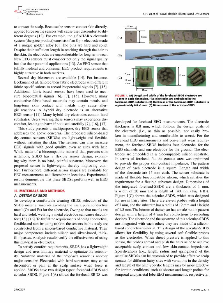
To satisfy comfort requirements, SBDS has a lightweightdesign and uses limitary material to optimize its sensitiv-ity. Substrate material of the proposed sensor is anothermajor consider. Electrodes with hard substrates may causediscomfort or pain at the scalp surface when force isapplied. SBDSs have two design types: forehead-SBDS andacicular-SBDS. Figure 1(A) shows the forehead-SBDS was
FIGURE 1. (A) Length and width of the forehead-SBDS electrode are15 mm in each dimension. Five electrodes are embedded in theforehead-SBDS substrate. (B) Thickness of the forehead-SBDS substrate isapproximately 0.8∼1 mm. (C) Dimensions of the acicular-SBDS.
developed for forehead EEG measurements. The electrodethickness is 0.8 mm, which follows the design goals ofthe electrode (i.e., as thin as possible, not easily bro-ken in manufacturing and comfortable to users). For theforehead EEG measurements and convenient wear require-ment, the forehead-SBDS includes four electrodes for theEEG channels and one electrode for the ground. The elec-trodes are embedded in a biocompatible silicon substrate.In terms of forehead fit, the contact area was optimizedto provide the proper skin-contact impedance. The patterndesign of each electrode is a square. Length and widthof the electrode are 15 mm each. The sensor substrate ismade of flexible biocompatible silicon, which satisfies therequirement for a flexible substrate design. Dimensions ofthe integrated forehead-SBDS are a thickness of 1 mm,a width of 20 mm and a length of 140 mm (Fig. 1(B)).Figure 1(C) shows the acicular-SBDS, which was designedfor use in hairy sites. There are eleven probes with a heightof 7 mm, and the substrate has a radius of 12 mm and a heightof 1.5 mm. The bottom of the sensor has a male button patterndesign with a height of 4 mm for connections to recordingdevices. The electrode and the substrate of this acicular-SBDSare integrated with each other; they are made of a silicon-based conductive material. This design of the acicular-SBDSallows for flexibility by using several soft flexible probesas the electrodes. When direct pressure is applied to thissensor, the probes spread and push the hairs aside to achieveacceptable scalp contact and low skin-contact impedance.Specifications (i.e., length, radius and springiness) of theacicular-SBDSs can be customized to provide effective scalpcontact for different hairy sites with variations in the densityand length of the hair. Specific lengths may be more effectivefor certain conditions, such as shorter and longer probes fortemporal and parietal lobe EEG measurements, respectively.
2700307 VOLUME 2, 2014
Y.-H. Yu et al.: Novel Flexible Silicon-Based Dry Sensors
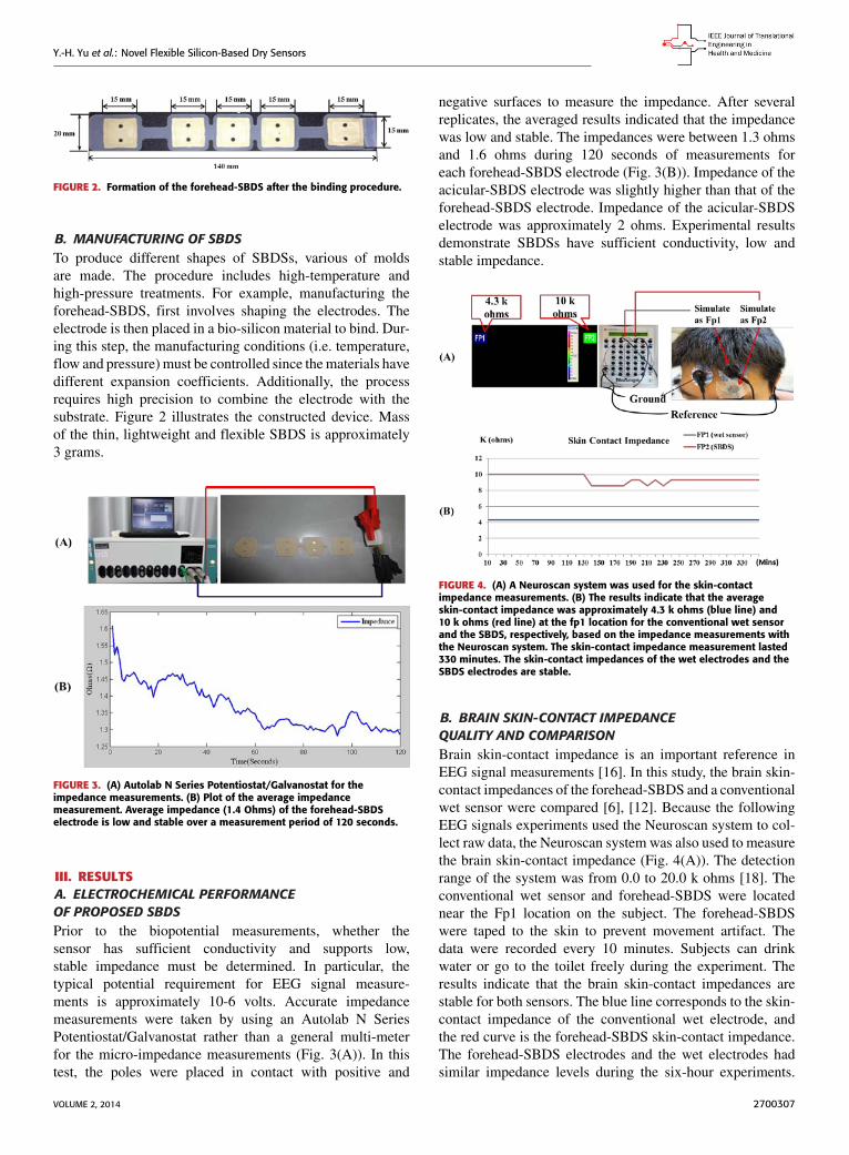
FIGURE 2. Formation of the forehead-SBDS after the binding procedure.
B. MANUFACTURING OF SBDSTo produce different shapes of SBDSs, various of moldsare made. The procedure includes high-temperature andhigh-pressure treatments. For example, manufacturing theforehead-SBDS, first involves shaping the electrodes. Theelectrode is then placed in a bio-silicon material to bind. Dur-ing this step, the manufacturing conditions (i.e. temperature,flow and pressure) must be controlled since thematerials havedifferent expansion coefficients. Additionally, the processrequires high precision to combine the electrode with thesubstrate. Figure 2 illustrates the constructed device. Massof the thin, lightweight and flexible SBDS is approximately3 grams.
FIGURE 3. (A) Autolab N Series Potentiostat/Galvanostat for theimpedance measurements. (B) Plot of the average impedancemeasurement. Average impedance (1.4 Ohms) of the forehead-SBDSelectrode is low and stable over a measurement period of 120 seconds.
III. RESULTSA. ELECTROCHEMICAL PERFORMANCEOF PROPOSED SBDSPrior to the biopotential measurements, whether thesensor has sufficient conductivity and supports low,stable impedance must be determined. In particular, thetypical potential requirement for EEG signal measure-ments is approximately 10-6 volts. Accurate impedancemeasurements were taken by using an Autolab N SeriesPotentiostat/Galvanostat rather than a general multi-meterfor the micro-impedance measurements (Fig. 3(A)). In thistest, the poles were placed in contact with positive and
negative surfaces to measure the impedance. After severalreplicates, the averaged results indicated that the impedancewas low and stable. The impedances were between 1.3 ohmsand 1.6 ohms during 120 seconds of measurements foreach forehead-SBDS electrode (Fig. 3(B)). Impedance of theacicular-SBDS electrode was slightly higher than that of theforehead-SBDS electrode. Impedance of the acicular-SBDSelectrode was approximately 2 ohms. Experimental resultsdemonstrate SBDSs have sufficient conductivity, low andstable impedance.
FIGURE 4. (A) A Neuroscan system was used for the skin-contactimpedance measurements. (B) The results indicate that the averageskin-contact impedance was approximately 4.3 k ohms (blue line) and10 k ohms (red line) at the fp1 location for the conventional wet sensorand the SBDS, respectively, based on the impedance measurements withthe Neuroscan system. The skin-contact impedance measurement lasted330 minutes. The skin-contact impedances of the wet electrodes and theSBDS electrodes are stable.
B. BRAIN SKIN-CONTACT IMPEDANCEQUALITY AND COMPARISONBrain skin-contact impedance is an important reference inEEG signal measurements [16]. In this study, the brain skin-contact impedances of the forehead-SBDS and a conventionalwet sensor were compared [6], [12]. Because the followingEEG signals experiments used the Neuroscan system to col-lect raw data, the Neuroscan system was also used to measurethe brain skin-contact impedance (Fig. 4(A)). The detectionrange of the system was from 0.0 to 20.0 k ohms [18]. Theconventional wet sensor and forehead-SBDS were locatednear the Fp1 location on the subject. The forehead-SBDSwere taped to the skin to prevent movement artifact. Thedata were recorded every 10 minutes. Subjects can drinkwater or go to the toilet freely during the experiment. Theresults indicate that the brain skin-contact impedances arestable for both sensors. The blue line corresponds to the skin-contact impedance of the conventional wet electrode, andthe red curve is the forehead-SBDS skin-contact impedance.The forehead-SBDS electrodes and the wet electrodes hadsimilar impedance levels during the six-hour experiments.
VOLUME 2, 2014 2700307
Y.-H. Yu et al.: Novel Flexible Silicon-Based Dry Sensors
The skin-contact impedance of the forehead-SBDS wasapproximately 10.0 k ohms, and the skin-contact impedanceof the wet sensor was approximately 4.3 k ohms (Fig. 4(B)).Although the contact impedance of the wet sensor waslower than that of the SBDS, the performance of SBDS(an impedance of 10.0 k ohms) is sufficient for generalEEG device applications. Conventional wet sensors mustbe applied with conductive gels. When the wet electrodeand the SBDS electrode were applied at room temperatureand the contact-impedance measurements were repeated afterseveral days, the SBDS skin-contact impedance remainedbelow 100 k ohm. However, the wet sensor had become toodry to be used. These skin-contact impedance measurementsdemonstrated that SBDS can provide low, stable skin-contactimpedances for long-term EEG measurements.
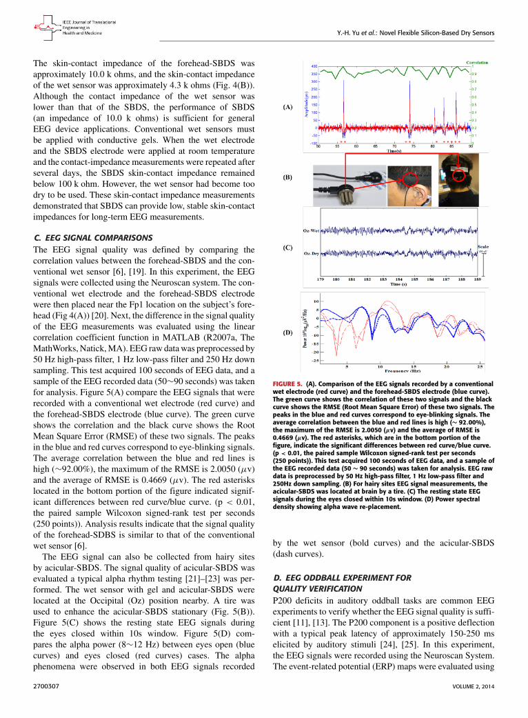
C. EEG SIGNAL COMPARISONSThe EEG signal quality was defined by comparing thecorrelation values between the forehead-SBDS and the con-ventional wet sensor [6], [19]. In this experiment, the EEGsignals were collected using the Neuroscan system. The con-ventional wet electrode and the forehead-SBDS electrodewere then placed near the Fp1 location on the subject’s fore-head (Fig 4(A)) [20]. Next, the difference in the signal qualityof the EEG measurements was evaluated using the linearcorrelation coefficient function in MATLAB (R2007a, TheMathWorks, Natick,MA). EEG raw datawas preprocessed by50 Hz high-pass filter, 1 Hz low-pass filter and 250 Hz downsampling. This test acquired 100 seconds of EEG data, and asample of the EEG recorded data (50∼90 seconds) was takenfor analysis. Figure 5(A) compare the EEG signals that wererecorded with a conventional wet electrode (red curve) andthe forehead-SBDS electrode (blue curve). The green curveshows the correlation and the black curve shows the RootMean Square Error (RMSE) of these two signals. The peaksin the blue and red curves correspond to eye-blinking signals.The average correlation between the blue and red lines ishigh (∼92.00%), the maximum of the RMSE is 2.0050 (µv)and the average of RMSE is 0.4669 (µv). The red asteriskslocated in the bottom portion of the figure indicated signif-icant differences between red curve/blue curve. (p < 0.01,the paired sample Wilcoxon signed-rank test per seconds(250 points)). Analysis results indicate that the signal qualityof the forehead-SDBS is similar to that of the conventionalwet sensor [6].
The EEG signal can also be collected from hairy sitesby acicular-SBDS. The signal quality of acicular-SBDS wasevaluated a typical alpha rhythm testing [21]–[23] was per-formed. The wet sensor with gel and acicular-SBDS werelocated at the Occipital (Oz) position nearby. A tire wasused to enhance the acicular-SBDS stationary (Fig. 5(B)).Figure 5(C) shows the resting state EEG signals duringthe eyes closed within 10s window. Figure 5(D) com-pares the alpha power (8∼12 Hz) between eyes open (bluecurves) and eyes closed (red curves) cases. The alphaphenomena were observed in both EEG signals recorded
FIGURE 5. (A). Comparison of the EEG signals recorded by a conventionalwet electrode (red curve) and the forehead-SBDS electrode (blue curve).The green curve shows the correlation of these two signals and the blackcurve shows the RMSE (Root Mean Square Error) of these two signals. Thepeaks in the blue and red curves correspond to eye-blinking signals. Theaverage correlation between the blue and red lines is high (∼ 92.00%),the maximum of the RMSE is 2.0050 (µv) and the average of RMSE is0.4669 (µv). The red asterisks, which are in the bottom portion of thefigure, indicate the significant differences between red curve/blue curve.(p < 0.01, the paired sample Wilcoxon signed-rank test per seconds(250 points)). This test acquired 100 seconds of EEG data, and a sample ofthe EEG recorded data (50 ∼ 90 seconds) was taken for analysis. EEG rawdata is preprocessed by 50 Hz high-pass filter, 1 Hz low-pass filter and250Hz down sampling. (B) For hairy sites EEG signal measurements, theacicular-SBDS was located at brain by a tire. (C) The resting state EEGsignals during the eyes closed within 10s window. (D) Power spectraldensity showing alpha wave re-placement.
by the wet sensor (bold curves) and the acicular-SBDS(dash curves).
D. EEG ODDBALL EXPERIMENT FORQUALITY VERIFICATIONP200 deficits in auditory oddball tasks are common EEGexperiments to verify whether the EEG signal quality is suffi-cient [11], [13]. The P200 component is a positive deflectionwith a typical peak latency of approximately 150-250 mselicited by auditory stimuli [24], [25]. In this experiment,the EEG signals were recorded using the Neuroscan System.The event-related potential (ERP) maps were evaluated using
2700307 VOLUME 2, 2014

Y.-H. Yu et al.: Novel Flexible Silicon-Based Dry Sensors
FIGURE 6. (A) Schematic of the EEG signals pre-processing routine for theERP test. (B) An ERP map with a conventional wet sensor located at Fp1.(C) An ERP map with a forehead-SBDS located near Fp1. (D) An ERP mapwith a wet sensor located at T3. (E) An ERP map with acicular-SBDSlocated near T3. Plots B-C and D-E display the same trials in the sameorder, at the respective electrodes. Comparatively, SBDSs’ effects wereweaker than the wet sensors, but the P200 phenomena were present inthe ERPs from both wet and dry channels.
EEGLAB (v10.2.5.5b) and MATLAB [11], [26], [27]. Forthe forehead EEG signal measurements, the conventional wetsensor and forehead-SBDS were placed near the Fp1 locationon the forehead. For hairy sites EEG signal measurements, thewet sensor and acicular-SBDS were placed near the tempo-ral (T3) position. An EEG pre-processing procedure includedartifact removals (removal of trials with extraordinary move-ment and muscle artifact) was performed before evaluatingthe ERPmaps (Fig 6(A)). After pre-processing, the ERPmapswere drawn based on the EEG channel ERP image functionof EEGLAB. Figures 6(B-C) show the EEG results for theconventional wet sensor and the forehead-SBDS, respectively.Clear P200 components (i.e. the peaks indicated as p200)were detected by the conventional wet sensor and forehead-SBDS while acquiring the ERP maps. Figures 6(D-E) showthe EEG results for the wet sensor and the acicular-SBDSlocated at the temporal (T3) position nearby also detectedP200 components. The acicular-SBDS was less effective inthis test. These findings validate the use SBDSs for EEGmeasurements [17].
E. SGS CERTIFICATIONTo determine whether the SBDSs are biocompatible sensorsthat do irritate skin, SGS Taiwan Ltd. was commissionedto perform a series of tests for the prototype SBDSs (thewhole sensor was made of the same silicon-based materialas the acicular-SBDS). There were no significant findingsof erythema or edema in either the control or the treatmentgroup, and there was no mortality. This result indicates thata single topical application of 0.5 ml of the silicon-basedelectrode extract will not cause skin irritation.
Additionally, cytotoxicity tests were conducted, such asmorphological evaluation, observation of monolayer conflu-ence and relative inhibition of cell viability. SGS test resultscan be summarized as follows. The silicon-silver-based dry-contact EEG sensors (conductive poly rubber with silverparticles) extract did not irritate skin or induce non-cytotoxiceffects on L929 cells according to ISO10993-5.
FIGURE 7. Resting state EEG signals with 3 times of grinding.
F. MECHANICAL TESTINGThe life cycles of SBDSs are difficult to identify exactly.According to previous experiences, if the surface of SBDSwas kept clean, the repetition frequency of use was over tentimes (5 hours/times). The responses to artifact and recoverytime of SBDS and wet sensors are almost the same. In thistest, the signals were recorded using the Neuroscan system.Figure 7 shows the recovery and eventual shave of artifactsfor jaw artifact recorded by wet sensor and acicular-SBDS atOz location. The subject ground his teeth three times within3 to 6 seconds. The average recovery time is around 1 second.
IV. CONCLUSIONThis study describes a novel, lightweight, flexible and non-skin irritating SBDS, including its design, fabrication andtesting. The proposed SBDSs have the following advantages.First, the sensor material has passed the standard SGScertifications, showing that it was biocompatible and didnot irritate skin or induce cytotoxic effects in the test.Second, SBDSs have an ergonomic design with a flexiblebase and can be used in non-gel applications. The proposedSBDBs are also comfortable to wear without causing pain.Third, the proposed SBDSs perform similarly to wet con-ventional sensors in the EEG measurements. The proposedSBDSs are superior to wet sensors in their use in long-term EEG measurements. Fourth, the proposed SBDSs aremade for two different brain sites for the EEGmeasurements,especially for the hairy sites. Finally, the proposed SBDSscan achieve the necessary skin-sensor interface impedance toprovide EEG signals of sufficient quality for brain dynam-ics research. Experimental results indicate that the designedsensors are applicable for EEG signals measurement in thehuman brain.Despite the above advantages of SBDSs, we recom-
mend that future research attempt to improve their life
VOLUME 2, 2014 2700307

Y.-H. Yu et al.: Novel Flexible Silicon-Based Dry Sensors
cycle, since major components of SBDSs include silver-based and SiO2. After several uses, SBDSs may oxidize andreduce conductivity. Although the proposed sensors cannotbe abraded or chemically treated to reveal fresh material,they can be washed with soap and sterilized by alcohol forreuse.
REFERENCES[1] P. L. Nunez, Electric Fields of the Brain: The Neurophysics of EEG.
New York, NY, USA: Oxford Univ. Press, 1981.[2] N. V. Thakor, ‘‘Biopotentials and electro-physiologymeasurement,’’ in The
Measurement, Instrumentation and Sensors Handbook. Boca Raton, FL,USA: CRC Press, 1999.
[3] N. V. Thakor, ‘‘Translating the brain-machine interface,’’ Sci. Transl. Med.,vol. 5, p. 210–217, Nov. 2013.
[4] C.-T. Lin et al., ‘‘Noninvasive neural prostheses using mobile and wire-less EEG,’’ Proc. IEEE, vol. 96, no. 7, pp. 1167–1183, Jul. 2008.
[5] C.-T. Lin, F.-C. Lin, S.-A. Chen, S.-W. Lu, T.-C. Chen, and L.-W. Ko,‘‘EEG-based brain-computer interface for smart living environmental auto-adjustment,’’ J. Med. Biol. Eng., vol. 30, no. 4, pp. 237–245, Aug. 2010.
[6] C.-T. Lin, L.-D. Liao, Y.-H. Liu, I.-J. Wang, B.-S. Lin, and J.-Y. Chang,‘‘Novel dry polymer foam electrodes for long-term EEG measurement,’’IEEE Trans. Biomed. Eng., vol. 58, no. 5, pp. 1200–1207, May 2011.
[7] L.-D. Liao, I.-J. Wang, S.-F. Chen, J.-Y. Chang, and C.-T. Lin, ‘‘Design,fabrication and experimental validation of a novel dry-contact sensor formeasuring electroencephalography signals without skin preparation,’’ Sen-sors, vol. 11, no. 6, pp. 5819–5834, Jun. 2011.
[8] T.-H. Huang, H.-P. Huang, Y.-H. Liu, Z.-H. Kang, and J.-Y. Kuan, ‘‘Devel-opment of a brain-controlled rehabilitation system (BCRS),’’ J. Neurosci.Neuroeng., vol. 2, no. 2, pp. 79–89, 2013.
[9] B. J. Lance, S. E. Kerick, A. J. Ries, K. S. Oie, and K. McDowell,‘‘Brain–computer interface technologies in the coming decades,’’Proc. IEEE, vol. 100, no. 5, pp. 1585–1599, May 2012.
[10] L.-D. Liao et al., ‘‘Gaming control using a wearable and wirelessEEG-based brain-computer interface device with novel dry foam-basedsensors,’’ J. NeuroEng. Rehabil., vol. 9, pp. 1–12, Jan. 2012.
[11] L.-D. Liao et al., ‘‘Biosensor technologies for augmented brain–computerinterfaces in the next decades,’’ Proc. IEEE, vol. 100, no. 5, pp. 1553–1566,May 2012.
[12] R. Merletti, ‘‘The electrode–skin interface and optimal detection of bio-electric signals,’’ Physiol. Meas., vol. 31, no. 10, p. preceding S157,Oct. 2010.
[13] L.-D. Liao and C.-T. Lin, ‘‘Novel trends in biosensors used for electroen-cephalographymeasurements in neurocognitive engineering applications,’’J. Neurosci. Neuroeng., vol. 1, no. 1, pp. 32–41, 2012.
[14] C. Grozea, C. D. Voinescu, and S. Fazli, ‘‘Bristle-sensors–low-cost flexiblepassive dry EEG electrodes for neurofeedback and BCI applications,’’J. Neural Eng., vol. 8, no. 2, p. 025008, Apr. 2011.
[15] L. Beckmann et al., ‘‘Characterization of textile electrodes and conductorsusing standardized measurement setups,’’ Physiol. Meas., vol. 31, no. 2,pp. 233–247, Feb. 2010.
[16] A. Searle and L. Kirkup, ‘‘A direct comparison of wet, dry and insu-lating bioelectric recording electrodes,’’ Physiol. Meas., vol. 21, no. 2,pp. 271–283, May 2000.
[17] K. Gramann et al., ‘‘Cognition in action: Imaging brain/body dynamics inmobile humans,’’ Rev. Neurosci., vol. 22, no. 6, pp. 593–608, 2011.
[18] T. C. Ferree, P. Luu, G. S. Russell, and D. M. Tucker, ‘‘Scalp electrodeimpedance, infection risk, and EEG data quality,’’ Clin. Neurophysiol.,vol. 112, no. 3, pp. 536–544, Mar. 2001.
[19] J. R. Estepp, J. C. Christensen, J.W.Monnin, I. M. Davis, and G. F.Wilson,‘‘Validation of a dry electrode system for EEG,’’ Proc. Human FactorsErgonom. Soc. Annu. Meeting, vol. 53, no. 18, pp. 1171–1175, Oct. 2009.
[20] H. Iguchi, K. Watanabe, A. Kozato, and N. Ishii, ‘‘Wearable electroen-cephalograph system with preamplified electrodes,’’Med. Biol. Eng. Com-put., vol. 32, no. 4, pp. 459–461, Jul. 1994.
[21] G. Gargiulo et al., ‘‘A new EEG recording system for passive dry elec-trodes,’’ Clin. Neurophysiol., vol. 121, no. 5, pp. 686–693, 2010.
[22] G.D.Gargiulo, P. Bifulco,M. Cesarelli, A. Fratini, andM.Romano, ‘‘Prob-lems in assessment of novel biopotential front-end with dry electrode:A brief review,’’Machines, vol. 2, no. 1, pp. 87–98, 2014.
[23] G. Mueller-Putz, R. Scherer, C. Brunner, R. Leeb, and G. Pfurtscheller,‘‘Better than random: A closer look on BCI results,’’ Int. J. Bioelectro-magn., vol. 10, no. 1, pp. 52–55, 2008.
[24] B. A. Brett-Green, L. J. Miller, W. J. Gavin, and P. L. Davies, ‘‘Multi-sensory integration in children: A preliminary ERP study,’’ Brain Res.,vol. 1242, pp. 283–290, Nov. 2008.
[25] F. Ferreira-Santos, C. Silveira, P. R. Almeida, A. Palha, F. Barbosa, andJ. Marques-Teixeira, ‘‘The auditory P200 is both increased and reducedin schizophrenia? A meta-analytic dissociation of the effect for standardand target stimuli in the oddball task,’’ Clin. Neurophysiol., vol. 123, no. 7,pp. 1300–1308, Jul. 2012.
[26] E. W. Sellers, D. J. Krusienski, D. J. McFarland, T. M. Vaughan, andJ. R. Wolpaw, ‘‘A P300 event-related potential brain–computer inter-face (BCI): The effects of matrix size and inter stimulus interval onperformance,’’ Biol. Psychol., vol. 73, no. 3, pp. 242–252, Oct. 2006.
[27] H. Zhang, C. Guan, and C. Wang, ‘‘Asynchronous P300-basedbrain–computer interfaces: A computational approach with statisticalmodels,’’ IEEE Trans. Biomed. Eng., vol. 55, no. 6, pp. 1754–1763,Jun. 2008.
YI-HSIN YU received the B.S. degree in computerscience and information engineering from TunghaiUniversity, Taichung, Taiwan, in 1995, and theM.S. degree in computer science and informationengineering from National Dong Hwa University,Hualien, Taiwan, in 1998. He is currently pur-suing the Ph.D. degree with the Brain ResearchCenter, Institute of Electrical Control Engineer-ing, National Chiao Tung University, Hsinchu,Taiwan. His research interests include brain
research, biomedical signal processing, brain–computer interfaces, andbiomedical circuits and systems.
SHAO-WEI LU received the B.S. degree in physicsand the M.S. degree from the Institute of Molec-ular Medicine, National Tsing Hua University,Hsinchu, Taiwan, in 2007. He is currently anAssistant Researcher with the Brain ResearchCenter, National Chiao Tung University, Hsinchu,where he is involved in electroneurophysiologyand biomedical signal processing.
2700307 VOLUME 2, 2014
Y.-H. Yu et al.: Novel Flexible Silicon-Based Dry Sensors
LUN-DE LIAO received the Ph.D. degree in elec-trical engineering from National Chiao Tung Uni-versity (NCTU), Hsinchu, Taiwan, in 2012, wherehe was a Post-Doctoral Researcher with the BrainResearch Center (BRC). He proposed the worldfirst bioinspired dry EEG sensors and their cor-responding circuit to the intelligent image humanbrain under the guidance of Prof. C.-T. Lin at BRC.He is currently a Senior Research Scientist andthe Head of the Neurovascular Imaging Laboratory
with the Singapore Institute for Neurotechnology, National University ofSingapore, Singapore. He has authored over 45 peer-reviewed SCI jour-nal papers, including the Nature: Journal of Cerebral Blood Flow andMetabolism, the PROCEEDINGS OF THE IEEE, and Neuroimage journals,and holds 10 issued patents. He has selected/nominated for over 30 inter-national awards since 2004. He was a recipient of the Young Investigator’sAwards from the World Association for Chinese Biomedical Engineers forhis contributions onmedical imaging and bioelectronics domain, in 2011, andan Outstanding Research Award from NCTU, in 2012, for his outstandingresearch performance. He served on Organization Committee and TechnicalProgram Committee of many flagship international conferences and work-shops. He serves as the Co-Editors-in-Chief of Journal of Neuroscience andNeuroengineering and also an Associate Editor of four SCI-index journals.His research interests include neuroimaging, cerebral neuroscience, cogni-tive neuroscience, in vivo optical microscopy, advanced sensing techniques,and design of optical system.
CHIN-TENG LIN (S’88–M’91–SM’99–F’05)received the B.S. degree fromNational Chiao TungUniversity (NCTU), Hsinchu, Taiwan, in 1986, andthe M.Sc. and Ph.D. degrees in electrical engineer-ing from Purdue University, West Lafayette, IN,USA, in 1989 and 1992, respectively. He is cur-rently the Provost, the Chair Professor of Electricaland Computer Engineering, a Professor with theInstitute of Imaging and Biomedical Photonics,and the Director of the Brain Research Center,
NCTU. He is currently the Editor-in-Chief of the IEEE TRANSACTIONSON FUZZY SYSTEMS.
VOLUME 2, 2014 2700307