dermatology & cosmeceuticals - patterson park … & cosmeceuticals solving problems with...
TRANSCRIPT
Dermatology & CosmeceuticalsSolving Problems with Customized Medications
2245 Eastern Ave. • Baltimore, MD 21231
Phone 410.675.6046Toll Free 855.859.2517
Fax 410.563.1147

1
©Storey Marke ng, All rights reserved.
Table of Contents Wrinkles and Photoaged Skin ................................................................................................ 2
Acne and Rosacea ................................................................................................................. 8
Psoriasis and Eczema ........................................................................................................... 10
Pigmenta on Abnormali es (Melasma, Vi ligo) ................................................................. 13
Scarring and Keloids ............................................................................................................ 16
Skin and Wound Care .......................................................................................................... 17
Warts, Plantar Warts, and Molluscum ................................................................................ 20
Head Lice and Scabies ......................................................................................................... 24
Hyperhydrosis ...................................................................................................................... 26
Fungal Infec ons of the Skin and Nails ............................................................................... 27
Topical Anesthe cs .............................................................................................................. 28
Miscellaneous ...................................................................................................................... 29
How to Write a Prescrip on for a Compounded Medica on .............................................. 36
2245 Eastern Ave. • Bal more, MD 21231
Phone 410.675.6046
Toll Free 855.859.2517
Fax 410.563.1147
www.pa ersonparkpharmacy.com contact@pa ersonparkpharmacy.com

2
©Storey Marke ng, All rights reserved.
Customized Medica ons—Compounding for Dermatology
We can prepare customized dermatologics that can improve therapeu c outcomes, increase pa ent compliance, decrease side effects, and save me and money. Prepara ons are compounded by prescrip on to contain the needed medica ons in the best vehicle (base) or dosage form (gels, creams, ointments, lip balms, powders, sprays, etc.) to most efficiently deliver
the drug to the affected area. Our compounding pharmacy u lizes state‐of‐the‐art equipment and the finest quality bases to improve both the aesthe c and therapeu c aspects of compounded medica ons. Cosme cally appealing prepara ons can contain numerous synergis c medica ons, with drug par cles that are fine enough to be absorbed through the skin, and penetrant enhancers when appropriate. Therapies for Resistant Problems
Specific therapies for resistant acne, recalcitrant viral warts and molluscum contagiosum. Immunotherapy to treat alopecia. Novel therapeu c agents to treat abnormal dermal scarring. Topical sodium cromoglycate for pyoderma gangrenosum. Natural an viral (2‐deoxy D‐glucose) to inhibit the mul plica on of herpes virus. Repigmenta on therapy for pa ents with vi ligo. Commercial products are limited, and some mes unavailable. When a customized prepara on is needed, such as a combina on of topical anesthe cs in a specialized base or dosage form, compounding pharmacists can work together with physicians to provide the most appropriate therapy. Examples include fast‐ac ng dermal anesthe cs, for fied prepara ons for use prior to therapeu c ta ooing for radia on therapy, and sprays which can be applied to wounds prior to dressing changes. The efficacy of any compounded medica on is influenced by the purity and quality of the ingredients, choice of vehicle (base), proper use of addi ves such as penetra on enhancers, and the technique and equipment used during formula on.
We work together with prac oners and their pa ents to provide innova ve solu ons to challenging medical problems. Please contact our compounding pharmacist for more informa on or to discuss customized therapies to meet your pa ents
specific needs.
T W P S Many “bio‐cosmeceu cals” are now available to treat aging skin, including re noids, an oxidants, hydroxy acids, bleaching agents, moisturizers, and sunscreens. Cosmeceu cals containing an oxidants are among the most popular an aging remedies. Topically applied an oxidants exert their benefits by offering protec on from damaging free radicals produced when skin is exposed to ultraviolet light or allowed to age naturally. Appropriate formula on and use which is supervised by a knowledgeable healthcare professional will maximize the benefits while minimizing any poten al side effects of these therapies. When a proper analysis reveals areas of dry, oily, or aging skin, we can provide the appropriate correc on for each skin type in a cosme c base containing the exfoliants, emollients, and micronutrients necessary for cellular repair. An oxidants such as Alpha Lipoic Acid and Vitamin C Ester are vital to the energy produc on of skin cells and forma on of collagen. Amino acids such as DMAE tone and add firmness to the skin, prevent age spots, and aid in healing the micro‐scarring which causes wrinkles. Coenzyme Q10 to Prevent Photoaging of Skin Oxida ve stress (UV irradia on, free radicals) and cellular oxida on play a significant role in the processes of aging and photoaging. This may be in part due to a decline in the levels of the endogenous cellular an oxidant Coenzyme Q10 (ubiquinone, CoQ10). Hoppe et al. inves gated whether topical applica on of CoQ10 has the beneficial effect of preven ng
3
©Storey Marke ng, All rights reserved.
photoaging. They were able to demonstrate that CoQ10 penetrated into the viable layers of the epidermis and reduced the level of oxida on measured by weak photon emission. Furthermore, a reduc on in wrinkle depth following CoQ10 applica on was shown. CoQ10 was determined to be effec ve against UVA mediated oxida ve stress in human kera nocytes in terms of thiol deple on, ac va on of specific phosphotyrosine kinases and preven on of oxida ve DNA damage. CoQ10 was also able to significantly suppress the expression of collagenase (which is responsible for wrinkle forma on) in human dermal fibroblasts following UVA irradia on. In vivo inves ga ons by Bla et al. have also shown that wrinkles around the region of the eyes ("crow’s feet") could be reduced by long‐term applica on of CoQ10. These results indicate that topical applica on of CoQ10 has the efficacy to prevent many of the detrimental effects of photoaging. Biofactors 1999;9(2‐4):371‐8 Z Gerontol Geriatr 1999 Apr;32(2):83‐8 Coenzyme Q10 (ubiquinone, CoQ10) is an important an oxidant that is taken orally as a supplement to strengthen immune and cardiac func on. The processes of aging and photoaging of the skin (due to sunlight) are associated with an increase in cellular oxida on, which may occur as the body’s own levels of CoQ10 decline. A study was done to determine if topical applica on of CoQ10 0.3% could prevent photoaging. A reduc on in wrinkle depth following CoQ10 applica on was shown, and results indicated that CoQ10 has the efficacy to prevent many of the detrimental effects of photoaging. Wrinkles around the region of the eyes (“crow’s feet”) may be reduced by long‐term applica on of CoQ10. Biofactors 1999;9(2‐4):371‐8 Z Gerontol Geriatr 1999 Apr;32(2):83‐8 Protec on and Reversal of Photodamage with Topical An oxidants Studies have shown that an oxidants can be delivered percutaneously to directly supplement the skin’s an oxidant reservoir and provide protec on from untraviolet radia on. (Oral supplementa on has not been successful in raising levels of an oxidants in the skin.) Topical vitamin C, when properly formulated, effec vely penetrates the skin and can produce a 20‐fold increase in endogenous levels of cutaneous vitamin C. At the North Carolina Biotechnology Center, Raleigh, Darr et al. reported that, in swine skin, vitamin C is capable of addi ve protec on against acute UVB damage when combined with a UVB sunscreen. A combina on of both vitamins E and C provided very good protec on from UVB radia on with the bulk of the protec on a ributable to vitamin E. However, vitamin C was significantly be er than vitamin E at protec ng against UVA‐mediated phototoxic insult. When vitamin C or a combina on of vitamin C and E is formulated with a commercial UVA sunscreen (oxybenzone), an apparently greater than addi ve protec on is noted against the phototoxic damage. These results confirm the u lity of an oxidants as photoprotectants and suggest the importance of combining the an oxidants with known sunscreens to maximize photoprotec on. Topical vitamins C and E, as well as topical selenium, protect skin against sunburn, suntan and skin cancer and also reverse the mo led pigmenta on and wrinkles of photoaging. However, only certain forms of these an oxidants are stable and ac ve a er percutaneous absorp on. Benefits of topical applica on are that the skin a ains far higher levels of each an oxidant than can be achieved by taking these vitamins orally and topical applica on arms the skin with a reservoir of an oxidants that cannot be washed or rubbed off, protec ng the skin for several days a er applica on. Acta Derm Venereol. 1996 Jul;76(4):264‐8 J Cosmet Dermatol. 2004 Jul;3(3):149‐55 Biofactors. 2003;18(1‐4):289‐97. The combined use of oral and topical lipophilic an oxidants increases their levels both in sebum and stratum corneum. Passi S, De Pità O, Grandine M, Simo C, Li arru GP. Centro Invecchiamento Cellulare, I.D.I. (IRCCS), Rome, Italy. [email protected] The concentra on of Vitamin E (vit E) and ubiquinone (CoQ10), which together with squalene (SQ), play a key role against external oxida ve insult, has been shown to decrease significantly during ageing. The aim of the present study is to inquire the effect of the combined use of topical bio‐cosme cs containing natural ac ve principles (including sebum‐like lipid frac ons, sebum and epidermal lipophilic and hydrophilic an oxidants), and oral an oxidant supplements on the an oxidant content of sebum and stratum corneum. We therefore treated the face and the back of 50 female volunteers aged 21‐40, daily for two months, with a base cream containing 0.05% ubiquinone, 0.1% vit E, and 1% squalene. In addi on 50 mg of CoQ10 + 50 mg of

4
©Storey Marke ng, All rights reserved.
d‐RRR‐alpha‐tocopheryl acetate + 50 microg of selenium were administered orally to half of the volunteers (Group A). Group B was represented by 25 volunteers who were treated only topically. Every 15 days during treatment the levels of CoQ10, vit E and SQ were verified in sebum, stratum corneum, and plasma. The daily topical applica on of the cream led to a significant increase, that peaked a er 60 days, of the levels of CoQ10, d‐RRR‐alpha‐tocopherol and SQ in the sebum (Group B), without significantly affec ng the stratum corneum or plasma concentra ons of the redox couple CoQ10H2/CoQ10 and vit E. The concomitant oral admistra on of an oxidants produced in Group A a significant increase of the levels of CoQ10H2/CoQ10 and vit E both in plasma and stratum corneum a er 15 and 30 days treatment respec vely, compared to Group B. However the sebum levels of lipophilic an oxidants and SQ did not show a significant increase. A er the treatments, the levels of CoQ10H2/CoQ10, vit E and SQ went back to basal levels within 6‐8 days in sebum, 12‐16 days in the stratum corneum, and 3‐6 days in plasma. Therefore topical applica on of the an oxidants was able to increase their level in sebum, while the concomitant oral administra on also affected the levels of vit E and CoQ10 in the stratum corneum. PMID: 14695946 J Cosmet Dermatol. 2005 Sep;4(3):167‐73. Links Clinical efficacy assessment in photodamaged skin of 0.5% and 1.0% idebenone. McDaniel D, Neudecker B, Dinardo J, Lewis J 2nd, Maibach H. Ins tute of An ‐Aging Research, Eastern Virginia Medical School, Norfolk, VA, USA. Idebenone is an an oxidant lower molecular weight analogue of coenzyme Q10. Previously, idebenone was shown to be a very effec ve an oxidant in its ability to protect against cell damage from oxida ve stress in a variety of biochemical, cell biological, and in vivo methods, including its ability to suppress sunburn cell (SBC) forma on in living skin. However, no clinical studies have been previously conducted to establish the efficacy of idebenone in a topical skincare formula on for the treatment of photodamaged skin. In this nonvehicle control study, 0.5% and 1.0% idebenone commercial formula ons were evaluated in a clinical trial for topical safety and efficacy in photodamaged skin. Forty‐one female subjects, aged 30‐65, with moderate photodamaged skin were randomized to use a blind labelled (either 0.5% or 1.0% idebenone in otherwise iden cal lo on bases) skincare prepara on twice daily for six weeks. Blinded expert grader assessments for skin roughness/dryness, fine lines/wrinkles, and global improvement in photodamage were performed at baseline, three weeks and six weeks. Electrical conductance readings for skin surface hydra on and 35 mm digital photography were made at baseline a er six weeks. Punch biopsies were taken from randomly selected subjects, baseline and a er six weeks, and stained for certain an bodies (interleukin IL‐6, interleukin IL‐1b, matrixmetalloproteinase MMP‐1, collagen I) using immunofluorescence microscopy. A er six weeks' use of the 1.0% idebenone formula, a 26% reduc on in skin roughness/dryness was observed, a 37% increase in skin hydra on, a 29% reduc on in fine lines/wrinkles, and a 33% improvement in overall global assessment of photodamaged skin. For the 0.5% idebenone formula on, a 23% reduc on in skin roughness/dryness was observed, a 37% increase in skin hydra on, a 27% reduc on in fine lines/wrinkles, and a 30% improvement in overall global assessment of photodamaged skin. The immunofluorescence staining revealed a decrease in IL‐1b, IL‐6, and MMP‐1 and an increase in collagen I for both concentra ons. PMID: 17129261 J Cosmet Sci. 2006 Nov‐Dec;57(6): Development of a w/o/w emulsion for chemical peeling applica ons containing glycolic acid. Yener G, Baitokova A. Istanbul University, Faculty of Pharmacy, Department of Pharmaceu cal Technology, Cosme cs Sec on, Istanbul, Turkey. Glycolic acid is a member of the AHA family, which occurs naturally in foods and has been used for centuries as a cutaneous rejuvena on treatment. It is used in many cosme c products as an exfoliant and moisturizer. When glycolic acid is used in greater amounts, however, there are greater cosme c benefits but also poten al for skin irrita on as far as burning increases. The aim of this work was to inves gate the feasibility of a topical delivery system as a mul ple emulsion combining glycolic acid, stron um nitrate, and dexpanthenol in order to op mize the acid's cosme c proper es and lowering its side effects. PMID: 17256078
5
©Storey Marke ng, All rights reserved.
Br J Dermatol. 2003 Oct;149(4):841‐9. Randomized, placebo‐controlled, double blind study on the clinical efficacy of a cream containing 5% alpha‐lipoic acid related to photoageing of facial skin. Beitner H. Department of Dermatology, Karolinska Hospital, 17176 Stockholm, Sweden. [email protected] BACKGROUND: alpha‐lipoic acid (LA) or the reduced form dihydrolipoate (DHLA) is a potent scavenger with an ‐inflammatory proper es. Previous uncontrolled studies with topical treatment with 5% LA‐containing creams indicate a beneficial effect on photoageing skin. OBJECTIVE: The purpose of this study was to inves gate whether a cream containing 5% LA showed any advantages concerning a number of the criteria associated with ageing of the facial skin, compared with an iden cal cream lacking LA. MATERIAL AND METHODS: Thirty‐three women, mean age 54.4 years, were included in this controlled study. A er randomiza on half the face was treated twice daily for 12 weeks with the LA cream and the other half with the control cream. The following methods of assessment were used: self‐evalua on by the test subjects, clinical evalua on, photographic evalua on and laser profilometry. Profilometry was performed before the start of treatment and at the end. RESULTS: All four methods of assessment showed a sta s cally significant improvement on the LA‐treated half of the face. Laser profilometry, the most objec ve method used, showed an average decrease in skin roughness of 50.8% (44.9‐54.0) on the LA‐treated side, compared with 40.7% (32.4‐48.7) on the placebo‐treated half of the face P < 0.001 (Wilcoxon matched pairs test). CONCLUSIONS: It is indicated that 12 weeks of treatment with a cream containing 5% LA improves clinical characteris cs related to photoageing of facial skin. PMID: 14616378 Clin Dermatol. 2008 Jul‐Aug;26(4):364‐6. Advancement in skin aging: the future cosmeceu cals. Giacomoni PU. Clinique Laboratories, 125 Pinelawn Road, Melville, NY 11747, USA. [email protected] Aging is a mul factorial process defined as the accumula on of damage. The aging of the skin is characterized by specific clinical end points, the cause of which is not always thoroughly understood. The skin is exposed to environmental aggressions and the reac ve oxygen species produced during cellular metabolism. Damage to the cellular and extracellular components of the skin can be avoided or removed by the appropriate topical applica on of ac ve ingredients. Sunscreens are essen al to avoid damage from the most important damaging environmental agent: solar radia on. Liposomes containing deoxyribonucleic acid repair enzymes and accelerate the endogenous removal of pyrimidine dimers a er exposure to ultraviolet radia on. Specific an oxidants reduce the rate of forma on of secondary ultraviolet‐induced damages, par cularly those induced by singlet oxygen. An ‐inflammatory agents, immunos mulants, and enhancers of molecular and cellular detoxifica on could enter the panoply of new cosmeceu cals to avoid age spots, dark circles, wrinkles, and other clinical aspects of skin aging. PMID: 18691516 Int J Cosmet Sci. 2004 Oct;26(5):231‐8. Links Topical niacinamide reduces yellowing, wrinkling, red blotchiness, and hyperpigmented spots in aging facial skin. Bisse DL, Miyamoto K, Sun P, Li J, Berge CA. The Procter & Gamble Company, Miami Valley Laboratories, Cincinna , OH, U.S.A. Previous clinical tes ng of topical niacinamide (vitamin B3) has revealed a broad array of improvements in the appearance of aging facial skin. The study reported here was done to confirm some of those previous observa ons and to evaluate addi onal end points such as skin an ‐yellowing. Caucasian female subjects (n = 50, aged 40‐60 years) par cipated in a 12‐week, double‐blind, placebo‐controlled, split‐face, le ‐right randomized clinical study assessing two topical products: moisturizer control product versus the same moisturizer product containing 5% niacinamide. Niacinamide was well tolerated by the skin and provided significant improvements versus control in end points evaluated previously: fine lines/wrinkles, hyperpigmenta on spots, texture, and red blotchiness. In addi on, skin yellowing (sallowness) versus control was significantly improved. The mechanism by which this array of benefits is achieved with niacinamide is discussed. PMID: 18492135
6
©Storey Marke ng, All rights reserved.
Acta Dermatovenerol Alp Panonica Adriat. 2008 Jun;17(2):47‐54. Skin aging. Puizina‐Ivić N. Department of Dermatovenerology, Split Clinical Hospital Center, Soltanska 1, 21 000 Split, Croa a. [email protected] There are two main processes that induce skin aging: intrinsic and extrinsic. A stochas c process that implies random cell damage as a result of muta ons during metabolic processes due to the produc on of free radicals is also implicated. Extrinsic aging is caused by environmental factors such as sun exposure, air pollu on, smoking, alcohol abuse, and poor nutri on. Intrinsic aging reflects the gene c background and depends on me. Various expressions of intrinsic aging include smooth, thinning skin with exaggerated expression lines. Extrinsically aged skin is characterized by photo damage as wrinkles, pigmented lesions, patchy hypopigmenta ons, and ac nic keratoses. Timely protec on including physical and chemical sunscreens, as well as avoiding exposure to intense UV irradia on, is most important. A network of an oxidants such as vitamins E and C, coenzyme Q10, alpha‐lipoic acid, glutathione, and others can reduce signs of aging. Further an ‐aging products are three genera ons of re noids, among which the first genera on is broadly accepted. A diet with lot of fruits and vegetables containing an oxidants is recommended as well as exercise two or three mes a week. PMID: 18709289 Vitamin C is a naturally occurring potent water‐soluble an oxidant. Accordingly, it has been incorporated into a variety of cosmeceu cals designed to protect and rejuvenate photoaged skin. Cutaneous benefits include promo ng collagen synthesis, photoprotec on from ultraviolet A and B, lightening hyperpigmenta on, and improvement of a variety of inflammatory dermatoses. Because of the diverse biologic effects of this compound, topical vitamin C has become a useful part of the dermatologist's armamentarium. Ascorbyl Palmitate (Vitamin C Ester) is a lipid soluble, neutral pH, non‐acidic (thus, non‐irrita ng and non‐s nging) form of Vitamin C which can reach cells within the skin rapidly in amounts greater than can be achieved by water soluble Vitamin C (L‐Ascorbic Acid). This proven an oxidant protects skin cells from damaging free radicals and provides essen al Vitamin C needed for collagen produc on. Ascorbyl Palmitate also inhibits the endogenous produc on of the inflammatory arachidonic acid, which plays a role in the development of psoriasis and the micro‐scarring that leads to the forma on of wrinkles. Unlike L‐ascorbic acid, Ascorbyl Palmitate can be mixed into creams and lo ons and remain stable for an extended period of me. Ascorbyl Palmitate s mulates the growth of fibroblasts which help to produce collagen and elas n in human skin. Dermatol Surg. 2005 Jul;31(7 Pt 2):814‐7 Alpha Lipoic Acid is a powerful an oxidant and scavenger with an ‐inflammatory proper es that is both water and lipid soluble and therefore can work on both the intercellular and intracellular levels. ALA is naturally present in the mitochondria, and promotes op mum efficiency for produc on of energy and removal of intracellular waste products, essen al for cellular healing and elimina on of wrinkles and facial scars. Glyca on (a achment of sugars to cellular proteins) causes the skin to lose elas city, and this can be prevented by ALA. A controlled study of thirty‐three women, mean age 54.4 years, inves gated whether a cream containing 5% ALA showed any advantages concerning a number of the criteria associated with aging of the facial skin, compared with an iden cal cream lacking ALA. A er randomiza on half the face was treated twice daily for 12 weeks with the ALA cream and the other half with the control cream. The following methods of assessment were used: self‐evalua on by the test subjects, clinical evalua on, photographic evalua on and laser profilometry. All four methods of assessment showed a sta s cally significant improvement on the ALA‐treated half of the face. Laser profilometry, the most objec ve method used, showed an average decrease in skin roughness of 50.8% on the ALA‐treated side, compared with 40.7% on the placebo‐treated half of the face. The study concluded that 12 weeks of treatment with a cream containing 5% ALA improves clinical characteris cs related to photoaging of facial skin. Br J Dermatol. 2003 Oct; 149(4): 841‐9 Topical niacinamide (vitamin B3) reduces yellowing, wrinkling, red blotchiness, and hyperpigmented spots in aging facial skin. Proctor and Gamble conducted a study to confirm previous observa ons and evaluate addi onal end points such as skin an ‐yellowing. Caucasian female subjects (n = 50, aged 40‐60 years) par cipated in a 12‐week, double‐blind, placebo‐controlled, split‐face, le ‐right randomized clinical study assessing two topical products: moisturizer control product versus the same moisturizer product containing 5% niacinamide. Niacinamide was well tolerated by the skin and provided significant

7
©Storey Marke ng, All rights reserved.
improvements versus control in end points evaluated previously: fine lines/wrinkles, hyperpigmenta on spots, texture, and red blotchiness. In addi on, skin yellowing (sallowness) versus control was significantly improved. Int J Cosmet Sci. 2004 Oct;26(5):231‐8. An ‐Wrinkle Effect of Topical DMAE DMAE (2‐dimethylaminoethanol, deanol) is an an oxidant found in abundance in fish, par cularly salmon. Applied topically to the skin, DMAE may improve the appearance of sagging skin. DMAE boosts the effects of other an oxidants, increases smoothness, reduces fine lines and gives the facial muscles a leaner look. In a randomized clinical study, 3% DMAE facial gel applied daily for 16 weeks has been shown to be safe and efficacious in the mi ga on of forehead lines and periorbital fine wrinkles, and in improving lip shape and fullness and the overall appearance of aging skin. These effects did not regress during a 2‐week cessa on of applica on. Beneficial trends were noted in the appearance of coarse wrinkles, under‐eye dark circles, nasolabial folds, sagging neck skin, and neck firmness. Applica on was found to be well tolerated, with no differences in the incidence of erythema, peeling, dryness, itching, burning, or s nging between the DMAE and placebo groups. The cosmeceu cal agent 2‐dimethylaminoethanol (deanol; DMAE) is a ter ary amine found in high concentra on in numerous topical an wrinkle prepara ons. At the University of Quebec, Morisse e et al. hypothesized that 3% DMAE applied to the skin could maintain a millimolar drug concentra on within a certain depth of the skin layers, and that cell expansion could account for the very rapid effect on the apparent skin fullness. Br J Dermatol. 2007 Mar;156(3):433‐9. Am J Clin Dermatol. 2005;6(1):39‐47. Transdermal Delivery of Amino Acids and An oxidants Enhances Collagen Synthesis One of the most visible changes associated with the aging process in humans relates to a progressive thinning of the skin. This results from a decline in both collagen and glycosaminoglycans, as well as from changes in their chemical structure and 3‐dimen onal organiza on. Transdermal administra on of an oxidants, alpha‐lipoic acid (ALA) 0.5% and proanthocyanidin (PA) 0.3% (a bioflavonoid found in grape seed extract) in a standard cosme c vehicle base formula on supplemented with 2% benzyl alcohol as a penetra on enhancer significantly enhanced collagen synthesis and deposi on. An 0.2% mixture of essen al amino acids was added to mimic serum concentra ons, with supplemental methionine added for addi onal sulfur. Department of Surgery, Keck School of Medicine, and Biomedical Engineering, University of Southern California, Los Angeles, CA. Connect Tissue Res. 2005;46(4‐5):251‐7. Topical estrogens may reverse some of the changes in the aging skin. The coincidence of menopausal symptoms and the beginning of skin aging suggests that estrogen deficiency may be a common and important factor in the perimenopausal woman. The effects of topical applica on of 0.01% estradiol and 0.3% estriol compounds were compared in premenopausal women with skin aging symptoms. A er treatment for 6 months, elas city and firmness of the skin had markedly improved and pore sizes had decreased substan ally in both groups. Furthermore, skin moisture had increased and the measurement of wrinkles revealed significant decreases of wrinkle depth. Eur J Obstet Gynecol Reprod Biol. 2007 Feb;130(2):202‐5. Int J Dermatol. 1996 Sep;35(9):669‐74. Topical 2% progesterone increases elas city and firmness in the skin of peri‐ and postmenopausal women. Because it is typically well‐tolerated, progesterone may be considered as a possible treatment agent for slowing down the aging process of female skin a er onset of the menopause. Br J Dermatol. 2005 Sep;153(3):626‐34.
8
©Storey Marke ng, All rights reserved.
Topical DHEA for Aging Skin Dehydroepiandrosterone (DHEA) is a steroid hormone involved in physiological aging. When administered by oral route, it has been shown to posi vely affect the skin condi on of aging people. The purpose of a pilot study, conducted in France, was to observe the effects on skin aging of topical DHEA 1%. The DHEA formula on or placebo (the cream without DHEA) was topically applied for 4 months to facial and hand skin in two groups of 20 post‐menopausal women. The efficacy of the treatment was evaluated on the basis of clinical and biophysical signs linked to skin aging. Results showed that DHEA treatment increased the rate of sebum (oil) produc on, which was posi vely received by a menopausal popula on usually affected with a declining sebum level and dry skin. Topical DHEA tended to improve skin brightness, and to counteract papery appearance of skin and breakdown characteris c of hormone‐related skin aging. Topical DHEA may also act on skin process related to wrinkles, but this remains to be confirmed. In conclusion, this study showed that DHEA has beneficial effects on skin that are rarely provided by other topical treatments. Maturitas. 2008 Feb 20;59(2):174‐81. Chemical peelings with kojic acid, glycolic acid, and trichloroace c acid, alone or in combina on, are available for treatment of hyperpigmenta ons. Dermatologists should have a choice of formula ons to sa sfy individual pa ent needs. Chemical Peel For Photo‐Aged Skin Indica ons for medium‐depth chemical peels include both medical condi ons, such as diffuse photodamage, and cosme c condi ons, such as the aging face and solar len ginosis. Trichloroace c acid (TCA) alone or in combina on with other agents is the mainstay of medium‐depth chemical peels. Medium‐depth chemical peeling with TCA is rela vely simple and has a favorable risk/benefit ra o. Chemosurgical peel is a technique that has been used widely by plas c surgeons and dermatologists to remove fine and deep wrinkles of the skin. At Stanford University Medical School, researchers compared the reac on of elas c ssue to the cutaneous applica on of commonly used chemical peeling agents, including 25% and 50% TCA, and dermabrasion. Skin analyzed at five intervals over 6 months showed there was no change in the quality, structure, or arrangement of elas c fibers in skin treated with a single applica on of 25% and 50% TCA or dermabrasion when compared with untreated skin. A controlled chemical peel technique for nonfacial skin using 70% glycolic acid gel combined with 40% TCA has given consistently good results on the skin of the neck, chest, arms, hands, back, and other nonfacial skin. At Coronado Skin Medical Center, Inc. (California), more than 3100 pa ents were given skin peels of the neck, chest, and other areas of the body. 70% glycolic acid gel was applied to the areas to be peeled, then immediately augmented with 40% TCA. Each area was carefully monitored for the end point and then neutralized with copious amounts of 10% sodium bicarbonate solu on. Clinical results were excellent, with smoother skin texture, decreased wrinkling and striae, and fading of pigmentary abnormali es. There was excellent blending into peeled facial skin and adjacent areas of nonpeeled skin. The researchers concluded that this technique can provide the benefits of skin peeling to nonfacial skin with excellent cosme c results and minimal complica ons. Plast Reconstr Surg 1997 Aug;100(2):489‐98; discussion 499‐500 J Am Acad Dermatol 1996 Apr;34(4):638‐44 Dermatol Surg 2000 Nov;26(11):994‐9
A , R Tea Tree Oil for Acne ‐ Tea‐tree oil (an essen al oil of the Australian Melaleuca tree) has long been regarded as a useful topical an sep c agent and has been shown to have a variety of an microbial ac vi es. Basse et al performed a single‐blind, randomized clinical trial on 124 pa ents to evaluate the efficacy and skin tolerance of 5% tea‐tree oil gel in the treatment of mild to moderate acne when compared with 5% benzoyl peroxide lo on. The results of this study showed that both 5% tea‐tree oil and 5% benzoyl peroxide had a significant effect in ameliora ng the pa ents' acne by reducing the number of inflamed and non‐inflamed lesions (open and closed comedones), although the onset of ac on in the case of tea‐tree oil was slower. Encouragingly, fewer side effects were experienced by pa ents treated with tea‐tree oil. J Fam Pract 1994 Jun;38(6):601‐5

9
©Storey Marke ng, All rights reserved.
Topical Applica on of NADH for the Treatment of Rosacea and Contact Derma s Among many important physiological func ons played by NADH (the reduced form of beta‐nico namide adenine dinucleo de), its an oxida ve proper es are remarkable. Ac ng directly as an an oxidant, NADH can effec vely protect the cell and its membrane from destruc on by free radicals. NADH can be stabilized as a suspension in hydrophobic ointments prepared in a way that prevents contact with atmospheric oxygen and water. Wozniacka et al. presented the first report of NADH as a treatment for some inflammatory dermatoses. It was found that topical applica on of 1% NADH in hydrophobic ointment can be very effec ve in the treatment of rosacea and contact derma s. Since no adverse effects were observed, therapy with NADH can be viewed as a poten al alterna ve to other established treatments. Clin Exp Dermatol 2003 Jan;28(1):61‐3 Indian J Dermatol Venereol Leprol. 2007 Jan‐Feb;73(1):22‐5. The efficacy of 5% topical tea tree oil gel in mild to moderate acne vulgaris: a randomized, double‐blind placebo‐controlled study. Enshaieh S, Jooya A, Siadat AH, Iraji F. Department of Dermatology, Skin Diseases and Leishmaniasis Research Center, Isfahan University of Medical Sciences, Isfahan, Iran. BACKGROUND: Finding an effec ve treatment for acne that is well tolerated by the pa ents is a challenge. One study has suggested the efficacy of tea tree oil in treatment of the acne vulgaris. AIM: To determine the efficacy of tea tree oil in mild to moderate acne vulgaris. METHODS: This was a randomized double‐blind clinical trial performed in 60 pa ents with mild to moderate acne vulgaris. They were randomly divided into two groups and were treated with tea tree oil gel (n=30) or placebo (n=30). They were followed every 15 days for a period of 45 days. Response to treatment was evaluated by the total acne lesions coun ng (TLC) and acne severity index (ASI). The data was analyzed sta s cally using t‐test and by SPSS program. RESULTS: There were no significant differences regarding demographic characteris cs between the two groups. There was a significant difference between tea tree oil gel and placebo in the improvement of the TLC and also regarding improvement of the ASI. In terms of TLC and ASI, tea tree oil gel was 3.55 mes and 5.75 mes more effec ve than placebo respec vely. Side‐effects with both groups were rela vely similar and tolerable. CONCLUSION: Topical 5% tea tree oil is an effec ve treatment for mild to moderate acne vulgaris. PMID: 17314442 Topical Dapsone for Acne and Rosacea Dapsone has an bacterial ac vity along with an independent potent an ‐inflammatory effect, making it a rac ve as a therapeu c modality for moderate to moderately severe acne. Studies have found that use of topical dapsone resulted in rapid improvement in the appearance of inflammatory lesions and ul mately a decrease in lesion count. Topical 5% gel has been very well tolerated in research at Oregon Health Sciences University. However, dapsone is highly insoluble in the aqueous solvents tradi onally used in dermatological prepara ons; therefore, proper formula on is cri cal to efficacy of this prepara on. The goal is delivery through the skin in two stages, with preferen al uptake of the drug immediately in the skin oil near the pilosebaceous follicle, followed by slower release from a suspension of micropar cles in the surrounding region. Dapsone blood levels were measured in a Phase I/II trial where 48 pa ents with moderate to severe acne applied dapsone 5% gel twice daily from the jawline to the hairline for 28 days. Among those subjects, the maximum concentra on of dapsone in the blood reached just a few ng/mL. Overall, blood levels of dapsone were 600‐fold lower than the 10 mcg/mL concentra on that would pose a toxicity concern in pa ents being treated with oral drug, according to Dr. David Osborne, who presented these results at the 2000 annual mee ng of the American Academy of Dermatology, Washington. In a phase III trial (500 pa ents, mul ‐center, double blind, vehicle controlled) dapsone 5% administered twice‐a‐day in proprietary topical drug delivery technology was found to be clinically and sta s cally superior to vehicle (placebo) in inflammatory lesion count, reduc on in non‐inflammatory lesion count, reduc on in overall lesion count, and improvement of overall appearance. Rosacea fulminans is a rare disease with female predominance characterized by abrupt onset of pustules, papules, and confluent nodules on the face. The conven onal treatment consists of systemic glucocor coids and isotre noin. For one 56‐year‐old woman with a marked facial papulopustular erup on that had followed an ini al period of severe seborrhea,
10
©Storey Marke ng, All rights reserved.
conven onal treatment produced no clear improvement. Dapsone treatment achieved complete healing in 5 weeks. Two pa ents with granulomatous rosacea and another pa ent with granulomatous perioral derma s also responded well to dapsone. Dapsone's postulated mechanism of allevia ng itch is by inhibi ng neutrophil infiltra on. In addi on to severe acne, dapsone has been prescribed for derma s herpe formis, vasculi s, and subcorneal pustular derma s (a form of psoriasis), all of which are characterized by the migra on of neutrophils into the skin. J Eur Acad Dermatol Venereol 2001 Sep;15(5):465‐7 Hautarzt 1997 Apr;48(4):246‐8 Dermatol Surg. 2008 Jul;34(7):891‐9; discussion 899 Excellent clinical results with a new prepara on for chemical peeling in acne: 30% salicylic acid in polyethylene glycol vehicle. Dainichi T, Ueda S, Imayama S, Furue M. Department of Dermatology, Graduate School of Medical Sciences, Kyushu University, Fukuoka, Japan. BACKGROUND: Chemical peeling by salicylic acid in ethanol or another vehicle may be accompanied by s nging and burning followed by pos nflammatory hyperpigmenta on in the treated area, or salicylism. We have developed a new formula on: 30% salicylic acid in polyethylene glycol (SA‐PEG). A topical applica on of SA‐PEG remodels photodamaged skin in mice and humans, without systemic absorp on. OBJECTIVE: The objec ve was to evaluate the safety and efficacy of SA‐PEG for clinical use in the treatment of acne. MATERIALS AND METHODS: We evaluated the effects of the prepara on histologically in mice and its safety and efficacy in 44 volunteers with normally aged skin and in 436 pa ents with acne. RESULTS: Histologic studies in animals showed no inflammatory changes in the skin following topical applica on of SA‐PEG. Volunteers noted an improved skin texture. In the acne pa ents, the comedones and papules disappeared, resul ng in an excellent outcome. There was a notable absence of s nging and burning, edema, bleeding, or crus ng in the treated area. CONCLUSION: The SA‐PEG prepara on appeared to be safe and effec ve, with minimal associated inflamma on or adverse effects, even in Asian pa ents who tend to develop hyperpigmenta on or keloids. This prepara on is thus ideal for chemical peeling. PMID: 18363720
P , E “For pa ents with localized psoriasis, and for many of those with moderate psoriasis as well, the mainstay of treatment is s ll topical therapy. The quality of life is greatly affected in such pa ents, and they o en express high levels of dissa sfac on with current treatment op ons. Safe, convenient, and effec ve topical regimens, such as combina on therapy with topical tacrolimus and salicylic acid, can be of great benefit in this large popula on.” Arch Dermatol. 2005;141:43‐6 Topical Tacrolimus and Salicylic Acid Combina on for Plaque Psoriasis Treatment The efficacy of tacrolimus ointment for the treatment of facial and intertriginous psoriasis suggests that if tacrolimus penetra on can be increased, the ointment could be used for effec ve treatment of plaque psoriasis. To assess whether tacrolimus ointment is an effec ve psoriasis treatment when used in a combina on regimen with the penetra on‐enhancer salicylic acid, Carroll et al (Wake Forest University and University of Texas) treated 30 adult subjects with generally symmetrical plaque‐type psoriasis with 6% salicylic acid gel plus vehicle or 6% salicylic acid gel plus 0.1% tacrolimus ointment in a randomized 12‐week le ‐right comparison study. There was greater improvement of the sum score in the tacrolimus plus salicylic acid–treated target plaques than in the vehicle plus salicylic acid–treated plaques at weeks 1, 2, and 8. The efficacy of the tacrolimus plus salicylic acid
combina on was clinically significant as evidenced by the frequency of treatment success (defined as >75% disease clearing) on the salicylic acid plus tacrolimus–treated side. Combina on therapy with the salicylic acid and tacrolimus ointments was well tolerated. Despite the small size of this exploratory study, tacrolimus treatment resulted in greater improvement than vehicle treatment in erythema, scale pruritus, and inves gator and subject global assessments, and the benefit reached sta s cal significance. Salicylic acid has been used alone as a treatment for psoriasis, but is most commonly used to increase the penetra on of other
topical prepara ons, primarily cor costeroids. In this small study, the use of 6% salicylic acid gel in conjunc on with tacrolimus ointment showed sta s cally significant improvement for the treatment of plaque psoriasis compared with the use of salicylic acid alone.

11
©Storey Marke ng, All rights reserved.
“For pa ents with localized psoriasis, and for many of those with moderate psoriasis as well, the mainstay of treatment is s ll topical therapy. The quality of life is greatly affected in such pa ents, and they o en express high levels of dissa sfac on with current treatment op ons. Safe, convenient, and effec ve topical regimens, such as combina on therapy with topical tacrolimus and salicylic acid, can be of great benefit in this large popula on.” Arch Dermatol. 2005;141:43‐46 Topical Vitamin B12 for Eczema & Psoriasis Treatments for eczema (atopic derma s) aim to control inflamma on, decrease itching, and manage infec ons that may occur as a result of repeated skin irrita on. Common treatments can cause significant side effects; for example, topical cor costeroids used to decrease inflamma on and control itching may cause skin thinning and prolong the healing me of damaged skin, and topical tacrolimus can cause a burning sensa on or itching. Topical vitamin B12 offers a new therapeu c approach for eczema. Vitamin B12 inhibits produc on of inflammatory cytokines and can trap nitric oxide (NO). The effect of a topical applica on of a vitamin B12 cream (0.07% cyanocobalamin) on eczema severity was evaluated in 49 people aged 18 to 70 years, in a prospec ve, randomized and placebo‐controlled phase III mul center trial. Vitamin B12 cream was applied to affected areas on one side of the body, and a placebo cream to affected areas on the other side of the body two mes per day for eight weeks. A ribbon of cream of approximately 2 cm in length was recommended for an area of roughly the size of the palm. The severity and extent of eczema was rated at the beginning of the study and at two, four, six, and eight week intervals therea er. Both physicians and study par cipants rated the vitamin B12 cream as significantly superior to the placebo cream in effec veness, and the treatment was very well tolerated. Avocado oil has been added to improve the formula on so that vitamin B12 cream can be distributed more easily on the surface of the skin, or we can use a specialized base that is easily applied and cosme cally appealing. Dermal vitamin B12 levels are reduced in psoria c plaques and in apparently healthy skin in pa ents with psoriasis. Vitamin B12 cream has considerable poten al as a well‐tolerated, long‐term topical therapy of psoriasis. A prospec ve clinical trial demonstrated the efficacy of topically applied vitamin B12 in the therapy of psoriasis. There were no significant differences in efficacy compared to that of calcipotriol. However, a er 4 weeks of therapy, there was a marked decrease in the efficacy of calcipotriol while the frequency and severity of skin irrita on increased, whereas the efficacy of the vitamin B12 cream remained largely constant throughout the observa on period. It can therefore be proposed that vitamin B12 cream may be suitable for long‐term therapy of psoriasis. Vitamin B12 has very poor systemic bioavailability (rapid elimina on of up to 90% of a single dose of 1 mg), and oral administra on of vitamin B12 for the treatment of psoriasis appears to produce no evidence of a reliable therapeu c effect. With topical applica on, the excellent depot characteris cs of the skin ensure that a large percentage of the vitamin B12 present in the cream base remains con nuously available. Topical vitamin B12 prepara on has caused local skin irrita ons (itching, burning and redness) in some pa ents. All adverse events were reversible within several days. No acneiform erup ons were reported. The red color of vitamin B12 produces a red‐colored cream, but it has been accepted well by pa ents because the cream is rapidly absorbed and does not stain the skin. Bri sh Journal of Dermatology 2004; 150: 977–983. Dermatology 2001;203:141–147 Topical Methotrexate for Psoriasis Vulgaris “Methotrexate has been used as an effec ve systemic chemotherapeu c drug for psoriasis by dermatologists for over 30 years. Nevertheless, pharmacokine c data indicate that oral methotrexate can cause a decrease in red and white blood cell and platelet counts and can also cause severe liver damage, diarrhea, and stomach irrita on, as dose‐related drug‐induced side effects. Such indica ons have limited its prescrip on by physicians. However, [Syed and Nordstrom of the Department of Dermatology, University of California‐San Francisco, and researchers from three other loca ons note that] if its incorpora on in a gel as a topical agent, in a proper dosage… imparts be er results without the cited side effects, then such a formula on appears to jus fy a clinical evalua on. Furthermore, published data have indicated that 70% of pa ents prefer topical therapy for trea ng psoriasis.” Therefore, Syed et al. conducted a placebo‐controlled double‐blind study to evaluate the clinical efficacy and tolerability of methotrexate 0.25% in a hydrophilic gel (hydroxyethylcellulose 1%) applied topically to treat pa ents afflicted with psoriasis
12
©Storey Marke ng, All rights reserved.
vulgaris. Sixty adult pa ents with slight to moderate chronic plaque‐type psoriasis and PASI (Psoriasis Area and Severity Index) scores between 5.3 and 17.5 joined the study. The mean dura on of the disease at entry was 9.6 years. Each pa ent was allocated a precoded tube (ac ve or placebo) with instruc ons on how to self‐administer the trial medica on topically (without occlusion) to their lesions two mes daily for 5 consecu ve days per week with 4 weeks of ac ve treatment. Pa ents were examined on a weekly basis and those showing total clearing or remission of lesions were considered effec vely treated. At the end of the treatment, the code was broken and disclosed that methotrexate 0.25% gel had significantly treated more pa ents than placebo (83.3% vs. 6.7%), reduced the PASI score to a mean of 2.2, and cleared more plaques (82.2% vs. 4.3%). Laboratory evalua ons, including CBC with differen al and platelet count, renal func on, liver chemistry (SGOT and SGPT), and serum crea nine, were within the normal limits. The treatment was well‐tolerated by all the pa ents, with no adverse drug‐related symptoms and no dropouts. The study was followed up for 12 months from the first day of the treatment; two cured pa ents had relapsed a er 8 months. The conclusion: methotrexate 0.25% in a hydrophilic gel is well tolerated and significantly more effec ve than placebo as a pa ent‐applied topical medica on to treat psoriasis vulgaris. J Cutan Med Surg 2001; 299‐302 Therapy for Palmoplantar Psoriasis A total of 14 adult pa ents diagnosed clinically as plaque type of palmoplantar psoriasis (>30% of the palm and/or sole areas involved) applied topical methotrexate 0.25% in a hydroxygel base twice daily to the lesions for twelve weeks. The average me taken for improvement was at least six weeks, but none of the pa ents had complete clearance of lesions. The study concluded that methotrexate 0.25% in a hydrophilic gel is well tolerated but is not very effec ve in controlling the lesions of psoriasis on the palms and soles; however, a higher concentra on in a different base with be er penetra on could possibly provide be er results. Tiwari, Kumar, et al. published a case report of topical methotrexate delivered by iontophoresis for the treatment of recalcitrant palmoplantar psoriasis. In a 46 y.o. male with well‐defined bilateral palmar plaques of 6 years dura on which were resistant to several therapies, the right palm was treated, as it had more severe lesions. Iontophoresis was performed using co on gauze soaked in 4 to 6 ml of methotrexate disodium solu on 10 mg/ml, once a week for four weeks. The researchers reported 75% improvement a er four weeks of therapy. Iontophoresis allows high concentra ons of drug to be delivered to a limited area, and may offer a method of reducing total drug accumula on and reduced side effects. J Dermatol 2004 Oct;31(10):798‐801 Int J Dermatol 2003 Feb;42(2):157‐9 Topical Calcitriol for Psoriasis The use of vitamin D analogues for the treatment of psoriasis is well documented. Calcitriol (1,25‐dihydroxyvitamin D3; calcipotriol) acts not only to inhibit cell prolifera on and enhance cell differen a on in the skin of pa ents with psoriasis, but also appears to have effects on inflammatory mediators and immunologic markers that are thought to play a role in the e ology of the disease. Studies provide evidence of the benefit of combining calcitriol with other an psoria c therapies. Combina on with ultraviolet (UV) B phototherapy was as effec ve as UVB alone over an 8‐week period; however, the combina on had a radia on dose‐sparing effect, thus reducing the risk of adverse events. Research has shown that concurrent topical calcitriol poten ates the efficacy of PUVA in the treatment of vi ligo, and that this combina on is well‐tolerated and achieves earlier pigmenta on with a lower total UVA dosage. Likewise, calcitriol combined with betamethasone valerate (each applied separately, once daily) was as efficacious as twice‐daily betamethasone, thereby achieving a cor costeroid‐sparing effect. Calcitriol applied twice daily has been found to be as effec ve as short‐contact dithranol in terms of global improvement. However, pa ents favoured calcitriol over dithranol when both quality of life and treatment acceptability were assessed. In a mul center, prospec ve, observa onal cohort study, 3,396 pa ents with psoriasis of the scalp were treated with calcipotriol solu on (50 microg/ml) twice daily over an 8‐week period either alone or in combina on with other treatments. All psoriasis severity parameters measured were reduced with a significant decrease in psoriasis scalp severity index (PSSI) scores from 18.4 to 5.6 a er 8 weeks of therapy. About 80% of the pa ents showed very good or good clinical improvement. Side effects (e.g. irrita on) occurred in only 2.4% of the pa ents. Extensive clinical experience, along with several short and long term clinical trials, has shown calcitriol ointment to be an effec ve and well tolerated topical agent in adult pa ents with psoriasis. Studies have demonstrated that at a concentra on of 3 micrograms/gram of ointment, topical calcitriol has no discernible photosensi zing or phototoxic poten al and no skin irritant or allergic poten al in healthy volunteers. Its low systemic absorp on through human skin is unlikely to significantly affect calcium homeostasis. Notably, there have been very few reports of pa ents developing hypercalcemia or hypercalciuria during

13
©Storey Marke ng, All rights reserved.
topical calcitriol therapy, with most occurring in pa ents who applied in excess of 100 grams of ointment per week. Although data in children is limited, the drug was well tolerated with the nature and incidence of adverse effects similar to those observed in adult pa ents. Am J Clin Dermatol 2001;2(2):95‐120 Dermatology 2001;203(2):153‐6 Br J Dermatol 2001 Apr;144 Suppl 58:3‐10 & 21‐5 Br J Dermatol 2001 Sep;145(3):472‐5 & 476‐9 Novel Therapy for Atopic Eczema Case Report by Jorge Crespo, M.D.: B is a wonderful, bright, professional woman with atopic eczema who has been one of my most challenging pa ents. At one me or another I have treated her with P.U.V.A. photochemotherapy, mul ple courses of systemic an bio cs, topical and systemic cor costeroids, azathioprine, etc. Many years ago, during a severe flare‐up and out of frustra on, she sought the input of a homeopath. Unfortunately, her flare‐up was due to one of the most feared complica ons of atopic eczema, specifically eczema herpe cum. In B's case, Herpes simplex disseminated to her eyes, down her ear canals, and involved most of her head and neck. B almost died. She was hospitalized on I.V. acyclovir and fortunately pulled through. One of the central themes of atopic eczema is a decreased immunity to common pathogens. B has an especially rare sub type of atopic eczema with a predilec on for the head and neck. She's prone to not only recurring herpe c infec ons but also more o en to recurrent staphylococcal infec ons. For years she has had standing orders for oral dicloxacillin, oral valacyclovir, etc. In this ongoing ba le with her disease, there have been some recent significant victories. First, several years ago, I came across ar cles on the use of topical tacrolimus. I was able to call my compounding pharmacist and have this compounded for her at least two years prior to it becoming commercially available. This worked very well for her. She was able to significantly reduce her dependence on cor costeroids. Unfortunately, there appears to be an increased incidence of Staph infec ons in pa ents using topical tacrolimus. Pa ents with atopic eczema are already suscep ble and frequently colonize with Staph. It is felt that this is one of the more common causes of disease flare‐ups. B presented with a resistant Staph infec on involving most of her nose, cheeks and ear. I placed her on a course of dicloxacillin and rifampin and she cleared. Over the next few months she developed recurrences and was again treated with dicloxicillin and rifampin. She finally developed a recurrence that was resistant to dicloxaillin and rifampin. Several months prior to this, my compounding pharmacist had shared with me an ar cle discussing the use of tea tree extract in the treatment of Staph infec ons. I called my compounding pharmacist and had another one of our countless exhilara ng conversa ons. We again examined the components of B's disease and a empted to come up with a topical treatment. The formula that we decided to use included tea tree extract for Staph infec ons, tacrolimus, and 2‐deoxy D‐glucose (a natural an viral) and micronized neomycin/bacitracin/polymixin B USP. For over six months, any recurrences have been treated effec vely with this topical compound and B has been clear of lesions. Editor’s note: Sixty‐six isolates of Staphylococcus aureus were tested and shown to be sensi ve to the essen al oil of Melaleuca alternifolia, or tea tree oil, in disc diffusion and modified broth microdilu on methods. Of the isolates tested, 64 were methicillin‐resistant S. aureus (MRSA) and 33 were mupirocin‐resistant. Using a TLC‐bioautographic technique, three tea‐tree oils ‐ terpinen‐4‐ol, alpha‐terpineol and alpha‐pinene ‐ were found to be ac ve against S. aureus, Staph. epidermidis and Propionibacterium acnes. The combina on of a 4% tea tree oil nasal ointment and 5% tea tree oil body wash appeared to perform be er than the standard combina on of 2% mupirocin nasal ointment and triclosan body wash for the eradica on of methicillin‐resistant S. aureus carriage. J An microb Chemother 1995 Mar;35(3):421‐4 Le Appl Microbiol 1995 Oct;21(4):242‐5 J Hosp Infect 2000 Nov;46(3):236‐7
P A : M , V Chemical Peelings for the Treatment of Cutaneous Hyperpigmenta ons Melasma is a circumscribed brown macular hyperpigmenta on of areas of the face and neck that are exposed to light, and is aggravated by sunlight, birth control pills, and pregnancy. Although hydroquinone is effec ve as a bleaching agent, recent studies have shown that at high concentra ons, hydroquinone is associated with irrita on, leukoderma, and ochronosis. Kojic acid has the advantage of being pharmaceu cally more stable and is also a tyrosinase inhibitor. 39 pa ents were treated with kojic acid on one side of the face and hydroquinone on the other. 51% of the pa ents responded equally to hydroquinone

14
©Storey Marke ng, All rights reserved.
and kojic acid. 28% had a more drama c reduc on in pigment with kojic acid; whereas 21% had more improvement with hydroquinone. Chemical peelings with kojic acid, glycolic acid, and trichloroace c acid, alone or in combina on, have been introduced for treatment of hyperpigmenta ons. Twenty pa ents with diffuse melasma were treated with a gel of 50% glycolic acid (for its peeling ac on) and 10% kojic acid (for its whitening proper es). Complete regression was observed in 30% of pa ents, a par al regression in 60%, and no regression in 10% of pa ents treated with 50% glycolic acid and 10% kojic acid. Twenty pa ents with localized hyperpigmenta ons (len go) were treated with 15%‐25% trichloroace c acid. Complete regression of localized hyperpigmenta ons was observed in 40%, a par al regression in 50%, and no regression in 10%. Based on these findings, both peelings can be considered effec ve in the treatment of cutaneous hyperpigmenta ons. Researchers concluded that dermatologists should have a choice of formula ons to sa sfy individual pa ent needs. Pseudocatalase Cream to Treat Vi ligo Vi ligo is a spontaneous irregular depigmenta on of skin. Pa ents with vi ligo have low catalase levels in their involved and uninvolved epidermis in associa on with high levels of hydrogen peroxide. These pa ents cannot efficiently remove H2O2 due to low catalase, glutathione peroxidase, and thioredoxin reductase levels. Pa ents with vi ligo also have problems maintaining calcium balance in their skin. Pseudocatalase cream is an externally applied UVB‐ac vated product containing calcium chloride, manganese chloride, and sodium bicarbonate, which func ons like catalase by removing peroxides from vi ligo affected skin and inhibits the progression of pigment loss in vi ligo. This approach leads to recovery of the oxida ve damage in the epidermis and remarkable repigmenta on in this disorder. Melanocytes are s ll present in long‐standing (> 25 years) depigmented skin of pa ents with vi ligo, and these melanocytes can recover their func onality in vivo and in vitro upon the removal of hydrogen peroxide. Thirty‐three pa ents with the depigmenta on disorder vi ligo were successfully treated with a new topical applica on of pseudocatalase, calcium and short‐term UVB light exposure. First repigmenta on occurred in the majority of cases a er 2‐4 months. Complete repigmenta on on the face and dorsum of the hands appeared in 90% of the group. In all pa ents, ac ve depigmenta on was arrested. None of them developed new lesions during treatment. No recurrence of the disease was observed during a 2‐year follow‐up. Skin Pharmacol Appl Skin Physiol 1999 May‐Jun;12(3):132‐8 J Pathol 2000 Aug;191(4):407‐16 J Inves g Dermatol Symp Proc 1999 Sep;4(1):91‐6 Dermatology 1995;190(3):223‐9 Int J Dermatol. 2008 Jul;47(7):743‐53. From basic research to the bedside: efficacy of topical treatment with pseudocatalase PC‐KUS in 71 children with vi ligo. Schallreuter KU, Krüger C, Würfel BA, Panske A, Wood JM. Department of Biomedical Sciences, University of Bradford, Bradford, UK. [email protected] BACKGROUND: The epidermal accumula on of hydrogen peroxide (H(2)O(2)) has been documented in vi ligo. AIM: To assess the effect on disease cessa on and repigmenta on of the reduc on/removal of H(2)O(2) using low‐dose, narrow‐band, ultraviolet‐B (UV‐B)‐ac vated pseudocatalase PC‐KUS in 71 children with vi ligo. METHODS: This uncontrolled and retrospec ve study included 45 girls and 26 boys (mean age, 10.3 years) who applied topical PC‐KUS twice daily to the en re body surface without narrow‐band UV‐B dose increments. The affected body areas were documented by special photography at the first visit and a er 8‐12 months. The response was evaluated by two independent physicians as > 75% vs. < 75% total repigmenta on of the face/neck, trunk, extremi es, and hands/feet. Generalized (n = 61) and segmental (n = 10) vi ligo were evaluated as different en es. The effect of total‐body, low‐dose, narrow‐band UV‐B (0.15 mJ/cm(2)) monotherapy once daily without any increments and without applica on of PC‐KUS was tested over 6 months in 10 children with vi ligo vulgaris (mean age, 8.4 years). RESULTS: One hundred per cent cessa on was observed in 70 of the 71 children. More than 75% repigmenta on was achieved in 66 of 71 pa ents on the face/neck, 48 of 61 on the trunk, and 40 of 55 on the extremi es; however, repigmenta on on the hands/feet was disappoin ng (five of 53). The response was independent of skin color, age of onset, dura on of disease, other demographic features, and previous treatments. The follow‐up a er narrow‐band UV‐B monotherapy showed no significant repigmenta on in all areas. Seven of 10 pa ents showed progression of their vi ligo. CONCLUSION: A reduc on in epidermal H(2)O(2) using low‐dose, narrow‐band UV‐B‐ac vated pseudocatalase PC‐KUS is an effec ve treatment for childhood vi ligo which can be safely performed at home. PMID: 18613887

15
©Storey Marke ng, All rights reserved.
Treatment Op ons for Vi ligo Vi ligo is a spontaneous irregular depigmenta on of skin which can occur at any stage in life with a worldwide prevalence ranging from 0.5% to 4%. Conserva ve therapies include photochemotherapy, phototherapy with UVB radia on, systemic steroids and pseudocatalase. Modern op ons include treatment with topical immunomodulators (tacrolimus, pimecrolimus), analogues of vitamin D3, laser and surgery. The face and neck respond best, while the acral areas are least responsive. No single therapy for vi ligo can be regarded as the most effec ve as the success of each treatment modality depends on the type and loca on of vi ligo. Melanocytes may s ll be present in long‐standing (>25 years) depigmented skin of pa ents with vi ligo. L‐phenylalanine uptake and turnover in the pigment forming melanocytes is vital for ini a on of melanogenesis. Phenylalanine hydroxylase ac vi es increase linearly with inherited skin color yielding eigh old more ac vi es in black skin compared to white skin. Camacho and Mazuecos performed an uncontrolled retrospec ve survey of a group of 193 pa ents (171 par cipants a er screening) with evolving vi ligo who were treated with oral (50 or 100 mg/kg daily) and topical (10% gel) phenylalanine plus sun exposure . When the study closed, 100% repigmenta on was achieved in 122 pa ents on the face, 35 on the trunk, and 33 on the limbs. Pa ents who were treated during the months of high solar radia on (and therefore also used the topical phenylalanine) achieved greater repigmenta on. No side effects were reported. J Drugs Dermatol 2002 Sep;1(2):127‐31 To evaluate the effec veness of topical and oral L‐phenylalanine in combina on with light plus 0.025% clobetasol propionate at night, an open trial studied a group of 70 pa ents. 90.9% of par cipants showed improvement, with 68.5% of pa ents achieving an improvement of 75% or more (most effec ve on the face). Arch Dermatol. 1999;135:216‐217 Peroxides are responsible for the destruc on of melanocytes (pigment cells). Pa ents with vi ligo cannot efficiently remove hydrogen peroxide (H2O2) due to low catalase, glutathione peroxidase, and thioredoxin reductase levels. Pa ents with vi ligo also have problems maintaining calcium balance in their skin. Pseudocatalase cream is an externally applied UVB‐ac vated product containing calcium chloride, manganese chloride, and sodium bicarbonate, which func ons like catalase by removing peroxides from vi ligo affected skin and inhibits the progression of pigment loss. On the Na onal Vi ligo Founda on website (www.nvfi.org/pages/info_pseudo.html ‐ Accessed 1/15/08), Dennis P. West, Ph.D., Professor of Dermatology at Northwestern University in Chicago, IL, reported that a specific compounded prescrip on containing pseudocatalase, calcium chloride, manganese chloride, sodium bicarbonate, and dis lled water in a vanishing cream base could be applied externally to inhibit the progression of pigment loss. Pseudocatalase cream is usually applied twice daily to the en re skin surface. Light treatments or sun exposure are usually used to restore pigment. Dr West notes that pa ents usually begin to see control of pigment loss in 2‐4 months. The dura on of treatment is indefinite, or as determined by a physician, and there are no known systemic side effects. The use of pseudocatalase cream can lead to recovery of the oxida ve damage in the epidermis and remarkable repigmenta on in this disorder. Melanocytes can recover their func onality in vivo and in vitro upon the removal of hydrogen peroxide. Thirty‐three pa ents with the depigmenta on disorder vi ligo were successfully treated with a new topical applica on of pseudocatalase, calcium and short‐term UVB light exposure. First repigmenta on occurred in the majority of cases a er 2‐4 months. Complete repigmenta on on the face and dorsum of the hands appeared in 90% of the group. In all pa ents, ac ve depigmenta on was arrested. None of them developed new lesions during treatment. No recurrence of the disease was observed during a 2‐year follow‐up. Skin Pharmacol Appl Skin Physiol 1999 May‐Jun;12(3):132‐8 J Pathol 2000 Aug;191(4):407‐16 J Inves g Dermatol Symp Proc 1999 Sep;4(1):91‐6 Dermatology 1995;190(3):223‐9 Skin Discolora on Increases or decreases in skin pigmenta on can be due to many condi ons (such as acne, pregnancy, cirrhosis, chronic renal failure, celiac disease) or use of medica on. Most types of skin discolora on are harmless from a medical viewpoint, but they may be cosme cally unacceptable. The goal of therapy in hyperpigmenta on disorders is to lighten the skin so it blends into the surrounding normal skin. Most prepara ons that are used to lighten the skin contain the drug hydroquinone. Other commonly used medica ons include azelaic acid, glycolic acid, hydrocor sone, kojic acid, and tre noin. Our compounding pharmacy can prepare customized dermatologics to meet each person’s specific needs. It may take three to six months of therapy before improvements in pigmenta on are no ced. These prepara ons may increase sensi vity to the sun, so ask our pharmacist about an effec ve sunscreen. Int J Pharm Comp. 2005 Sep/Oct ; 8(5):376‐380

16
©Storey Marke ng, All rights reserved.
A double‐blind randomized trial of 5% ascorbic acid vs. 4% hydroquinone in melasma Melasma is an acquired treatment‐resistant hyperpigmenta on of the skin. Sixteen women with idiopathic melasma were included in a randomized trial. They were instructed to use 5% ascorbic acid cream on one side of the face and 4% hydroquinone cream on the other side, nightly for 16 weeks. Sunscreen was applied daily throughout the period of observa on. They were evaluated every month by colorimetry, digital photography, and regular color slides. Subjec ve evalua on by each pa ent was also taken into account. The best subjec ve improvement was observed on the hydroquinone side with 93% good and excellent results, compared with 62.5% on the ascorbic acid side; however, colorimetric measures showed no sta s cal differences. Side‐effects were present in 68.7% (11/16) with hydroquinone vs. 6.2% (1/16) with ascorbic acid. The authors concluded that although hydroquinone showed a be er response, ascorbic acid may play a role in the therapy of melasma as it is almost devoid of side‐effects. Ascorbic acid can be used alone or in combina on therapy. Int J Dermatol. 2004 Aug;43(8):604‐7
S K Tranilast Transdermally for Treatment of Keloid and Hypertrophic Scars and for Relief of Pain and Itching ‐ Shigeki et al of the Department of Orthopedic Surgery, Hiroshima University School of Medicine, Japan, evaluated the feasibility of transdermal delivery of tranilast [N‐(3,4‐dimethoxycinnamoyl) anthranilic acid], an inhibitor of collagen synthesis, for the treatment of keloid and hypertrophic scars in hairless rats and humans. Tranilast was effec vely delivered transdermally (using iontophoresis) into the restricted skin ssues of hairless rats and the affected parts of four pa ents with hypertrophic scars with no skin damage. In four other pa ents, tranilast given iontophore cally for a period of 30 minutes a week reduced the pa ents' complaints of pain and itching a er only one or two treatments. Murakami et al studied the transdermal delivery of tranilast using an ethanol solu on containing oleic acid and propylene glycol as penetra on enhancers. Results of these two studies indicate that transdermal delivery of tranilast using a properly compounded vehicle is a useful treatment for keloid and hypertrophic scars, par cularly for relieving pain and itching, and is more beneficial than tranilast given orally. Tranilast: inhibits chemical mediators by macrophages and inflammatory cells (membrane stabilizing) inhibits migra on and prolifera on of smooth muscle cells has an an ‐inflammatory effect restores cytokine‐induced nitric oxide produc on Scand J Plast Reconstr Surg Hand Surg 1997 Jun;31(2):151‐8 J Pharm Pharmacol 1998;50: 49‐54 h p://www.mayo.edu/cme‐rst/dec2000/07‐Tilbury/tsld008.htm Topical Tamoxifen for Dermal Scarring & Keloids Abnormal dermal scarring affects a large number of people, is aesthe cally disfiguring, and can be func onally disabling. Keloids are a type of excessive scar ssue forma on characterized by fibroblast overproduc on of collagen types I and III. Exis ng medical and surgical strategies to prevent or to treat scars are frequently disappoin ng. Tamoxifen, a synthe c, nonsteroidal an ‐estrogen has been shown to inhibit the prolifera on of fibroblasts and decrease collagen produc on. Studies of the topical use of a tamoxifen analog showed that concentra ons in the skin were many mes greater than levels in other
ssues, including the eyes. Current studies indicate that tamoxifen has poten al as a novel therapeu c agent in trea ng abnormal dermal scarring. Br J Plast Surg 1998 Sep;51(6):462‐9

17
©Storey Marke ng, All rights reserved.
S W C Dry skin can be treated with topical applica on of moisturizers such as silicone or urea. We u lize specialized equipment, such as an ointment mill, that allows us to compound formula ons containing much higher concentra ons of ac ve ingredients than can be prepared using tradi onal methods. Decubitus ulcers, venous stasis and diabe c ulcers, trauma c wounds, and burns may heal more quickly if treated topically. Medica ons which improve capillary blood flow can be added to a compounded medica on to enhance circula on at the wound margins and promote healing of the injured area, and topical anesthe cs can be added to relieve pain. Skin Irrita on Numerous topical prepara ons containing cholestyramine or sucralfate have been applied for their protectant proper es or for treatment of a variety of dermatologic and mucosal problems, including oral and esophageal ulcers, peristomal and perineal excoria on, decubitus ulcers, and radia on‐induced rectal and vaginal ulcera ons. Topical Dexpanthenol for Skin Disorders “Pantothenic acid (vitamin B5) is essen al to normal epithelial func on. It is a component of coenzyme A, which serves as a cofactor for a variety of enzyme‐catalyzed reac ons that are important in the metabolism of carbohydrates, fa y acids, proteins, gluconeogenesis, sterols, steroid hormones, and porphyrins. The topical use of dexpanthenol, the stable alcoholic analog of pantothenic acid, is based on good skin penetra on and high local concentra ons of dexpanthenol when administered in an adequate vehicle, such as water‐in‐oil emulsions. Topical dexpanthenol acts like a moisturizer, improving stratum corneum hydra on, reducing transepidermal water loss and maintaining skin so ness and elas city... Dexpanthenol has been shown to have an an ‐inflammatory effect on experimental ultraviolet‐induced erythema. Beneficial effects of dexpanthenol have been observed in pa ents who have undergone skin transplanta on or scar treatment, or therapy for burn injuries and different dermatoses. The s mula on of epitheliza on, granula on and mi ga on of itching were the most prominent effects of formula ons containing dexpanthenol. In double‐blind placebo‐controlled clinical trials, dexpanthenol was evaluated for its efficacy in improving wound healing. Epidermal wounds treated with dexpanthenol emulsion showed a reduc on in erythema, and more elas c and solid ssue regenera on... Adjuvant skin care with dexpanthenol considerably improved the symptoms of skin irrita on, such as dryness of the skin, roughness, scaling, pruritus, erythema, erosion/fissures, over 3 to 4 weeks. Usually, the topical administra on of dexpanthenol prepara ons is well tolerated, with minimal risk of skin irritancy or sensi za on.” Dexpanthenol, at concentra ons of 2 to 5% is used topically as an ointment, emulsion, or solu on, as an adjunct in the treatment of various skin and mucosal lesions. Topical formula ons marketed in Europe usually contain 5% concentra on. Dexpanthenol 2% to 5% s mulates the regenera on of injured human skin, ac vates fibroblast prolifera on (important for wound healing), and also accelerates epitheliza on, which may be the reason it has been used to treat leg ulcers and anal fissures. “In a mul center study, 483 pa ents requiring adjuvant skin care received dexpanthenol in topical formula ons.[34] Most pa ents had atopic derma s (41.8%), ichthyosis (19.7%), psoriasis (9.3%), or contact derma s (9.3%). All symptoms (dryness of the skin, roughness, scaling, pruritus, erythema, erosion/fissure) improved considerably over 3 to 4 weeks. All symptoms improved by >80%, in the case of dryness of the skin and desquama on, improvement was as high as >90%. Local irrita on was observed in 1.9% of the cases only, and the cosme c proper es of the dexpanthenol formula ons were rated as good or very good by >90% of the pa ents.” Am J Clin Dermatol. 2002;3(6):427‐33 Compounded Therapies for Decubitus Ulcers commonly include drugs such as phenytoin, topical protectants, and misoprostol. The choice of topical proctectants is dependant on formula on. Secundum Artem, Vol. 9, No. 2, describes a “useful” topical formula on called “decubitus ulcer gel” which contains phenytoin, lidocaine, and misoprostol. Misoprostol is a prostaglandin E1 analog that is most commonly used to prevent and treat NSAID‐induced gastroduodenal damage. However, misoprostol can “promote homeostasis in ssues in addi on to the stomach by similarly inhibi ng the ac vity or release of various injurious molecules and inflammatory cytokines such as interleukin‐1 and thromboxane... It is reasonable to conclude that [misoprostol’s] therapeu c and adverse effects depend on route of administra on...” Pharmacotherapy 2001 Jan;21(1):60‐73
18
©Storey Marke ng, All rights reserved.
Peristomal Dermatoses: a novel indica on for topical steroid lo ons Dermatoses that interfere with the normal use of a stoma appliance are common. When preventable causes, such as infec on or allergy, are not iden fied, barrier prepara ons or topical steroids have been used. However, topical medica ons formulated in a cream or ointment base will cause stoma bags to detach, resul ng in leaks. Lyon et al. of the Dermatology Centre, University of Manchester School of Medicine, UK, inves gated the efficacy and suitability of cor costeroids in aqueous/alcohol lo ons in the management of peristomal dermatoses. Sixty pa ents with a variety of noninfec ve, inflammatory dermatoses (irritant derma s, pyoderma gangrenosum, psoriasis, and cons tu onal eczema) were treated with topical cor costeroid aqueous/alcohol lo ons for up to a maximum of 4 weeks, which proved to be par cularly useful. It was determined that a er the ini al treatment course, occasional reapplica ons, approximately every 2 weeks, may be necessary to control skin disorders. This low frequency of applica on minimizes the risk of side effects so that the authors did not iden fy local or systemic side effects in any of the pa ents treated. They concluded that topical cor costeroids formulated in aqueous/alcohol lo ons are effec ve and acceptable treatments for peristomal dermatoses, and if these prepara ons are used appropriately, the risk of side effects is low. J Am Acad Dermatol 2000 Oct;43(4):679‐82 Debridement of Necro c Eschar with 40% Urea Paste Speeds Healing Four cases have been described by Pelle and Miller of the Department of Dermatology, Geisinger Medical Center, Danville, PA, detailing the beneficial use of 40% urea paste to speed healing of residual limbs and avoid further surgery. 50 y.o. male with diabetes S/P le below‐knee amputa on (BKA) for a nonhealing infected foot ulcer, referred
postopera vely for wound care prior to a planned above‐knee amputa on (AKA). 62 y.o. male with diabetes S/P underwent bilateral BKA for gangrene secondary to foot ulcera ons, referred for wound
care of necro c eschars of both distal residual limbs. 56 y.o. male with diabe c neuropathy affec ng both upper and lower extremi es who developed celluli s and gangrene
following thermal burns to the le sole. One month following BKA, the wound dehisced, and an adherent necro c eschar formed at the distal residual limb.
62 y.o. female with diabetes with ischemic gangrene of the le toes underwent BKA. A er amputa on, her residual limb developed ischemic necrosis with painful adherent eschar forma on.
Painful ischemic ulcera ons with adherent eschar forma on complicate and prevent healing of BKA residual limbs and precluded mechanical debridement of these pa ents' residual limbs. BKA is preferable to AKA because preserva on of the knee allows for a more func onal prosthesis. However, in a study of 50 pa ents undergoing BKA, the wounds of 14% of pa ents never healed, and reamputa on above the knee was required. Faced with this consequence, Pelle, Miller et al. a empted to avoid mechanical debridement by using 40% urea paste. Applica on of 40% urea paste to a necro c eschar involves outlining the wound to protect the surrounding skin and use of an occlusive dressing, which is removed at 72 hours, followed by immediate debridement, as the eschar hardens within minutes if not debrided immediately. Urea paste provides fast and effec ve so ening of large and small eschars. The rapid ac on of urea results from its strong osmo c effect on the skin. Urea rehydrates the stratum corneum by drawing water from deeper epidermal and dermal ssues. In these pa ents, complete healing of wounds was achieved between 8 and 16 weeks following urea applica on. None of the ulcers required a second surgical or chemical debridement to remove necro c ssue. The occluded wounds remained pain free, and a prosthesis was successfully fi ed for each pa ent. Chemical debridement of necro c eschars with enzyma c agents, including papain‐urea, fibrinolysin‐desoxyribonuclease, and collagenase is marginally and slowly effec ve and may be complicated by increased inflamma on and pain. Chemical debridement with urea is painless, avoids wound trauma za on, and is inexpensive. Possible adverse effects of 40% urea paste (not observed in these cases) include irritant derma s caused by contact with surrounding healthy skin or contact allergy to the vehicle. These cases demonstrate the ability of urea to so en and debride necro c or devitalized skin. From their experience, these authors believe that debridement with 40% urea paste is the preferred efficient and effec ve method to remove adherent eschars, prevent AKA, and enhance quality of life. Arch Dermatol. 2001 Oct;137(10):1288‐90
19
©Storey Marke ng, All rights reserved.
Biomed Pharmacother. 2004 Dec;58(10):588‐97. Healing of diabe c foot ulcers in L‐arginine‐treated pa ents. Arana V, Paz Y, González A, Méndez V, Méndez JD. Department of Pathological Anatomy, Hospital General de Zona # 47, IMSS, Mexico City, Mexico. Experimentally, we demonstrated the beneficial effects of L‐arginine on regula on of hyperglycemia and dyslipidemia in experimental diabetes, in addi on to a posi ve an ‐aggrega ng effect in platelets in animals and humans. Here, the effect of L‐arginine on foot ulcers from diabe c pa ents was studied. Three groups of diabe c pa ents were included: 11 pa ents without ulcer received neither treatment and served as controls. Eleven pa ents with diabe c ulcer received the standard treatment, this group served as diabe c control with diabe c ulcer. Eleven remain pa ents with diabe c ulcer received 10 mM L‐arginine subcutaneously on the site of the wound. Biopsy with punch number 5 on wound site comprising both ulcera ve and con guous undamaged skin were performed in all pa ents with ulcera ve lesions before any treatment. Pa ents with intact skin had biopsy performed with punch number 5 on external malleolar region of right lower limb. Biopsies were examined by light and confocal microscopy u lizing histochemical and immunohistochemical methods. Ini al and final blood samples were collected to determine glucose, triglycerides, total cholesterol, glycated hemoglobin (HbA(1c)), low (LDL), and high density lipoproteins (HDL). Significant differences (P < 0.05) were observed between ini al and final serum glucose levels for treated pa ents, and ini al serum glucose levels between treated and control pa ents without diabe c ulcer. Glycated hemoglobin, triglycerides, cholesterol, and lipoprotein levels showed no significant changes. Eight pa ents treated with L‐arginine reached total wound healing and the remaining three who abandoned the study because of change of residence showed relevant improvement. Histochemistry and immunohistochemistry methods have shown vascular impairment in both pa ents with diabe c ulcer (prior to treatment) and control pa ents without diabe c ulcer. Our observa ons strongly support efficacy of L‐arginine for successful wound healing of diabe c ulcers. PMID: 15589068 Transdermal L‐Arginine Improves Circula on and Temperature in Diabe c Feet Pa ents with diabetes have abnormally low levels of L‐arginine and elevated levels of endothelial nitric oxide synthase (eNOS). Normally, nitric oxide is generated in the endothelium through the oxida on of L‐arginine by eNOS. Nitric oxide then causes smooth muscle to relax, resul ng in increased blood flow. To determine if transdermal administra on of L‐arginine would improve vascular circula on and temperature of the feet of diabe c pa ents, 16 subjects with diabetes and impaired foot circula on were enrolled and 13 completed a double‐blind, vehicle‐controlled, two period crossover protocol with washout periods of one week. The ac ve cream contained L‐arginine in a proprietary transdermal cream base. Due to the long‐las ng effect of the L‐arginine cream, the analysis was altered to determine the effect from cumula ve exposure to L‐arginine throughout the protocol. In feet which received an applica on of L‐arginine cream twice daily for two weeks, average Doppler flow increased 33% at the metatarsal and 35% at the Achilles tendon, and average temperature increased 5 degrees Fahrenheit at the metatarsal and 8 degrees Fahrenheit at the great toe. Impaired circula on is a major cause of such diabetes‐related complica ons as cold, painful feet and foot ulcers. Restora on of blood flow in the feet of people with diabetes may prevent ulcers and amputa ons. Addi onal research is needed to determine if L‐arginine cream has any clinical benefit in preven ng or reducing amputa ons or other foot complica ons. Diabetes Care. January 2004; 27(1):284‐5 Cracked Dry Skin on the Feet and Heels Fungal infec ons of the feet are commonly associated with dry, cracked skin surrounding the plantar surface and heel fissures. Hyperkeratosis can have various e ologies, such as moccasin nea pedis, or ichthyosis vulgaris. These are o en chronic condi ons that are quite difficult to treat. Moccasin nea pedis is typically resistant to topical an fungal therapy when used as sole therapy, because the scale on the plantar surface of the foot impedes or limits the absorp on of the an fungal agent. Elewski et al of the Department of Dermatology, University of Alabama at Birmingham, evaluated the efficacy of 40% urea cream as an adjunct to topical an fungals in the treatment of moccasin nea pedis. Pa ents with untreated moccasin nea pedis were selected from the general dermatology clinic. The diagnosis of moccasin nea pedis was made clinically and confirmed with a potassium hydroxide test or a posi ve fungal culture. A total of 12 pa ents with moccasin nea pedis were treated with 40% urea cream once daily and an fungal cream twice daily. Pa ents then were evaluated a er 2 to 3 weeks of treatment for the presence of erythema, scaling, and pruritus. A er 2 to 3 weeks, a 100% cure rate was achieved in the 12 pa ents treated with topical 40% urea cream and an fungal cream concomitantly. Cu s 2004 May;73(5):355‐7

20
©Storey Marke ng, All rights reserved.
Sucralfate Topically for Treatment of Chronic Venous Ulcers, Peristomal Irrita on, and “Diaper” Derma s Topical sucralfate has been successfully studied in peristomal and perineal dermatoses, in moist desquama on during radiotherapy, in erosion and ulcera on of the perineal area, in vaginal ulcera on, in dystrophic epidermolysis bullosa, and in second and third degree burns. Venous leg ulcers are an important medical issue due to their high incidence in the elderly and the lack of a standard cura ve approach. A placebo‐controlled, randomized study sought to determine the effec veness, safety and tolerability of sucralfate gel (25 g sucralfate per 100 g gel) for local treatment of non‐healing, full‐thickness venous stasis ulcers refractory to 8 weeks of conven onal therapy. Before topical applica on, the ulcers were cleaned with isotonic saline and iodine solu on following the surgical removal of debris. The gels were applied daily at the bo om of the ulcers. The ulcers were then covered with sterile dry gauze and in a few cases with an elas c bandage. Before the following day's treatment, the old gel was cleared from the ulcers. Results indicated that the daily applica on of sucralfate gel to non‐infected post‐phlebi s/vascular ulcers, for a median period of 42.0 days led to complete healing in 95.6% of pa ents against only 10.9% of cases with matched placebo. Sucralfate is able to s mulate the synthesis and release of epidermal growth factor which in turn s mulates healing and affects prostaglandin synthesis. Research demonstrated by ultrastructural analysis that the topical use of sucralfate gel was able to affect neoangiogenesis, increase wound contrac on and re‐epithelializa on of the wound area, and diminish the inflammatory reac on.1 Vaginal ulcera on has been treated successfully with vaginal douches of 10% sucralfate suspension administered twice daily. “A 10% aqueous solu on of sucralfate, given as a rectal enema or vaginal douche, was also used successfully to treat radia on‐induced rectal and vaginal ulcers.2,3 Dermatoses affec ng the skin around stoma sites are common and difficult to treat. Apart from forming a physical barrier to further irrita on, sucralfate binds to basic fibroblast growth factor preven ng its degrada on and thereby promotes healing. The effec veness of topical sucralfate in the management of peristomal dermatoses was evaluated in adults using an open study design. In 8 of 9 pa ents with fecal or urine erosions, daily topical sucralfate treatment was associated with healing within 4 weeks. Topical sucralfate represents a safe, inexpensive and effec ve therapeu c interven on, par cularly for those pa ents with high output or short stomas where repeated stoma leakage may be unavoidable.4 1 Int J Mol Med. 2008 Jul;22(1):17‐23. 2 Arch Dermatol. 2000 Oct;136(10):1199‐200. 3 Ann Pharmacother. 1999 Dec;33(12):1274‐6 4 Clin Exp Dermatol. 2000 Nov;25(8):584‐8. Sex Hormones and Ur caria Chronic ur caria is characterized by mast cell/basophil ac va on which ini ates the inflammatory response. Altered func on of the neuro‐endocrine‐immune system due to stress and other factors has also been implicated in the pathogenesis of ur caria. Sex hormones modulate immune and inflammatory cell func ons, including mast cell secre on, and are regarded as responsible for gender and menstrual cycle phase‐associated differen al suscep bility and severity of some autoimmune and inflammatory diseases. Chronic ur caria is approximately twice as frequent in women as in men. In addi on, ur caria may be associated with some condi ons characterized by hormonal changes, including the menstrual cycle, pregnancy, menopause and hormonal contracep ves or hormone replacement therapy. Lower serum dehydroepiandrosterone sulfate (DHEA‐S) concentra ons have been observed in pa ents with chronic ur caria with both posi ve and nega ve responses to autologous serum skin tests. Thus, the influence of fluctua ons in the hormonal milieu and altered sex hormone expression on the suppression, maintenance or aggrava on of ur caria should be taken into account. In addi on, the possible impact of estrogen mime cs, in the environment and in food, on the development of disease associated with mast cell ac va on must be considered. J Dermatol Sci. 2008 May 14. [Epub ahead of print]
W , P W , M
2% Sodium Salicylate for Plantar Warts Twenty pa ents with 104 plantar verrucae received 2% sodium salicylate solu on administered iontophore cally (22.5 mA‐minute/electrode, 3 treatments at 6‐ to 9‐day intervals). Nineteen subjects were followed. Verrucae area declined in 15 subjects (78.9%) and increased in 2 subjects (10.5%). One subject (5.3%) no longer had verrucae, and 1 subject (5.3%) exhibited no change. Overall, the number of verrucae and total area decreased. Four of 6 subjects with ini al complaints of load‐bearing pain reported diminished pain a er treatment. Two subjects whose verrucae's size increased reported an increase in pain at the end of the study.
21
©Storey Marke ng, All rights reserved.
Sodium salicylate iontophoresis appeared to compare favorably with other office‐based interven ons in diminishing the size of plantar warts and their associated pain. Applica on of iontophoresis to weight‐bearing surfaces in some subjects appeared to decrease the pain and scarring associated with freezing and electrocautery and the fixa on problems associated with medicated patches. Phys Ther. 2002 Dec;82(12):1184‐91 Squaric Acid Dibutylester (SADBE) for Cutaneous Warts in Children Warts are a common pediatric skin infec on caused by human papillomavirus (HPV). Spontaneous clearance of warts involves an ‐HPV immunity, which may be enhanced by contact sensi zers. Squaric acid dibutylester (SADBE) is a nonmutagenic sensi zing agent useful for immunotherapy of alopecia areata. Contact immunotherapy with SADBE is rela vely safe and an effec ve alterna ve in the management of mul ple and resistant cutaneous warts in children. An open‐label, retrospec ve study of 61 children with warts was performed. Sensi za on with 2% SADBE on the forearm was followed with home applica on of 0.2% SADBE to warts 3 to 7 nights per week for at least 3 months. Complete clearing occurred in 34 pa ents (58%), with a mean dura on of therapy of 7 weeks. Par al clearing occurred in 11 (18%), and no response in 14 (24%). Clearance correlated with plantar distribu on, wart dura on under 2 years, and first‐line therapy with SADBE. Mild side effects occurred in 1/3 of pa ents, and were limited most commonly to mild erythema at the site of sensi za on, and necessitated discon nua on of therapy in only 2 pa ents. To evaluate treatment with twice weekly applica ons of serial dilu ons of SADBE (0.03‐3%) for no more than 10 weeks, children were enrolled who sa sfied at least two of the following criteria: single or mul ple sites with several warts, warts resistant to repeated medical and/or surgical treatments, recurrent mul ple warts, and pa ent or parent refusal to undergo destruc ve or surgical treatment. Of the 148 children who completed the study, 124 (84%) showed complete clinical resolu on with no significant side effects. Of those with total clinical resolu on, 101 completed a 24‐month follow‐up with no relapses. Twenty‐four (16%) children were nonrespondent. No apparent correla on between treatment response and age, gender, anatomic site, lesion type, or atopy was found. In an earlier study, 29 pa ents who had warts for a mean dura on of 2.1 years were treated with SADBE for warts that were resistant to other therapies. Pa ents were sensi zed with 1% or 2% SADBE in acetone under occlusion, then treated with 0.5% to 5% SADBE applied to their warts every 2 to 4 weeks in the office. Clearing of all warts was seen in 20 of 29 pa ents (69%), improvement in 3 pa ents (10%), and no change in 6 pa ents (21%). For the cured pa ents, mean dura on of treatment was 4.2 months (range, 1 to 12 months) and mean number of treatments was 5.7 (range, 2 to 15). Adverse effects included acute contact derma s, with 6 pa ents experiencing blisters and one experiencing hypopigmenta on. The researchers concluded that SADBE treatment is worth considering in pa ents with recalcitrant warts, especially in those who tolerate painful procedures poorly. J Am Acad Dermatol. 2000 May;42(5 Pt 1):803‐8 Pediatr Dermatol. 2000 Jul‐Aug;17(4):315‐8 J Am Acad Dermatol. 1999 Oct;41(4):595‐9 Topical Treatment of Resistant Warts with Glutaraldehyde Therapy with glutaraldehyde was used to treat twenty‐five pa ents with selec vely resistant warts. The pa ents were categorized as having one or more of the following condi ons: 1) the loca on of the warts was either periungual, palmar, or plantar, 2) the age of the pa ent was five years or younger, 3) the number of warts was two or more. Eighteen (72%) out of twenty‐five cases were cured, with cure rates of 80%, 60%, and 68.5% respec vely for the three condi ons. Pigmentary changes occurred immediately a er the ini al topical applica on of glutaraldehyde, and the surface of the verruca hardened. Soon debris began to drop off of the verruca ssue li le by li le, and final healing was completed in less than twelve weeks without disagreeable marks. This therapy was found to be extremely useful, not only because the cure rate was high, but also due to the following advantages: 1) no pain or pruritus, 2) no evidence of scarring or permanent pigmentary change, 3) good penetra on in any loca on, 4) no need for special instruments or reagents except the solu on, and 5) no special technique required (possible home treatment). This therapy is superior to cryotherapy in that it is useful for warts on any loca on, regardless of the number of lesions, and it is good for young children, although the cure rates for cryotherapy and glutaraldehyde are almost equal. J Dermatol 1994 Apr;21(4):248‐53

22
©Storey Marke ng, All rights reserved.
Recalcitrant Viral Warts Treated by Diphencyprone (DPCP) Immunotherapy An altera on in the immune status of the skin is thought to contribute to a number of common dermatological problems, including viral warts, alopecia areata, and some cutaneous tumors. Topical immunotherapy with a contact sensi zer offers a possibility of complete remission, or cure in some cases. Diphencyprone (DPCP; diphenylcyclopropenone) has advantages over dinitrochlorobenzene (DNCB) and squaric acid dibutyl ester (SADBE). Immunotherapy with DPCP has been used successfully in the treatment of resistant hand and foot warts, but pa ents must be mo vated to a end for sequen al applica ons and must be warned about poten al uncomfortable side‐effects. One study involved 8 weekly applica ons of DPCP to 134 pa ents with periungual and/or palmoplantar warts. The scheduled treatment course and follow‐up were completed by 111 pa ents. There were 49 complete and 18 par al remissions, for a posi ve response rate of 60%. In another study, 60 pa ents with a 3‐year median dura on of warts were sensi zed to DPCP. Of 48 individuals who completed therapy, 42 (88%) cleared of all warts. The median number of treatments to clear was 5 (range 1 to 22) and the median me to clear was 5 months (range 0.5‐14). Adverse effects occurred in 56% of pa ents, most commonly local blistering, pompholyx‐like reac ons, and eczematous erup ons. 25 pa ents were followed up for periods of 1 month to 8 years (median 2 years) and none had a recurrence. Sensi za on of medical, nursing and pharmacy staff may be avoided by careful handling of the DPCP solu ons. “Induc on of a tolerable degree of eczema is a desired part of treatment and is not considered a side‐effect, although a number of pa ents with warts may clear without any symptoms at all.” Pa ents should have ready access to medical advice in the event of development of untoward side‐effects. Unsightly pigmenta on at the sensi za on site occurs in many pa ents so sensi za on for warts is best carried out on a 1‐cm2 area of skin on the inner upper arm. DPCP is then applied to the warts (pared down where possible) at an ini al concentra on of 0.1% on the fingers, periungual regions, palms, toes and heel and 2.0% on the sole. The warts are carefully covered with adhesive dressings for 48 hours to avoid passive transfer of DPCP. When any reac on to DPCP has se led, pa ents are instructed to begin paring the warts and can use keratoly c agents. Repeat treatments are carried out at intervals of 1‐4 weeks. Most clinicians do not give pa ents topical sensi zers for unsupervised use at home, although some allow self‐treatment once pa ents are familiar with the mode of applica on. Br J Dermatol 2001 Sep;145(3):385‐405 Br J Dermatol 1999 Aug;141(2):292‐6 Int J Clin Pract 1999 Mar;53(2):130‐7 Dermatology 1996;193(3):236‐8 Pediatr Dermatol. 2008 May‐Jun;25(3):398‐9. Compara ve study on the sustained efficacy of diphencyprone immunotherapy versus cryotherapy in viral warts. Choi MH, Seo SH, Kim IH, Son SW. Department of Dermatology, Korea University College of Medicine, Seoul, South Korea. This study compared the sustained clearance rate of viral warts treated with topical diphencyprone (DCP) therapy (group A) versus cryotherapy (group B). A er 12 months follow‐up, 93.3% (42/45) of group A and 76.3% (29/38) of group B presented sustained clinical clearance. Our data suggest that topical DCP therapy may lead to the induc on of the long‐term immunity to human papillomavirus (HPV). PMID: 18577059 Australas J Dermatol. 2006 Aug;47(3):182‐5. Treatment of palmoplantar warts with a diphencyprone and salicylic acid ointment. Armour K, Orchard D. Department of Dermatology, Royal Children's Hospital, Parkville, Victoria, Australia. We report on 50 consecu ve suitable pa ents with one or more palmoplantar warts who were treated with a pa ent‐applied ointment comprising 0.1% diphencyprone and 15% salicylic acid in white so paraffin. All pa ents sensi zed to diphencyprone were followed up clinically and assessed by pa ent ques onnaire. The inten on to treat success rate in this series was 88%. The
me to wart clearance ranged from less than 4 weeks to 4 months. In our pa ent group, 90% rated their treatment as 'excellent' or 'good', whereas 10% stated that the reac on induced by diphencyprone was 'too severe'. Our results are compared with those previously published using diphencyprone in the treatment of palmoplantar warts. PMID: 16866999

23
©Storey Marke ng, All rights reserved.
J Dermatolog Treat. 2002 Jun;13(2):47‐50. An interes ng response to diphencyprone (DPC) sensi za on on facial warts: review of DPC treatment for viral warts. Pollock B, Highet AS. York District Hospital, York, England. [email protected] BACKGROUND: This paper highlights the some mes impressive effect of diphencyprone (DPC) sensi za on on warts resistant to other treatments and is interes ng in view of the fact that all the warts apparently responded, despite only a very small area being treated. METHODS: A 31‐year‐old woman with a 5‐year history of widespread facial plane warts that had proved resistant to repeated treatments with cryotherapy and topical prepara ons was sensi zed to diphencyprone. RESULTS: A er applica on of DPC to the warts within only a 1‐cm(2) area of the face, all the facial warts became inflamed and resolved, including those not ac vely treated. Complete clearance occurred with no recurrence. CONCLUSION: DPC appears to be a valuable, safe and well‐tolerated treatment for resistant viral warts and can be considered as a first line treatment. We review its use and ac on in this paper PMID: 12060501 KOH 5% Solu on for Molluscum Contagiosum MC is a common non‐cancerous skin growth caused by a viral infec on, and transmi ed by skin‐to‐skin contact. Children tend to get MC more o en than adults. Many dermatologists advise trea ng molluscum because they spread. If there are many growths, mul ple treatment sessions may be needed every 3 to 6 weeks un l the growths are gone. Discomfort is associated with freezing, scraping, electrocautery and laser therapy. Romi et al. evaluated 35 children with MC to determine the effec veness of treatment with topical 10% KOH (potassium hydroxide) aqueous solu on. The solu on was applied with a co on swab twice daily, on each MC lesion, by the parents of affected children. Thirty‐two of 35 pa ents achieved complete clinical cure a er a mean treatment period of 30 days. Three children discon nued treatment: two reported severe s nging of the lesions and refused further applica ons; the other, with giant MC lesions, developed a secondary infec on. A s nging sensa on was reported by most children during the trial and some developed hyper‐ or hypopigmenta on a er treatment at the site of the lesions. In an a empt to reduce those side effects, a new trial of 20 children used a less concentrated KOH solu on (5%). All MC cleared completely within 6 weeks using a 5% KOH aqueous solu on, twice a day. The s nging sensa on was absent or minimal during treatment and in no case were disturbances of pigmenta on observed. Perivaginal and perianal lesions were treated the same way and severe irrita on was not observed. Children with periorbital lesions were excluded from this trial. A 5% KOH aqueous solu on proved to be as effec ve and less irrita ng than 10%, and spared children from more aggressive physical modali es of treatment. Pediatr Dermatol 2000 Nov‐Dec;17(6):495 Pediatr Dermatol 1999 May‐Jun;16(3):228‐31 Pediatr Dermatol. 2006 May‐Jun;23(3):279‐81. Double‐blind, randomized, placebo‐controlled trial of the use of topical 10% potassium hydroxide solu on in the treatment of molluscum contagiosum. Short KA, Fuller LC, Higgins EM. Department of Dermatology, King's College Hospital, Denmark Hill, London, United Kingdom. [email protected] Molluscum contagiosum is a common viral infec on of the skin that frequently affects children. Lesions take between 6 and 18 months to resolve spontaneously and are a source of great embarrassment to both caretakers and children, o en affec ng a endance at school and limi ng social ac vity. Treatment op ons to date have been poorly tolerated by children but recent studies have suggested that potassium hydroxide may be beneficial. This double‐blind, randomized, placebo‐controlled study compared 10% potassium hydroxide with placebo (normal saline). Twenty pa ents, aged 2 to 12 years, were recruited. Parents applied a solu on twice daily to lesional skin un l signs of inflamma on appeared. Children were examined by the same observer on days 0, 15, 30, 60, and 90. Seventy percent of children receiving topical potassium hydroxide cleared, compared with 20% in the placebo group. Further dosing studies are required to iden fy whether weaker concentra ons of potassium hydroxide are as efficacious, with less irritancy. PMID: 16780480
24
©Storey Marke ng, All rights reserved.
H L S Head lice affect an es mated 12 million people in the United States each year, and are rapidly becoming resistant to over‐the‐counter and prescrip on medica ons. Addi onally, prescrip on products such as lindane and malathion have recognized health risks. Therefore, a need exists for a safe, easy‐to‐use prepara on that is effec ve against mul ple strains of suscep ble and/or treatment‐resistant head lice. Researchers at the University of Massachuse s Amherst have found that ivermec n, a compound used to treat intes nal worms and plant parasites, was 100 percent effec ve in killing head lice resistant to many standard treatments. Ivermec n is not well‐absorbed through the skin, which makes it suitable for products that are used externally. Researchers applied a topical prepara on of ivermec n to the skin or scalp of school children in southern Florida with resistant head lice. Formula ons containing 1.0, 0.5 and 0.25 percent ivermec n were found to be 100 percent effec ve in killing newly hatched lice following 10 minutes of exposure. A topical formula on containing a mixture of natural ingredients was also more effec ve than 0.5 percent ivermec n alone, indica ng that the mixture may allow ivermec n to penetrate more easily into the lice. The effec veness of ivermec n could save children from mul ple applica ons of toxic chemicals. "Since most people find head lice intolerable, they o en repeatedly apply insec cides without realizing their poten al for harm if overused or misapplied," said J. Marshall Clark, lead inves gator. Ivermec n for Head Lice and Scabies Due to reports of permethrin‐resistant head lice1, many physicians have been searching for effec ve treatment alterna ves for pediculosis. Ivermec n has demonstrated promise in elimina ng head lice and has been successfully u lized both orally and topically. In a clinical study evalua ng the efficacy of ivermec n in the treatment of pediculosis, a single ivermec n dose of 200 micrograms/kg body weight was administered to 26 pa ents (2 male, 24 female, aged 5‐17 years) with head lice infesta on. On day 14, twenty pa ents were improved. On day 28, seven pa ents were considered healed (complete disappearance of eggs and all clinical symptoms), eight pa ents showed no nymph infesta on, and 26 pa ents showed no adult infesta on or excoria on. No side effects were reported.2 Approximately 100 pa ents with scabies received oral ivermec n in a single dose of 200 mcg/kg. A er four weeks, 83% of the pa ents showed marked improvement. A severe headache was noted in one pa ent. The researchers concluded that oral ivermec n is easy to administer as a single oral dose, and induces an early and effec ve improvement in signs and symptoms associated with scabies.3 In another study, 75 pa ents were treated with a single topical applica on of ivermec n to inves gate the effect of this drug against the human ectoparasites Sarcoptes scabiei and Pediculus humanus capi s which are responsible for scabies and head lice. Ivermec n was found to have a cura ve effect on head lice within 48 hours of a single topical applica on. In pa ents with scabies, the drug was also found to be effec ve a er a single applica on. However, in 50% of scabies cases, another applica on was needed five days later.4 There is no commercially available topical ivermec n product. 1 Archives of Pediatrics and Adolescent Medicine, 1999; 153:969‐973 2 Trop Med Parasitol 1994 Sep;45(3):253‐4 3 J Dermatol 2001 Sep;28(9):481‐4 4 Am J Trop Med Hyg 1995 Dec;53(6):652‐3 J Cutan Med Surg 2000 Oct;4(4):199‐201 J Infect Dis. 2006 Feb 1;193(3):474‐6. Epub 2005 Dec 19. Oral ivermec n in the treatment of body lice. Foucault C, Ranque S, Badiaga S, Rovery C, Raoult D, Brouqui P. Unite des Ricke sies CNRS UMR 6020, Laboratoire de Parasitologie‐Mycologie INSERM UMR 399, Faculte de Medecine, Universite de la Mediterranee, Marseille, France. The mainstays of treatment of body‐louse infesta on in humans in a community se ng are insec cides and the removal of infested clothing. We report here the drama c effect that 3 doses of oral ivermec n (12 mg each), administered at 7‐day intervals, have in reducing the total number of body lice in a cohort of homeless men from a shelter in Marseilles, France. We iden fied a baseline total of 1898 lice in the cohort. Over a 14‐day period, this number fell to 6 lice; the prevalence of infested

25
©Storey Marke ng, All rights reserved.
individuals fell from 84.9% to 18.5%. Although this effect was not sustained at day 45, it establishes that ivermec n plays a novel role in the control of body‐louse infesta on in humans. PMID: 16388498 J Med Entomol. 2008 Jan;45(1):75‐81. Links A new ivermec n formula on topically kills permethrin‐resistant human head lice (Anoplura: Pediculidae). Strycharz JP, Yoon KS, Clark JM. Department of Veterinary and Animal Science, University of Massachuse s, Amherst, MA 01003, USA. This study examines the effec veness of a new ivermec n formula on for the topical treatment of the human head louse, Pediculus humanus capi s De Geer (Anoplura: Pediculidae). Permethrin‐resistant lice originally obtained from south Florida and maintained on an in vitro rearing system were 100% suscep ble to ivermec n formula ons by using a semiclinical hair tu bioassay. The formula on was 100% effec ve at killing lice using 1, 0.5, and 0.25% ivermec n concentra ons a er 10‐min exposures. As judged by the lethal me (LT)50 and LT95 values, 0.5% formulated ivermec n was 3.8 and 3.2 mes faster at killing lice, respec vely, than 0.5% nonformulated ivermec n, indica ng that the formula on may facilitate the penetra on of ivermec n into the louse. The hair tu ‐based bioassay in conjunc on with the in vitro rearing system provides a standardized method to assess the compara ve efficacy of pediculicide formula ons in a reproducible format that mimics the exposure scenario that occurs on the human scalp. PMID: 18283945 Mayo Clin Proc. 2004 May;79(5):661‐6. Rela onship of treatment‐resistant head lice to the safety and efficacy of pediculicides. Burkhart CG. Department of Internal Medicine, Medical College of Ohio at Toledo, 5600 Monroe St, No. 106‐B, Sylvania, OH 43560, USA. Head lice infesta on is a common and growing problem that primarily affects school‐aged children. Most cases of head lice are diagnosed and treated by nonphysicians. Misdiagnosis may lead to treatment when no lice are present. Treatment failure may lead to repeated use of and improperly applied pediculicides, poten ally resul ng in overexposure to pes cides. These treatment failures are primarily due to the emergence of treatment‐resistant lice. In regions where resistant lice are common, pa ents may self‐treat numerous mes with over‐the‐counter pediculicides before seeking treatment from a physician. Resistance has decreased the efficacy of lindane, a prescrip on pediculicide that has been used for decades. In addi on, the Food and Drug Administra on recently warned of poten ally serious adverse effects associated with lindane and recommended strict controls for its use. Malathion, recently reintroduced in the United States as a prescrip on pediculicide, has not been associated with treatment resistance or notable adverse effects, although it is flammable due to its alcohol base. Because of concerns about decreasing efficacy due to resistance and safety concerns about over‐the‐counter products and some prescrip on pediculicides, a reassessment of pediculicide safety is warranted. The safety and efficacy of commonly used over‐the‐counter and prescrip on pediculicide products are discussed, along with the safety and efficacy of other treatments, such as ivermec n, that are not indicated for the treatment of head lice but are being used increasingly. PMID: 15132409 Natural Remedy for Head Lice Head louse infesta ons are prevalent worldwide. Researchers at Department of Parasitology, Hebrew University Medical School, examined the pediculicidal efficacy and safety of a natural remedy and compared it with a known pes cide spray in an open clinical study involving 119 children. The natural remedy, which contains coconut oil, anise oil and ylang ylang oil, was applied to the hair of infested children three mes at 5 day intervals. Each treatment lasted for 15 minutes. The control pediculicide was a spray formula on containing permethrin, malathion, piperonyl butoxide, isododecane and propellant gas, which was applied twice for 10 minutes with a 10 day interval between applica ons. Treatment was successful with the natural remedy in 60 children (92.3%) and with the control pediculicide in 59 children (92.2%). There were no significant side effects associated with either formula on. Isr Med Assoc J. 2002 Oct;4(10):790‐3

26
©Storey Marke ng, All rights reserved.
H Primary hyperhidrosis (excessive perspira on) is a physically and emo onally distressing condi on which involves mainly the palms, soles, and axillae. Hyperhidrosis is a concern for many athletes, as well as members of dance teams and marching bands. Oral an cholinergic agents and beta‐blockers may be effec ve for controlling or reducing profuse swea ng, but also carry significant side effects. Topical therapies may be the most prac cal and most common treatment for hyperhidrosis, but many agents that have proven useful in clinical trials are not commercially available. Placebo‐controlled trials have shown that topically applied 20% aluminum chloride hexahydrate significantly reduces the symptoms of hyperhidrosis in 60%‐100% of pa ents. Skin irrita on can be minimized with 1% hydrocor sone cream or by compounding 20% aluminum chloride in a 4%salicylic acid gel base, instead of in anhydrous alcohol base (as is the commercial product Drysol®). Luh and Blackwell of the Dept. of Internal Medicine, University of Texas Medical Branch at Galveston describe a healthy, ac ve 27‐year‐old male resident physician who had excessive facial swea ng with minimal exer on or stress. The swea ng was especially pronounced on the forehead, nose, and upper lip. Daily topical applica on of a 0.5% glycopyrrolate solu on to the face and forehead significantly reduced facial swea ng a er the first treatment, without any discomfort to the skin. No loss of efficacy was seen a er mul ple face washings. Facial hyperhidrosis recurred a er withdrawal of the glycopyrrolate for 2 days, confirming its therapeu c effect. Two years later, he con nues to use glycopyrrolate as needed. Mayo Clin Proc 1986 Dec;61(12):951‐6 South Med J 2002 Jul;95(7):756‐8 Ann Pharmacother 1995 May;29(5):489‐92 Intl J Pharm Comp 2001 Jan/Feb;5(1):28‐9 Dermatol Ther. 2008 Sep‐Oct;21(5):406‐8. A medical alterna ve to the treatment of compensatory swea ng. Cladellas E, Callejas MA, Grimalt R. Department of Thoracic Surgery, University of Barcelona, Barcelona, Spain. Compensatory swea ng a er sympathectomy does not have a sa sfactory, free‐of‐secondary‐effects treatment. Glycopyrrolate has been successfully used to treat other types of hyperhidrosis. Compensatory swea ng a er sympathectomy could respond to the topical applica on of glycopyrrolate. Ten pa ents were selected with compensatory swea ng a er sympathectomy. One milliliter of a 2% water solu on of topical glycopyrrolate was applied once a day over the affected area and massaged for 30 seconds. Treatment was maintained for 6 weeks. The results were rated using a scale from 1 to 10 of sa sfac on at the end of the study. Eight of the 10 treated pa ents drama cally improved with the topical applica on of glycopyrrolate. Two pa ents quit the treatment due to secondary effects (accommoda ve failure and dry mouth). The results of the study demonstrated that local applica on of glycopyrrolate might be the treatment of choice for compensatory hyperhidrosis. PMID: 18844718 Yonsei Med J. 2003 Aug 30;44(4):579‐82. Treatment of compensatory gustatory hyperhidrosis with topical glycopyrrolate. Kim WO, Kil HK, Yoon DM, Cho MJ. Department of Anesthesiology and Pain Medicine, Yonsei University College of Medicine, Seoul, Korea. Gustatory hyperhidrosis is facial swea ng usually associated with the ea ng of hot spicy food or even smelling this food. Current op ons of treatment include oral an cholinergic drugs, the topical applica on of an cholinergics or aluminum chloride, and the injec on of botulinum toxin. Thirteen pa ents have been treated to date with 1.5% or 2% topical glycopyrrolate. All pa ents had gustatory hyperhidrosis, which interfered with their social ac vi es, a er transthroacic endoscopic sympathectomy, and which was associated with compensatory focal hyperhidrosis. A er applying topical glycopyrrolate, the subjec ve effect was excellent (no swea ng a er ea ng hot spicy food) in 10 pa ents (77%), and fair (clearly reduced swea ng) in 3 pa ents (23%). All had reported incidents of being very embarrassed whilst ea ng hot spicy foods. Adverse effects included a mildly dry mouth and a sore throat in 2 pa ents (2% glycopyrrolate), a light headache in 1 pa ent (1.5% glycopyrrolate). The topical applica on of a glycopyrrolate pad appeared to be safe, efficacious, well tolerated, and a convenient method of treatment for moderate to severe symptoms of gustatory hyperhidrosis in post transthoracic endoscopic sympathectomy or sympathicotomy pa ents, with few side effects. PMID: 12950111
27
©Storey Marke ng, All rights reserved.
Br J Dermatol. 2008 May;158(5):1094‐7. Epub 2008 Feb 22. Topical glycopyrrolate for pa ents with facial hyperhidrosis. Kim WO, Kil HK, Yoon KB, Yoon DM. Department of Anaesthesiology and Pain Medicine, Anaesthesia and Pain Research Ins tute, Yonsei University College of Medicine, CPO Box 8044, Seoul, Korea. BACKGROUND: Facial hyperhidrosis may nega vely impact the quality of life. Although various conserva ve modali es have been suggested, the condi on is not o en treated successfully. OBJECTIVES: To examine whether topical glycopyrrolate could be an effec ve and safe treatment for facial hyperhidrosis. METHODS: Twenty‐five pa ents with facial hyperhidrosis were enrolled and treated with 2% topical glycopyrrolate on one half of the forehead while the other half of the forehead was treated with a placebo. RESULTS: The sweat produc on rate of the half of the forehead treated with topical glycopyrrolate was significantly reduced to 37.6+/‐2.8 mg min(‐1) (mean+/‐SEM) compared with 102.2+/‐5.5 mg min(‐1) at the placebo‐treated half of the forehead (P<0.001). Pa ents evaluated their degree of anhidrosis as excellent in six (24%) pa ents, good in 16 (64%), fair in two (8%) and poor in one (4%). Twenty‐four pa ents (96%) were par ally or fully sa sfied with their fair to excellent anhidrosis; only one pa ent (who developed a transient headache a er treatment) was dissa sfied with its therapeu c effect. Only seven pa ents (28%) experienced recurrence within 1 day while 17 pa ents (68%) had recurrence within 2 days. One pa ent (4%) remained stable for up to 4 days. CONCLUSIONS: Topical glycopyrrolate applica on appears to be effec ve and safe for the treatment of excessive facial swea ng in primary craniofacial and secondary gustatory hyperhidrosis following sympathectomy. PMID: 18294315
F I S N Topical Therapy for Nail Infec ons Yeast and bacterial infec ons of the nails are usually the result of microscopic damage to the nail plates. The nails will have either a white, thin discolora on at the p of the nail that starts to extend toward the cu cle, or may have a greenish‐black color to the nail. A mixture of 4% thymol in alcohol used twice daily un l the affected area has grown out is excellent for this condi on.1 Thymol is an an bacterial and an fungal, and alcohol also reduces moisture in skin folds and cu cles. For treatment of onychomycosis, penetra on of the topical an fungal agent through the nail plate from the surface of the nail and diffusion of the systemic an fungal drug through the nail bed may increase the total amount of an fungal ac vity at the site of infec on. Results from an ini al study in pa ents with onychomycosis suggest that this approach can enhance the overall efficacy of therapy. Using a combina on of an fungal drugs in this manner may poten ally reduce the dura on of therapy and allow a reduc on in dose of the oral agent, which may reduce systemic adverse effects. Physicians may also consider combining topical an fungal therapy with topical urea. Urea degrades protein, including kera n ‐‐ a major component of the nail plate. Poten ally, urea can so en the nail plate, making it more porous and penetrable to topical an fungal drugs.2 Urea ointment (40 to 55%) can be applied to the nail twice daily for two weeks. Then, topical formula ons such as clotrimazole 2% and ibuprofen 2% in DMSO USP (“apply to affected nails BID for 6 weeks”)3 can be applied to the affected nail. 1 Audrey Kunin, M.D. h p://www.AAAskindoctor.com/nailfungus.html 2 h p://www.medscape.com/viewar cle/452687_8 3 Timothy J. Sco , DPM, FACFAS, Clarion, PA Therapy for Onychomycosis (Fungal Nail) Studies have shown that an fungal agents can be of benefit in trea ng the elderly, children, and immunocompromised individuals (e.g., transplant pa ents, Down's pa ents, HIV pa ents, and diabe cs) with onychomycosis. The treatment modality of onychomycosis in special pa ent popula ons should take into account the clinical presenta on of onychomycosis, causa ve organism, pa ent and physician preference, concomitant medica ons that the pa ent is taking, and the poten al for adverse events associated with an fungal therapy. Topical treatment of onychomycosis, as opposed to oral therapies, offers a dis nct advantage by allowing the pa ent to apply medica on directly to the affected area, thus decreasing the poten al for serious adverse events, such as drug toxicity and drug interac ons.
28
©Storey Marke ng, All rights reserved.
At the Nail Disease Centre, Cannes, France, 13 pa ents with onychomycosis, aged 25‐78 years, most with involvement of the matrix region, were treated with a solu on of 1% fluconazole and 20% urea in a mixture of ethanol and water, applied once daily at bed me. In four pa ents there was complete resolu on of the condi on; four pa ents who had involvement of one nail only demonstrated a 90% improvement. Of the four pa ents who had presented with involvement of both big toenails, two showed 50% improvement bilaterally and in the remaining two pa ents there was a 90% improvement in one nail and a 50% improvement in the other. J Dermatolog Treat. 2005 Feb;16(1):52‐5 J Cutan Med Surg. 2004 Jan‐Feb;8(1):25‐30. Epub 2004 Jan 23 40% Urea Cream for Moccasin Tinea Pedis Moccasin nea pedis is a chronic dermatophyte infec on of the foot that is recalcitrant to topical an fungal therapy. Furthermore, most pa ents with moccasin nea pedis also have onychomycosis, thus adding to the recalcitrant nature of the infec on. Topical an fungals used as sole therapy are generally ineffec ve because the scale on the plantar surface impedes or limits the absorp on of the an fungal agent. A study at the Department of Dermatology, University of Alabama at Birmingham evaluated the efficacy of 40% urea cream as an adjunct to topical an fungals in the treatment of moccasin nea pedis. Pa ents with untreated moccasin nea pedis were selected from the general dermatology clinic, and the diagnosis was made clinically and confirmed with a potassium hydroxide test or a posi ve fungal culture. A total of 12 pa ents with moccasin nea pedis were treated with 40% urea cream once daily and ciclopirox cream twice daily. Pa ents then were evaluated a er 2 to 3 weeks of treatment for the presence of erythema, scaling, and pruritus. A er 2 to 3 weeks, a 100% cure rate was achieved in all 12 pa ents treated with topical 40% urea cream and ciclopirox cream concomitantly. Cu s. 2004 May;73(5):355‐7 Clotrimazole 2% “An ‐Fungal Nail Solu on” Timothy J. Sco , DPM, FACFAS, regularly prescribes a compounded prepara on containing clotrimazole 2% and ibuprofen 2% in DMSO (to enhance penetra on through the nail) for the treatment of fungal nail with direc ons to “apply to affected nails BID for 6 weeks”, dispense 30 ml with two refills. Pa ents (n=32) who originally received prescrip ons for the prepara on over one year ago were interviewed. Ten pa ents reported a “cure” defined as “100% improvement”. Twenty addi onal pa ents noted par al improvement. Only 2 of 32 pa ents reported treatment failure, and both said they did not comply with the direc ons for use. The podiatrist and his pa ents note that they prefer this therapy to oral therapy with an ‐fungal agents because lab work is not needed at baseline and to monitor therapy, the “An ‐Fungal Nail Solu on” is not problema c when a pa ent has pre‐exis ng liver disease, and it is easy to apply.
T A Laser and cosme c procedures require adequate analgesia for pa ent comfort and tolerability of op mal treatment se ngs necessary for maximal results. A topical anesthe c that has a faster onset of ac on may result in less systemic absorp on and a higher safety profile. Convenience of applica on without need for occlusion is another important considera on. Triple anesthe c gel “containing permea on enhancers can provide effec ve cutaneous anesthesia as early as 15 minutes a er applica on without occlusion." Cosme c Dermatology 2003 Apr;16(4):35‐7 “BLT” Gel Prior to Laser Therapy A study evaluated the clinical efficacy of a triple‐anesthe c gel containing benzocaine, lidocaine, and tetracaine (“BLT”), and compared it with three other topical anesthe cs for induc on of local anesthesia prior to treatment with a 532‐nm KTP laser. Some pa ents were also treated with an 810‐nm diode laser to standardize responses to different wavelengths. The other anesthe cs included a eutec c mixture of lidocaine 2.5% and prilocaine 2.5%, with and without occlusion; lidocaine 5% cream; and lidocaine 4% microemulsion gel; versus a control. At all intervals (15, 30, 45, and 60 minutes a er applica on), pain scores

29
©Storey Marke ng, All rights reserved.
were significantly lower with the BLT gel than with the 3 other topical anesthe cs, and all topical anesthe cs were superior to the control. BLT gel produced the fastest anesthesia and no adverse side effects. BLT gel which is properly formulated can provide effec ve cutaneous anesthesia as early as 15 minutes a er applica on without occlusion, reaching a maximum effect 30 minutes a er applica on. BLT gel was used to treat thousands of pa ents over a 3‐year period with no allergic reac ons or signs of systemic toxicity. Cosme c Dermatology 2003 Apr;16(4):35‐7 Lidocaine‐Adrenaline‐Tetracaine Gel for Topical Anesthesia in Linear Scalp and Facial Lacera ons “The search con nues for a topical anesthe c that affords painless, safe applica on, does not contain narco cs or controlled substances, and has a maximum safety with complete anesthesia.” A randomized, prospec ve, double‐blinded clinical trial compared topical LAT gel (4% lidocaine, 1:2000 adrenaline, 0.5% tetracaine) to topical TAC gel (0.5% tetracaine, 1:2000 adrenaline, 11.8% cocaine) for analgesic efficacy, side effects, and cost in 95 adults as well as 95 children aged 5 to 17 years with linear lacera ons of the face or scalp. Gels were applied into and around the wound edges using a co on‐ pped applicator, taking care to avoid mucous membranes. Onset of anesthesia was 10 to 30 minutes. Evalua on by physicians and pa ents/parents indicated that LAT was sta s cally equal to TAC in effec veness. Follow‐up of over 90% of pa ents revealed no reported complica ons for either medica on. Considering the advantages of using a non‐controlled substance, avoidance of poten al serious side effects associated with cocaine administra on, and substan ally less expense with LAT, LAT gel appears to be be er suited than TAC gel for topical anesthesia in lacera on repair. Clinical experience has shown that adrenaline (epinephrine) in the gel helps to stop bleeding secondary to injury, and applica on of the gel makes it much easier to cleanse the wound. LAT can also be compounded as a spray. Pediatrics 1995 Feb;95(2):255‐8 Am J Emerg Med 1995 Mar;13(2):151‐4 Topical Piroxicam vs. EMLA Cream to Relieve Pain and Inflamma on A er Laser Hair Removal Fi y female volunteers were enrolled in a prospec ve, randomized, double‐blind, clinical study over a 6‐month period to compare the efficacy of piroxicam 0.5% gel and EMLA cream on pain control and subsequent inflamma on in neodymium:y rium‐aluminum‐garnet (Nd:YAG) 1,064 nm laser hair removal. Pa ents were randomly assigned to receive topical piroxicam (group P) or EMLA cream (group E). Topical analgesics were applied to the treatment sites for 60 minutes. The pain scores and side effects were recorded using a visual analog scale (VAS) before the hair removal, during the hair removal, at the end of the hair removal, and 1 hour, 2 hours and 24 hours a er the hair removal. Pa ents' characteris cs and the treatment se ngs of the Nd:YAG 1,064 nm laser were similar in the two groups. The pain scores (VAS) were similar, and sa sfac on was high in both groups a er the hair removal. The number of blanching and erythema episodes were significantly higher in group E than in group P. Inflammatory side effects were less frequent in group P than in group E a er the procedure. This study showed that topical piroxicam and EMLA provided adequate and similar pain relief a er Nd:YAG 1,064 nm laser hair removal in female volunteers. Topical piroxicam was associated with fewer inflammatory side effects than was EMLA cream, because of its an ‐inflammatory effect a er the procedure. Lasers Med Sci. 2008 Aug 21. [Epub ahead of print]
M Sunscreens We can compound sunscreen formula ons with low‐allergenicity UVB protectors in combina on with chemicals such as oxybenzone that extend protec on to UVA wavelengths. For broad spectrum coverage that blocks or reflects UVA and UVB, physical sunscreens containing tanium dioxide or zinc oxide may be preferable.

30
©Storey Marke ng, All rights reserved.
Topical NSAID for Sunburn Pain & Inflamma on Diclofenac is a non‐steroidal an ‐inflammatory drug used for a variety of inflammatory and painful condi ons. A low‐dose, topical gel form of diclofenac sodium has been developed in Europe for pain relief and reduc on of redness a er sunburn. A randomized, double‐blind, vehicle‐controlled clinical trial was conducted to assess the efficacy and tolerability of diclofenac sodium 0.1% gel in 172 subjects suffering from acute first‐degree natural sunburn. Pa ents with skin phototypes II‐IV were randomized in a ra o of 2:1 to receive two applica ons of either diclofenac sodium 0.1% gel or its base (placebo), 6 hours and 10 hours a er the end of sun exposure. Subjects were drawn from a target popula on of healthy volunteers and outdoor sunbathers with normal tolerance to ultraviolet light and the sun. Previously untanned areas were exposed to carefully determined standardized doses of sun (2.8 individual minimal erythema doses) on 15% body surface area to induce first‐degree sunburn. A er administra on of diclofenac sodium 0.1% gel, subjects reported a significant reduc on in spontaneous pain intensity compared with those who received only the gel. Pain relief was rapid with a reduc on in erythema, which was apparent within the first few hours a er the first applica on of the trial medica on with a maximum effect observed up to 30 hours a er sun exposure. A “good”, “very good”', or “excellent”' cooling effect was recorded by 85% of subjects a er treatment. Reported treatment‐emergent adverse effects were infrequent, generally mild and none were considered to be related to the trial medica on. A study was done to compare the difference in blood and urine levels between oral diclofenac and diclofenac 0.1% topical gel, a er applica on to normal skin and skin with ultraviolet‐induced erythema. Results showed that the systemic exposure a er topical applica on of 25 mg diclofenac sodium on sunburned skin was similar to that of normal skin and less than 3% of the systemic levels produced by 75 mg oral diclofenac sodium. The study concluded that diclofenac sodium 0.1% gel can be applied safely to superficially sunburned skin as well as to normal skin. In a clinical study, diclofenac sodium 0.1% gel was applied to the bu ock skin of adult male subjects, 6 and 10 hours a er sunburn had been induced by irradia on with UVA + UVB rays. The gel was efficacious in allevia ng pain as well as reducing erythema, edema and skin temperature. In a single‐ versus 2‐applica on comparison study, a single applica on of 0.1% gel was sufficient to alleviate the pain and accompanying symptoms of sunburn with an onset of ac on 2 hours a er applica on. A second applica on of gel 4 hours a er the first maintained the analgesia and reduc on of other symptoms for a period of up to 48 hours a er irradia on. Eur J Dermatol. 2004 Jul‐Aug;14(4):238‐46 Eur J Clin Pharmacol. 2004 Oct;60(8):591‐4. Epub 2004 Sep 29 Skin Pharmacol Physiol. 2005 May‐Jun;18(3):144‐52 Itching Relieved with Topical Naltrexone Pruritus is a very common and distressing skin problem. Currently available therapies are not very effec ve, so there is a need for new effec ve topical drugs against itching. Two studies used a topical formula on of 1% naltrexone (or placebo) for 2 weeks to treat pa ents with localized and generalized atopic derma s with severe itching. More than 70% of the pa ents using the 1% naltrexone cream experienced a significant reduc on of pruritus. The cream containing naltrexone had an overall 29.4% be er effect than placebo. The naltrexone formula on required a median of 46 minutes to reduce itching by 50%; the placebo, 74 minutes. The an pruri c effects of the placebo emphasizes the importance of rehydra on for dry skin. Aquagenic pruritus is an intense prickling sensa on that develops in affected individuals immediately a er contact with water at any temperature. It is most commonly associated with polycythemia rubra vera. Common but o en ineffec ve treatments include an cholinergics and an histamines. Other moderately successful treatments include capsaicin cream, UVB phototherapy, and sodium bicarbonate bath water. Endogenous opiates, like naltrexone, can modify pruritus by influencing the peripheral and central sensa on of itch, and have been found to be successful in suppressing the percep on of pruritus from many diverse origins including aquagenic pruritus. J Am Acad Dermatol. 2007 Jun;56(6):979‐88 J Cutan Med Surg. 2005 Oct;9(5):215‐6 Topical Sodium Cromoglycate for Treatment of Moderate‐to‐Severe Atopic Derma s Atopic derma s is a common inflammatory allergic disease of children. The primary therapy is topical steroids; however, it would be desirable to have an effec ve treatment that did not produce the topical and systemic adverse effects of cor costeroids.
31
©Storey Marke ng, All rights reserved.
In a double‐blind, controlled study, children aged 2‐12 years with atopic derma s were randomized to 12 weeks of treatment with a lo on containing 4% sodium cromoglycate (n=58) or the lo on base (n=56). Topical steroid usage was reduced in both groups but more significantly in the sodium cromoglycate‐treated pa ents. Treatment‐related adverse events (primarily moderate irrita on, redness and burning at the site of applica on) were reported in 11 subjects (drug 7, placebo 4). The study showed a clinically useful benefit of sodium cromoglycate lo on in children with moderately severe atopic derma s. Unsa sfactory treatment results for severe atopic derma s have led to inves ga on of alternate therapies, including cromolyn sodium in various vehicles at concentra ons ranging from 1% to 10%. Results suggest that the vehicle used to deliver the cromolyn is relevant to its effec veness. Moore et al. of the Department of Pediatrics, LSU Medical Center, New Orleans, tested the efficacy of low concentra ons of cromolyn sodium (0.21%) in a water‐soluble emollient cream base for the treatment of moderate‐to‐severe atopic derma s in a double‐blind, placebo‐controlled crossover study. Twenty‐six pediatric pa ents who had failed to respond to conven onal therapy were randomized into 2 treatment groups. Upon enrollment and at each follow‐up visit, every pa ent was given a severity score based on extent and severity of skin involvement. At enrollment, there were no significant differences between groups A and B in severity scores, age, sex, race, skin test and/or RAST posi vity, eosinophil levels, IgE concentra ons, or the presence of concomitant rhini s or asthma. A er the first phase of the study treatment, severity scores had decreased for both groups with a significant difference between group A (cromolyn) and group B (placebo). A er crossover, both groups had significantly lower severity scores than at entry into the study. The study concluded that treatment with topical cromolyn in a hydrophilic emollient vehicle has a significant an ‐inflammatory effect on moderate‐to‐severe atopic derma s, and the physicians incorporated the treatment into their clinical prac ce. Br J Dermatol. 2005 Feb;152(2):334‐41 Ann Allergy Asthma Immunol. 1998 Nov;81(5 Pt 1):452‐8 Topical Niacinamide for Treatment of Inflammatory Skin Condi ons Various skin disorders with an inflammatory component o en have been treated with steroids and/or oral an bio cs. However, long‐term use of these agents may induce numerous serious side effects so alterna ve treatments have been inves gated. Many clinical reports have iden fied niacinamide (also known as nico namide) as a beneficial agent in the treatment of a variety of inflammatory skin disorders such as acne vulgaris and rosacea, and its excep onal safety profile makes it a poten ally ideal long‐term therapy. Topical applica on of niacinamide (such as in a 2% cream) has a stabilizing effect on epidermal barrier func on, seen as a reduc on in transepidermal water loss and an improvement in the moisture content of the horny layer, and it may be used as a treatment adjunct in atopic derma s. Niacinamide leads to an increase in protein synthesis (e.g. kera n), has a s mula ng effect on ceramide synthesis, speeds up the differen a on of kera nocytes, and raises intracellular NADP levels. In aging skin, topical applica on of niacinamide improves the surface structure and pigmentary disorders, smoothes out wrinkles and inhibits photocarcinogenesis. Cu s 2006 Jan;77(1 Suppl):11‐6. Int J Dermatol 2005 Mar;44(3):197‐202. J Cosmet Dermatol 2004 Apr;3(2):88‐93 Stretch Marks Topical applica on of tre noin (re noic acid) has been shown to significantly improve the appearance of pregnancy‐related stretch marks. In a double‐blind, randomized, vehicle‐controlled study, 22 women with early, clinically ac ve stretch marks applied either 0.1% tre noin or vehicle daily for 6 months to the affected areas. Pa ents were evaluated by physical exam monthly and by analysis of biopsy specimens of stretch marks obtained before and at the end of therapy in comparison with untreated normal skin. A er 2 months, pa ents treated with tre noin had significant improvements in severity scores of stretch marks compared with pa ents who received vehicle. A er 6 months, 8 of the 10 tre noin‐treated pa ents had definite or marked improvement compared with one of the 12 vehicle‐treated pa ents. An open‐label, mul center, prospec ve study of 20 women found that tre noin cream 0.1% applied daily for 3 months to pregnancy‐related stretch marks in the abdominal area resulted in significantly improved clinical appearance. Another study reported that elas n content within the re cular and papillary dermis can increase with topical 20% glycolic acid combined with 0.05% tre noin emollient cream therapy. Arch Dermatol. 1996 May;132(5):519‐26 Adv Ther. 2001 Jul‐Aug;18(4):181‐6 Dermatol Surg. 1998 Aug;24(8):849‐56

32
©Storey Marke ng, All rights reserved.
Diaper Rash (Derma s) Approximately two‐thirds of infants experience diaper rash. Customized diaper rash prepara ons ‐ointments, powders, or creams‐ tailored to treat each baby’s specific symptoms, can be compounded using ingredients which will protect the skin from addi onal irrita on, soothe and encourage healing, and prevent secondary infec ons. Skin protectants (zinc oxide, petrolatum) provide a physical barrier against external irritants such as urine or gastrointes nal enzymes in stool. An fungal creams can be used when a yeast (Candida) infec on is suspected. Topical steroids (such as hydrocor sone 1%) should be reserved for severe diaper rash, because a baby’s skin can absorb enough medica on to lead to systemic effects. Decreased gastrointes nal transit me can mean less me for bile acid resorp on in the distal ileum, and high concentra ons of bile acids in the stool can irritate the anus and bu ocks in a manner similar to the skin irrita on associated with ostomies. When applied topically, cholestyramine, a bile acid sequestrant, can irreversibly bind the bile and bring relief to the pa ent. Annals of Pharmacotherapy 30(9):954‐956 reported the case study of a two‐month old boy with reflux and regurgita on who was treated with a promo lity agent. He developed a rash on his bu ocks and anal irrita on that progressed in severity despite the use of numerous topical products and extended diaper‐free periods. A compounded topical cholestyramine ointment was administered and resulted in complete resolu on within three days. Cholestyramine Ointment for Severe Diaper Rash If high concentra ons of bile acids are contained in the stool, they can irritate the anus and bu ocks in a manner similar to the skin irrita on experienced by pa ents with ostomies. When applied topically, cholestyramine, a bile acid sequestrant, can irreversibly bind the bile and bring relief to the pa ent. A two‐month old boy with reflux and regurgita on was treated with the promo lity agent cisapride. He developed a rash on his bu ocks and anal irrita on that progressed in severity despite the use of numerous topical products and extended diaper‐free periods. A topical cholestyramine ointment was compounded and administered, resul ng in complete resolu on within three days. Six pa ents status post con nent reservoir opera on and ileoanal anastomosis developed severe perianal skin inflamma on resistent to ordinary therapy. Twice daily treatment with cholestyramine ointment resulted in a “cure” within ten days. Sucralfate Topically for Treatment of Chronic Venous Ulcers, Peristomal Irrita on, and “Diaper” Derma s Topical sucralfate has been successfully studied in peristomal and perineal dermatoses, in moist desquama on during radiotherapy, in erosion and ulcera on of the perineal area, in vaginal ulcera on, in dystrophic epidermolysis bullosa, and in second and third degree burns. Venous leg ulcers are an important medical issue due to their high incidence in the elderly and the lack of a standard cura ve approach. A placebo‐controlled, randomized study sought to determine the effec veness, safety and tolerability of sucralfate gel (25 g sucralfate per 100 g gel) for local treatment of non‐healing, full‐thickness venous stasis ulcers refractory to 8 weeks of conven onal therapy. Before topical applica on, the ulcers were cleaned with isotonic saline and iodine solu on following the surgical removal of debris. The gels were applied daily at the bo om of the ulcers. The ulcers were then covered with sterile dry gauze and in a few cases with an elas c bandage. Before the following day's treatment, the old gel was cleared from the ulcers. Results indicated that the daily applica on of sucralfate gel to non‐infected post‐phlebi s/vascular ulcers, for a median period of 42.0 days led to complete healing in 95.6% of pa ents against only 10.9% of cases with matched placebo. Sucralfate is able to s mulate the synthesis and release of epidermal growth factor which in turn s mulates healing and affects prostaglandin synthesis. Research demonstrated by ultrastructural analysis that the topical use of sucralfate gel was able to affect neoangiogenesis, increase wound contrac on and re‐epithelializa on of the wound area, and diminish the inflammatory reac on.1 Vaginal ulcera on has been treated successfully with vaginal douches of 10% sucralfate suspension administered twice daily. “A 10% aqueous solu on of sucralfate, given as a rectal enema or vaginal douche, was also used successfully to treat radia on‐induced rectal and vaginal ulcers.2,3 Dermatoses affec ng the skin around stoma sites are common and difficult to treat. Apart from forming a physical barrier to further irrita on, sucralfate binds to basic fibroblast growth factor preven ng its degrada on and thereby promotes healing. The effec veness of topical sucralfate in the management of peristomal dermatoses was evaluated in adults using an open study design. In 8 of 9 pa ents with fecal or urine erosions, daily topical sucralfate treatment was associated with healing within 4 weeks. Topical sucralfate represents a safe, inexpensive and effec ve therapeu c interven on, par cularly for those pa ents with high output or short stomas where repeated stoma leakage may be unavoidable.4

33
©Storey Marke ng, All rights reserved.
1 Int J Mol Med. 2008 Jul;22(1):17‐23. 2 Arch Dermatol. 2000 Oct;136(10):1199‐200. 3 Ann Pharmacother. 1999 Dec;33(12):1274‐6 4 Clin Exp Dermatol. 2000 Nov;25(8):584‐8. Prurigo Nodularis is a poorly understood disease with the central feature of intense pruri s. In prurigo nodularis, a person feels intense pruritus at discrete points and cannot control the urge to rub or scratch these points on the body. The results are mul ple nodular, hyperpigmented/purpuric lesions with surfaces that are scaly, excoriated, and may be crusted. This disease is o en termed “Picker’s nodules”. Prurigo nodularis can become debilita ng‐ the intense itching and extent of body surface area involved can become so severe that some pa ents no longer feel func onal for work or other everyday ac vi es. Serial biopsies at various stages of lesional development u lizing immunoelectron microscopy and immunofluorescence have demonstrated that nerve fibers within lesional skin have mul ple dense‐core granules, many of which expressed calcitonin gene‐related pep des. In addi on, these axonal fibers are frequently found in proximity to mast cells and eosinophils, sugges ng a neurogenic e ology for the inflamma on seen in this condi on. The number of granules, nerves and mast cells within lesional skin are markedly greater than that seen in the non‐lesional skin in these subjects or in the skin of the control subjects. The prac oner discussed this problem with his local compounding pharmacist and a formula on was developed containing the mast cell stabilizer tranilast, beta 1,3 glucan, and meclofenoxate which has helped many pa ents with this disease. Pyoderma Gangrenosum is a probable auto‐immune disease of unknown e ology which many mes results in severe spontaneous ulcera on and skin hyperreac vity (pathergy) at the site of trauma. This condi on is poten ally associated with many other diseases, notably inflammatory bowel diseases such as Crohn's and ulcera ve coli s but also rheumatoid arthri s, lymphoma, etc. A significant percentage of cases are idiopathic. Case Report: Twenty‐two year old white male presented with acute toxic megacolon secondary to ulcera ve coli s. Following hemicolectomy, he developed spontaneous ulcera on at the surgical site. Within the following few weeks this extended rapidly to involve the perineum and scrotum. Over the next two years numerous interven ons were a empted including systemic and intralesional cor costeroids, Cyclosporin A, and thalidomide. He was referred for hyperbaric oxygen treatments. During these numerous interven ons, side effects included significant weight gain, hypertension. and peripheral neuropathy. I found an ar cle on the use of topical disodium cromoglycate in the management of pyoderma gangrenosum. Cromolyn is an effec ve stabilizer of mast cell membranes and has a direct effect on neutrophils. In pyoderma gangrenosum, the infiltrate consists predominantly of neutrophils and mast cells. Topical cromolyn may suppress leukocyte hyperac vity, inflamma on and subsequent ssue destruc on. I immediately called my compounding pharmacist and explained that due to the anatomic loca on of his disease, this pa ent would have great difficulty applying the cromolyn solu on. The compounding pharmacist was able to prepare a paste containing cromolyn 8% and ibuprofen. Within a couple of weeks of beginning applica on, this pa ent began to heal. Editor’s note: Tamir et al evaluated the effect of topical 1% sodium cromoglycate solu on on pyoderma gangrenosum in 5 hospitalized pa ents 25‐30 years of age, including 2 who were receiving systemic steroid treatment. Ini al improvement was noted in all 5 pa ents a er 3‐7 days of sodium cromoglycate treatment. Systemic cor costeroids were further added in 2 pa ents whose ini al improvement was inadequate. Complete healing of the ulcers occurred within 5‐8 weeks. The authors concluded that topical treatment with sodium cromoglycate can be effec ve as adjunc ve or sole treatment in pyoderma gangrenosum. Dermatology 1996;192(3):252‐4 Thiabendazole Topically for Creeping Erup on (cutaneous larva migrans) Cutaneous larva migrans is a dis nc ve serpiginous erup on caused by a reac on to burrowing hookworm larvae, which are in the feces of infected dogs and cats. The condi on occurs mainly in the Caribbean and New World, and is the most frequent skin disease among travelers returning from tropical countries. Anyone walking barefoot or si ng on a contaminated beach is at risk. The infec on is usually self‐limited, normally las ng 2‐8 weeks, but may persist for more than a year if misdiagnosed. The lesions, called creeping erup ons, are characteris cally erythematous, raised and vesicular, linear or serpen ne. Complica ons (impe go and allergic reac ons), together with the intense pruritus and the significant dura on of the disease, make treatment mandatory. Biopsies rarely reveal an organism. Thus, it is important for the infec on to be recognized clinically, so that effec ve treatment may begin.
34
©Storey Marke ng, All rights reserved.
One study of 44 pa ents found that lesions mainly affected the feet (39%), bu ocks (18%) and abdomen (16%), but the lower leg, arm and face were also involved. Mul ple lesions were seen in seven of 44 cases (16%). Laboratory abnormali es were absent in all pa ents. Thirty‐one pa ents received oral albendazole 400 mg daily for 3‐5 days and 24 were cured (77%). Four pa ents received oral thiabendazole 1.5 g daily for 3 days and all required further therapy. Five pa ents received 10% thiabendazole cream topically for 10 days and four were cured (80%). Physicians at the Department of Dermatology, Mount Sinai School of Medicine, NY, NY found topical thiabendazole to be fast and effec ve in trea ng a case of cutaneous larva migrans of six months' dura on. Cu s. 2003 Aug;72(2):111‐5 Br J Dermatol. 2001 Sep;145(3):434‐7 Mt Sinai J Med. 1999 Oct‐Nov;66(5‐6):334‐5 Topical Estrogen Therapy for Androgene c Alopecia in Menopausal Females Androgene c alopecia (AGA) is a common, cosme cally disfiguring and o en difficult‐to‐treat medical problem with a significant psychosocial impact. Commercially available topical or systemic modali es achieve only retarda on of the hair loss process and, possibly, minimal regrowth. Another management op on is the topical applica on of lo ons containing estrogens with or without the addi on of cor costeroids. To evaluate safety and efficacy, 75 postmenopausal females aged 48‐71 years were studied. Those at high risk for breast cancer were excluded from the study. Treatment groups received a lo on containing estradiol valerate 0.03% and were instructed to apply 15 drops of the lo on to the affected area of the scalp every evening for 4 weeks, and then every other evening. Group 1 was treated for 12 weeks, and G2 for 24 weeks, and women were followed for 6 months a er comple on of therapy. No significant adverse effects were noted. Among women treated for 12 weeks, no systemic side effects were observed. However, 2 women (9.1%) of those treated for 24 weeks experienced postmenopausal uterine bleeding and were withdrawn from the study, a er which estradiol levels returned to normal. Results suggest that topical estrogen therapy is significantly more effec ve than placebo in AGA of menopausal females, as far as the improvement of the growth to res ng phase ra o and reduc on of hair shed. Side effects are probably dose‐dependent and associated with the form of estrogen used, extent of systemic absorp on, and dura on of therapy, and may be more prominent in females with menopause of recent onset. A 3‐month course of estradiol valerate 0.03% did not differ significantly in efficacy from a 6‐month course. AGA is a con nuing process; therefore, repeated courses of therapy are necessary. The authors recommend, “in this context, 3‐month courses of topical estrogen therapy would be safer and more cost effec ve than 6‐month courses [and] a 12‐week treatment achieves considerable therapeu c value with minimal side effects.” Dermatology. 2004;208(2):178‐9 Topical Colchicine Therapy for Ac nic Keratoses Topical colchicine has been reported to be an effec ve treatment for ac nic keratoses. To inves gate the op mal concentra on, and to compare the efficacy and safety of 0.5% and 1% colchicine cream, 16 pa ents with ac nic keratoses were enrolled in a compara ve randomized study. Pa ents applied either 0.5% or 1% colchicine cream, twice daily on their lesions for 10 days. Pa ents were examined before treatment and at 10 days, and followed up at 1, 2 and 6 months of treatment. Both groups showed significant clinical improvement. Complete healing of ac nic keratoses were observed in six of the eight pa ents in the 1% colchicine group, and in seven of the eight pa ents in the 0.5% colchicine group. The reduc on rate in number of ac nic keratoses at the end of treatment in the 1% colchicine group was 73.9% (48/65), and the reduc on rate in the 0.5% colchicine group was 77.7% (52/67). Systemic side effects were not seen in either concentra on. Twenty pa ents with ac nic keratoses on the scalp, most of which had been previously treated with 5‐fluorouracil or cryotherapy, were included in a double‐blinded protocol. Ten pa ents applied a hydrophilic gel (placebo) or colchicine 1% twice daily on the forehead. A complete healing of the solar keratoses was observed in 7/10 pa ents treated with 1% colchicine gel; these showed no recurrence a er 2 months of follow‐up. Burning and itching occurred only in the colchicine group a er 2 or 3 days of applica on. Repeated blood controls showed that there was no systemic absorp on. J Dermatolog Treat. 2001 Dec;12(4):199‐203. Dermatology. 2000;200(4):346‐8.

35
©Storey Marke ng, All rights reserved.
“Rhus Tox”: Desensi za on for Poison Ivy, Oak, and Sumac Urushiol is the oleoresin that causes the severe allergic reac on seen in individuals sensi ve to poison ivy, oak, and sumac. “Allergenic extracts that contain offending an gens have been used for almost 90 years for the diagnosis and therapy of various allergic condi ons. Hyposensi za on with poison‐ivy extract was first inves gated in the 1930s. The use of commercially available oral and parenteral products for hyposensi za on became common prac ce... [The FDA] announced its inten on to revoke licensure of all injectable toxicodendron oleoresinous prepara ons in February of 1986.” The effects of an oral solu on of Rhus Toxicodendron extract taken by pa ents during 2002 for the preven on of contact allergic derma s were reviewed. The 0.0001% solu on (6X/12X) was prepared from a mother ncture provided by Boiron. A total of 73 pa ents, ages 12 to 75 years, received a prescrip on for 30 ml of Rhus Toxicodendron oral solu on from their physicians, with instruc ons to administer a sublingual dose of 3 ml on days 0, 7, and 14, followed by a maintenance dose of 3 ml administered at monthly intervals for 7 addi onal doses. Each dose was to be held under the tongue for 30 seconds a er which any remaining dose was swallowed. Since “Rhus Tox oral solu on” is used to desensi ze individuals prior to the allergy season and maintain the protec on throughout the exposure period, therapy was intended to be ini ated prior to March and con nued through September, although this was not always the case. Of 58 pa ents available for follow‐up in November, two did not suffer from an urushiol reac on the previous year (2001) and so were excluded. Analysis of 56 remaining pa ents showed that 25 (44%) had no “poison‐ivy reac on” in the year they took the oral solu on. An addi onal 27 pa ents (48.2%) reported fewer or less severe poison‐ivy reac ons while using the oral solu on. 96.6% of respondents said they would use the prepara on again, and 93% said they would recommend Rhus Tox oral solu on to their family and friends, and to their physicians for use in other pa ents. Side effects were minimal. The development of pruri s ani has been reported in other studies which used oil‐based oleoresinous prepara ons. However, alcohol‐based prepara ons such as those used in this study appear to be be er tolerated, and no occurrences of pruri s ani were reported. Although this study was small and lacked a control group, reported reduc ons of severity of allergic symptoms in 92.9% or respondents indicated that this therapy is useful. Int J Pharm Comp. 7(4); Jul/Aug 2003:273‐6 Hydrocor sone Oral Suspension & Solu on Hydrocor sone suspensions of both 1 mg/mL and 2 mg/mL extemporaneously compounded in a 1:1 mixture of Ora‐Sweet and Ora‐Plus (for oral administra on) were found to be physically and chemically stable for a period of up to 91 days, with or without refrigera on. Another report noted that a “beyond use date of 30 days should be appropriate” for a specific formula on of Hydrocor sone Oral Liquid (refrigerated). J Inform Pharmacother 2003;13:100‐110 Int J Pharm Comp. Jan/Feb 2004; 8(1):56
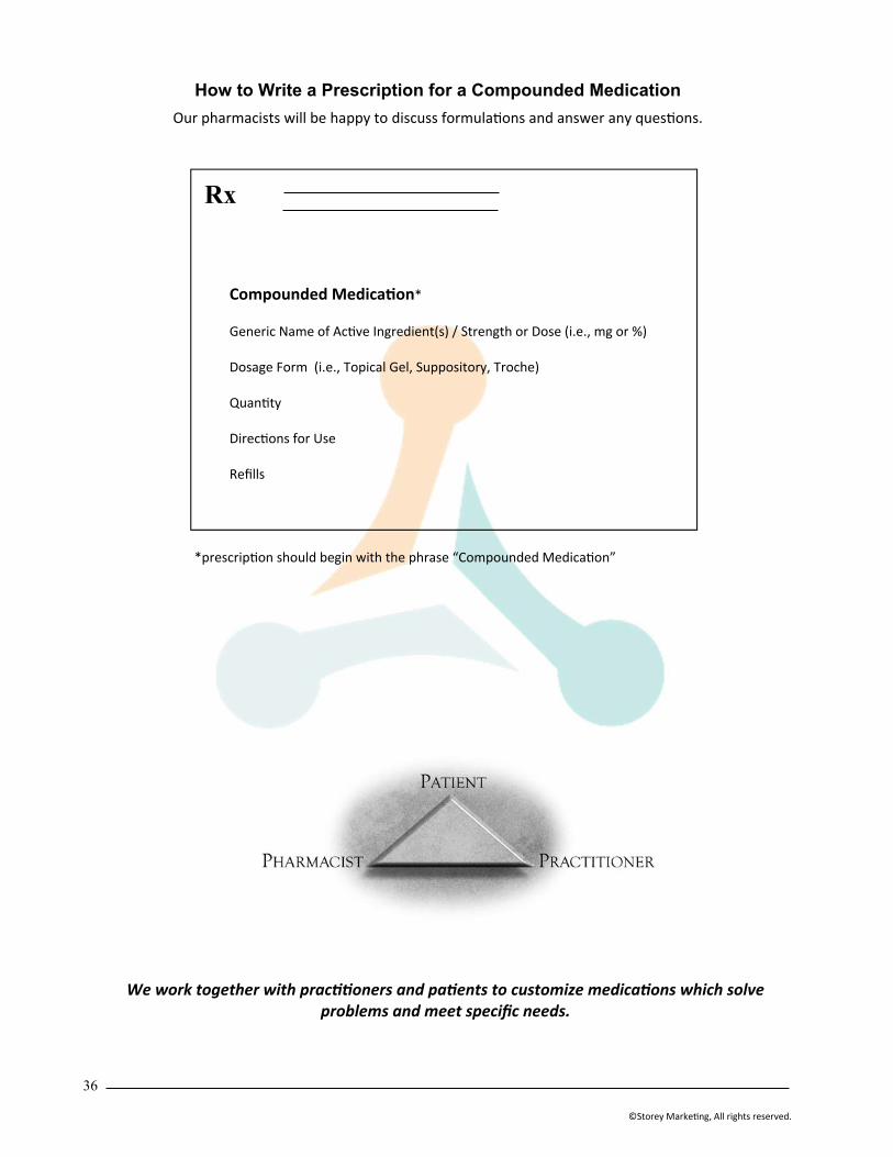
36
©Storey Marke ng, All rights reserved.
How to Write a Prescription for a Compounded Medication
Our pharmacists will be happy to discuss formula ons and answer any ques ons.
Compounded Medica on* Generic Name of Ac ve Ingredient(s) / Strength or Dose (i.e., mg or %) Dosage Form (i.e., Topical Gel, Suppository, Troche) Quan ty Direc ons for Use Refills
*prescrip on should begin with the phrase “Compounded Medica on”
Rx
We work together with prac oners and pa ents to customize medica ons which solve problems and meet specific needs.