dermatologia cosmetologica .ouarterly review of … di dermatologia cosmetologica .ouarterly review...
TRANSCRIPT
Trimestrale di Dermatologia Cosmetologica .Ouarterly Review of Cosmetic Dermatology Volume 3 - Number 3 July-September 1985 ISSN 0392-8543 Sped. a bb. post. gr. IV 0 nO
INTERNATIONAL EDIEMME
'C..
Dalla continuità nella ricerca scientifica
'•
una risposta più completa ·
BIOE$E®
Integratore alimentare di · Gelatina-Cistina, Fe, Cu, Zn ·
DIETETICO REG. MIN. SAN. N. 706/4971
Per Campioni Medici e Documentazione Scientifica: Mavi s.r.l. · Via Filippo Bernardini, 22 · 00165 Roma (Italy) · Tel. (06) 63.84.348

OLIO di AVEENO® usato nel bagno, per la doccia o direttamente su lla cute , svolge un 'importante azione lenitiva ed emolliente, previene gli arrossamenti, idrata e deterge, evitando l'uso del sapone e dei syndets. È contenuto in 8 dosi «self closing» da 30 ml. che ne permettono l'uso anche parziale.
~· • • - 2 1042 Carenno (VA) V•a Milano 160 · Tel {02) 96590311213

PROTEO GEL
)ferule di gelatina da 500 mg
TIOGEL: ~ferule di gelatina e e-cistina da
TIOGEI:
- - -PROTEO PROTEO
a17. mav1
TIOGEI: .. ~El: -TIOGEf j . ..~Ei:-TIOGEt:-s•UIJll 90 snauu
Per Campioni Medici e Documentazione Scientifica: Mavi s.r.l. - Via Filippo Bernardini, 22 - 00165 Roma (Italy) - Tel. (06) 63.84.348
Mavl soe· DOPO
.,. .... --
6
...... --

Per documentazione tecnica e campioni medici: Mavi s.r.l. - Via Filippo Bernardini, 22 - 00165 Roma (Italy) - Tel. (06) 63.84.348

• mav1 LA RICERCA SCIENTIFICA NELLA DERMOCOSMESI
MAVIGEN LATTE MAVIGEN GEL ALFA 4 MAVIGEN SAPONE
IDROSKIN NORMOSKIN SEBO A
ALFA STRIA ACROMOS
MAVISOLE 3 MAVISOLE 6 MAVISOLE 9 MAVISOLE 15 MAVISOLE DOPO
MAVIGEN SEBO MA VIGEN SHAMPOO MA VIGEN FORFORA AMINO PIL
BALSAMO ZPT BALSAMO LPO
PROTEOGEL TIOGEL BIOESSE BETAEFFE .
PRODOTTI PER VISO E CORPO Detersione
Detergente fisiologico Tonico analcolico Biosoluzione acida Detergente al collagene
Protezione e normalizzazione
Idratante al D-coUagene Nutriente alle vitamine Ph e sebonormalizzante
Trattamenti specifici Smagliature e rughe lperpigmentazioni cutanee
Sole e pelle
Per pelli normali ad effetto rapido Idratante per pelli delicate Filtro totale nelle iperpigmentazioni anomale Per le labbra e le zone più sensibili Arrossamenti cutanei post-sole
PRODOTTI PER CAPELLI
Nella eccessiva secrezione sebacea Al collagene per lavaggi frequenti Contro il ristagno della forfora Aminoacidi solforati - Uso topico
Protezione e normalizzazione
Antiforfora Protettivo: elimina le doppie punte
INTEGRATORI DELLA DIETA
Sferule di gelatina da 500 mg Sferule di gelatina e 1-Cistina da 200 mg Tavolette di Fe, Cu, Zn, gelatina e 1-Cistina Capsule di_ P-carotene, vit. E e F

POSTE ITALIANE
P'P.1"40 010~QO-lMISSIONl
During the International Meeting «A New Look at Old Skin: A Challenge to Cosmetology» progressively numbered special cards were issued in a limited number of copies (1500). This card has a stamp, especially designed for the occasion, that is now part of the museum of the Italian Post-office. A limitated number of both cards and covers «first day issue» are stili available at EDIEMME.
Cover «First day issue» Special card «First day issue»
L. 2.000 $ 2 L. 5.000 $ 3

A new look at old skin : A challenge to cosmetology lst lnternational Meeting on Cosmetic Dermatology, Rome,ltaly, March 7-9, 1985
Editors: P.Morganti, W Montagna
Readership: Third year undergraduates, research workers in the field of Cosmetic Chemistry, Biochemistry, Medicine, Pharmacy and Pharmacology, researchers and managers working in the cosmetic and pharmaceutical industries.
A NEW LOOK AT OLD SKIN: A CHALLENGE TO COSMETOLOGY
Editors: P. Morganti. W. Montagna
The. proceedings contained in this vo lume provide a comprehensive view of the different aspects of the skin aging with i ts cosmetological implicantions
Contents (main chapters) The problems of the aged (R. Butler) Nutrition and aging (M. Proja) Common structural changes in aging human skin ( W.
Montagna) An overview of phypsiological changes (B.A. Gi/chrest) The skin as a barrier and a homeostat ic compartment
of the body (G. Esposito) Skin collagen cross links natu ral and unnatura l (J.P.
Bentley) Aging changes in the mucus membranes (A. Jarrett) Changes in cutaneous appendages (F.J.G. Ebling) Sebum secretion rates in relat ion to age: A new look (J.S.
Strauss) Pharmacology and the aging cutaneous system (R.
Paoletti) Sunlight, age and skin cancer (J.C. van der Leun) Stereology of the skin surface: a comparison between
ageing and UV-induced damages (P. Corcuff) Cosmetic wrinkle smoothing (A. Meybeck) Collagen in cosmetic formulations: A contribulion to
research on aging skin (I Beyssac) The cosmetic maneuver in elderly women (A.M. Kligman) Essential fatty acids and skin aging (P. Morganti, S.D.
Randazzo) Treatment cosmetics and aging (L.C. Calvo)
Proceeding of 1. st lnternational Meeting on Cosmet ic Dermatology, Rome, ltaly, March 7-9, 1985 1985, 17-24 cm. Approx '.100 riages. Hard-bound U.S. S 40 Ready on December 1985 ISSN 0393-5779
VII

X
INFORMAZIONI PER L'ABBONAMENTO
L'abbonamento annuale comprende quattro numeri. È possibile ottenere abbonamenti a prezzo ridotto da parte dei ricercatori che lavorano presso Istituti che abbiano sottoscritto almeno un abbonamento a prezzo normale. L'Editore potrà fornire a richiesta notizie piu dettagliate. Le sottoscrizioni di abbonamento possono essere effettuate mediante assegni postali, bancari, di conto corrente o pe r contanti indirizzandoli a:
L'IVA è a carico dell'editore, 11011 detraibile dall'abhonato a norma art. 74 lctt. C DPR 633/72 .
INTERNATIONAL EDIEMME · Via Filippo Bernardini 22, 00165 ROMA
{e/e bancario n. 27/4328 Banco di Napoli Ag. 20)
SOTTOSCRIZIONI ANNUALI
Italia L. 60.000 · Altre Nazioni $ SO
Numero singolo L. 16.000
Numero arretralo L. 20.000
Per la pubblicità di questa rivista: Concessionaria Italiana Pubblicità G & V s.r.l.· Via Savona, 94 · 20144 Milano · Tel. (02) 4225907 -4231910
SUBSCRIPTION INFORMATION
Subscriptions are entered on a calendar year basis only and include four regular quarterly issues. Half-price su bscriptions are available to research scienlists whose institutions already subscribe al full rate. De tails on a pplication from publisher. Payment must be made in U.S. dollars using bank draft, internalional postai money order only. Italian res idents only may pay by persona! check:
INTERNATIONAL EDIEMME · Via Filippo Bcrnardini 22, 00165 ROMA
(C.C. banca rio n° 27/4328, Banco di Napoli, Roma)
ANNUAL SUBSCRIPTION RATE:
Italy, Lit. 60.000 · Other Counlries, $ 50 Additional Air Mail postage rate: Africa and Middle East $ 12. North, Centrai and South America$ 14, Far East $ 15, Oceania$ 19.50
Statements and opinions expressed in the articles and communicalions herein are those of the author(s) and not necessarily those of the Editor(s), or publisher. The Editor(s) and publisher, di sclaim any responsibili ty or liabi li ty for such materiai and do not guarantee, warrant, 01· endor:;e any produc t or service adverised in this publication nor do guarantee any claim made by the manufacturer of such product or service.
XI
GENERAL INFORMA TION
The JOURNAL OF APPLIED COSMETOLOGY is an in ternational journal devoted to publishing originai papers, reviews and other materiai which represent a useful conlribution to research on the skin and on cosmetics. It is aimed at cosmelic chemists, dermatologisls, microbiologists, pharmacists, experimental biologisls, toxicologists, plastic surgeons, and ali other scientists working on products which will come into contact with the skin and its appendages. The Journal is published qua1·terly both in English and Ilalian. It is dislribu ted to cosmetic chemists, dermatologists, plastic surgeons, medicai and pharmaceutical schools, medicai libraries, selected hospitals and research institutions th rought the world, and by subscription to any other interested individuals or organizations. Stalemenls and opinions expressed are persona! to the respective contributors and are not necessari ly endorsed by the E<litor(s), Adviscrs, Publishers or Dis lributors of this Journal.
COPYRIGHT
Submitted materiai must be the originai work of the author(s) and musi noi havc been submitted fo1· publ icalion elsewhere. By submitting a manuscript, the aulhors agree that the copyright for their articles is transferred to lhe publisher if and when the a rlic le is accepted f0t· publicalion. None of the conten i of lhis publicat ion may be repi-oduced in whole or in pari, translated, s tored in a retrieval system, or lransmilled or dislributed in any form or by any means (electronic, mechanical, photocopy, recording or olherwise) withoul the prior written permission of the Puhlishers.
Sections of Journal
The following sections will be features of the Journal: Originai Laboratory S1udies: descriptions of originai investigative laboratory research in cosmetics and related areas. Special Reports: ltems of special interest to the readers, including reports on meetings, societies, legislat ion, etc. Generai Articles: scientific articles of generai interest to our readers will l;ie considered for publication. These arlicles should be concerned with ncwer developments in such related fie lds as derma1ology, biology, toxicology, etc. Short Communicalions: the lenght should noi exceed 5 typewritten pages with not more than 3 figures included. Headings ("Materials", "Discussion", etc.) as well as Summaries are to be omitted. If accepted, these submission will appear in print in a very short time. Lelter to Lhe Editor: comments on Journal art icles are invited as well as brief contributions on any aspects of cosmelic science. Letters may include figures, and/or references, hut brevi ty is nccessary. Guest Editoria/s: concise, authoritative, substantiated commentary on s pecific topics of contemporary interest. Book Reviews: books and monographs (domeslic and foreign) will be reviewed depending on their in terest and value to subscribers. Send materiai for review to the Editor, Dr P. Morganti. No such materiai will be returned.
Address: all papers should be submitted to: Dr. P. Morganti INTERNATIONAL EDIEMME Via F . Bernardini. 22 00165 ROME - I taly Tel. 06/637.87.88

Xli
INFORMATION FOR AUTHORS
Papers musi be submitted in English. Authors whos e mother tongue is not English shou ld a rrange for their manuscripts to be written in proper Englis h prior lo submission. Italian authors mus i submit their manuscripts bolh in Italian and in English.
Procedure o/ Submission of Manuscripts: submit three copies of both the manuscdpt and a li illus tra tive materiai to the above address.
Organization o/ the Manuscript: investigative s tudies s hould be organized as follow: title, abstra ct page, int rod uction, materi a] and methods , results, di scuss ion, acknowledgments, references, legends for figures, ta bles . Ali pages should be numered consecutively s tarting with the abst ract. The enti re manuscript is to be typewritten, double-spaced, a nd with 3 cm margins. Tr ade na mes must be capitalized: the common narne for compounds may be used if the formai chemical na me a s established by inte rnational convention is g iven afte r the fi r st use. Any a bbreviations other than those which a re gene ral ly accepted mus t be defined. In the tex t, refe rences lo dual a uthors will use both surnames throughout. For multiple a uthors, use the surnames of ali autho1·s at the firs t 1·eference and only the firs t author followed by "et a l. " thereaftei-. Please ma1·k in the margin of the manuscrip t the desired position of the figures a nd tables. To allow fas ter publication on ly set of proofs will be furnished to the author including the figures and tables in their final pos ition.
Tir le page: li s t the title, name(s) and degree(s) of author(s), dcpartment(s) a nd institu t ion(s) at which the work was clone, city, state, and pos tai code. Any preliminary report or abstract of the wo1·k should be refe rred to as a footnote lo the t it le.
S ummary: each paper mus i be headed by an English language title of not aver 70 characters (inc luding spaces) suita ble for use as a running head and mus i al so be preceded by an Engl ish summa ry noi exceeding 300 words typed double·s paced. The summary will include statements of the proble m, me thocl of s tucly, results, and conclus ions. Since this summary will be used by abstract ing journa ls, it mus t be se lf-expla na lo ry a nd should not include abb1·eviations , foo tnotes, and references.
Foot notes: should be li s lecl consecut ively al thc bo ttom of the page on which they fai!, designated by the following symbols in o rder: *, +, +, §, II, ** , etc.
Key Words: key words for compute rised storage and retrieval of information should be incorporateci in the summai·y.
R eferences: the references have to be abbrevia ted as listed in the Index Medicus. The s tyle of the references mus t conform to the examples given below: I) Robbins CR, Kellych (1970) Aminoacid composition of human hair. Text Res J 40:891-896. 2) Strehler BL (1977) Time, cell s a nd aging 2nd edn. Academic Press , New York 3) Ebling FJ, Rook (1972) Ciclic activity of the follicle. In: Textbook of clermatology 11, Blackwell,
Oxford, p. 1567-1573.
Illusrmtions: figures shou ld be numbered consecutively using Arab ic numerals , Tab les should be numbered consecu tively, using Roman numerals. Ali photographs should be black and white, glossy a nd unmoun ted. The number and size of illustration shou ld be restrictecl to the min im um needed to clarify the text. Authors requiring extra s pace fo r il lus tra tions w ill be charge accordingly. This is a lso the case fo1- color illustrat ions. Ali figures, photographs, graphs, or diagrams shoulcl be submitted on sepa rate sheets.
Animai Experimen ts: descriptions of animai experiments shoudl include full details of the types of animai used (inbred, etc .) and the conditions under which they were kept (standard d iet, etc.).
Trade Names: ali common cosmetic ingredients should be referred to by their generic names, as indicated in the latest eclition of CTFA Cosmet ic Ingredient Dictionary, and the European Pharmacopeia. lf a materia i is not listed, then the trademar ked name can be used, with the chemical composition given in footnotes .

XII!
Trimestrale di Dermatologia Cosmetologica Quarterly Review of Cosmetic Dermatology Contents
Generai articles
187 Cutaneous elastin degradation in agein g and inflammation W.E. Pa rish
Originai Lahoratory Studies
211 Essential Fatty acids and skin aging P. Morganti, S.D. Randazzo
Guest editoria!
223 Contact Dermatitis due to Cosmetics G. Angelini, G.A. Vena, G. Giglio, F. Fiordalisi. C.L Meneghini
Historical Special Report
234 Marvelous secrets W. Montagna, P. Morganti
238 To make art? B. Lisi
Io be orto Realise
240 Book Review
241 Letter to the editor
XXI Announcements


J. Appl. Cosmetol., 3, 187-210 (J uly/September 1985)
Cutaneous elastin degradation in ageing and inflammation W.E. PARISH, Head of Toxicology - Unilever Research Colworth House Sharnbrook - Bedford MK44 1 LO England
Received: March 8, 1985. Presented ar rhe lnternational Meering: A New Look ar O/d Skin: a Challenge to Cosmero/ogy, March 7-9, 1985 Rome - ltaly
Key words: Elastic Fibres in Skin; Ageing of Elastin; Degeneration of Elastin; Elastase Inhibitors; Actinic Elastosis.
Synopsis
Elastic fibres are an essential component of skin and connective tissue. Some fibres persist for many years and turnover of elastin is slow. The amount and dist ribut ion of elastic fibres within a tissue normally remains remarkably constant; the nature of the contro! of this constancy is unknown. Ageing changes of elastic fibres in skin protected from sunlight show some decrease in t he sub-epidermal fibr ils and slight thickening and fragmentation of the deeper fibres. Frequent exposure of persons with pale skin to bright sunlight results in loss of the sub-epidermal fibrils. much hyperplasia of the reticular and deeper elastic fibres followed by fragmentation and, histologically, deposition of amo1-phous elastin in dense masses, degradable by elastase, weakly by chymotrypsin but not by collagenase. There is some mild chronic inflammation which is more likely to result from the other damaging e ffects of sunlight, and distortion of the de rmis by the elastin masses than to a primary st imulus to elas tic fibre degeneration. A summary of studies on elast ic fibre degeneration in acute and chronic vasculi t is is presented, in which in acute vasculitis induced by immune complexes, neut rophils degrade the internal elastic lamina in proportion to their numbers and time. In human chronic vasculitis lesions there appears to be an unstable balance between synthesis of elas tic fibres and their degradation by elastases shown to be secreted by cells in the lesion. Plasma inhibitors of elastases detected in the lesions by immunofluorescence, and probably others from macrophages, fai[ to comrletely contro! the degradation. Fractions o rabbit macrophages were found to contain two distinct inhibitors for rabbit neutrophil elastases.
Riassunto
Le fibre elatiche sono una componente essenzia le dei tessuti cutaneo e connettivo. Alcune fibre persistono per molti a nni e il ricambio dell 'elastina è lento. La quantità e la distribuzione delle f ibre elastiche in un tessuto r imangono di norma considerevolmente costanti; la natura dei fattor i che determ inano questa cos tanza non è nota. I cambiamenti dovut i all'invecchiamento delle fibre elas tiche della pelle protetta dalla luce del sole mostrano una certa diminuzione delle fibrille sotto-epidermiche e un leggero ispessimento e frammentazione delle fibre più profonde. L'esposizione ripetuta di persone da lla pelle chiara alla luce sola re porta come risultato alla perdita delle fibrille sotto-epidermiche, ad una consistente iperplasia delle fibre elastiche reticolari e profonde, a cui fa seguito un processo di frammentazione e, dal punto di vista istologico, una deposizione di elastina amorfa in masse dense, degradabile dall'elastasi e dalla chimotrips ina ma non dalla collagenasi. Si verifica un leggero p rocesso infiammatorio cronico che più probabilmente è il risultato di a ltri effetti dannosi della luce solare e della distorsione del derma ad opera delle masse di elastina, piuttosto che di uno stimolo primario alla degenerazione della fibra elastica. Viene presentato un compendio di s tudi sulla degenerazione della fibra elastica nella vasculite acuta e cronica; nella vascu li te acuta indotta da imunocomplessi, i neutrofil i degradano la lamina e lastica interna in proporzione a l loro numero ed al tempo. Nelle lesioni umane dovute a vasculite cronica sembra esistere un equilibrio instabile t ra la sintesi delle fibre elastiche e la loro degradazione ad opera delle elastasi che si ritiene siano secrete dalle ce llule lese. Gli inibitori delle elastasi presenti nel plasma, rilevati nelle lesion i mediante immunofl uorescenza e probabilmente altri inibitori, originati dai macrofagi, non riescono a controllare completamente la degradazione. In frazioni di macrofagi di coniglio si è rilevata la presenza di due inibitori delle elastasi neutrofi le del coniglio.
188 Cutaneous elasti-n degradation in ageing and inflammation
Elastic fibres in skin
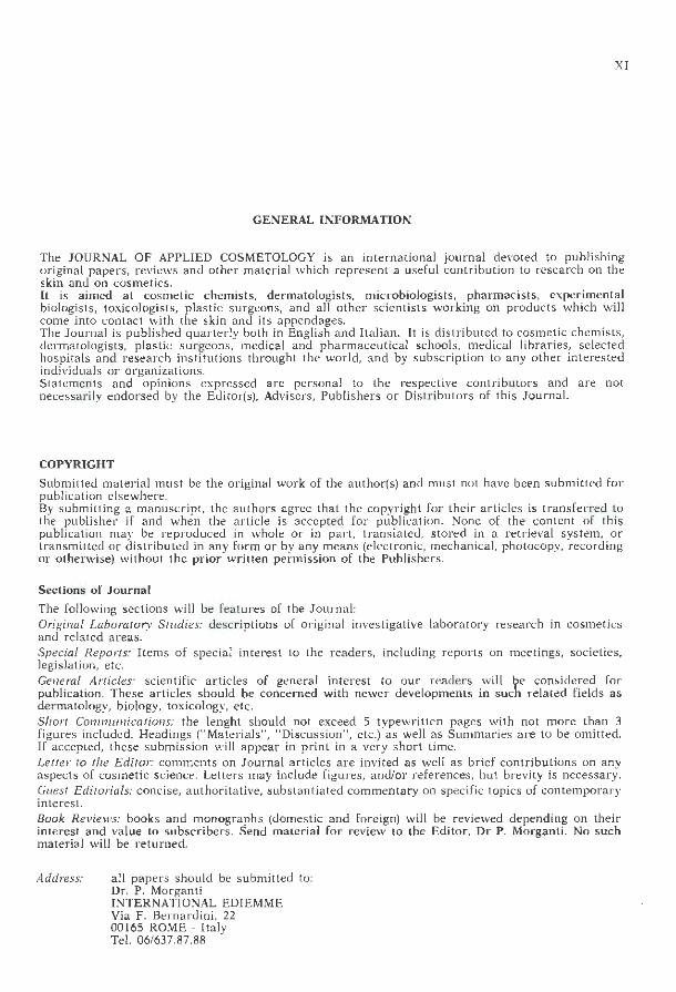
In skin e las tic fibres show four genera] patterns of distribution. They occur in large number s in the subcutaneous connective tissue binding the dermis, (the panniculus carnosus) enabling free movement of the skin aver the body. In the deep dermis thick elastic fibres tend to follow the piane of the collagen fibres and pa rallel to the epidermis. A less regular reticular distribution of thinner fibres lies in the more superficia l dermis. From the reticular fibres fine fibrils pass towards and at r ight angles to the dermoepidermal junction, dividing into a plexus of even finer fibrils which meet the junctional layer (Fig. 1). Fine elastic fibres, usually in reticulate pattern, surround the eccrine and apocri-
Fig. 1: Human skin, dermo-epidermaljunction. Elastic fibres emerging from the reticular Jayer to lie a t right-angles to the junction zone and divide into a plexus of fine fibrils. which contact the junction membrane. (Gomori aldehyde fuchsin; counterstain Light green and Orange G).
Fibre elastiche nella pelle
Le fibre elastiche cutanee mostrano quattro modelli generali di distribuzione. Esse sono presenti in gran numero nel tessuto connettivo sottocutaneo che lega il derma (il pannicolo carnoso), il quale rende possibile il libero movimento della pelle sul corpo. Nella parte più profonda del derma le spesse fibre elastiche tendono a seguire il piano delle fibre di collagene, parallelamente all'epidermide. Sul derma più superficiale è presente una distribuzione reticolare meno regolare. Fibrille sottili si trasferiscono ad angolo retto dalle fibre reticolari a lla giunzione derma-epidermica, dividendosi in un plesso d i fibrille ancora più sottili che vanno ad incontrare lo strato di congiunzione (Fig. 1).
Fig. I: Pelle umana, giunzione dermo-epidermica. Fibre elastiche che emergono dallo strato reticolare: sono disposte ad angolo retto rispetto alla zona di congiunzione e si dividono in un plesso di sottil i fihrill e, a cont11tto con al membrana di congiunzione. (Aldeide-fucsina di Gomori; controcolorazione verde chiaro e arancione G).

W.E. Parish
ne glands, and also portions of the hair follicles. The arrector pili smooth muscle of the hair also contains wefts of reticulate elastin, particularly at its junction with the follicle. Of particular interest to our studies is the distribution of elastic fibres in blood vessels. In small arterioles, with a diameter of 300 µm or less, the tunica intima is comprised of the endothelium with the internal elastic lamina just beneath it, which in fixed histological specimens has a fine undulating or scalloped form, probably resulting from contraction, outside which lies two or three layers of smooth muscle cells. In the connective tissue adventitia surrounding the small arterioles, dispersed elastic and collagen fibres lie mainly parallel to the course of the vessel. The elastic lamina becomes
Fig. 2: Guinea-pig subcutaneous tissue. Arteriole (right) thick undulating internal elastic lamina. Venule {left) thinner elastic lamina, and discontinuous appearance of spirai fibres in adventitia. Elastic fibres support dilated lymphatic vessels bclow and above blood vessels. (Stain as in Fig. 1).
189
Fibre elastiche sottili circondano le ghiandole eccrine ed apocrine ed anche parti dei follicoli piliferi, di solito secondo un modello reticolato. Anche il muscolo liscio erettore del pelo contiene piccole quantità di elastina reticolata, in particolare nel suo punto di congiunzione con il follicolo. La distribuzione delle fibre elas tiche nei vasi sanguigni presenta un interesse particolare per i nostri studi. Nelle piccole arteriole, aventi un diametro di 300 µm o meno, la tunica intima comprende l'endotelio con la lamina elastica interna subito al di-sotto di esso, che in campioni istologici fissati ha una sottile forma ondulata o dentellata, probabilmente risultato di contrazione, al di fuori del quale si trovano due o tre strati di cellule di muscolo liscio. Negli avventizi del tessuto
Fig. 2: Tessuto sottocutaneo di cavia. Spessa lamina elastica interna ondulata di arteriola (a dest ra). Più sottile lamina elastica di venula (a sinistra), e aspetto discontinuo di fibre a spirale negli avventi;li. Fibn: dasliche suslenguno vasi linfatici dilatati al di sotto e al di sopra dei vasi sanguigni. (Colorazione come nella Fig. I).
190 Cutaneous elastin degradation in ageing and inflammation
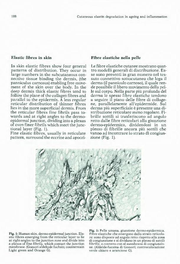
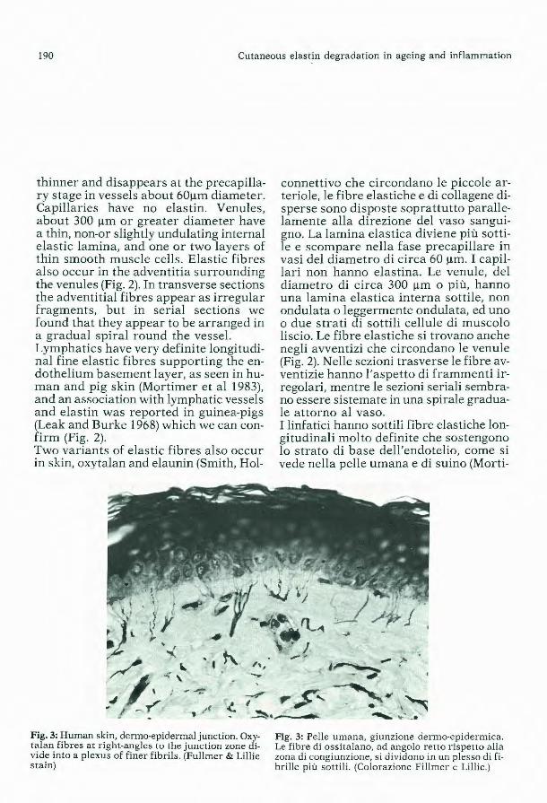
thinner and disappears at the precapillary stage in vessels about 60µm diameter. Capillaries have no elastin. Venules, about 300 µm or greater diameter have a thin, non-or slightly undulating internal elastic lamina, and one or two layers of thin smooth muscle cells. Elastic fibres also occur in the adventitia surrounding the venules (Fig. 2). In transverse sections the adventitial fib res appear as irregular fragments, but in serial sections we found that they appear to be arranged in a gradual spira! round the vessel. Lymphatics have very definite longitudinal fine elastic fibres supporting the endothelium basement layer, as seen in human and pig skin (Mortimer et al 1983), and an association with lymphatic vessels and elastin was reported in guinea-pigs (Leak and Burke 1968) which we can conf irm (Fig. 2). Two variants of elastic fibres also occur in skin, oxytalan and elaunin (Smith, Hol-
•I t\, ,,, -Fig. 3: Human skin, dermo·epidermal junction. Oxytalan fibres at right·angles Lu Lhe junction zone di· vide into a plexus of finer fibrils. (Fullmer & Lillie stain)
connettivo che circondano le piccole arteriole, le fibre elastiche e di collagene disperse sono disposte soprattutto parallelamente alla direzione del vaso sanguigno. La lamina elastica diviene più sottile e scompare nella fase precapillare in vasi del diametro di circa 60 µm . I capillari non hanno elastina. Le venule, del diametro di circa 300 µm o più, hanno una lamina elastica interna sottile, non ondulata o leggermente ondulata, ed uno o due strati di sottili cellule di muscolo liscio. Le fibre elastiche si trovano anche negli avventizi che circondano le venule (Fig. 2). Nelle sezioni trasverse le fib re avventizie hanno l'aspetto di frammenti irregolari, mentre le sezioni seriali sembrano essere sistemate in una spirale graduale attorno al vaso. I linfatici hanno sottili fibre elastiche longitudinali molto definite che sostengono lo strato di base dell'endotelio, come si vede nella pelle umana e di suino (Morti-
Fig. 3: Pelle umana, giunzione derma-epidermica. Le fibre di ossitalano, ad angolo retto rispetto alla zona di congiunzione, si d ividono in un p lesso di fibrille più sottili. (Colorazione Fillmer e Lillie.)
W.E. Parish
brook and Byers 1982). Oxytalan fibres stain like elastin fibres with routine histological elastic tissue stains but only after exposure to a strong oxidizing agent, and are very resistant to strong acids (Fullmer and Lillie 1958). They occur in much connective tissue, tendons, and the adventitia of blood vessels. In the skin they are particularly evident in the dermo-epidermal junction (Hasegawa 1960) where they form a plexus of radiating fine fibrils at right angles to the junction zone (Fig. 3) in the same manner as the mature elastic fibres (Fig. 1). Elaunin fibres also in skin, have intermediate properties between elastin and oxytalan, staining with orcein without pretreatment oxidation, but requiring prior oxidation before staining with aldehyde fuchsin (Gawlik 1965). It is possible that these are immature forms of elastin. In surface-damaged skin, e.g. an abraision, during resolution or healing with scar formation, the fine elastic fibres beneath the dermo-epidermal junction are frequently absent, but oxytalan fibres are detectable (Hasegawa 1960; Parish unpublished). Elastic fibres are comprised mainly of amorphous elastin surrounded by a thin sheath of microfibrils which have no elasticity. Good contrast of elastin in histological sections is achieved with the semiselective stains acid orcein, Weigert's acidic resorcin-fuchsin, Verhoef's ironhaematoxylin, which is not effective for fine fibrils as in the dermo-epidermal junction, and Gomori's aldehyde-fuchsin, which provides good definition for photography but is less selective in that mast cells, mucin, and «ground substance», possibly mast cell products, in chronic infiamma tory lesions also bind the stain but can be identified as not susceptible to weak pancreatic elastase treatment. Elastic fibres also stain histochemically for ribonuclease, deoxyribonuclease and
191
mer et. al. 1983) nelle cavie è stata osservata (Leak e Burke, 1968) una associazione dei vasi linfa ti ci con l'elastina che possiamo confermare (Fig. 2). Nella pelle si trovano, inoltre, due varianti delle fibre elastiche, l'ossi ta lano e l'elaunina (Smith, Holbrook e Byers, 1982). Le fibre di ossitalano si colorano come le fibre di elastina in colorazioni istologiche di routine del tessuto elastico, ma soltanto dopo esposizione ad un agente fortemente ossidante; sono, inoltre, molto resistenti agli acidi forti (Fullmer e Lillie, 1958). Esse si trovano in gran parte del tessuto connettivo, nei tendini, e negli avventizi dei vasi sanguigni. Nella pelle, sono particolarmente evidenti nella giunzione dermo-epidermica (Hasegawa, 1960) dove formano un plesso di sottili fibrille radianti ad angolo retto rispetto a lla zona di congiunzione (Fig. 3) analogamente alle fibre elastiche mature (Fig. 1). Le fibre di elaunina, anch'esse presenti nella pelle, hanno proprietà intermedie tra l'elastina e l'ossitalano, si colorano con l'orceina senza un pre-trattamento di ossidazione, ma richiedono una ossidazione preliminare per la colorazione Fucsinaldeide (Gawlik, 1965). È possibile che vi siano forme immature di elastina. Nelle lesioni cutanee superficiali, per esempio nelle abrasioni, durante la risoluzione o guarigione con formazione di cicatrice, le sottili fibre elastiche al di sot to della giunzione dermo-epidermica sono solitamente assenti, mentre si possono trovare fibre di ossitalano (Hasegawa, 1960; Parish, non pubblicato). Le fibre elastiche sono costituite essenzialmente da elastina amorfa circondata da un sottile rivestimento di microfibrille che non possiedono elasticità. Nelle sezioni istologiche si ottiene un buon contrasto di elastina con l'orceina acida a colorazione scmisclcttiva, con la rcsorcinafucsina aciclica di Weigert, con la ferroematossilina di Verhoef, che non è effi-

192 Cutaneous elastin degradation in ageing and inflammation
acid phosphatase; another feature that differentiates them from collagen fibres (Jarrett and Hardy 1968; Jarret 1974). Elastic fibres are formed from at least four types of cell, the most active being smooth muscle cells and fibroblasts. The most conclusive evidence is derived from culture of purified celi preparations with histochemical and electron microscopica! examination of the protein fibres synthesised. The following are the cell types shown to synthesise elastin. 1. Smooth muscle cells, i.e. myoblasts or myofibroblasts from immature guineapigs, or aortic smooth muscle (Ross 1971; Fisher-Dzoga, Jones, Vesselinovitch and Wissler 1973; Vitto, Hoffman and Prockop 1976; Abraham, Hart, Winge and Carnes 1977; Burke and Ross 1979). 2. Fibroblasts , especially those from the ligamentum nuchae (J ones, Sear and Grant 1980; Mecham, Lange, Maderas and Starcher 198 1) 3. Chondrocytes in vitro (Thyberg and Hinek 1977; Quintarelli, Starcher, Vacaturo, DiGianfilippo Gotte and Mecham 1979), and a l so by transplan ts in rabbi ts (Thyberg and Hinek 1977) 4. Endothelium (Carnes, Abraham and B uonassisi 1979) Synthesis of elastin is closely associated with that of collagen, though the two products are distinct. It has been reported that fibroblasts in vitro may synthesise elastin and collagen simultaneously (Kao, Gray, Bressan and Prockop 1980). In the intima interna and media of venules four weeks after mild damage by immune complexes, fine strands of elastin appear on the inner surface of the smooth muscle cells, and fine granules are apparent in enlarged endothelia l cells. These stain with orcein or aldehyde-fuchsin and are degradable by elastase but not collagenese. Discrete fibrils leading to thickening of the elastic intima are evidence of synthesis, but the granules in the endo-
cace per fibrille sottili quali quelle presenti nella giunzione derma-epidermica, e con la fucsin-aldeide di Gomori, che fornisce una buona definizione per la fotografia ma è meno selettiva. Infatti, anche le mast cell, la mucina, e la «Sostanza di base», probabilmente prodotti della mast celi, nelle lesioni croniche da infiammazione legano il colorante ma possono essere identificate come non suscettibili ad un debole trattamento pancreatico di elastasi. Anche le fibre elastiche si colorano istochimicamente per la ribonucleasi, la deossiribonucleasi e l'acido fosfatasi; altra caratteristica che le differenzia dalle fibre di collagene (Jarret e Hardy, 1968; Jarret, 1974). Le fibre e lastiche si formano da almeno quattro tipi di cellula, tra cui le più attive sono le cellule di muscolo liscio e i fibroblasti. La prova più decis iva è derivata dalla coltura di preparati cellulari purificati con esame istochimico ed elettromicroscopico delle fibre di proteina sintetizzate. Qui di seguito sono riportati i tipi di cellule che hanno mostrato di sintetizzare l'elastina: 1) Cellule di muscolo liscio, cioè mioblasti o miofibroblasti di cavie immature, o muscolo liscio aortico (Ross, 1971; FisherDzoga, J oines, Vesselinovitch e Wissler, 1973; Vitto, Hoffman e Prockop, 1976; Abraham, Hart, Winge e Carnes, 1977; Burke e Ross, 1979). 2) I fibroblasti, specialmente quelli del ligamentum nuchae (Jones, Sear e Grant, 1980; Mecham, Lange, Maderas e Starcher, 1981). 3) Condrociti in vitro (Thybergee Hinek, 1977; Quintarelli, Starcher, Vacaturo, Di Gianfilippo Gotte e Mecham, 1979), ed anche per mezzo di trapianti eseguiti suconigli (Thyberg e Hinek, 1977). 4) Endotelio (Carnes, Abraham e Buonassisi, 1979). La sintesi dell'elastina è strettamente associata con quella del collagene, sebbene
W.E. Parish
thelium may represent degradation rather than synthesis.
Ageing of elastin in protected normai tissu
The turnover of mature elastin in norma! undamaged tissue is very slow; individua! fibres appear to persist for severa! years. There must exist complex systems that protect fibres from endogenous degradation, and in normai protected tissue maintain approximately the same amount of elastin throughout !ife. C14
- labelled glycine incorporateci into the aortic elastin of rats 28 days old, showed some d ecline un ti! the age of 120 days, and a steady state thereafter, up to 930 days. It was concluded that there was no turnover of formed elastin in adult rats or during pregnancy (Walford, Carter and Schneider 1964). Different results, but stili evidence of long duration of e lastin , were observed following treatment of suckling rats with C14 1-proline. In the lungs elastin decreased by one third in 510 days, but by two thirds in skin. Loca! tissue conditions apparently influence the longevity of elastin, and some replacement is likely because the weights of lungs and dry weights of their elastin did not change between 122 and 510 days (Pierce, Besnick and Henry 1967). With increasing age in rabbit a nd human elastic fibres, the polar amino acids, particularly aspartic and glutamic acids, and the amount of cross linking amino acids desmosine and isodesmosine decrease, with a consequent reduction in elasticity. This may progress to fragmentation . The ageing fibres become more fluorescent and yellow (see Sandberg 1976). In examination of human skin by electron microscopy, Braverman a nd Fonferko (1982) showed that in the sun-protected skin of the buttock, there was a small
193
i due prodotti siano distinti. Si è osservato ch e i fibroblasti in vitro possono s intetizzare l'elastina e il collagene simul taneamente (Kao, Gray, Bresson e Prockop, 1980). Nella intima interna e media delle venule, quattro settimane dopo un lieve danno da immuno-complessi, sottili fili di elas tina compaiono sulla superficie interna delle cellule di muscolo liscio, e, nelle cellule endoteliali allargate, si rileva la presenza di piccoli granuli. Questi si colorano con l'orceina e la fucsin-adeide e sono degradabili dall'elastasi ma non dalla collagenasi. Fibrille distinte che portano all'ispessimento della intima elastica sono prova di sintesi, ma i granuli nell'endotelio possono rappresentare una degradazione piuttosto ch e una sintesi.
Invecchiamento dell'elastina in un tessuto normale protetto
Il r icambio dell'elastina matura in un tessuto normale non leso è mol to lento; le fibre individuali sembrano pers istere per vari anni. Esistono evidentemente dei sistemi complessi che proteggono le fibre contro la degradazione endogena, e che in un normale tessuto protetto mantengono pressappoco la stessa quantità di e lastina nel corso di tutta la vita. La glicina marcata C14 incorporata nell'elastina aortica di ratti di 28 giorni, ha mostrato un certo declino fino all'età di 120 giorni e successivamente, fino a 930 giorni, uno stato di stabilità. Si è concluso che nei ratti adulti o durante la gravidanza non vi è un r icambio di elastina (Walford Carter e Schneider, 1964). Trattando con 1-prolina C14 ratti lattanti, si sono avuti risultati differenti, anche se è stata posta in evidenza la lunga durata dell'elastina. Nei polmoni, l'elastina è diminuita di un terzo in 5 IO giorni, mentre è diminuita di due terzi nella pelle. Le condizioni locali del tessuto sembrano influenzare la !on-
194 Cutaneous elastin degradation in ageing and inflammation
amount of fibre disintegration, but at 50 to 70 years of age gaps appeared in the fibres, some empty, others containing granular materiai. Some elastin synthesis occurs (Kligman 1969} but with increasing age the fibres are loosely assembled (Braverman and Fonferko 1982). After 50 years old, there is some loss of the plexus of fine fibri ls beneath the dermoepidermal junction (Montagna and Carlisle 1979}. No inflamma tory cell infiltration is associateci with the age-associated degenera tion (Braverman and Fonf erko 1982).
Light activated degeneration or elastin (elastosis)
The ageing process is much accelerated, and accompanied by other degenerative changes when skin is frequently exposed to strong sunlight, resulting in chronic solar or actinic elastosis. The clinica! manifestations are wrinkles, deep furrows, thinning of the skin and loss of pliability. The dermis contains large amounts of amorphous materia! binding elastinselective stains which atone time was not believed to be derived entirely from elastin, hence elastosis. The early onset in young adults, and severe degeneration in the face of persons 30 to 40 years old exposed to bright surlight, the regional differences in the face according to exposure, and the possible activity of the long ultraviolet waves in 340 to 400nm band, have been well described by Kligman (1969; 1974). The first histological change in elastosis is seen in the papillary dermis with transient thickening of the fibrils perpendicular to the dermo-epidermal junction, which then disappear with loss of the fine plexus or palmate structure. In the reticular zone thickening and h yperplasia proceeds to intertwisting and fragmenta-
gevità dell'elastina, ed è probabile che avvenga qualche sostituzione dal momento che il peso dei polmoni e il peso secco della loro elastina non è cambiato tra 122 e 510 giorni (Pierce, Besnick e Henry, 1967}. Nelle fibre elastiche di coniglio e di uomo, gli aminoacidi polari in particolare l'acido aspartico e glutammico, aumentano con l'avanzare dell'età, e la quantità di desmosina e di isodesmosina, amminoacidi a legame crociato, diminuisce, con una conseguente riduzione di elasticità. Questo processo può portare alla frammentazione. Con l'invecchiamento, le fibre diventano più fluorescenti e gialle (vedi Sandberg, 1976). Braverman e Fonferko (1982}, in uno studio su cute umana condotto con microscopia elettronica, hanno mostrato che nella pelle dei glutei protetta dal sole il livello di disintegrazione delle fibre è b asso, mentre tra SO e 70 anni appaiono tra le fibre lacune vuote o con materiale granuloso. Avviene una sin tesi di elastina (Kligman, 1969} ma con l'avanzare dell'età le fibre si dispongono in modo non compatto (Braverman e Forfenko, 1982). Dopo i 50 anni di età, si verifica una certa perdita del plesso delle sottili fibrille che si trovano al di sotto della giunzione derma-epidermica (Montagna e Carlisle, 1979). Nessuna infiltrazione cellulare di tipo infiammatorio è associata con la degenerazione dovuta all'età (Braverman e Forfenko, 1982).
Degenerazione dell'elastina attivata dalla luce (elastosi)
Il processo di invecchiamento è molto accelerato, ed accompagnato da altri cambiamenti degenerativi quando la pelle viene frequentemente esposta alla luce solare forte, che porta ad una elastosi cronica solare o attinica. Le manifestazioni cliniche sono rughe, pieghe profonde, assottigliamento della pelle e perdita di ela-
W.E. Parish
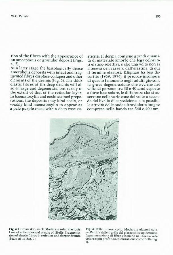
t ion of the fibres with the appearance of an amorphous or granular deposit (Figs. 4, 5). At a later stage the histologically dense amorphous deposits with intact and fragmented fibres dispiace collagen and other elements of the dermis (Fig. 6). The thick elastic fibres of the deep dermis will also enlarge and degenerate, but rarely to the extent of that of the reticular layer. In haematoxylin and eosin stained preparations, the deposits may bind eosin, or weakly b ind haematoxylin to appear as a pale purple mass wi th a deep rose co-
Fig. 4: Human skin, neck. Moderate solar elastosis. Loss of subepidermal plex4s of fibrils, fragmentation of elastic fibres in reticular and deeper dermis. (Stain as in Fig. I)
195
sticità. Il derma contiene grandi quantità di materiale amorfo che lega coloranti elstino-selettivi, e che una volta non si riteneva derivassero dall'elastina, di qui il termine elastosi. Kligman ha ben descritto (1969, 1974), il precoce insorgere di questo fenomeno negli adulti giovani, la grave degenerazione che avviene nel volto di persone tra 30 e 40 anni esposte a forte luce solare, le differenze che si osservano nelle varie zone del volto a seconda del livello di esposizione, e la possibile attività delle onde u ltraviolette lunghe comprese nella banda tra 340 e 400 nm.
Fig. 4: Pe lle umana, collo. Moderata elastosi solare. Perdita delle fib rille del plesso sotto-epidermico, frammentazione di fibre elastiche nel derma reticolare e più profondo. (Colorazione come nella Fig. l).

198 Cutaneous elastin degradation in ageing and inflammation
mass enlarges, the cell change is probably a response to the abnormal tissue enlargement and to other sunlight-mediated effects in the skin, and does not significantly participate in the elastotic process. The particular stimulus to elastic fibre hyperplasia, and the source of degrading enzymes or other mediators of degeneration have not been ascertained. Elastosis has been induced in mice (Sams, Smith and Burk 1964) and rats (Nakamura and Johnson 1968) by longterm intermittent exposure to ultraviolet light. Protection against the effec t of strong sunlight may be natural or acquired by topica! application of suitable light filters. The melanin of negro skin affords much protection; no elastic hyperplasia may be present in the skin of the young, and in the elderly the hyperplasia is less than in caucasians (Kligman 1969). Thus melanin provides much but not complete protection. In white subjects a tan that is not accompanied by appreciable thickening of the s tratum corneum does not provide substantial protection against sunburn (Kaidbey and Kligman 1978). Thickening of the stratum corneum in response to sunburn increases protection (J ohnson 1978) and lightly pigmented skin scatters light, reducing absorption. Additional protection can be acquired by topica! application of sunscreens. The degree of protection can be determined in the laboratory, but the use of a light source that simulates the UV spectrum of sunlight is essential; the common mercury vapour lamps can give false results because of their excessive emissions of short UV wavelenghts. Most sunscreens act by absorbing the burning UVB wavelengths. Formulations that penetrate into the stratum corneum and resist removal by sweating may be more effective (Willis and Kligman 1969).
ne il trattamento produca delle zone che si colorano meno fortemente con l'orceina o con la fucsinaldeide dopo il trattamento. Il materiale elastosico è soprattutto elastina amorfa. Il derma elastosico contiene una quantità in peso di elastina da quattro a sei volte maggiore di quella del derma normale (Smith, Davison e Clark, 1962) e sebbene alcuni ammino-acidi siano presenti nell'elastina normale e elastosica in quantità diverse, essi sono tuttavia simili per composizione ma differiscono significativamente per gli amminoacidi del collagene (Smith, Davison, Sams e Clark, 1962). L'elastosi sembra risultare dalla iperplasia e dalla degenerazione delle fibre elastiche. Non vi è prova consistente di infiammazione cronica. Braverman e Fonferko (1982) descrivono un aumento del numero di macrofagi e di mas t cell. Un certo aumento di queste cellule si verifica, ma in considerazione della distorsione e sostituzione dell' interno del derma a mano a mano che la massa elastosica si allarga, il cambiamento cellulare è probabilmente una risposta all'allargamento abnorme del tessuto e ad altri effetti mediati dalla luce del sole sulla pelle, e non partecipa in modo significativo al processo elastotico. Il particolare stimolo all'iperplasia delle fibre elastiche, e la fonte di enzimi responsabili di degradazione o di altri mediatori di degradazione non sono state accertati. L'elastosi è stata indotta nei topi (Sams, Smith e Burk, 1964) e nei ratti (Nakamura e Johnson, 1968) per mezzo di esposizione, intermittente e a lungo termine, alla luce ultra violetta. La protezione contro l'effetto della luce solare forte può essere naturale o acquisita dopo applicazione di filtri solari adatti. La melanina della pelle nera esplica una maggiore protezione, la iperplasia elastica non può presentarsi nella pelle

W.E. Parish
Elastase and Elastase inhibitors
Elastases are secreted by severa} types of cell apart from the digestive pancreatic enzymes. Neutrophil granule elastases of which at least three separable forms occur in human cells and which comprise one sixth of the total soluble protein in the granules, degrade elastin in inflammatory disorders of the lungs, joints and blood vessels, but there is little evidence of participation in skin spontaneous disease (J anoff 1972; Ohlsson and Olsson 1977; Werb, Banda, McKerrow and Sandhaus 1982). Macrophages also secrete elastase, particularly when stimulated by phagocytosis, but the amounts secreted are either much less than released by netrophils or they are less readily detected (Janoff, Rosenberg and Galdston 1971: Werb and Gordon 1975; Werb et al 1982) and may not be detected at all (Levine, Senior and Butler 1976). However, neutrophils release their considerable amo un t of preformed elastase in a short time, even within minutes, whereas macrophages appear to have little preformed elastase, but on stimulation secrete it forsevera! days. It is possible that macrophage elastase is latent, and becomes activated by plasmin (Chapman and Stone 1984). Human skin fibroblasts also secrete an elastase degrading oxytalan and elaunin fibres r apidly, but mature elastic fibres more slowly (Szendroi, Meimon, Bakala, Frances, Robert, Godeau and Hornebeck 1984). It did not degrade elastin isolateci from bovine ligamentum nuchae, indicating a different substrate specificty from neutrophil elastase. The significance of this enzyme in ageing is still to be determined. Elastase activity has also been isolateci from platelets. Neutrophil elastase probably mediates most elastic fibre degradation in acute inflammation. It is a neu-
199
dei giovani, e tra gli anziani l'iperplasia è meno frequente di quanto non lo sia t ra i caucasici (Kligman, 1969). La melanina, dunque, rappresenta una maggiore, ma non una completa protezione. Nei soggetti a pelle bianca una abbronzatura che non sia accompagnata da un significativo ispessimento dello strato corneo non fornisce una protezione sufficiente a evitare le scottature (Kaidbey e Kligman, 1978). L'ispessimento dello strato corneo in risposta alla scottatura aumenta la protezione (Johnson, 1978) e la pelle leggermente pigmentata disperde la luce, r iducendo l'assorbimento. Si può conseguire una ulteriore protezione con l'applicazione topica di fil tri solari. Il grado di protezione può essere determinato in laboratorio, ma è essenziale l'uso di una fonte di luce che simula lo spettro UV della luce solare; le comuni lampade a vapore di mercurio possono dare risultati falsi a causa della loro eccessiva emissione di UV a lunghezza d'onda corta. L'azione della maggior parte dei filtri solari consiste nell'assorbimento delle lunghezze d'onda UVB che producono la scottatura. Le formulazion i che penetrano nello strato corneo e non vengono eliminate con il sudore risultano essere più efficaci (Willis e Kligman, 1969).
Elastasi e inibitori delI'elastasi
Le elastasi sono secrete da vari tipi di cellula, oltre che dagli enzimi pancreatici della digestione . Le e1astasi granulari neutrofile che si riscontrano nelle cellule umane in almeno tre forme distinte e che rappresentano un sesto del totale delle proteine solubili dei granuli, degradano l'elastina nei processi infiammatori dei polmoni, delle giunture e dei vasi sanguigni, ma esiste scarsa evidenza della loro partecipazione alle malattie spontanee della pelle (Janoff, 1972; Ohlson e Olsson,
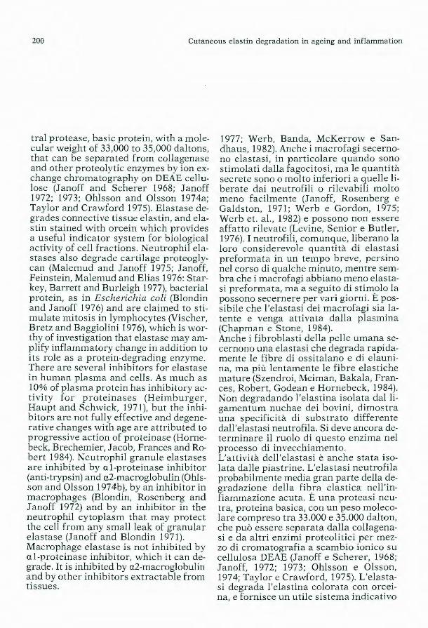
200 Cutaneous elastin degradation in ageing and inflammation
tral protease, basic protein, with a molecular weight of 33,000 to 35,000 daltons, that can be separateci from collagenase and other proteolytic enzymes by ion exchange chromatography on DEAE cellulose (Janoff and Scherer 1968; Janoff 1972; 1973; Ohlsson and Olsson 1974a; Taylor and Crawford 1975). Elastase degrades connective tissue elastin, and elastin stained with orcein which provides a useful indicator system for biologica! activity of cell fractions. Neutrophil elastases also degrade cartilage proteoglycan (Malemud and Janoff 1975; Janoff, Feinstein, Malemud and Elias 1976: Starkey, Barrett and Burleigh 1977), bacterial protein, as in Escherichia coli (Blondin and Janoff 1976) andare claimed to stimulate mitosis in lymphocytes (Vischer, Bretz and Baggiolini 1976), which is worthy of investigation that elastase may amplify inflammatory change in addition to its role as a protein-degrading enzyme. There are severa! inhibitors for elastase in human plasma and cells. As much as 10% of plasma protein has inhibitory activi ty for proteinases (Heimburger, Haupt and Schwick, 1971), but the inhibitors are not fully effective and degenerative changes with age are attributed to progressive action of proteinase (Hornebeck, Brechemier, Jacob, Frances and Robert 1984). Neutrophil granule elastases are inhibited by al-proteinase inhibitor (anti-trypsin) and a2-macroglobulin (Ohlsson and Olsson 1974b), by an inhibitor in macrophages (Blondin, Rosenberg and J anoff 1972) and by an inhibitor in the neutrophil cytoplasm that may protect the cell from any small leak of granular elastase (Janoff and Blondin 1971). Macrophage elastase is not inhibited by al-proteinase inhibitor, which it candegrade. It is inhibited by a2-macroglobulin and by other inhibitors extractable from tissues.
1977; Werb, Banda, McKerrow e Sandhaus, 1982). Anche i macrofagi secernono elastasi, in particolare quando sono stimolati dalla fagocitosi, ma le quantità secrete sono o molto inferiori a quelle liberate dai neutrofili o ri levabili molto meno facilmente (Janoff, Rosenberg e Galdston, 1971; Werb e Gordon, 1975; Werb et. al., 1982) e possono non essere affatto rilevate (Levine, Senior e Butler, 1976). I neutrofili, comunque, liberano la loro considerevole quantità di elastasi preformata in un tempo breve, persino nel corso di qualche minuto, mentre sembra che i macrofagi abbiano meno elastasi preformata, ma a seguito di stim_olo la possono secernere per vari giorni. E possibile che l'elastasi dei macrofagi sia latente e venga attivata dalla plasmina (Chapman e Stone, 1984). Anche i fibroblasti della pelle umana secernono una elastasi che degrada rapidamente le fibre di ossitalano e di elaunina, ma più lentamente le fibre elastiche mature (Szendroi, Meiman, Bakala, Frances, Robert, Godean e Hornebeck, 1984) . Non degradando l'elastina isolata dal ligamentum nuchae dei bovini, dimostra una specificità di substrato differente dall'elastasi neutrofila. Si deve ancora determinare il ruolo di questo enzima nel processo di invecchiamento. L'attività dell'elastasi è anche stata isolata dalle piastrine. L'elastasi neutrofila probabilmente media gran parte della degradazione della fibra elastica nell'infiammazione acuta. È una proteasi neutra, proteina basica, con un peso molecolare compreso tra 33.000 e 35.000 dal ton, che può essere separata dalla collagenasi e da altri enzimi proteolitici per mezzo di cromatografia a scambio ionico su cellulosa DEAE (Janoff e Scherer, 1968; Janoff, 1972; 1973; Ohlsson e Olsson, 1974; Taylor e Crawford, 1975). L'elastasi degrada l'elastina colorata con orceina, e fornisce un utile sistema indicativo
W.E. Parish
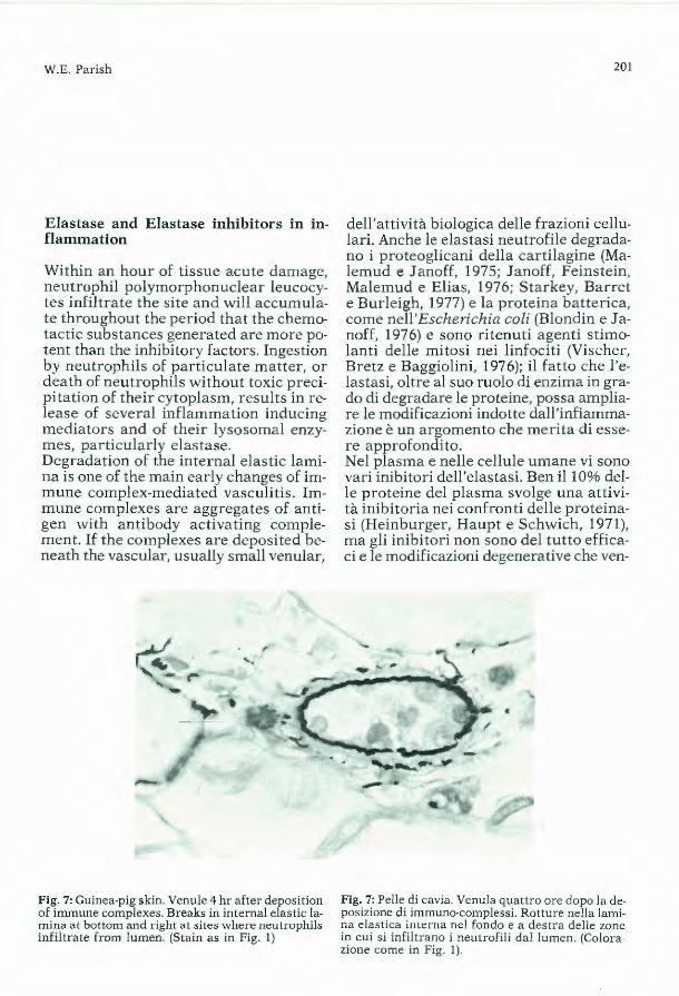
Elastase and Elastase inhibitors in inflammation
Within an hour of tissue acute damage, neutrophil polymorphonuclear leucocytes infiltrate the site and will accumulate throughout the period that the chemotactic substances generated are more potent than the inhibitory factors. Ingestion by neutrophils of particulate matter, or death of neutrophils without toxic precipitation of their cytoplasm, results in release of severa! inflammation inducing mediators and of their lysosomal enzymes, particularly elastase. Degradation of the internal elastic lamina is one of the main early changes of immune complex-mediated vasculitis. Immune complexes are aggregates of antigen with antibody activating complement. If the complexes are deposited beneath the vascular, usually small venular,
Fig . 7: Guinea-pig skin. Venule 4 hr after deposition of immune complexes. Breaks in internal elastic lamina at bottorn and right at sites wheno rn::ulrophils infiltrate from lumen . (Stain as in Fig. 1)
201
dell'attività biologica delle frazioni cellulari. Anche le elastasi neutrofile degradano i proteoglicani della cartilagine (Malemud e J anoff, 197 5; J anoff, Feinstein, Malemud e Elias, 1976; Starkey, Barret e Burleigh, 1977) e la proteina batterica, come nell'Escherichia coli (Blondin e Janoff, 1976) e sono ritenuti agenti stimolanti delle mitosi nei linfociti (Vischer, Bretz e Baggiolini, 1976); il fatto che l'elastasi, oltre al suo ruolo di enzima in grado di degradare le proteine, possa ampliare le modificazioni indotte dall 'infiammazione è un argomento che merita di essere approfondito. Nel plasma e nelle cellule umane vi sono vari inibitori dell'elastasi. Ben il 10% delle proteine del plasma svolge una attività inibitoria nei confronti delle p roteinasi (Heinburger, Haupt e Schwich, 1971), ma gli inibitori non sono del tutto efficaci e le modificazioni degenerative che ven-
Fig. 7: Pelle di cavia. Venula quattro ore dopo la deposizione di immuno-complessi. Rotture nella lamina clastica interna nel fondo e a dest ra delle zone in cui si infiltrano i neu trofili dal lumen. (Colorazione come in Fig. 1).
202 Cutaneous elastin degradation in ageing and inflammation
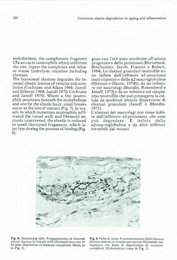
endothelium, the complement fragment CSa attracts neutrophils which infiltrate the site, ingest the complexes and release tissue hydrolytic enzymes including elas tase. The lysosomal elastase degrades the internal elastic lamina of venules and a r terioles (Cochrane and Aiken 1966; J anoff and Scherer 1968; J anoff 1970; Cochrane and J anoff 197 4). Where a few neutrophils penetrate beneath the endothelium and overlie the elastic layer, small breaks occur at the site of con tact (Fig. 7). In vessels in which numerous neutrophils infiltrateci the vessel wall, and fibrinoid necrosis supervened, the elas tin is reduced to small disrupted fragments, which later lyse during the process of healing (Fig. 8).
gono con l'età sono attribuite all'azione progressiva della proteinasi (Hornebeck, Brechemier, Jacob, Frances e Robert, 1984). Le elastasi granulari neutrofile sono inibite dall'inibitore a.1 -proteinasi (anti-tripsina) e dalla a.2-macroglobulina (Ohlsson e Olsson, 1974b), da un inibitore nei macrofagi (Blondin, Rosemberd e Janoff, 1972) e da un inibitore nel citoplasma neutrofilo che può proteggere la cellula da qualsiasi piccola dispersione di elastasi granulare (Janoff e Blondin, 1971 ). L'elastasi dei macrofagi non viene inibita dall' inibitore al-proteinasi, che essa può degradare. È inibita dalla a2-macroglobulina e da altri inibitori estraibili dai tessuti.
.. ~"2~ ... •
~
- -·---...... :: ·'
.,__ ~
~ - .,.-"' fl"' ... _ Fig. 8: Guinea-pig skin. Fragmentation of internal elastic la111im1 in venule with fibrinold necrosis 24 hr after deposition of immune complexes. (Stain as in Fig. I)
Flg. 8: Pelle di cavia. Frammentazione della lamina elastica interna in venula con necrosi fibrinoide ventiquattro ore dopo la deposizione di immunocomplessi. (Colorazione come in Fig. I).
W.E. Parish
Neutrophil proteinases degrade both the elastic lamina and basement membrane enabling more infiamma tory cells to emigrate, with further tissue damage while the complexes and tissue debris persist. In experimental lesions resolution starts as soon as the immune complexes are removed. In spontaneous vasculi6s in man, even when initiated by immune complexes, several phenomena perpetuate the tissue damage resulting in chronic inflammation (Parish 1977; 1980). In studies on vasculitis some aspects of elastin degradation and elastase inhibitors were examined. 1. Fragmentation of elastin in the blood vessel (venular) wall as a feature to score the severity of vasculitis. 2. The indistinct loose wefts of fibres and diffuse histological staining for elastin in chronic vasculitis as evidence of imbalance between hyperplasia and degradation
-" /
' .,
,•
' • - ..
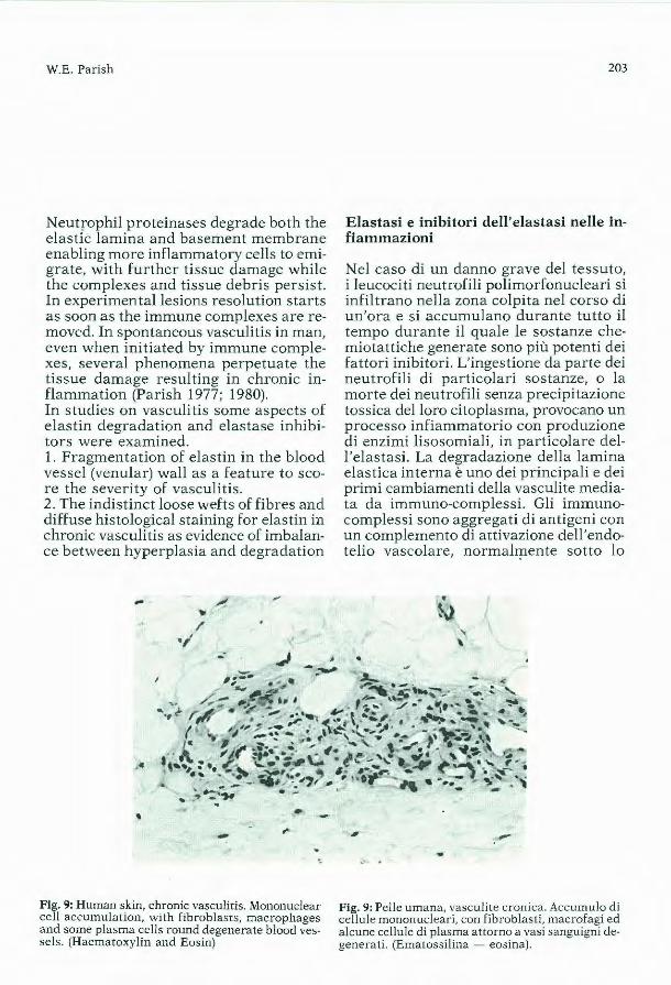
Fig. 9: Human skin, chronic vasculitis. Mononuclear cell accumulation, with fibroblasts, macrophages and some plasma cells round degenerate blood vessels. (Haematoxylin and Eosin)
203
Elastasi e inibitori dell' elastasi nelle infiammazioni
Nel caso di un danno grave del tessuto, i leucociti neutrofili polimorfonucleari si infiltrano nella zona colpita nel corso di un'ora e si accumulano durante tutto il tempo durante il quale le sostanze chemiotattiche generate sono più potenti dei fattori inibitori. L'ingestione da parte dei neutrofili di particolari sostanze, o la morte dei neutrofili senza precipitazione tossica del loro citoplasma, provocano un processo infiammatorio con produzione di enzimi lisosomiali, in part icolare dell'elastasi. La degradazione della lamina elastica interna è uno dei principali e dei primi cambiamenti della vasculite mediata da immuno-complessi. Gli immunocomplessi sono aggregati di antigeni con un complemento di attivazione dell'endotelio vascolare, normahp.ente sotto lo
' '
-Fig. 9: Pelle umana, vasculite cronica. Accumulo d i cellule mononucleari, con fibroblasti, macrofagi ed alcune cellule di plasma attorno a vasi sanguigni degenerati. (Ematossilina - eosina).
204 Cutaneous elastin degradation in ageing and inflammation
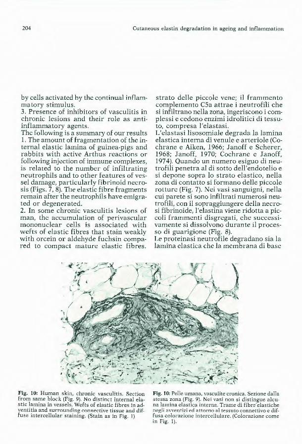
by cells activated by the continua! inflammatory stimulus. 3. Presence of inhibitors of vasculitis in chronic lesions and their role as antiinflammatory agents. The following is a summary of our results 1. The amount of fragmentation of the internal elastic lamina of guinea-pigs and rabbits with active Arthus reactions or following injection of immune complexes, is relateci to the number of infiltrating neutrophils and to other features of vessel damage, particularly fibrinoid necrosis (Figs. 7, 8). The elastic fibre fragments remain after the neutrophils have emigrateci or degenerateci. 2. In some chronic vasculitis lesions of man, the accumulation of perivascular mononuclear cells is associateci with wefts of e lastic fibres that stain weakly with orcein or aldehyde fuchsin compared to compact mature elastic fibres.
Fig. 10: Human skin, chronic vasculitis. Section from same block (Fig. 9). No d istinct internal e]astic lamina in vessels. Wefts of elastic fibres in adventitia and surrounding connective tissue and diffuse intercellular staining. (Stain as in Fig. 1)
strato delle piccole vene; il frammento complemento CSa attrae i neutrofili che si infiltrano nella zona, ingeriscono i complessi e cedono enzimi idrolitici di tessuto, compresa l'elast asi. L'elastasi lisosomiale degrada la lamina elastica interna di venule e arteriole (Cochrane e Aiken, 1966; J anoff e Scherer, 1968; Janoff, 1970; Cochrane e Janoff, 1974). Quando un numero esiguo di neutrofili penetra al di sotto dell'endotelio e si depone sopra lo strato elastico, nella zona di contatto si formano delle piccole rotture (Fig. 7). Nei vasi sanguigni, nella cui parete si sono infiltrati numerosi neutrofili, con il sopraggiungere della necrosi fibrinoide, l'elastina viene ridotta a piccoli frammenti disgregati, che successivamente si dissolvono durante il processo di guarigione (Fig. 8). Le proteinasi neutrofile degradano sia la lamina elastica che la membrana di base
Fig. 10: Pelle umana, vasculite cronica. Sezione dalla stessa zona (Fig. 9). Nei vasi non si distingue alcuna lamina elastica interna. Trame di fibre' elast iche n<>gli avventizi ed attorno al tessuto connet tivo e diffusa colorazione intercellulare. (Colorazione come in Fig. 1).
W.E. Parish
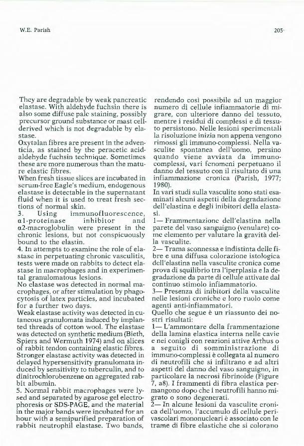
They are degradable by weak pancreatic elastase. With a ldehyde fuchsin there is also some diffuse pale staining, possibly precursor ground substance or mast cellderived which is not degradable by elastase. Oxyta lan fibres are present in the advent icia, as stained by the peracetic acida ldehyde fuchsin technique. Sometimes these are more numerous than the mature e lastic fibres. When fresh tissue slices are incubated in serum-free Eagle's medium, endogenous elastase is detectable in the supernatant fluid when it is used to treat fresh sections of nonTial skin. 3 . Using immunofluorescence, al-proteina se inhibitor and a2-macroglobulin were present in the chronic les ions, but not conspicuously bound to the elastin. 4. In attempts to examine the role of elastase in perpetuating chronic vasculiti s, tests were made on rabbits to detect elastase in macrophages and in experimental granulomatous lesions. No elastase was detected in norma! macrophages, or after stimula tion by phagocytosis of la tex particles, and incubated for a further two days. Weak elastase activity was detected in cutaneous granulomata induced by implanted threads of cotton wool. The elastase was detected on synthetic medium (Bieth, Spiers and Wermuth 1974) and on slices of rabbit tendon containing elastic fibres. Stronger elastase activity was detected in delayed hypersensit ivi ty granulomata induced by sensitivity to tuberculin, and to dinitrochlorobenzene on aggregated rabbit albumin. 5. Normal rabbi t macrophages were lysed and separated by agarose gel electrophoresis or SDS-PAGE, and the ma teria! in the major bands were incuba ted for an hour with a semipur ified preparation of rabbit neutrophil elastase . Two bands,
205 ·
rendendo così possibile ad un maggior numero di cellule infiammatorie di migrare, con ulteriore danno del tessuto, mentre i residui di complessi e di tessuto persistono. Nelle lesioni sperimentali la risoluzione inizia non appena vengono rimossi gli immuno-compless i. Nella vasculi te spontanea de ll 'uomo, persino quando viene avviata da immunocomplessi, vari fenomeni perpetuano il danno del tessuto con il r isultato di una infiammazione cronica (Parish, 1977; 1980). In vari studi sulla vasculite sono stati esaminati alcuni aspetti della degradazione dell'elastina e degli inibitori della elastasi. 1- Frammentazione dell'elas tina nella parete del vaso sanguigno (venulare) come elemento per valutare la gravità della vasculi te. 2- Trama sconnessa e indistinta delle fibre e una diffusa colorazione istologica dell'elastina nella vasculite cronica come prova di squilibrio t ra l'iperplasia e la degradazione da parte di cellule a ttivate dal continuo stimolo infiammatorio. 3- Presenza d i inibitori della vasculite nelle lesioni croniche e loro ruolo come agenti anti-infiammatori. Quello che segue è un riassunto dei nostri risultati: 1- L'ammontare della frammentazione della lamina elastica interna nelle cavie e nei conigli con reazioni attive Arthus o a seguito di somministrazio n e di immuno-complessi è collegata al numero di neutrofili che s i infiltrano e ad altri aspetti del danno del vaso sanguigno, in particolare la necrosi fibrinoide (Figure 7, a8). I frammenti di fibra elastica permangono dopo che i neutrofili hanno migrato o sono degenerati. 2- In alcune lesioni da vasculite cronica dell'uomo, l'accumulo di cellule perivascolari mononucleari è associato con le trame di fib re elastiche che si colorano
206 Cutaneous elastin degradat ion in ageing and inflammation
which did not contain proteolytic activity far casein, were found which reduced the neutrophil elastase potency. These findings are interpreted to indicate that in acute immune-complex mediated damage, elastic fibre fragmentation is almost entirely due to the neutrophils, though other cells participate in later dissolution during resolution. In chronic vasculitis lesions there is an indication of an unstable balance between increased synthesis of elastin, and degradation by elastases. The mononuclear cells in the lesion are continually stimulated by the inflammatory process. It is likely that fibroblasts and smooth muscle cells synthesise the elastin fibres; at the same time elastases are secreted which degrade the new elastin/fibres. The inhibitors in serum, and probably in the macrophages, do not fully control the activity of the elastases.
Acknowledgements
I thank Professor E. Wilson Jones of St. John's Hospital, London for the human skin preparations used as illustrations. I am grateful to Mr. M.J . Cooke and to Mr. A. Shaw respectively for the high standard of his tological preparation and photography.
debolmente con l'orceina o la aldeidefucsina, diversamente dalle fibre elastiche mature compatte. Esse possono essere degradate da elastasi pancreatica debole. Con la fucsin-aldeide si ha anche una certa colorazione diffusa e pallida, provocata probabilmente da una sostanza precursore o derivata da mast cellula non degradabile da parte dell 'elastasi. Le fibre di ossitalano sono p resenti negli avventizi, quando si applica la tecnica di colorazione con acido peracetico e d fucsin-aldeide. Talvolta queste sono p iù numerose delle fibre elastiche mature. Quando sezioni di tessuto fresco vengono incubate nel mezzo di Eagle privo di siero, l'elastasi endogena può essere identificata nel supernatante, quando questo viene usato per trattare sezioni fresche di pelle normale. 3- Utilizzando l' imunofluorescenza, l'inibitore della al-prote inas i e l a a2-macroglobulina risultavano presenti nelle lesioni croniche, ma non lega t i in modo significativo all'elastina. 4- Nel tentativo di esaminare il ruolo dell'elastasi nella va'sculite cronica, sono stati condotti esami sui conigli per identificare l'elastasi nei macrofagi e nelle lesioni sperimentali granulomatose. Non è stata rilevata elastasi nei macrofagi normali o dopo stimolo con fagocitosi di particelle latex e dopo incubazione per altri due giorni. È stata rilevata una debole attività dell 'elastasi nei granulomi cutanei indotti per impianto di filamenti di ovatta. L'elastasi è stata identificata nel mezzo sintetico (Bieth, Spiers e Wermuth, 1974) e su sezioni di tendine di coniglio contenen ti fibre elastiche. Una più forte attività dell'elastasi è stata rilevata nei granulomi a ipersensibilità r itardata, indotti dalla sensibilità alla tubercolina, e al dinitroclorobenzene su albumina di coniglio aggregata. 5- Macrofagi normali di coniglio sono

W.E. Parish 207
stati dissolti e separati per mezzo di elettroforesi con agarose gel o di SDS-PAGE, e il materiale contenuto nelle bande principali è stato incubato per un'ora in un preparato semipurificato di elastasi neutrofila di coniglio. Sono state rilevate due bande, che non contenevano attivi tà proteolitica per la caseina, le quali r iducevano la potenza dell'elastasi neutrofila. Queste rivelazioni vengono interpretate come indicazioni del fatto che, nel danno acuto mediato da immuno-complesso, la frammentazione della fibra elastica è quasi interamente dovuta ai neutrofili, sebbene altre cellule partecipino alla successiva dissoluzione durante la risoluzione. Nelle lesioni da vasculite cronica vi è una indicazione di equilibrio instabile tra un'aumentata sintesi dell'elast ina e la degradazione da parte delle elastasi. Nella lesione, le cellule mononucleari sono continuamente stimolate dal processo infiammatorio. È probabile che i fibroblasti e le cellule dì muscolo sintetizzino le fibre di elastina; nello stesso tempo vengono secrete le elastasi che degradano le nuove fibre di elastina. Gli inibi tor i presenti nel siero, e probabilmente nei macrofagi, non controllano completamente l'attività delle elastasi.
Ringraziamenti
Ringrazio il Prof. E. Wilson Jones del St. John Hospital, Londra, per aver fornito i preparati d i pelle umana usati come illustrazioni. Desidero esprimere la mia gratitudine ad M.J. Cooke e A. Shaw rispettivamente per l'alto livello delle preparazioni istologiche e del materiale fotografico fornito.
208 Cu taneous elastin degradation in ageing and inflammat ion
REFERENCES
1. Abraham PA, Hart KL, Winge R. Carnes WH (1977) Biosyn thesis of e lastin by an aortic media] celi cul ture. Adv. Exp. Med. Biol. 79: 397-4 11
2. Bieth J, Spiers B, Wermuth CG (1974) The synthesis and analytical use of a highly sensitive and convenient substrate of elastase. Biochem. Med. li : 350-357
3. Blondin J , Rosenberg R, Janoff A (1972) An inhibitor in human lung macrophages active against human neutrophil elastase. Amer. Rev. of Resp. Disease 106: 477-479
4. Blondin J , Janoff A (1976) The role of lysosomal elastase in the digestion of Escherichia coli proteins by human polymorphonuclear leukocytes. Experiments with living leukocytes. J. clin. Invest. 58: 971-979
5. Braverman IM, Fonferko E (1982) Stu dies in cutaneous ageing: 1. The elastic fibe r network. J. invest De rm. 78: 434-443
6. Burke JM, Ross R (1979) Synthes is of connective tissue macromolecules by smooth muscle. Int Rev. Connect Tissue Res. 8: 119-157
7. Carnes WH, Abraham PA, Buonasslsl V (1979), Biosynthesis of elasti n by an endothelial celi culture. Biochem Biophys. Res. Comm. 90: 1393-1398
8. Chapman JA, Stone OL (1984) Co-operation between plasmin and elastase in elastin degradation by intact murine macrophages. Biochem. J 222: 721 -728
9. Cochrane CG, Aiken BS (1966) Polymorphonuclear leukocytes in immunologie reactions. The destruction of vascular basement membrane in vivo and in vitro. J. exp. Med. 124: 733-752
10. Cochrane CG, Janoff A (1974) The Arthus react ion. A model of neutrophil and complement-mediated njury. In The l nflammatory Process. Eds Zweifach BW, Grant L, McClusky RT. Academic Press, New York/Liondon 2nd edn. Voi. 3, p. 85-162
11. Fisher-Dzoga K, Jones RM, Vesselinovltch D, Wissler RW (1973) Ultras tructural a nd immunohis tochemical studies of pr imary cultu res of aortic mediai cells. Exp. Mo[. Patho/. 18: 162-176
12. Fullmer HM, Lillie RD (1958) The oxytalan fiber: a previously undesc ribed connective tissue fiber. J. Histochem. Cytochem. 6: 425-430
13. Gawlik Z (1965) Morphological and morphochemical propert ies of the elastic system in the motor organ of man. Folia Histochem Cytochem 3: 233-251
14. Hasegawa J (1960) «Oxytalan» fibers of the dermo-epidermal junction. Arch. Dermatol 82: 250-253 15. Heimburger N, Haupt H, Schwick S (1971) Proteinase inhibition in human plasma. In Proceedings
of the lruernational Research Conference on Proteinase inhibition, (eds) Fritz H. Tschesche H. Walter de Gr uyla, New York, p.1-21
16. Hornebeck W, Brechemier, D. Jacob, M.P. Frances, C. Robert L. (1984) On the multiplic ity of cellular e lastases and their inefficient contro! by natural inhibitors. Adv. exp. Med. Biol. 167: 111-119
17. Jones CD, Sear CH, Grant ME (l 980) An ultras tructura l study of f ibroblasts derived from bovine ligarnen th um nuchae and their capacity fo r elastogenesis in culture. J. Path. 131: 35-53
18. Janoff A, Scherer (1968) Mediators of inflammation in leukocyte lysosornes IX. E lastinolytic activity in granules of human polymorphonuclear leukocytes. J. Exp. Med. 128: 11 37-1 15 1
19. Janoff A (1970) Mediators of tissue damage in leukocyte enzymes. X. Furthe r studies on human granulocyte elastase. Lab. !nvest. 22: 228-236
20. Janoff A, Blondin J (1971) Inhibition of the e lastase-like esterase in human leukocyte granules by hurnan leukocyte celi sap. Prue. Sci exp Biol Med 136: I 050· 1053
21. Janoff A, Rosenberg R, Galdston M (1971) Elastase-like , esteroprotease activity in human and rabbit alveola r rnacrophaye granules (35426). Proc. Soc. Exp. Biol. Med. 136: 1054-1 058
22. Janoff A (1972) Human granulocyte elastase. Furthe r delineation of its role in connective tissue darnage. Amer. J. Path. 68: 579
23. Janoff A (1973) Purification of human granulocyte elastase by affinity chromatography. Lab. !nvest. 29: 458-464
24. Janoff A, Blondin J (1973) The effect of hurnan granulocyte elastase on bacterial suspensions. Lab. !nvest. 29: 454-457
25. Janoff A, Feinstein G, Malemud CJ, Elias JM (1976) Degradation of cart ilage proteoglycan by human leukocyte granule neutra! p roteases; A model of joint injury I. Penetrat ion of enzyme into rabbit arti· cular cartilage and release of 35S04-labelled materiai from tissue. J. clin. l nvest. 57: 615-624
26. Jarrett A (1974) The elastic tissue of the dermis. In The Physiology and pathophysiology of the skin. Ed Jarrett A. Acadernic Press London/New York. Vol. 3 p. 847-872
27. Jar rett A, Hardy JA (1968) Acid nucleases and other hyd rolases in human skin w it h special reference to elastic tissue. Histochem J. 1: 18
W.E. Parish 209
28. Johnson BE (1978) Changes in sunburn and mechanisms of protection. J. Soc. Cosmet. Chem. 29: 31-44 29. Jones CD, Sear CH, Grant ME (1980) An ulstrastructural study of fibroblasts derived from bovine li
gamentum nuchae and their capacity for elastogenesis in culture. J. Path. 131: 35-53 30. Kaidbey KH, Kligman AM (1978) Sunburn protection by longwave ul traviolet radiat ion-ind uced p ig
mentation. Arch. Dermatol 114: 46-48 31. Kao W-KK, Gray S, Bressan GM, Prockop DJ (1980) Demonstra t ion by immunofluorescence that the
cells from chick embryo aortas syn thesize both elastin and collagen types I and III. Arte ry 7: 176-190 32. Kligman AM (1969) Early destructive effect of sunlight on human skin. f. Amer. Med. Assn. 210: 2377-2380 33. Kligman AM (1974) Solar elastosis in relation to pigmentation. In Sunlight and man. Eds Pa thak MA,
Harber LC, Seij i M, Kukita A. Univ. of Tokyo Press p. 157-163 34. Leak LV, Burke JF (1968) Ultrastructural studies o n the lymphatic anchoring filament s. f . Celi Biol.
36: 129-149 35. Levine EA, Senior RM, Butler JV (1976) The elastase activity of alveola r macrophages: Measurements
using synthetic substrates and e lastin. Amer. Rev. of Resp. Disease 113: 25-50n 36. Malemud CJ, Janoff A (1975) Human polymo rphonuclear leucocyte elastase and cathe ps in G mediate
the degradation of !apine articular cartilage proteoglycan. Ann N. Y. Acad Sci 256: 254-262 37. Mencham RP, Lange G, Maderas J , Starcher B (1981) Elastin synthesis by ligamentum nuchae fib ro
blasts: Effect of culture conditions and extracellular matrix on elastin production. J Celi Biol. 90: 332-338 38. Montagna W, Carlisle K (1979) Structural changes in ageing huma n skin. J. Invest Denn . 73: 47-53 39. Mortimer PS, Cherry GW, Jones RL, Barnhill RL, Ryan TS (1983) The importance of elast ic fib res
in skin lymphatics. Br. J. Derm. 108: 561-566 40. Nakamura K, Johnson WC (1968) Ultraviolet light induced connective tissue changes in rat skin: a
histopathologic and histochemical study. J. invest. Derm. 51: 253-258 41. Ohlsson K, Olsson I (1974a) The neutra! proteases of human granulocytes. Isola t ion and partial cha
racterisation of granulocyte elastases. Europ. f. Biochem. 42: 519-527 42. Ohlsson K, Olsson I (1974b) Neutra! proteases of human g ranulocytes III Interac tion between granu
locyte elastase and plasma protease inhibitors. Scand J. clin Lab. Invest. 34: 349-355 43. Parish WE (1977) Features of human spontaneous vasculitis reproduced experimentally in animals.
Effects of antiglobulins, C-reactive protein and fibrin. Bayer-Symposium VI. Experimental models of chronic inflammatory diseases. Springer-Verlag, New York/Berlin p. 117-151
44. Parish WE (1980) St imuli perpetuating chronic vasculitis. In Vasculitis. Eds Wolff K, Winkelman RK. Lloyd-Luke, London p. 159-172
45. Pierce JA, Bernick H, Henry PH (1967) Collagen and elastin metabolism in the lungs skin and bo nes of adult rats. ]. Lab clin Med. 69: 485-493
46. Quintarelli G, Starcher BC, Vacaturo A, DiGianfilippo FD, Gotte L, Mecham RP (1979) F ib rogencsis and biosynthesis of elastin in cart ilage. Connect. Tissue Res. 7: 1-8
47. Ross R (1971) The smooth muscle celi II Growth of smooth muscle in culture and fo rmation of elast ic fib res. J Celi Biol. 50: 172-186
48. Sams WM, Smith JG, Burk PG (1964) The experimental production of elastosis with ult ra v iolet light. J. invest. Derm. 43: 467-47 1
49. Sandberg LB (1976) Elastin structure in health and disease. Int Rev. Conn Tiss. Res 7: 159-21 0 50. Szendroi M, Meimon G, Bakala H, Frances C, Robert L, Godeau G, Hornebeck W. (1984) On the pre
sence of a metalloprotease in human skin fibroblasts that degrades the human skin elastic fiber sys tem. J Invest. Derm. 83: 224-229
51. Smith JG, Davidson EA, Sams WM, Clark RD (1962) Alterations in human derma! connective ti ssue with age and ch ronic sun damage. f. invest. Derm 39: 347-350
52. Smith JG, Davidson EA, Clark RD (1962) Derma! elastin in actinic elastosis and pseudoxanthoma elasticum. Nature 195: 716
53. Smith LT, Holbrook KA, Byers PH (1982) Structure of the derma! matrix during development and in the adult. J. invest. Derm. 79: 93s-I04s
54. Starkey PM, Barrett AJ, Burleigh MC (1977) The degradation of articular collagen by neut roph il proteinases. Biochim. Biophys. Acta 483: 386-397
55. Szendroi M, Meimon G, Bakala H, Frances C, Robert L, Godeau G, Hornebeck W. (1984) On the presence of metalluprotease in human skin fibroblasts that degrades the human skin elastic fiber system. J Invest. Derm. 83: 224-229
56. Taylor JC, Crawford IP (1975) Purifica tion and preliminary characterisation of human leukocyte elastase. Arch. Biochem. Biophys. 169: 91-101
57. Thybet-g J, Hinek A (l 977) Fine structure of rabbit ear chondrocytes in vitro and after autotransplantation Celi Tissue Res. 180: 341-356

210 Cutaneous elastin degradation in ageing and inflammation
58. Uitto J, Hoffmann H·P, Prockop DJ (1976) Synthesis of elastin and procollagen by cells from embryonic aorta. Differences in the role of hydroxyproline and the effects of proline analogs on the secret ion of the two proteins. Arch. Biochem. Biophys. 173: 187-200
59. Vischer TL, Bretz U, Baggiolini M (1976) In vitro stimulation of lymphocytes by neutra! proteinases from human polymorphonuclear leukocyte granules. J. exp. Med. 144: 863-872
60. Walford RL, Carter PK, Schneider RB (1964) Stability of labelled aortic elastic tissue with age and pregnancy in the rat. Arch. Path. 78: 43-45
61. Werb Z, Gordon S (1975) Elastase secretion by stimulated macrophages. J. Exp. Med. 142: 361-377 62. Werb Z, Banda MJ, McKerrow JH, Sandhaus RA (1982) Elastases and elastin degradation. J. invest.
Derm. 79: 154s-159s 63. Willis I, Kligman AM (1969) Evaluation of sunscreens by human assay. J. Soc. Cosmet. Chem. 20: 639-651

f. Appl. Cosmetol., 3, 211-222 (July/September 1985)
Essential Fatty Acids and skin aging P. MORGANTI 1, S.D. RANDAZZ02
I University School far Cosmetologists Catholic University of Rome - Via F. Bernardini, 22 - 00 165 Roma 2 Department of Experimental Dermatology University of Catania
Received: March 9, 1985. P1·esented at the /111ema1ional Meeting: A New Look at Old Skin: A Cha//a11ge 10 Cosmetology, March 7-9, 1985 Rome-ltaly.
Kcy words: EFA, Skin Anging, Esscntial Fatty Aci<ls and Skin, EFA Protective Aclivity, Moisturizing Activity, Single-Dose Cream.
Synopsis
The agenls that bring about aging of human skin are the changes and alteretions produced by UV rays and those induced by biologica I and tempora! factors. These modifications are characterized by the thinning of the epidermis and a diminution of sebum production with consequent qualitative and quantitative insufficiency of the superficial hydrolipidic film. To evaluate the possible act ivity of essential fatty acids applied in a topica( mixture incOI-porated in a cosmetic vehicle, in vivo testing was performed to estimate the water-binding capacity of aged human skin treated with the cosmetic under examination. Moreover, il was assessed the protective action of this special cosmetic against damages in<luced, at the leve! of skin surface markings, by cor tisone-like compounds, which are well known skin aging agents. The water binding capacity of horny layer was evaluate<l by the Corneometer CM 420, based on dielectric constant measurements. Through the water sorption-desorption test of young and old skin, the hydrating activity of the s ingledose under examination was evaluated. Fit-st results, as well as other data available, suggest that EFA, incorporated in a cosmetic vehicle, are capable of protecting aged skin against environmental insults, when applied topically, thus also improve the skin's hydration capacity.
Riassunto
Come è noto la cute umana invecchia essenzia lmente a causa degli effetti lesivi provocati dai raggi U.V. e per le trasformazioni organiche c funzionali legate alla cronobiologia di ogni singo lo individuo. Queste modificazioni, evidenti soprattutto a livello delle zone cutanee scoperte, s i traducono tra l'a ltro in un assottigliamento dell'epidermide ed in una d iminuita produzione sebacea con insufficienza qualitativa e q uan titativa <lei film idrolipidico di superficie. Durante l 'invecchiamento si osserva inoltre la diminuzione quantitativa degli acidi grassi cutanei, con riduzione della loro incorporazione nei mitocondri delle cellule dello strato hasale, e la progressiva disidratazione sia dell'epidermide che de l derma. Onde valutare l'eventuale attività svo lta dagli aci<li grassi essenziali, applica ti in miscela per uso topico mediante veicolo cosmetico, si è voluto controllare «in vivo» il potere di legare acqua della cute umana senile trattata con il cosmetico in studio. È stata controllata, inoltre, l'azione protettiva svolta da questo speciale cosmetico nei confronti dei danni provocati dai cortisonici, noti invecchiant i cutanei, a livello del reticolo a losanghe che solca la superficie cutanea. Dopo aver determinato il gra<lo <li assorbimentocessione dell'acqua de lla cute giovane e senile per verificare la validità dell 'apparecchio ut il izzato, è stata valutata, «in vivo», l'attività idratante della monodose in esame. Le misurazioni sono state eseguite in doppio ceco su un gruppo di 40 donne volontarie di età compresa tra 30 e 45 anni, dopo tre mesi di utilizzazione del prodotto. L'attività protettiva del prodotto è stata valutata sullo stesso gruppo mediante r ilevazioni fotografiche effettuate direttamente sulla cute e su repliche della stessa. Dai primi dati ottenuti sembra che gli acidi grassi insaturi, inserit i ne lla monodose cosmetica, svolgano un importante ruolo protettivo 111a11tt:nendo e migliorando le strutture biologiche cellu lari della cute umana invecchiata.
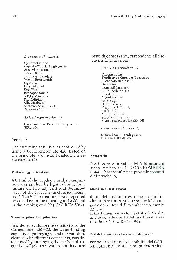
214
Base cream (Produci A)
Cyclomethicone Caprylic/Capric Triglyceride Stearyl Heptanoate Decyl Oleate Isopropyl Lanolate Wheat Bran Lipids Squalene Cetyl Alcohol BeesWax Benzophenone-3 A,E,B6 Vitamins Phosfolipids Alfa-Bi sa bolo! Sorbitan Sesquioleate Ceteareth-20
Active Cream (Produci B)
Base cream + Essential fatty acids (EFA) 3%
Apparatus
The hydrating activity was controlled by using a Corneometer CM 420, based on the principle of constant dielectric measurements (5).
Methodology of treatment
A 0.1 ml of the products under examination was applied by light rubbing for 1 minute on two adjacent and delimited areas of the forearm. Each area measured 2.5 cm2 • The treatment was repeated twice a day: in the morning at 10.00 and in the evening at 6.00 (18°C RH~50%).
Water sorption-desorption test
In arder to evaluate the sensitivity of the Corneometer CM-420, the water-binding capacity of young, aged and normai skin, cleaned with different detergents, was determined by employing the method of Tagami et all (6). The results obtained are
Essential Fatty acids and skin aging
privi di conservanti, rispondenti alle seguenti formulazioni:
Crema Base (Prodotto A)
Ciclometicone Trigliceride Caprilico/Caprinico Eptanoato di stearile Dee il alea to Isopropil Lanolato Lipidi della crusca Squalene Alcool cetilico Cera d'api Benzofenone-3 Vitamina A, E e B6 Fosfolipidi Alfa-Bisabololo Sorbitan sesquioleato Alcool cetilstearilico (20) OE
Crema Attiva (Prodotto B)
Crema base + acidi grassi Essenziali (EFA) 3%
Apparecchi
Per il controllo dell'attività idratante è stato utilizzato il CORNEOMETER CM-420 basato sul principio delle costanti dielettriche (5).
Metodica di trattamento
0,1 ml dei prodotti in esame sono stati frizionati per 1 min. su due superfici contigue e delimitate dell'avambraccio, ampie 2,5 cm2 .
Il trattamento è stato ripetuto due volte al giorno: alle ore 10 del mattino e la sera alle 18 (18°C RH~50%).
Test dell'assorbimento/cessione dell'acqua
Per poter valutare la sensibilità del CORNEOMETER CM 420 è stata determina-

P. Morganti - S.D. Randazzo
shown in Figures 1 and 2. Afterwards, double-blind measurements were carried out on three adjacent areas of the right forearm on a group of 40 volunteer women ranging 35 to 45 years old, by employing a Base cream (Product A) and an Active Cream (Product B) far 30 consecutive days. Neither the operator nor the patients were able to identify the Active Product. The results obtained are shown in Figure 3.
Evaluation of cutaneous hydration on human skin
To forty female volunteers 35 to 45 years old, were given 120 monodoses of the contro! Base cream (Product A) or of the Acti ve Cream (Product B). Neither the operator nor the patients were able to identify the product considered active. The product was applied twice a day, in the morning and in the evening, far two months, from October to January
215
ta la capacità di legare acqua della cute sia giovane che senile, detersa con differenti detergenti, utilizzando la metodica di Tagami e Coli. (6). I risultati ottenu ti sono riportati nelle Figure 1 e 2. Successivamente sono state eseguite, a doppio cieco, misurazioni in tre zone contigue dell'avambraccio destro, su un gruppo di 40 donne volon tarie con età compresa tra 35 e 45 anni, utilizzando la Crema Base (Prodotto A) e la Crema Attiva (Prodotto B) per 30 giorni consecutivi. Nè l'operatore né le pazienti erano in grado di identificare il prodotto attivo. I r isultati ottenuti sono riportati nella Figura 3.
Valutazione dell'idratazione cutanea su cute umana
A 40 soggetti volontari di sesso femminile, di età compresa tra i 35 e 45 anni, sono state consegnate 120 monodosi della
single-dose cosmetic+EFA
single-dose cosmetic
control t = 18'C RH!> 50/.
-oi 120
~ 110
:. hygroscopicity .l. · • • • water holding capacity
CJ
··-r- ..... ... • T
. : li'', • r ........
100 :/ ............ ....., T
." I ?----
... ... ... • •' • • • • T .I
- ; I ------ -
~t:~~! I--..........__ -------~ 80 J .............__!_~~~~~~~~-;/ J. T
J.
t i i i i i i water 10 20 30 40 50 60 70- time(sec)
Fig. 3: Values of sorption-desorption test after application of a single-dose cosmetic cream enriched with EFA (mean for 40 subjects x 30 days).
Fig. 3: Valori del test dell'assorbimento-cessione dopo applicazione di una crema monodose arricchita con EFA (media d i 40 soggetti per 30 giorni).

216
(1984-85). During the 30 days prior to testing, cont ro! measurements were made on a previously delimited area of the right forearm, as described earlier. Following the Mosler method (5) which we modified slightly, the mean of three measurements carried out during daylight hours (at 10.00 a.m., 2.00 p.m. and 6.00 p.m.) was recorded as value of the day. Daily measurements were taken three times per week (Monday, Wednesday, and Friday) up to a tota! of 30 measurements for each subjects tested. The data obtained, considered as base data, are recorded in Figure 4.
90
85
Essential Fatty acids and skin aging
crema di con trollo (Prodotto A) o della crema attiva (Prodotto B). Né l'oper a tore né le pazienti erano in grado di identificare il prodotto considerato attivo. II prodotto è stato applicato due volte al giorno, al mattino e alla sera per due mesi, nel periodo compreso tra ottobre e gennaio (1984-85). Durante i 30 giorni che hanno preceduto le sperimentazioni, sono state eseguite misurazioni di controllo in una zona p recedentemente delimitata dell'avambraccio, come descritto precedentemente nella metodica di trattamento. Seguendo la metodica di Mosler (5), da
I ~ 80 ----------1-----------1 u
5 10 15
Fig. 4: Water binding capacity of aged skin (Mean for 40 subjects x 30 days).
20 25 30 - time(days)
Fig. 4: Capacità di legare acqua della cute senile (media di 40 soggetti per 30 giorni).
____ single -dose cosmetic+ EFA
•. . . . •• single-dose cosmetic
_ control
"' Gl :I 16 > .... Gl
~ ~ o u
105
100
95
90
85
80
RH .. 50
l=1B'C
10 20 30 40 50 60 - time(days)
Fig. 5: Effect of topically applied EFA on moisture holding capacity of horny layer (mean for 40 subjects x 60 days).
Fig. 5: Effetti dell'applicazione topica degli EFA sulla capacità di legare acqua dello strato corneo (media di 40 soggetti per 30 gio rni).

P. Morganti - S.D. Randazzo
Ten days prior to and during the treatment, patients did not use any cosmetic for cleaning their forearm, except a normai cleaning-milk, formulated by us, used in the evenings. During the tested period, after having recorded the base data, measurements were taken three times a week for two consecutive months up to a total of 60 measurements for each subjects. The data obtained are shown in Figure 5.
The Durhing Chamber test
The Durhing Chamber was employed in order to evaluate the possible protective action performed by the test cosmetic against the damages provoked by cortisone-like compounds (4). Clobetasol propionate, a cortisone-like compound, was selected and used for this testing as a model of active substance able to accelerate skin aging process. In order to assess the protective action performed by
Fig. 6: Intact contro! skin surface markings: normai pattern polisulfide rubber replica.
217
noi leggermente modificata, è stata registrata come valore del giorno la media delle 3 misurazioni effettuate durante le ore diurne (rispettivamente alle 1 O, alle 14 ed alle 18). Le misure giornaliere sono state effettuate 3 volte la settimana (lunedì, mercoledì, venerdì) p er un totale di 30 misurazioni per ogni soggetto sottoposto a sperimentazione. I dati ottenuti, considerati dati di base, sono riportati nella Figura 4. Dieci giorni prima e durante il periodo di trattamento, le pazienti non h anno utilizzato alcun cosmetico per detergere il braccio ad eccezione di un normale latte detergente da noi formulato ed adoperato sempre di sera. Iniziata la sperimentazione, dopo aver registrato i dati di base, sono state eseguite le letture 3 volte la settimana per due mesi consecutivi e per un totale d i 60 m isurazioni per soggetto. I dati ottenuti sono riportati nella Figura 5.
Fig. 6: Reticolo cutaneo a losanghe: replica della cute normale.

218
the Active Cream (Product B), we evaluated and codified the type and extent of the destructive action developed on cutaneous skin-markings by the cortisone-like compound employed in occlusive patchtests. Occlusive conditions as well as an exact application of dosed quantities of the preparations can be otbained using the aluminum chamber. Chambers were fixed wi th a micropore to the volar part of the farearm. Products were renewed daily. The testing, which lasted three weeks, was carried out on 30 female volun teers.
Methodology of treatment
The cutaneous area selected far treatmen t was divided into three sub-areas: - the first was massaged daily without applying any product; - the second was massaged far 1 minute using 0.2 ml of Base Cream (Product A); ·
Fig. 7: Marked atrophy-skin surface markings: highly irregular pattern. Polysulfide rubber replica 3 weeks after treatment with Clobetasol propionate cream only.
Essential Fatty acids and skin aging
Test della Camera di Duhring
È stata utilizzata ]a Camera d i Durhing per valutare l'eventuale azione protettiva svolta da] cosmetico in stud io nei confronti dei danni da cortisonici (4). Per questa prova è stato scelto ed uti lizzato il Clobetaso] propionato preso a modello quale sostanza attiva capace di accelerare il processo di invecchiamento cutaneo. Il tipo e la portata dell'azione lesiva, provocata sul reticolo a losanghe della cute del derivato cortisonico utilizzato con Patch-test occlusivi, è stata valutata ecodificata per controllare ]'attività protettiva svolta dalla crema attiva in esame (Prodotto B). La camera di alluminio consente le c0ndizioni occlusive nonché l'esatta applicazione dei preparati in quantità dosata. Le camere sono state fissate con cerotto «micropore» al lato flessorio dell'avambraccio. I prodotti sono stati rinnovati ogni giorno.
Fig. 7: Marcata atrofia del reticolo cutaneo: re ticolo altamente irregolare. Replica cutanea 3 settimane dopo il trattamento con la crema Clobetasolpropionato.
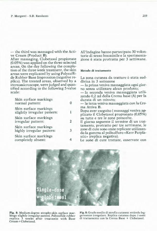
P. Morga nti - S.D. Randazzo
- the third was massaged with the Active Cream (Product B). After massaging, Clobetasol propionate (0.05%) was applied on the three selected areas. On the day following the completion of the three week treatment, the skin areas were replicated by using Polysulfide Rubber Base Impression (negative replica). The treated areas, observed by a stereomicroscope, were judged and quantified according to the following 5-value scale:
Skin surface markings normai patters: O
Skin surface markings slightly irregular pattern: 1
Skin surface markings irregular pattern: 2
Skin surface markings highly irregular pattern: 3
Skin surface markings completely absent: 4
Fig. 8: Med ium-degree atrophy-skin surface markings: slighùy irregular pa ttern. Polysulfide rubber replica 3 weeks afte r treatment with Base Cream+ Clobe tasol.
219
All'indagine hanno partecipato 30 volontarie di sesso femminile e la speriment azione è stata protratta per 3 settimane.
Metodo di trattamento
La zona cutanea da trattare è stata suddivisa in 3 sottozone - la prima veniva massaggiata ogni giorno senza utilizzare alcun prodotto; - la seconda veniva massaggiata utilizando 0,2 ml della Crema base (A) per la durata di un minuto; - la terza veniva massaggiata con la Crema Attiva B. Dopo aver eseguito i massaggi veniva applicato il Clobetasol p ropionato (0,05%) su tutte e tre le zone prescelte. Il giorno seguente il termine di un trattamento, protratto per tre settimane, le zone di cute sono state replicate utilizzando la gomma al poli solfuro «Kerr Perplastic» (replica negativa). Le zone di cute trattate, osservate con
Fig. 8: Grado medio di atrofia cutanea: ret icolo leggermente irregolare. Replica cutanea dopo 3 mesi di trattamento con la Crema Base + Clobetasol.

220
The results obtained are shown in Figures 6, 7, 8, 9 and 10.
Results and comments
As shown in Figure 3, incorporating Essential Fatty Acids into the Base Cream under examination improves considerably (by approximately 20%) the waterbinding capacity of human skin. In fact, the maximum base value expressed as mean value for the subjec1s examined increases from 90 to 11 O CV (Corneometer Values) (P < 0.05) when the EFA-free Cream (Product A) is employed. When the cream is enriched with essential fatty acids (Product B), the base vale of 80 reaches a value of 130 (P < 0.05). Analysis employing the Student-test, shows a highly significant difference between the Base Cream and the Active Cream (P<0.05). Similar results were obtained in longterm experiments. When comparing Figures 4 and 5, one can see that the treatment
Fig. 9: Intact skin-skin surface markings: normai pattern. Polysulfide rubber replica 3 weeks after treatment with Active Cream+ Clobetasol.
Essential Fatty acids and skin aging
uno stereomicroscopio, sono state giudicate e quantizzate secondo la seguente scala di valori:
Cute con reticolo a losanghe: Normale o Cute con reticolo a losanghe: Lievemente scompaginato
Cute con reticolo a losanghe: Scompaginato 2
Cute con reticolo a losanghe: Notevolmente scompaginato 3
Cute con reticolo a losanghe: Completamente assente 4
I risultati ottenuti sono riportati nelle Figure 6, 7, 8, 9 e 10.
Risultati e commenti
Come si può osservare dalla Figura 3, l'aggiunta di Acidi Grassi Essenziali
Fig. 9: Cute con reticolo a losanghe intatto: replica cutanea dopo 3 settimane di trattamento con Crema Attiva + Clobetasol.

P. Morganti · S.D. Randazzo
wi th the Base Cream improved the skin hydration capacity. The degree of hydration is decidedly enhanced when the Base Cream is enriched with essential fatty acids (EFA). A further evidence of the benefits imparted to human skin by these fatty acids, is given in Figures 6, 7, 8, 9 and 10. These figures clea rly show that the application of the cosmetic monodose under examination reduces by 30% the negative action developed by Clobetasol propionate against the skin surface markings. Such protective action is further enhanced by the monodose enriched with EFA. These first results, as well as other data available, suggest that EFA, incorporateci in a cosmetic vehicle, are capable of protecting aged skin against environmental insults, when applied topically, thus also improve the skin's hydration capacity.
221
(EF À) alla crema base in studio migliora , notevolmente (20% ca) la capacità d i le
gare acqua della cute umana. Infatti il valore massimo di base espresso come media dei soggetti esaminati passa da 90 a 110 CV (valori corneometrici) (P < 0,05), quando venga u tilizzata la crema priva di acidi grassi essenziali (Prodotto A). Quando si arricchisce la crema con acidi grassi essenziali (Prodotto B), si passa da un valore base di 80 ad un va lore di 130 (P < 0,05). Con il t di Student la differenza ottenuta tra la brema base e la crema attiva è risultata altamente significativa (P < 0,05). Analoghi r isultati sono stati ottenuti con lo studio a lungo termine. Se si confronta la Figura 4 con la Figura 5, si può osservare come il trattamento con la Crema Base migliora sensibilmente il grado di idratazione cu tanea. Il grado di idratazione migliora in mo~o ancora più
C::J single-dose cosmetic + EFA
r:::::::J single-dose cosmetic
4
3
c:::::J clobetasol propionate 0,05 Y.
n=30
Fig. 10: Evaluation of skin surface markings. Fig. 10: Valutazione del reticolo cutaneo a losanghe.

222
REFERENCES
Essential Fatty acids and skin aging
netto quando si arricchisce la crema base con gli acidi grassi essenziali. Una ulteriore verifica dei benefici apportati da questi acidi grassi nei confronti della cute umana la possiamo osservare dalle Figure, 6, 7, 8, 9 e 10. Si nota chiaramente come l'applicazione della monodose cosmetica in studio riduca del 30% circa l'azione negat iva svolta dal Clobetasol propionato nei confronti del reticolo a losanghe. Tale azione protettiva aumenta ulteriormente e in modo netto quando arricchiamo la monodose con acidi grassi essenziali. Da questi primi dati e da altri dati in nostro possesso sembra si possa desumere come gli acidi grassi essenziali siano in grado di proteggere la cute senile dalle aggressioni esterne, migliorandone anche il grado di idratazione, se u tilizzat i per uso topico ed inseriti in un veicolo cosmetico adeguatamente protetto dai fenomeni ossidativi.
1) R. Jackson (1972) «Solar and senile skin: changes caused by aging and habitual exposure to the sun» 27, 106.
2) B.A. Gilchrest (1981) «A quantitative approach to measuring actinic aging in human skin» J. soc. Cosmet. Chem. 32, 153.
3) Prottey (1976) »Essential fatty acids and the skin» Br. J. Dermatol. 94, 579. 4) Frosch P.J., Wendt H. and Kligman A.M. (1980) «The durhing chamber test far assaying corticoste
roid atrophy in humans» in: Mauvais-Jarvis P, Vickers CFH and Wepierre J «Percutaneous abso rption of steroids, p. 185-198, London Academic Press;
5) K. Mosler (1983) «Haufteuchtigkeitsmessung kein problem mit dem Cornometer CM-420» Parf. und Kosmetik 64, 375.
6) H. Tagami, Y. Kanamaru, K. Inoue, S. Svehisa, F. Inove, K. Iwatsuki, K. Yoshikum e M. Yamada (19!!2) «Water sorption-desorption test of the skin «in vivo» for functional assessment of the stratum corneum» J. Invest. Dermatol. 78, 425.

J. Appl. Cosmetol., 3, 223-236 (July/September 1985)
Contact Dermatitis Due to Cosmetics G. ANGELINI. G.A. VENA, G. GIGLIO, F. FIORDALISI , C.L. MENEGHINI Clinica Dermatologica dell'Università di Bari
Received: fune 11, 1985
Key words: Contact Allergy, Hair Cosmetics, Eyelid Cosmetics. Essen tial Oils. Guaiazulene
Synopsis
Even though cosmetics are widely used, they do no t frequently cause serious side effects. Recently, however, the incidence of documented dermatitis due to cosmetics has become higher due to the considerable increase in the use of cosmetics of every type. A better understanding of the responsible allergens, particularly regarding perfume ingredients, has also facilitated detection. This paper deals with contact dermatitis due to hair, eyelid, face and lip cosmetic products. Scalp reactions to hair products are relatively ra re and mainly due to ha ir dyes. Eye make-up intolerance seems more frequent, and is caused by the components of eyeshadows and mascara. Contact allergy of the face is mainly related to certain components of perfumes, especially oak moss and cinnamaldehyde. Allergie contact cheilitis is rarely induced by lipstik, but more frequently by components of toothpastes, mouthwashes and dental preparations.
Introduction
Cosmetics, though in daily use b y enormous numbers of individuals, do not produce untoward effects on a large scale. This is confirmed by the recent, though scarce, data on the incidence of cosmetics-induced dermatitis in the papula ti on a t large. Skog ( 17) recorded 41 cases of cosmetics-induced dermatitis in
Riassunto
I cosmetic i, sebbene usati giornalmente da un gran numero d i individui, non producono effett i indesiderati su larga sca la. Negli ultimi anni, tuttavia, l'incidenza di allergia da contatto con cosmetici si è fatta più elevata per l'enorme aumen to di consumo dei cosmetici di ogni tipo e la gradua le più approfondita conoscenza dei singoli componenti degli s tessi, specie per quanto r iguarda le numerose essenze profumate. Saranno qui prese in cons iderazione in particolare le dermatiti da contatto con cosmet ici per capelli, per le palpebre, per il viso e per le labbra. L'incidenza di alle rgia da contatto del cuoio capelluto da cosmetici per capelli è piu ttosto bassa ed appare legata nella gran parte dei casi a coloranti. Più frequente sembra invece la dermat ite da contatto delle palpebre per l'uso soprattutto di ombretti e mascara. Le essenze profumate sono particolarmente in causa nella dermatit e allergica da contatto del viso, legata in gran parte a l muschio di guercia ed alla cinnamaldeide. La cheilite allergica da contatto risulta oggi raramente legata ai rossetti, più spesso ai dentifrici, collutori e preparati denta ri.
Introduzione
I cosmetici, sebbene u sati giornalmente da un gran numero di individui, non producono effetti indesiderati su larga scala. Lo confermano anche gli scarsi recenti dati sull'incidenza delle derma t it i d a cosmetici nella popolazione in genere. Skog (17) ha osservato 41 casi di dermatiti da
224
a total of 70, 126 dermatological patients, corresponding roughly to a population of 250,000. The North American Contact Dermatiti s Group (5) found that of a total of 8093 patients with contact dermatitis, cosmetics-induced disease accounted for 487 cases, with 80% of these showing evidence of a llergie pathogenesis. An appreciable increase in the incidence of dermatitis induced by perfume essences documented in recent years has been a result more of the increased knowledge of individu al ingredients of cosmetics, especially as regards the essential oils (8; 10; 16), than of the increased use of cosmetics of a ll types. Diagnostic possibilities in cosmetics-induced dermatitis have been considerably increased by the use of specific allergens of perfume essences in allergological studies . As far back as 1968, tests for allergy to de tergent and soap perfumes carried out in 1943 consecutive patients with contact dermatitis proved positive in 78 cases (approx. 4 %), especially to benzyl salicylate (15). Even higher figures but only to certain perfume constituents were reported more recently (2; 11) especially after Larsen (10), collaborating with a firm producing perfume essences, succeeded in isolating a group of allergens, almost all in a chromatographically pure form. These agents, very useful for etiological diagnosis by patch testing, are as follows: amyl cinnamic alcohol, cinnamic alcohol, cinnamic aldehyde, isoeugenol, geranio!, h ydroxycitronellal, oakmoss essence. In this paper we present our own data on certain forms of cosmetics-induced contact dermatitis, as follows: 1. Contact dermatitis induced by hair co
smetic products. 2. Contact dermatitis induced by eye ma
keups. 3. Contact dermatitis induced by essen
tial oils . 4. Contact cheilitis.
Contact Derrnatitis due to Cosmetics
cosmetici su 70.126 dermopazienti, riferibili a grandi linee ad una popolazione di 250.000 abitanti. Il North American Contact Dermatitis Group (5), s u 8093 casi di dermatiti da contatto, riporta 487 casi di dermatiti da cosmetici, di cui 1'80% a patogenesi allergica. Un sensibile aumento di dermatiti da essenze profumate si sta verificando negli ultimi anni non tanto per l'aumento del consumo dei cosmetici di ogni tipo, quanto per la aumentata conoscenza dei singoli componenti dei cosmetici, specie per quanto riguarda gli oli essenziali (8, 10, 16). L'indagine allergologica con gli specifici allergeni delle essenze profumate aumenta le possibilità diagnos tiche delle d ermatiti da cosmetici. Già nel 1968, su 1943 pazienti con dermati te da contatto consecutivamente esaminat i per allergia a profumi da saponi e detergenti, 78 (4% circa) mostravano reazioni positive ai profumi, specie al benzil salicilato (15). Incidenze più elevate anche soltanto ad alcuni costituenti di profumi sono state riportate più di recente (2, 11), specie dopo che Larsen (10), con la collab orazione di una casa produttrice di essenze profumate, ha potuto isolare un gruppo di sostanze allergeniche quasi tutte cromatograficamente pure. Queste ultime, molto utili per procedere alla diagnosi causale mediante patch tests, sono le seguenti: alcool amilcinnamico, alcool cinnamico, aldeide cinnamica, eugenolo, isoeugenolo, geraniolo, idrossicitronella, muschio di quercia. In questa nota presentiamo i dati personali riguardanti alcuni gruppi d i dermatiti da contatto con cosmetici: 1. dermatiti da contatto da prodotti co
smetici per capelli 2. dermatiti da contatto da cosmetici per
palpeb re · 3. dermatit i da contatto indotte da oli es
senziali 4. cheiliti da contatto.

G. Angelini - G.A. Vena - G. Giglio - F. Fiordalisi - C.L. Meneghini 225
Materiai and methods
In relation to contact dermatitis induced by hair cosmetics we reviewed 1968-1983 data far 8230 consecutive patients with eczematous forms of dermatitis of various types. With regard to contact dermatitis induced by eye makeups we scrutinized the data far 6160 patients with eczematous dermatitis. Tests with essential oils were carried out in 3037 patients with eczema, while potential allergens in cheilitis were tested in only 54 patients presenting with eczematous cheilitis. All the patients were patch tested with the standard European series (Trolle Lassen, Copenhagen, Denmark) and with severa! of the most common ingredients of cosmetic preparations. In addition, depending on the history, tests were carried out from time to time with severa! essential oils (fragrance-mix and its constituents) and, in cases of contact dermatitis of the scalp and lids, with various potentially allergenic ingredients of the relevant cosmetic preparations. Patients with contact cheilitis were also tested I with the dental screening test-series (kemoteknik, Malmo, Sweden), as well as with several toothpaste and mouthwash ingredients (table I). Most of the required test substances were supplied by Trolle Lassen (Copenhagen), some were prepared by dissolving pure compounds in standard vaseline (Carlo Erba, Milan). The method of patch test application and reading was that suggested by Fregert et al (7).
Results and comment
Contact dermatitis induced by hair cosmetics. Primary contact dermatitis due to hair cosmetics was diagnosed in only 52 patients (47 women and 5 men), or in 0.63% of 8230 patients with eczema coming con-
Materiale e Metodi
Per le dermatiti da contatto con prodotti cosmetici per capell i sono stati presi in considerazione i dati relativi al periodo 1968-1983 su 8230 pazienti consecutivamente osservati per dermatiti eczematose di vario tipo. Per quanto riguarda le dermatiti da contatto con cosmetici delle palpebre sono stati considerati i dati relativi a 6160 casi di dermatiti eczematose. Gli oli essenziali sono sta ti testificati in 3037 pazienti eczematosi, mentre i potenziali allergeni in causa nelle cheiliti sono stati testificati soltanto in 54 pazienti selezionati affetti da cheili te eczematosa. In tutti i pazienti sono stati condotti patch tests con la serie standard europea (Trolle Lassen, Copenhagen, Danimarca) e con alcuni fra i più comuni cos tituenti di cosmetici. Inoltre, in accordo con la storia, sono sta ti di volta in volta testificati alcuni oli essenziali (fragrance-mix e suoi costituenti) e vari componenti dei cosmetici noti come potenziali allergeni in caso di dermatiti da contatto del cuoio capelluto e delle palpebre. Nel caso di cheiliti da contatto sono state inoltre testificate la dental screening test-series (Kemoteknik, Malmo, Svezia) ed alcuni componenti di dentifrici e collutori (tab. I). Gran parte delle sostanze in esame sono state fornite dalla Ditta Trolle Lassen (Copenhagen), altre sono state preparate dissolvendo le sostanze pure in vaselina standard (Carlo Erba, Milano). La metodica di applicazione e di lettura dei patch tests è stata quella suggerita da Fregert et al. (7).
Risultati e commento
Dermatite da contatto da cosmetici per capelli.
I pazienti con primitiva dermatite da contatto da prodotti cosmetici per capelli so-

226 Contact Dermatitis due to Cosmetics
0798-Tab/P - disco 472
Table I
Substances patch tested in subjects witb contaci dermatitis to cosmetics.
Substances*
European standard series (Trolle Lassen) Dental screening test series (Kemoteknik) Fragrance-mix Cinnamyl alcohol Cinnamaldehyde Eugenol Amylcinnamaldehyde Hydroxycitronellal Geranio! Isoeugenol Oak moss absolute Imidazolidinyl urea (Germall I 5) Quaternium 15 Bronopol Sorbic acid Benzyl alcohol Guai azulene Turpentine Triethanolamine Resorcinol Butylhydroxyanisole Butylhydroxytoluene Di-tert .-butyl-hydroquinone Propylene glycol Polyethylene glycol 300 Trichlorocarbani I ide Wood tar Coal tar p-Aminodiphenylamine o-ni tro-p-Phenylenediamine Pyrogallol Benzyl benzoate Jasmine absolute Musk ambrette
* Ali prepared in white petrolatum unless otherwise indicateci.
Table II
Concentra ti on
8 % 1 % 1 % 1 % 1 % 1 % 1 % I % I % 2 % 2 % I % 2,5% 5 % 1 %
10 % 5 % 2 % 2 % 2 % ! % 5 %, aqueous 3 % 2 %
12 % 4 %
0,25% 2 % 1 % 2 %
10 % 5 % , alcohol
Positive reactions to one or more substances in 34 patients with contact allergy to hair cosmetics.
Substances
Pa raphenylenediamine Diaminodiphenylmethane Benzocaine Formaldehyde Parabens Balsam of Peru
Cases n°
33 (97.0%) 15 (44.1%) 4 (1 1.8%) 2 ( 5.9%) 1 ( 2.9 %) 1 ( 2.9%)

G. Angelini - G.A. Vena - G. Giglio - F. Fiordalisi - C.L. Meneghini 227
Table III
Single or multiple sites of contaci dermatitis induced by hair cosmetics in 52 subjects.
Site
Scalp Face External ears Eyelids Borders of the scalp Neck
secutively under our observation. Positive tests were obtained in 34 cases (65.4%). The remaining 18 cases (34.6%) were considered as likely instances of contact irritation. Table II lists positive reactions to one or more compounds in 34 patients with contact a llergy. It shows that the substance responsible for the highest number of positive reactions was paraphenylenediamine and, in fact, of various hair dyes (4; 6) it was the synthetic organic type of preparation which proved the most frequent cause of contact dermatitis of the scalp and face. Positive reactions to diaminodiphenylmethane and to benzocaine are instances of the well known phenomenon of cross sensitization between compounds of the para group and can be therefore considered as indicators of allergy to the group. Parabens, formaldehyde and balsam of Peru are included in various hair cosmetics including shampoos. The skin areas involved in 52 patients with contact dermatitis due to hair cosmetics are shown in table III: they are mainly the scalp and the face, with the neck and the external ears being less freq uently affected. The above findings indicate that the incidence of contact dermatitis due to cosmetic hair preparations, in spite of their most \·videspread use, is rather low. In fact, the scalp is resistent to potential al-
Cases n°
20 12 IO 8 6 4
no stati soltanto 52 (47 donne e 5 maschi), cioè lo 0,63% degli 8230 pazienti eczematosi consecutivamente osservati. Sono state rilevate reazioni positive in 34 casi (65,4%). I rimanenti 18 casi (34,6%) sono stati considerati verosimilmente affetti da forme da contatto irritante. Nella tabella II sono riportate le reazioni positive ad una o più sostanze nei 34 pazienti con allergia da contatto. Come si può notare, la parafenilendiamina ha fornito il più alto numero di reazioni positive; infatti, fra i vari coloranti per capelli (4, 6), quelli sintetici organici rappresentano la causa più frequente di dermatiti da contatto del cuoio capelluto e del viso. Le reazioni positive al çliaminodifenilmetano ed alla benzocaina sono riportabili al ben noto fenomeno della sensibilizzazione crociata fra sostanze di gruppo «para»: esse, pertanto, possono essere considerate come reazioni «rivelatrici» dell'al lergia di gruppo. I parabens, la formaldeide ed il balsamo del Perù si ritrovano in vari prodotti per capelli, fra cui gli shampoos. Le sedi cutanee colpite nei 52 pazienti con dermatite da contatto da cosmetici per capelli sono riportate nella tabella III: appaiono prevalentemente interessati il cuoio capelluto ed il viso, con minore frequenza il collo ed i padiglioni auricolari. Dai dati sopra riportati risulta che l'incidenza di dermatiti da contatto da preparati per capelli, nonostante il loro lar-

228
lergens, probably mainly because of the thickness of its epidermis and, in particular, of the stratum corneum. Another important point is that the potential irritants and/or allergens contained in hair cosmetics, and generally more harmful on contact with other parts of the body, are only rarely applied to the scalp sufficiently and frequently for long periods and in high concentrations. In contrast, it would seem quite obvious that the same substances and paraphenylenediamine in particular would cause rather more frequently skin lesions of the hands in hairdressers (12). Contact dermatitis caused by hair preparations and by dyes in particular is by far more frequent in the middle aged and the aged. The age of our patients ranged from 30 to 67 years and approx. 64% were over 40. The contact dermatitis induced by hair cosmetics develops usually several hours after or the day after the use of the product. The clinica! picture varies considerably in severity and an important point is that it is the same in cases of irritant and of allergie contact dermatitis. The picture may include marked facial oedema, particularly of the lids with weeping of the scalp. Erythema and weeping may extend to the neck, to the upper part of the trunk and arms, but its generalization is exceptional. The facial oedema may be so severe as to be mistaken for angioneurotic oedema or for bilatera! erysipelas. Usually, however, the clinica! picture is not so dramatic and the patient complains of intermittent moderate swelling and itching of the lids, scalp and neck. Often the findings are limited to the scalp and retroaricular creases. It must be pointed out also that the hands may be affected as a consequence of domestic or persona] use of these preparations.
Contact Dermati tis due to Cosmetics
' ghissimo impiego, è piuttosto bassa. In effetti, il cuoio capelluto si dimostra resistente alle sostanze potenzialmente allergizzanti, verosimilmente e soprattutto per lo spessore dell'epidermide e dello strato corneo in particolare - si deve ancora tener conto del fatto che le sostanze potenzialmente irritanti e/o sensibilizzanti contenute nei prodotti cosmetici per capelli, in genere più nocive quando vengono a contatto con altre parti del corpo, raramente vengono applicate sul cuoio capelluto con frequenza adeguata, per lunghi periodi di tempo ed a concentrazioni alte. Al contrario, appare ovvio che le stesse sos tanze, ed in particolar modo la parafenilendiamina, debbano causare un più frequente danno cutaneo a livello delle mani nei parrucchieri (12) . La dermatite da contatto da prodotti per capelli, specie quella da coloranti, è di gran lunga più frequente in soggetti di età media o anziani. L'età dei nostri pazienti variava dai 30 ai 67 anni; il 64% circa di essi era di età superiore ai 40 anni. La dermatite da contatto da prodotti per capelli appare in genere dopo alcune ore dall'uso del prodotto o il giorno successivo. La gravità del quadro clinico varia considerevolmente e, fatto importante, lo stesso quadro clinico può accompagnare una dermatite da contatto irritante o una dermatite allergica da contatto. Vi può essere un intenso edema del viso, particolarmente alle palpebre, con essudazione a livello del cuoio capelluto. L'eritema e l'essudazione possono estendersi al collo, alla porzione superiore del tronco e a lle braccia; eccezionalmente sono generalizzati. L'edema del viso può essere ditale intensità da essere scambiato con un angioedema o una eresipela bilaterale. In genere, tuttavia, il quadro clinico è meno drammatico ed il paziente lamenta intermittenti modesti edema ed irritazione delle palpebre, del cuoio capelluto e del collo. Frequentemente, le manifestazioni
G. Angelini - G.A. Vena - G. Giglio - F. Fiordalisi - C.L. Meneghini 229
Table IV
Incidence of contact dermatitis of the eyelids in 6160 patients with eczematous forms of various type.
Dermatitis
Irr itant contact dermatitis Allergie contact dermatitis
Cases n°
84 (1.4%) 52 (0.8%)
Table V
Positive reactions to one or more substances in 52 patients with contact allergy of the eyelids.
Substances
Nickel sulfate Cinnamaldehyde Balsam of Peru Parabens Eugenol
Contact dermatitis of the lids
Table IV shows the incidence (2.2%) of contact dermatitis of the lids in 6160 consecutively tested patients with eczematous dermatitis of various types. Of 136 cases under observation, 84 (61.8%) had irritant contact dermatitis with a rather variable clinica! picture. In some cases the manifestations were mild and transient, with pinkish oedema and dandrufflike desquamation. In others they were more marked with severe red or brownish-purple oeqema. Allergie contact dermatitis of the lids was diagnosed in 52 patients (38.2%). the clinica! picture, generally more severe than in irritant contact cases, consisted of severe oedema and erythema, while vesicu lation was rather rare. Often the lesions spread to the surrounding skin. The responsible allergens are specified in table V. The susceptibility of the lids to exogenous factors and the special clinica! findings of contact dermatitis referred to above are connected with their specific anatomie structure. The long list of substances
Cases n °
23 (44.2 %) 15 (28.8%) 12 (23.0%) 4 ( 7 .7%) 1 ( 1.9%)
cliniche interessano soltanto i ma rgm1 del cuoio capelluto e le pieghe retroauricolari. Da notare infine che talora possono essere interessate anche le mani, a causa dell'uso domestico e personale, per esempio, del colorante.
Dermatite da contatto delle palpebre
La tabella IV mostra l'incidenza (2,2 %) della dermatite da contatto delle palpebre su 6160 pazienti consecutivamente testificati per d ermatiti eczematose di vario tipo. Dei 136 casi osservati, 84 (6 1,8%) erano forme di dermatite da contatto irritante con un quadro clinico-morfologico piuttosto variabile. Talora le manifestazioni erano modeste e transeunti, con er itema roseo e desquamazione furfuracea; in altri casi esse erano più evidenti con intenso edema ed eritema rosso o brunoporpora. La dermatite allergica da cont atto delle palpebre era rilevata in 52 casi (38,2%). Il quadro clin ico, in genere più pronun-

230
involved in the causation of contact dermatit is of the lids {4; 6; 14; 18) includes also n ail varnishes as a possible cause and the only example of «ectopic» eczema. Contact allergy to these varnishes only rarely affects the periungual skin b ut more often causes lesions of the lids and other facial areas. As regards the etiology of contact dermatitis of the lids it must be emphasized that skin tests with eye cosmetics are often negative, probably because of the great difference between the thickness of the palpebra! skin and the skin of the back, the usual site of tests. For this reason it may be useful to add to the test substance a drop of 50% dimethylsulphoxide, to e nhance the local absorption of the cosmetic (9).
Contact dermatitis due to essential oils.
Of 3037 patients with eczematous dermatitis consecutively tested with fragrancemix and with its individua} ingredients positive results were r ecorded in 144 (4.7%), including 113 women {78.5%) and 31 men (21.5%). Women obviously showed a greater incidence of essential oilsinduced allergie contact dermatitis of the face, axillae and neck (table VI). Positive reactions were also obtained in cases of ulcerative dermati tis of the legs related to topical use of medicaments containing the same compounds and to the well known susceptibility of these lesions to contact allergy. The h ands were a relatively less common single site of eczematous dermatitis. Of 144 patients with positive reactions to fragrance-mix only 123 (85.4%) showed positive reactions to one or more ingredients of the mixture and the commonest causes of this contact allergy were oakmoss (27.8%), cinnamic aldehyde (24.8%) and eu genol (16.5%) (table VII); 50% of
Contact Dermatitis due to Cosmetics
ciato di quello delle forme da contatto irritante, consiste più spesso di intensi edema ed eritema, mentre piuttosto rara è la componente vescicolare. Frequentemente le lesioni si espandevano alla cute circostante. Gli allergeni in causa sono ri- . portati nella tabella V. La suscettibilità delle palpebre ai fattori esogeni ed i particolari reperti clinici della dermatite da contatto prima considedrati sono indotti dalla peculiare struttura anatomica de lla regione. Nella lunga lista di sos tanze in causa nella dermatite da contatto delle palpebre (4, 6, 14, 18), si r icorda anche il possibile ruolo causale delle vernici per unghie, come unico esempio peraltro di eczema «ectopico». L'allergia da contatto a tali vernici, infatti, solo rar amente interessa la cute periungueale, mentre colpisce più spesso le palpebre o altre sedi del viso. Per quanto riguarda la diagnosi eziologica della dermatite da contatto delle palpebre, è necessario enfatizzare l'evenienza che i tests epicutanei con i cosmetici per occhi risultano spesso negativi, verosimilmente per la grande differenza nello spessore della cute fra le palpebre ed il dorso, sede usuale della testificazione. Per questi motivi si rit iene utile aggiungere alla sostanza in esame una goccia di dime tilsulfosside al 50%, allo scopo di favorire l'assorbimento cutaneo del cosmetico (9).
Dermatite da contatto da oli essenzia li
I risultati relativi ai 3037 pazienti affetti da dermatiti eczematose consecutivamente testificati con il fragrance-mix ed i suoi singoli componenti hanno messo in evidenza reazioni pos itive in 144 casi (4, 7%), di cui 113 donne (78,5 %) e 31 maschi {21,5%). La localizzazione dell'allergia da contatto con oli essenziali a livello del viso, delle ascelle o del collo è sta-
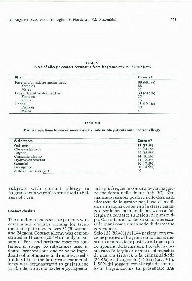
G. Angelini - G.A. Vena - G. Giglio - F. Fiordalisi - C.L. Meneghini 231
Table VI Sites of allergie contact dermatitis from fragrance-mix in 144 subjects.
Site
Face and/or axillae and/or neck Females Mal es
Legs (Ulcerative dermatitis) Females Mal es
Hands Females Males
Cases n ° 99 (68.7%) 88 Il 30 (20.8%) 15 15 15 (10.4%) IO 5
Table VII
Positive reactions to one or more essential oils in 144 patients with contact a llergy.
Suhstances Oak mass Cinnamaldehyde E ugenol Cinnamic alcohol Hydroxycitronellal Geranio! Isoeugenol Amylcinnamaldehyde
subjects with contact allergy to fragrance-mix were a lso sensitized to balsam of Perù.
Contact cheilitis.
The number of consecutive patients with eczem atous cheilitis coming for treatment and patch-tested was 54 (30 women and 24 men). Contact allergy was demonstrated in 11 cases (20.4%), mainly to balsam of Peru and perfume essences contained in rouge, to substances used in dental preparations and to some ingredients of toothpastes and mouthwaseehs (tahle VTU). In the latter case contact alle rgy was demonstrated to guaiazulene (1; 3), a derivative of azulene (cyclopenta-
Cases n°
37 (27.8%) 33 (24.8%) 22 (16.5%) 14 (10.5%) 11 ( 8.3%) 10 ( 7.5%) 6 ( 4.5%) o
t a la più frequente con una ovvia maggiore incidenza nelle donne (tab. VI). Non mancano reazioni positive nelle dermatiti ulcerose delle gambe per l'uso di medicamenti topici contenenti le stesse essenze e per la ben nota predisposizione all'allergia da contatto su lesioni di questo tipo. Con minore incidenza sono interessate le mani come unica sede di dermatite eczematosa. Solo 123 (85,4%) dei 144 pazienti con reazione positiva al fragrance-mix hanno mostrato una reazione positiva ad uno o più componenti della mistura. Prevale in questo caso l'allergia da contatto al muschio di quercia (27,8%), alla cinnamaldeide (24,8%) e all'eugenolo (16,5%) (tab. VII). Il 50% dei soggetti con allergia da conta tto al fragrance-mix ha presentato una

232 Contact Dermatitis due to Cosmetics
Table VIII
Positive reactions to one or more substances in 11 patients with allergie contact cheilitis.
Substances
Guaiazulene Balsam of Peru Fragrance-mix Meth ylme Lhacry late Parabens
cycloheptane) found in plants (13). In Italy, commercial guaiazulene is used in such toiletries as soaps, toothpastes, mouthwashes, shampoos, creams and lotions, for its soothing and antiinflammatory properties. It is also contained in certain inhalants and in denture adhesives. Patients with eczematous cheilitis complain of peeling, cracks and dryness of the pseudomucosa and the surrounding skin; occasionally they show more marked changes with erythema and weeping. Some complain also of symptoms suggestive of stomatitis, such as loss of taste and a burning feeling.
Cases n°
5 3 2 2 1
contemporanea sensibilizzazione anche al balsamo del Perù.
Cheilite da contatto
I pazienti consecutivamente osservati e testificati per cheilite eczematosa sono stati 54 (30 donne e 24 maschi). È stata evidenziata allergia da contatto in 11 casi (20,4%), prevalentemente a l balsamo del Perù ed essenze profumate contenute in rossetti, a sostanze utilizzate in preparati dentari e ad alcuni componenti d i dentifrici e collutori (tab. VIII). A quest'ultimo riguardo, è stata messa in evidenza una allergia da contatto al guaiazulene (1, 3). Quest'ultimo è uno dei derivati dell'azulene (ciclopentacicloeptano) che si ritrovano in varie piante (13). In Italia, il guaiazulene è la forma commerciale usata nei cosmetici, quali saponi, dentifrici, collutori, shampoos, creme e lozioni, a causa delle sue proprietà lenitive ed antiinfiammatorie. Esso è contenuto anche in alcuni inalanti e adesivi di dentiere. I soggetti con cheilite eczematosa lamentano disturbi rappresentati da desquamazione, ragadi e secchezza della pseudomucosa e della cute circostante; occasionalmente, presentano alterazioni più evidenti con eri tema ed essudazione. Alcuni di essi accusano anche sintomi soggettivi di s~omatite, quali perdita del gusto e bruciore.

G. Angelini - G.A. Vena - G. Giglio - F. Fiordalisi - C.L. Meneghini 233
REFERENCES
l. Angelini G. and Vena, G.A. (1984) Allergie contact cheilitis to guaiazulene. Contaci Dermati tis 10, 311 2. Calnan, C.D., Cronin, E. and Rycroft, R.J.G. (1980) Allergy to perfume ingredients. Contaci Dermatitis
6, 500. 3. Cronin, E. (1967) Contact dermatitis froim cosmetics. I. Soc. Cosm. Chem. 18, 681. 4. Cronin, E. (1980) Contact Deramtitis, Churchill Livingstone, Edimburg. 5. Eiermann, H.J., Larsen, W., Maihach, H.I. and Taylor, J.S. (1982) Prospective study of cosmetic reac
tions: 1977-1980. J. Am. Acad. Dermatol. 6, 909 6. Fisher, A.A. (1973) Contaci Dermatitis. Lea and Febiger, Philadelphia. 7. Fregert, S., Hjorth, N., Magnusson, B., Bandmann, H.J., Calnan, C.D., Cronin, E., Malten, K., Mene
ghini, C.L., Pirilii, V. and Wilkinson, D.S. (1969) Epidemiology of contact dermatitis . Trans. St. Jonh 's Hosp. Dermatol. Soc. 55, 17.
8. Hjorth, N. (1961) Eczematous allergy to balsarns, applied perfumes and flavouring agents. Munksagaard, Copenhagen.
9. Van Ketel, W.G. (1979) Patch testing with eye cosmetics. Contact Dermatitis, 5, 402. 10. Larsen, W.G. (1977) Perfume dermatitis. Arch. Dermatol. 113, 623. 11. Malten, K.E., van Ketel, W.G. Nater, J.P. and Liem, D.H. (1984) Reactions in selected patien ts t o 22
fragrance materials. Contact Dermatits 113, 623. 12. Meneghini, C.L. and Angelini, G. (1980). La paraphénilènidiamine dans les dermites allergiques de con
tact. Médicine et Hygiène 38, 1577. 13. Proserpio. G., Bardi, E. and Massera, A.M. (1981) Cosmetic Index. Sinerga, Milano. 14. Rantuccio, F. Angelini, G., Vena, G.A. and Conte, A. (1982) Manifes tazioni allerghiche della cute della
regione orbitaria. Atti XVI Congresso Società Oftalmologica Meridionale, Simposio su «Occhio e Cute», Viterbo 11·13 Giugno.
15. Rothenhorg, H.W. and Hjorth, N. (1968) Allergy to perfumes from toilet soap and detergents in pa-tients with dermatitis. Arch. Dermatol. 97, 417.
16. Rndzi, E., Grzywa, Z. and Brud, W. (1976) Sensitivity to 35 essential oils. Contact Dermatitis 2, 196. 17. Skog, e. (1980) Incidence of cosmetic dermat itis. Contact Dermatitis 6, 449. 18. Vena, G.A., Angelini, G. and Rantuccio, F. (1982) Eyelid contact dermatitis. It. Gen. Revie1v Dermato/.
19, 47.
J. Appl. Cosmetol., 3, 234-237 (July/September 1985)
Marvelous Secrets W. MONTAGNA. P. MORGANTI
Decoction to make hair grow (1)
Two manipol, each of agrimony and elm bark enough common water and made a decoction. Bai! unti! reduced to one third the origina! volume. Wash the hair in this decoction twice a week. It has been tested.
To make hair grow where there are none (2)
Rx. put equal parts of snail meat, wasps, bees, leeches, and burnt salt in a glazed jar with perforated bottom. Piace the jar into another glazed jar, without holes to collect the drippings; apply these juices to the ares where you want hair to grow. ith this remedy I regrew the beard of a canon in less than a month. Daily applications of honey water or honey spirit, or evenn warm oil of tartaro, achieve the same result.
In order not to deprive the reader of the real substance of this decoction we offer him the directions for the furmulation, translated literally and using the same punctuation.
To make hairs grow where there aren't any
Snail meat, wasps, bees, leeches, burn salt of each equa! parts, piace everything in a glazed vase, and perforated, which has many holes on the bottom, and under it, another glazed vase, to gather the juices, which will come out, and with this anoint where you want, that hair grow, and with this remedy I was able to grow the beard of a canon in less than a month, applying it every day: honey water or spirit does the same, as does also oil of tartaro slightly warmed, and then anoint the piace severa! times does the same.

W. Montagna - P. Morganti
i) L I B R ~o. _peoni t 1 a/Tara, afirolo~ia wnd.l (eme di fk nocchioTfo go di lticaclos,armoniJco,mar rubi o, gumma lacca,di eiafched.dr.due, ti facci decotto in lib. trè di acqua fecondo -l'arte-; la do(e dr. ·quattro la mattina. · Per I~ t•ffe, <:!i 11/11111, oht no11jù1
• i11Wuhi11111 • .• JJl, H I/'o~, b.aftlicl>, c11pel venère,'.m~r-
rubfo, regolitia,di ciafchedon.o dr.fei, Ci facchlect>tto in (ufficiente quan. · titl d'acqua f«"Ondo l'arte; nella coll.ttura metti miele Olle. tr~, ne·beva un bicch iero caldo (era, ·e mattina, e fl cuopra bene, che è mirabilé. :w "' -
· Yn'•lt,,..ri-.iiio j111' J'llf.,,,•. ~· G Entiana, hifopo, agarico, di eia.
fched. dr. mez.. mallici dr. i. col miele ilinne elettuario:. la dofe è da. trè ~
' cin,quedr.ènonci èfecretolimiJe. · :Elt1t1111rio_ptr J' ef 1111• •nti&tl, fl' 111/A
~ opnmiont Ji •ilz.&. / ~· A Sfara dram. una , fugo di marru.
bio, d'hifopo, tlorace, calloreo, di ~iaf~hedunod'ram. meza, pepe longo bianco, e nerO', aaW<di dafchedu no dra. due, con oximlele d.f.acci elettuario: la dofe è un cucchiaro fera , e mattin.i.
EIMt1t•rio' pt,.t'll/11111. lJt. M Anna Calario onc;i. capta ellrat.
· ta di nuovo .bila canna oncie meza,a ppeniti,oncie tre, ogUo d•altlaodoJe dolci o_avatodi fretto.~na po~c,.
. , ne
235
p R l . M o. 71 ne di volJ'e prep:ir3ta drl. 2.· fiori di (~lf? d. r: f--cc1' elettuario da ufarlo alh b1-ram.1.u.. . '' N ' {i . Con quello elettuario guan a iz.-ogm. .l' r.
a ContefTa in taaoco o uo meJe cou uun d .• fiupore di tutti quelli Me ict--; .
~n_gNtntt> ptr Jt hem0Yro1d• ,: . V NgU~nto-populeon ~r1.c. ~ roffi d JJt ouo num. due,oglio violato onc~ t canfora dr. t.op.p'odrr.meu, unguento r~fato onde men, ·ft &cci .ung~entfi. nel morta.ro.d\>piombo~ s'i{'plich!«<>n on· did•lattuca. •.J~ ,'
1 Dtcottri ptr lt h1mmo1"'' '. ..
·TAffo b:irbaffo manip. i: r~d1che~-lJi altra m:rhip. 1. {e~th hno o.ne~
.... .... ll a o"ni cofa: ;n un boccaLd.i tat-mez..:i ,.'VV f'>' - 1 ..- ro e-te· di oi 6 metta il tt~ Ort ca .. ,~ , • ·n; dc~va i\ fumo per da baff~, e gui!1ra ..
Dte1t.10 ptr f•r nlffc~e !• c•ptll• • "'
A Grimòniit' fcorze d olmo' verbe-
1'!• na.dicia{cheduno mani pol. 2.- ac-'1:1.tS C°'11 mu n.e qJJanto bafb , f~ nne decotto' che con6unirhen:o ,ed1 eff~decot· to lav:i li cappelli dµe v.otte la.Cc:ttJ_mana 1
·& è provato. r. _ Pt"" f.r: ,,11.fc.,,. ut•~lfidwe-•0111~"(: ..
e Arne-d1 Lumache ,,vcf P1: ,api,. 3.n-1'1 çui(ughe, {at~abb~~g1ato d.1 era~ {cheduno plrti uguah,mem il~utto '!1-u va(o virriato,e forato, ch'habb1 molu bud1i hl fondo, e {otto rli, effo u1.1·~~cro vafo "iaato • ·C'bctaccolga i hùmd1ta 1 che. ne
ufc1·
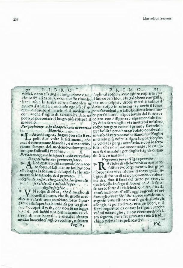
236 Marvelous Secrets
,,, L I n R ·o M;. ' p R I M o·. 73 ,. fcirà., e,co1~ elfa u n~i il l11ogo dove vuoi, l'oglin,i I wfpo vivo,e (ubico coprirlo co11
. <;he ~1afca!1 l1 capellr, et con CJuefio rimedio il foo coperchio_, e luttalo be~c ,con~all~ •• _-tccc1 u(~1r la barba acl un Canol)fco in che no n refpiri, dipoi metti ~-b~llireil
m:tnc'? d u.n.me(e·' · 01_1tando ogni di: l'ic- detto rofpo in c:impagna •. ac,c10 ti fu.mo "C]U:t, o fpmto d1 miele fa il medelimo non t'avueleni, e fallo bollire a lento tuo· com' anche. l' oglio. di tanaro ic.ildato ur; co perfei ho re, dipoi levalo dal fuoco' e poco, e poi ontare 11 luo~o più voir~fà il colato con diligenza, e(premen.dolo formedemo: . ..!• . 1,,, , ce,&indettooglio v.iri°'.1ettera1un'altro Per proh1b1rc, ç h~./, '"f!eWmJn Ji'fltntillo ro{po- p~r~:tto come il primo, f.1 ccnàolo
1!. • , bunch1. , 1 - ., , pur bollire per 6.hore,e 'colato con(~rva~? IJ! L Atte dt cagna, bagna con elfo li ca- in vafo di vetro come ba J(amo;que.ft oglto
. pelli due vo_Ice la/ettimana, che untando pil} voi re la ti~n~ la gua~t (ce,fat· mai deventera11110 b1anch1, e u mantene- ca prima l.l pur~ i necefiana, e co~t le fcroranno fempre del rnedemo coloi~uan- fole , che 110 n fo 110 ancor ~ott~ ~le con{utunq11e fo«eallài vecchio. m~: & è mirabile per doghe fogide ontaai. Per 11~1111111z.:1: .. ~t Jt tigno/t , çhf ç•rrod~o do {era , e ma ttin:t. .
J, c1tptitt.che non p_onnoo·t/t:tre, V11g(J(:nto per la T1gn~ pro1:Ato • •· Aloè epaticodiltemperalo'.co n ace- lJt· RAdiche di elleborobianc~, en~r?, •
to forte, e falli dar un bollo,e co·n (olfo vivo, orpimento, ltt.irg1r.io elfo bagna la (ommità de'capeJ1i, chf a.rn- d'oro e.tic-e viva, a lume di rocca,g3.\b .tu~ maz~arà le tignole, & è provato. ligi ne' di fo~no di ciafch. on.mez.. vc:rdera.
• Og/10 di ro/po, cheg1tAri/ct /111;_g114 , le me clra. due {j facci c,!el tu cto po~ vere? la l' /crofolt, & è mir11bile per qual< bolla in fu go di boragine, di f~:rb10-. tiog/iefrigide. . • fa, aceto fo rte di ciafched. onc.tre, 1111.all.1 ~- v N ro(po difelva, che è meglio di confumacione ò' effi, aggio ngc:nÒO'fl nel . . quell~ d'horco, c he lfa grolfo met- .fine aglio vt'cchi·o lib. t .pece n~v:ile <;>n. ~-
t1lo 111 va(o dt terra sbufciato fotto à pur· argento vivo ellinto con foga da f.ùv1a '~ g~re dalla (uperfiua humidicà per ere _gior- aflongia di porcod ra.:1,. cera un poco, e i1 n!, e ~uop~i il va_fo, che non {e a1e fugga facd unguento da ontare h tigna, µerche v1a: d1 .Poi habb1 una pignata nuova vj. vedrai maraviglie, e 11011 occorrerebbe a!-treat:i di due b~ccal~, e me~tici denrro ero fe~reto, per t'lfer ~rova:o.: ma .:i frad~~ mc~z.o boccalc<l .ogl10 ve~h1p •.e dencro chiJlo pdma li cape lh camv1. _ , .. J
l'og-lio, · •· - YllJ.

W. Montagna - P. Morganti 237
To prevent hair from becoming gray (3)
Rx. Wash hair in dog (bitch's) milk twice a week and they will never be gray. They will remain the same color even in old age.
To prohlbit, that hairs don't become white
Rx. ~ilk of bitch, bathe the hair with it two times a week, which never will become white, and they will remam always the same color even though very old.
To kill ringworm which corrodes hair so that it cannot grow (4)
Rx. Dissolve hepatic aloe in strong vineyar and boil it . Pour over the hair and ringworm will disappear. This has been tested.
Toad oil to cure ringworm and scrofula (tuhercular sores); it is also wonderful for chiblains. (5)
Rx. Piace a large woods toad (this is better than the garden variety) into an earthen jar with perforated bottom for three days to allow the toad to cleance itself of excess fluids. Cover the jar so that the toad doesn't escape. Put half a boccale of old oil in a new glazed two-boccale pot, shove the toad in the oil and quickly seal the lid with paste so tat it can't breathe. Boil the oil over a slow fire for six hours under a hood (the author says «in campagna», which means «in the country»; this is probably a misprint; perhaps he meant to say «in campana» which could mean under a bell) so that the smoke won't poison you. Remove the toad from the oil and drain it carefully by squeezing hard, and then put another toad, cleansed like the first one, in the oil, boiling it also for six hours. Repeat the performance as with the first toad and t hen keep the oil as a balsam in a glass jar. This oil cures ringworm, after one has taken the necessary purge, and also cures tubercular sores that have not yet opened. It is also marvelous for chilblain when apllied morning evening.
J. Appl. Cosmetol., 3, 238-239 (July/September 1985)
To make art
BRUNO LISI
To be or to realise
To m ake art should be to touch the horizon that separates the finite from the infinite. For example, if we piace the work of Mark Rothko (who I maintain to have been one of the greatest artists of recent years) within the vis ion of a creativity that goes beyond the object, we have to say that all Rothko's work is no more than a beginning (the painting no more than a window on the world or, worse an object of forniture). Christo actually wraps entire islands creating an alternative tò one's relationship with things and nature. But it nevertheless remains aesthetics resolving itself into «what a beautiful photo' of the operation». The potential content of the act of creat ion is too often reduced by those analyses of it which only underline professionalism , going against «pure creation», cut ting out the possibility of «seeing ins ide». I believe, however, that to make art is the only one of man's «activities» finalized in his growth; it is the «madness» of living beyond the time at ou r disposition. In other words, despite Melevic's Black Sq~are (1913) and the works 'Concep-
Fare arte dovrebbe essere toccare l'orizzonte che separa il finito dall' infinito. Ad esempio, se collochia mo l'operato di Mark Rothko (che ri tengo sia s tato uno dei più grandi artisti degli ultimi anni) nella visione di una creatività che superi l'oggetto, dobbiamo dire che tutta l'opera di Rothko è non più di un vagito (il quad ro all'occasione finestra sul mondo o p eggio oggetto d 'arredamento). Christo impacchetta addiri ttu ra interi isolotti cr eando una alternativa al rapportarsi con le cose e la natura, ma è pur sempre estetica che si risolve nel «Come è bella la foto a colori dell'operazione». Il contenuto potenziale del creare è troppo s pesso ridotto da lle analisi che sottolineando il professionismo vanno contro la creazione pura, togliendo la possibilità di «vedere dentro». Credo che comunque il fare arte sia l 'unica «attività» dell 'uomo finalizzata alla sua crescita, è la «follia» per vivere oltre il tempo a nostra disposizione. Insomma malgrado il quadrato nero di Malevic (1913) e le operazioni Concettuali, Minimal, Land, ecc., sostanzialmente non è cambiato nulla, fare arte rimane un modo di essere più che realizzare e il mistero del perché e come si arrivi al confi-
tuals', 'Minimal' and 'Land' etc., substantially nothing has changed. To make art remains more a way of "being» than of « realizing», and the mystery of why and how one arrives at the border between the finite and the infinite is probably incomprehensible because, in our reading of that mystery, it is indecipherable. It is, however, stili there wi thin the beginning and end of all things.
Roma, October, 5 1985
J. Appl. Cosmetol., 3, 238-239 (July/September 1985)
ne tra il finito e l'infinito è probabilmente incomprensibile in quanto indecifrabile, nella nostra lettura comunque all'interno dell'inizio e fine di ogni cosa.
Roma, 5 Ottobre 1985

Book Review J. Appl. Cosmetol., 3, 240 (July/September 1985)
Flavor and Fragrance Mate rials 1985 Allured Publishing company, Box 318, Wheaton, Illinois 60189-0318, USA $ 55.00 (Add $ 20.00 for airmail to any country in the World)
The 1985 Edition of Flavor & Fragrance Materials has just been published with extensive revisions from the 198 1 Edition.
Nearly a ll of the eleven categories of information have been revised because of changes in the status of these materials in the past four years. Changes include: The new materials of FEMA - GRAS list No. 13; revisions in the FDA section numbers; a major revision in the Council of Europe numbers; recent determinations of the «Nature Identica!» status of fl avor material s; revisions in the Scient ific Literatu re Reviews of flavor materi als; additions to the listings of CAS numbers; additions to the fragrance materials with published RIFM Monographs; and a revision and additions to the IFRA Guidelines status. Every individua! in cosmetic industry, including suppliers, traders, dealers, users of materials and dermatologists should have a persona! copy of thi s unique reference pubblication .
Questa edizione del 1985, aggiornamento dell'analogo testo del 198 1, elenca in ordine alfabetico tutte le materie prime utilizzate nelle formulazioni di composti profumati. Nell'ultima parte è r iportato inoltre sia l'elenco dei prodotti di materie prime profumate che le organizzazioni di vendita delle stesse materie prime. È un testo senz'altro utile sia per chi lavori presso industrie cosmetiche, che per gli studiosi del settore della Dermatologia cosmetologica, siano essi chimici o medici dermatologi.
Letter to the editor I. Appl. Cosmetol., 3, 241-242 (July/September 1985)
A clinical case of true acq uired leukonychia
The observation of a clinica! case of true acquired leukonychia offered to us the opportunity of a clinica! and therapeutic clarification. Leukonychia is a dyschromia characterized by a white colouring of nails, generally affecting hand fingers and exceptionally observed in feet-nails. There are severa! types of leukonychia, i è true and manifest, congenita! and acquired; the latter being divided into: tota!, subtotal, streaked, variegated and dotted (1). The statistica! incidence of the various forms is considerably low; no specific therapy exists and treatments applied are practically affectless. We have studieds the case of a 18 years old young man, suffering from true and acquired tota! leukonychia for the past six months. In the absence of possible etiopathogenetic moments, detectable through clinical-anamnestic inspection, and following an «ex juvantibus» criterium, we have treated the patient with therapy, based on 1-cystine (400 mg) associated with gelatin (1500 mg) jelly 1500 mg administered daily for three months. The surprisingly positive solution of this case supports the use of this food association as a possible therapy for the va-
L'osservazione di un caso clinico di Leuconichia vera acquisita ci offre lo spunto per una puntualizzazione clinica e terapeutica. La Leuconichia rappresenta una forma di discromia ungueale caratterizzata da una colorazione bianca delle unghie: essa interessa generalmente le dita delle mani mentre è eccezionale la localizzazione a quelle dei piedi; si distingue classicamente in leuconichia vera e apparente, congenita ed acquisita a sua volta suddivisa in totale, subtotale, striata, variegata e punteggiata. (1) L'incidenza statistica delle varie forme è notevolmente bassa; la terapia è aspecifica e risulta praticamente nulla. Il caso, venuto alla nostra osservazione riguarda un giovane di 18 anni, affetto da leuconichia vera acquisita, affetto da leuconichia vera acquisita totale, da circa sei mesi. In assenza di possibili momenti etiopatogenetici, evidenziabili ad un esame clinico-anamnestico, è stata instaurata terapia, con criteri «ex iuvantibus», a base di cistina levogina alla dose di 400 mg. pro die in associazione a gelatina alla dose di 1500 mg pro die, per la durata di 90 gg. La sorprendente risoluzione totale del caso clinico ci spinge a comunicare e

rious forms of acquired leukonychia. Regarding the action performed by the above mentioned food, one can suggest the hypothesis of the synergic capaci ty of gelatin-cystine towards keratinogenesis, with an increase in the proteic fraction of the nail-matrix. (2, 3)
Califano L., Cannavò S.P., Santoro G.
J. Appl. Cosmetol., 3, 241-242 (July/September 1985)
quindi suggerire questa associazione come possibile terapia delle varie forme di leuconichia acquisita. Per quanto riguarda il meccanismo d'azione si può prospettare l'ipotesi della capacità sinergica della associazione della cistina con la gelatina nei confronti della cheratinogenesi, con incremento della frazione proteica ad alto contenuto di zolfo della matrice delle unghie. (2,3)
Clinica Dermatologica Universitaria (Dir. Prof. B.Guarneri) Ospedale Piemonte MESSINA - ITALY
1) Baran R., Dawber R.P.R. (1984) «Diseases of the nails and the ir management» Blackwell Scientific Pub lications, Oxford. 2) Randazzo S.D. (1982) «La cistina nella terapia dermatologica» Fa rmacia Ottanta, 3, 35. 3) P. Mol'ganti (1984) Nutrition and Hair - J. Appl. Cosmetol., 2, 41-49

Announcements
Second international course on occupational dermatoses'
29 September - 3 October 1986
Course location Haaasaari Cultura! Centre, Espoo, Finland. 2nd announcement & application Available from February 1986 on. Application deadline Application forms must reach the course secretary by 23 June 1986. Background
xv
The first International Course on Occupational Dermatoses was arranged by the present organisers in 1982 in Helsinki, Finland. It turned out to be a great success with about 100 participants, and no wonder, as of ali occupational diseases, occupational dermatoses cause the highest percentage of industriai d isabilities and the greatest loss of workdays due to labour nonfatal disease. It is often exceedingly difficult even for an experienced dermatologist to determine the agents causing an oocupational skin d isease. The purpose of the course is to give the clinica! a better background in this detective job. Objective The aim of the course is to give practical up-to-date knowledge on occupational dermatoses. Partici pants will learn both bas ics in occupational derma tology as well as the latest information on subjects such as computers in occupational dermatology, new treatment methods, new allergens, and contact urticaria. A Hand-Clinic, with in-depth studies on the skin region most frequently affected by occupational dermatitis, will be presented. A pane! of experts will discus instructive cases from their own practices, and audience participation will be encouraged. Interesting cases of occupational dermatology can be presented as free communications or posters. Target group Dermatologists a nd clinicians in industriai medicine with a good basic knowledge of occupational dermatology Partici pants Approximate ly 100 Main topics - Contact dermatitis - Patch testing problems - Hand-Clinic - How to use computers in occupational dermatology - Contact urticaria - New allergens - Work-related di seases and the skin - Photocontact dermatitis - Specific causes of allergie contact dermatitis - Occupational dermatoses and atopy Abstracts/scientific papers Participants are encouraged to present their own pa pers or studies on occupational dermatology. They will be presented as free communications (3-10 minutes) or in poster form. The deadline far abs tracts is 2 May 1986. Contact person Lasse Kanerva, MD, Phd Institute of Occupational Health Section of Dermatology Haartmaninkatu 1 SF-00290 HELSINKI FINLAND Tel. (+358 O) 474 7288 Course secretary Outi Teperi

Journal of Applied Cosmetology pubblished quarterly by INTERNATIONAL EDIEMME, Via F. Bernardini 22, 00165 Roma I taly. Direttore Responsabile P. Morganti. Direzione, Redazione ed Amminist razione: Via F. Bernardini 22, 00165 Roma Ita ly. Fotocomposizione e s tampa EUTF.C:RA PHICA S.r.l. P rogetto grafico e copertina B. Lisi. Impaginazione e grafica: E. Francioso. Spedizione in abbonamento posta le gruppo IV/70. Autorizzazione del tribunale di Roma n. 3173183 del 8.7.1983.
Per Campioni Medici e Documentazione Scientifica: Lieder Mavì Distribntrice Via Filippo Bernardini, 22 00165 Roma Tel. (06) 63.84.348