deposition of au/tio 2 nanocomposite on ito surface by seed-mediated...
TRANSCRIPT

This content has been downloaded from IOPscience. Please scroll down to see the full text.
Download details:
IP Address: 129.119.67.237
This content was downloaded on 24/08/2014 at 10:53
Please note that terms and conditions apply.
Deposition of Au/TiO2 Nanocomposite on ITO Surface by Seed-Mediated Liquid Phase
Deposition Method
View the table of contents for this issue, or go to the journal homepage for more
2013 J. Phys.: Conf. Ser. 431 012011
(http://iopscience.iop.org/1742-6596/431/1/012011)
Home Search Collections Journals About Contact us My IOPscience

Deposition of Au/TiO2 Nanocomposite on ITO Surface by Seed-Mediated Liquid Phase Deposition Method
Suratun Nafisah1, Arfian Oktaviandi2, Akrajas Ali Umar1, Muhammad Mat Salleh1, Aamna Balouch1,3, and Siti Khatijah Md Saad1,4
1 Institute of Microengineering and Nanoelectronics (IMEN), Universiti Kebangsaan Malaysia, 43600 UKM Bangi, Selangor, Malaysia 2 Department of Chemistry, Faculty of Mathematics and Natural Science, Padjajaran University, Bandung, Indonesia 3 National Centre of Excellence in Analytical Chemistry, University of Sindh, Jamshoro, Pakistan 4 College of Engineering, Universiti Tenaga Nasional, Km 7, Jalan Kajang-Puchong, 43009 Kajang, Selangor, Malaysia E-mail: [email protected] Abstract. An efficient, simple and new procedure has been performed to synthesis Au/TiO2 nanocomposite thin film on solid surface of ITO by seed-mediated of liquid phase deposition (LPD) method. The deposition of Au seed was applied by our previously reported seed-mediated growth procedure. The solution was prepared by mixing HAuCl4 and (NH4)2TiF6 successfully deposited on to ITO substrate containing Au seed. After one hour well adhered film was obtained by mentioned approach of synthesis and nonocomposite with spherical rod like networks has been successfully grown. The resulting nanocomposites were confirmed by ultraviolet-visible (Uv-Vis) absorption spectroscopy. Further characterization and morphology was checked by field emission scanning electron microscopy (FESEM), energy dispersive X-ray spectroscopy (EDX) and X-ray diffraction (XRD).
1. Introduction Nanocomposite of metal and metal oxide have been intensively studied due their good optical, photocatalytic, electrical, magnetic and mechanical properties [1]. These metal and metal oxide nanocomposites are widely applied in gas sensor [2], catalyst [3],[4] and anti bacterial [5]. Nanocomposite of metal and metal oxide have been intensively studied due their good optical, phtocatalytic, electrical, magnetic and mechanical properties such as..Many studies have been reported in the literatures on the effect of doping metal like Au, Pd, Ag and Pt on the TiO2 [5],[6]. TiO2 is one of the most attractive semiconductor materials due to the lowest cost, non-toxicity, biologically and chemically stable properties [7]. The presence of metal in the TiO2 facilitates electron transfer from photoexcited semiconductor to the surroundings and decreases recombination rate between the electrons-holes so that good for application in photoelectrochemical and photocatalytic [8]. In recent years, various methods has been used in synthesis of composite TiO2-metal nanoparticles such as photoreduction [5], magnetron sputtering [1, 9], dip coating [10] and sol gel method [6, 11]. Owing to 1 [email protected]
3rd ISESCO International Workshop and Conference On Nanotechnology 2012 (IWCN2012) IOP PublishingJournal of Physics: Conference Series 431 (2013) 012011 doi:10.1088/1742-6596/431/1/012011
Published under licence by IOP Publishing Ltd 1

the simplicity of the process, catalytic and optical properties, the presence method could be potential approach for producing Au/TiO2 nanocomposite for application in photocatalysis, optical sensing and non-linear optical application.
Liquid phase deposition method (LPD) is widely used in the deposition of TiO2 on solid surface. In particular, LPD is promising technique for direct preparation of TiO2 thin film on to solid substrate using boric acid as scavenger. However, metals such as -Al and Fe has also been reported to be used as scavenger for fluoride ion[12]. In this study, we aims to develop a simple and efficient method to synthesis Au/TiO2 nanocomposite thin film by seed-mediated grow procedure using LPD technique. The obtained composite thin films were characterized by using Uv-Vis absorption spectroscopy, field emission scanning electron microscopy (FESEM), energy dispersive X-ray spectroscopy (EDX) and X-ray diffraction (XRD).
2. Experimental
2.1. Materials Analytical grade chemicals in this work were used without further purification. HAuCl4 and (NH4)2TiF6 were purchased from Sigma Aldrich while trisodium citrate was purchased from Wako Chemical and NaBH4 was from Fluka. The ITO substrate was purchased from CBC Ing. Co. Ltd., Japan. The ITO substrate was cleaned by distilled water and sonicated in acetone followed by 2-propanol for 30 minutes according to the standard procedure. The depositions of Au/TiO2 nanocomposite on to ITO substrate practically involved two steps, namely seeding process and growth process. The ITO substrate immersed into Au seed solution as reported in our previous work [13].The growth process was carried out by immersing the sample in standard growth solution which containing 5 ml of HAuCl4 1 mM and 5 ml of (NH4)2TiF6 50 mM. The reaction was performed in bath deposition to keep temperature unchanged during the reaction. The sample was then washed several times by distilled water and dried under nitrogen flow.
Effect of concentration (NH4)2TiF6 was performed by preparing four different of variation concentration namely 50, 100, 200 and 250 mM with the fixed concentration of HAuCl4 1 mM at temperature 90ºC for growth time 1 hour. The investigation of formation Au/TiO2 nanocomposite was then studied by varying the growth time of the synthesized nanocomposite for four different variations namely 15 min, 30 min, 1 hour and 3 hours.
2.2. Au/TiO2 nanocomposite characterization The optical absorption of the sample for fixed concentration, variation concentration of (NH4)2TiF6, and growth time were studied using Uv-Vis spectrophotometer (Lambda 900 PERKIN ELMER) with the wavelength range 300-900 nm. Morphology of nanocomposite for fixed concentration and variation of growth for synthesized nanocomposite were also studied using FESEM technique (ZEISS SUPRA 55VP). The elemental composition of nanocomposite was studied using energy X-ray dispersive (EDX) method (Oxford instruments INCA) at 30 kV using CuKα radiation (λ = 1.541 Ǻ). X-ray diffraction spectrum was obtained from X-ray diffractometer (BRUKER D8 Advance) with the diffraction angle of 0-60º.
3. Results and Discussions Au/TiO2 has been successfully deposited on the surface using the presence approach. As reported by Gutierrez et al. who successly produced TiO2 using trivalent Al(III) and Fe(III), in this work we tried to use trivalent Au(III) as scavenger ion in the absence of boric acid. The chemical reaction can be expressed as follows:
TiF + 2H O ↔ TiO + 4HF + 2F (1)
Au + 4HF ↔ [AuF ] + 4H (2)
3rd ISESCO International Workshop and Conference On Nanotechnology 2012 (IWCN2012) IOP PublishingJournal of Physics: Conference Series 431 (2013) 012011 doi:10.1088/1742-6596/431/1/012011
2

In typical process, the Au/TiO2 nanocomposite formed in standard growth solution at temperature of 90ºC for 1 hour. Figure 1a shows a typical FESEM image of Au/TiO2 nanocomposite on the ITO surface via seed-mediated LPD method. From the FESEM image, we can see that the shape Au/TiO2 nanocomposites was more towards spherical rod-like with a diameter of 250 nm. These spherical rod-like particles however tends to agglomerate with one another forming a micron size network. In high magnification (Figure 1b), it can be clearly seen that the roughness among the surface of nanoparticles, indicated that the nanoparticles was not only pure Au, but contain dopant (TiO2). The EDX study was performed on the sample to confirm the existence of TiO2. The EDX spectrum below (figure 1c) showed Au, Ti and O peak. This confirmed the formation of Au/TiO2 nanocomposite thin film. However as can be seen from the spectrum, Ti peak was not as high as Au indicates that the composition of nanocomposite was dominated by Au.
Figure 1. FESEM image of Au/TiO2 nanocomposite deposite on to ITO surface (a) at high
magnification (b) and it’s EDX spectrum (c)
Figure 2 showed the XRD spectrum of Au/TiO2 nanocomposite. The peaks observed match to the JCPDS of Au nanoparticles (file number 04-0784) where the peaks related at (111) and (200) corresponding with the diffraction angle 38.185º and 44.393º. However from the XRD spectrum the presence of TiO2 was not seen. This might due to the fact that the presence of TiO2 in the Au/TiO2 nanocomposite was very small. As also can be seen in figure 1, the distribution of investigated nanocomposite was not dense enough.
C
1 µm
A
100 nm
B
keV
3rd ISESCO International Workshop and Conference On Nanotechnology 2012 (IWCN2012) IOP PublishingJournal of Physics: Conference Series 431 (2013) 012011 doi:10.1088/1742-6596/431/1/012011
3

Figure 2. XRD spectrum of Au/TiO2 nanocomposite deposited on solid surface.
UV-Vis absorption in figure 3 showed that the absorption of Au/TiO2 nanocomposite centering at
wavelength 570 nm. This absorption is slightly difference with the absorption of Au nanoparticles which is normally exhibit plasmonic absorption around wavelength 520 – 530 nm [13]. There are two possible reasons that may be responsible for that bathocromic shift. First, the presence of TiO2 in the gold nanoparticles (Au/TiO2 nanocomposite) as it was previously reported that the absorption peak for thin film TiO2 was found to be at 350 nm[14]. Second, it was caused by the link of spherical nanocomposite that was contributed to the red shifted absorption spectrum.
Figure 3. UV-Vis absorption of Au/TiO2 nanocomposite.
The works was carried out further to optimize the effect of concentration (NH4)2TiF6 used on the
formation of nanocomposite. Four different concentration of (NH4)2TiF6 namely 50, 100, 200 and 250 mM were used in this study. Here, it can be seen when the molarity of (NH4)2TiF6 used increased, the optical absorption of the prepared samples seems to decrease. This might due to the formation of TiO2 complex has hindered the performance of Au in the nanocomposite as it is known better that Au has excellence optical performance contributed by its plasmonic effect. From the spectra profile (figure 4) of UV-Vis absorption, it can be conclude that the optimum (NH4)2TiF6 concentration is 50 mM gave
20 30 6050402 Theta
Inte
nsity
(a.u
)111
200
400 500 8007006000.00
0.04
0.02
0.08
0.06
Wavelength (nm)
Abs
orba
nce
(a.u
)
3rd ISESCO International Workshop and Conference On Nanotechnology 2012 (IWCN2012) IOP PublishingJournal of Physics: Conference Series 431 (2013) 012011 doi:10.1088/1742-6596/431/1/012011
4
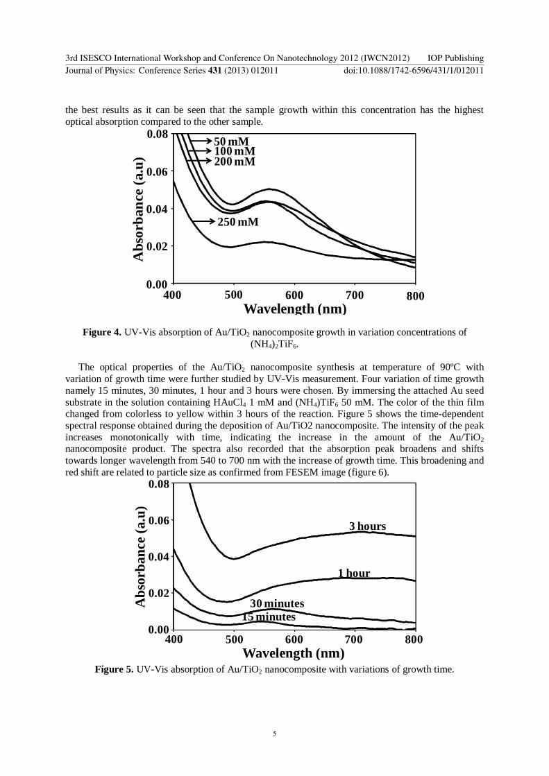
the best results as it can be seen that the sample growth within this concentration has the highest optical absorption compared to the other sample.
Figure 4. UV-Vis absorption of Au/TiO2 nanocomposite growth in variation concentrations of (NH4)2TiF6.
The optical properties of the Au/TiO2 nanocomposite synthesis at temperature of 90ºC with
variation of growth time were further studied by UV-Vis measurement. Four variation of time growth namely 15 minutes, 30 minutes, 1 hour and 3 hours were chosen. By immersing the attached Au seed substrate in the solution containing HAuCl4 1 mM and (NH4)TiF6 50 mM. The color of the thin film changed from colorless to yellow within 3 hours of the reaction. Figure 5 shows the time-dependent spectral response obtained during the deposition of Au/TiO2 nanocomposite. The intensity of the peak increases monotonically with time, indicating the increase in the amount of the Au/TiO2 nanocomposite product. The spectra also recorded that the absorption peak broadens and shifts towards longer wavelength from 540 to 700 nm with the increase of growth time. This broadening and red shift are related to particle size as confirmed from FESEM image (figure 6).
Figure 5. UV-Vis absorption of Au/TiO2 nanocomposite with variations of growth time.
400 500 8007006000.00
0.04
0.02
0.08
0.06
Wavelength (nm)
Abs
orba
nce
(a.u
)50 mM100 mM200 mM
250 mM
0
0.02
0.04
0.06
0.08
400 500 600 700 800400 500 8007006000.00
0.04
0.02
0.08
0.06
Wavelength (nm)
Abs
orba
nce
(a.u
)
15 minutes30 minutes
1 hour
3 hours
3rd ISESCO International Workshop and Conference On Nanotechnology 2012 (IWCN2012) IOP PublishingJournal of Physics: Conference Series 431 (2013) 012011 doi:10.1088/1742-6596/431/1/012011
5

The morphology of Au/TiO2 nanocomposite growth at different time intervals shows was presented by the FESEM image shown below. The shape of the nanocomposite formed was more into spherical shape and was bound to agglomerate towards each other. As the growth time increased, the linked agglomeration of the spherical particle of nanocomposite was elongated. This is due to the fact of the increased deposition time give rise to the formation of this nanocomposite. Thus, it is clear why when the growth time increased, the nanocomposite in micron size network increased. From the FESEM images obtained at the magnification of 20 kx, the grain size of the nanocomposite formed were 149, 172, 278 and 371 nm for the growth time 15 minutes, 30 minutes, 1 and 3 hours respectively.
Figure 6. FESEM image of Au/TiO2 nanocomposite with variation growth time i.e. 15 minutes (A), 30 minutes (B), 1 hour (C) and 3 hour (D) in obtained condition. Scale bars are 1 µm.
4. Conclusion Au/TiO2 nanocomposites thin film has been successfully prepared by seed-mediated LPD technique on the surface of ITO substrate. The UV-Vis spectrum shows band of nanocomposite thin films centering at wavelength 540 – 700 nm. FESEM image showed shape of nanocomposites were spherical like nanorods. EDX analysis confirmed the presence of Au, Ti and O atoms in the Au/TiO2 nanocomposite thin film. From the studies, the optimum growth condition for deposited Au/TiO2 nanocomposite thin films on the surface by seed-mediated liquid phase deposition for HAuCl4 of 1 mM is (NH4)TiF6 50 mM with the temperature of 90ºC grown 1 hour.
Acknowledgements This work was partially supported by the Universiti Kebangsaan Malaysia under research grant UKM-GUP-2011-377 and UKM-GUP-NBT-08-25-086. Author would like to acknowledge Center for Research and Instrumentation Management (CRIM) for the FESEM instrument.
References [1] Pal, M., T. Sasaki, and N. Koshizaki, 2001. 44(8–9): p. 1817-1820. [2] Jia, W., et al. The Journal of Physical Chemistry C, 2009. 113(37): p. 16402-16407. [3] Enache, D.I., et al., Science, 2006. 311(5759): p. 362-365.
A B
C D
3rd ISESCO International Workshop and Conference On Nanotechnology 2012 (IWCN2012) IOP PublishingJournal of Physics: Conference Series 431 (2013) 012011 doi:10.1088/1742-6596/431/1/012011
6

[4] Su, L., et al., Electrochemistry Communications, 2009. 11(11): p. 2199-2202. [5] Chen, S., et al., Nano Research, 2010. 3(4): p. 244-255. [6] Zhao, G., H. Kozuka, and T. Yoko, 1996. 277(1): p. 147-154. [7] Hashimoto, K., H. Irie, and A. Fujishima, AAPPS Bulletin, 2007. 17(6): p. 13. [8] Subramanian, V., E. Wolf, and P.V. Kamat, The Journal of Physical Chemistry B, 2001.
105(46): p. 11439-11446. [9] KIM, S.C., et al., Journal of the Korean Physical Society, 2005. 47(4): p. 700-704. [10] Hsiao, Y.C. and Y.H. Tseng, Micro & Nano Letters, IET, 2010. 5(5): p. 317-320. [11] Zhao, G., H. Kozuka, and S. Sakka, Journal of Sol-Gel Science and Technology, 1995. 4(1): p.
37-47. [12] Gutierrez-Tauste, D., et al., Journal of Materials Chemistry, 2006. 16(23): p. 2249-2255. [13] Umar, A.A. and M. Oyama, Applied Surface Science, 2006. 253(4): p. 2196-2202. [14] Umar, A.A., et al.,. Applied Surface Science,In Press.
3rd ISESCO International Workshop and Conference On Nanotechnology 2012 (IWCN2012) IOP PublishingJournal of Physics: Conference Series 431 (2013) 012011 doi:10.1088/1742-6596/431/1/012011
7