deployment of hagfish slime thread skeins requires the...
TRANSCRIPT
1235
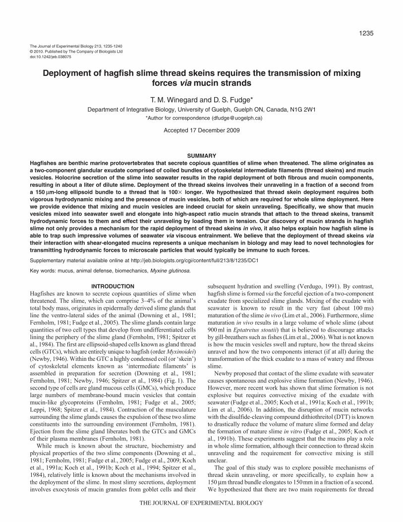
INTRODUCTIONHagfishes are known to secrete copious quantities of slime whenthreatened. The slime, which can comprise 3–4% of the animal’stotal body mass, originates in epidermally derived slime glands thatline the ventro-lateral sides of the animal (Downing et al., 1981;Fernholm, 1981; Fudge et al., 2005). The slime glands contain largequantities of two cell types that develop from undifferentiated cellslining the periphery of the slime gland (Fernholm, 1981; Spitzer etal., 1984). The first are ellipsoid-shaped cells known as gland threadcells (GTCs), which are entirely unique to hagfish (order Myxinoidei)(Newby, 1946). Within the GTC a highly condensed coil (or ‘skein’)of cytoskeletal elements known as ‘intermediate filaments’ isassembled in preparation for secretion (Downing et al., 1981;Fernholm, 1981; Newby, 1946; Spitzer et al., 1984) (Fig. 1). Thesecond type of cells are gland mucous cells (GMCs), which producelarge numbers of membrane-bound mucin vesicles that containmucin-like glycoproteins (Fernholm, 1981; Fudge et al., 2005;Leppi, 1968; Spitzer et al., 1984). Contraction of the musculaturesurrounding the slime glands causes the expulsion of these two slimeconstituents into the surrounding environment (Fernholm, 1981).Ejection from the slime gland liberates both the GTCs and GMCsof their plasma membranes (Fernholm, 1981).
While much is known about the structure, biochemistry andphysical properties of the two slime components (Downing et al.,1981; Fernholm, 1981; Fudge et al., 2005; Fudge et al., 2009; Kochet al., 1991a; Koch et al., 1991b; Koch et al., 1994; Spitzer et al.,1984), relatively little is known about the mechanisms involved inthe deployment of the slime. In most slimy secretions, deploymentinvolves exocytosis of mucin granules from goblet cells and their
subsequent hydration and swelling (Verdugo, 1991). By contrast,hagfish slime is formed via the forceful ejection of a two-componentexudate from specialized slime glands. Mixing of the exudate withseawater is known to result in the very fast (about 100 ms)maturation of the slime in vivo (Lim et al., 2006). Furthermore, slimematuration in vivo results in a large volume of whole slime (about900 ml in Eptatretus stoutii) that is believed to discourage attacksby gill-breathers such as fishes (Lim et al., 2006). What is not knownis how the mucin vesicles swell and rupture, how the thread skeinsunravel and how the two components interact (if at all) during thetransformation of the thick exudate to a mass of watery and fibrousslime.
Newby proposed that contact of the slime exudate with seawatercauses spontaneous and explosive slime formation (Newby, 1946).However, more recent work has shown that slime formation is notexplosive but requires convective mixing of the exudate withseawater (Fudge et al., 2005; Koch et al., 1991a; Koch et al., 1991b;Lim et al., 2006). In addition, the disruption of mucin networkswith the disulfide-cleaving compound dithiothreitol (DTT) is knownto drastically reduce the volume of mature slime formed and delaythe formation of mature slime in vitro (Fudge et al., 2005; Koch etal., 1991b). These experiments suggest that the mucins play a rolein whole slime formation, although their connection to thread skeinunraveling and the requirement for convective mixing is stillunclear.
The goal of this study was to explore possible mechanisms ofthread skein unraveling, or more specifically, to explain how a150 mm thread bundle elongates to 150mm in a fraction of a second.We hypothesized that there are two main requirements for thread
The Journal of Experimental Biology 213, 1235-1240© 2010. Published by The Company of Biologists Ltddoi:10.1242/jeb.038075
Deployment of hagfish slime thread skeins requires the transmission of mixingforces via mucin strands
T. M. Winegard and D. S. Fudge*Department of Integrative Biology, University of Guelph, Guelph ON, Canada, N1G 2W1
*Author for correspondence ([email protected])
Accepted 17 December 2009
SUMMARYHagfishes are benthic marine protovertebrates that secrete copious quantities of slime when threatened. The slime originates asa two-component glandular exudate comprised of coiled bundles of cytoskeletal intermediate filaments (thread skeins) and mucinvesicles. Holocrine secretion of the slime into seawater results in the rapid deployment of both fibrous and mucin components,resulting in about a liter of dilute slime. Deployment of the thread skeins involves their unraveling in a fraction of a second froma 150 mm-long ellipsoid bundle to a thread that is 100� longer. We hypothesized that thread skein deployment requires bothvigorous hydrodynamic mixing and the presence of mucin vesicles, both of which are required for whole slime deployment. Herewe provide evidence that mixing and mucin vesicles are indeed crucial for skein unraveling. Specifically, we show that mucinvesicles mixed into seawater swell and elongate into high-aspect ratio mucin strands that attach to the thread skeins, transmithydrodynamic forces to them and effect their unraveling by loading them in tension. Our discovery of mucin strands in hagfishslime not only provides a mechanism for the rapid deployment of thread skeins in vivo, it also helps explain how hagfish slime isable to trap such impressive volumes of seawater via viscous entrainment. We believe that the deployment of thread skeins viatheir interaction with shear-elongated mucins represents a unique mechanism in biology and may lead to novel technologies fortransmitting hydrodynamic forces to microscale particles that would typically be immune to such forces.
Supplementary material available online at http://jeb.biologists.org/cgi/content/full/213/8/1235/DC1
Key words: mucus, animal defense, biomechanics, Myxine glutinosa.
THE JOURNAL OF EXPERIMENTAL BIOLOGY
1236
skein unraveling: (1) thread skein deployment is dependent on therate of mixing within the water column, and (2) mucin vesicles arerequired for proper thread skein deployment. These hypothesespredict that increasing the mixing rate will increase the degree ofthread skein unraveling, and disruption of the mucin network willinhibit thread skein unraveling. We tested these predictions usinga number of in vitro unraveling experiments, as well as fluorescentmicroscopy to observe the behaviour of mucin vesicles in responseto flow. Here we present data that support both of these hypothesesand propose a new model for the mechanism of hagfish slimedeployment.
MATERIALS AND METHODSExperimental animals
Specimens of Atlantic hagfish (Myxine glutinosa Linnaeus) werecollected from the Huntsman Science Centre in St Andrews, NewBrunswick, Canada. At the University of Guelph, animals weremaintained in 2000l tanks at the Hagen Aqualab in artificial seawater(34‰, 10°C). Myxine glutinosa were fed squid once per month tosatiety (University of Guelph Animal Care Protocol 05R154).
Animal anesthesia and slime collectionA stock anesthetic solution was prepared by combining clove oil(Sigma-Aldrich, Oakville, ON, Canada; stock# C8392) and 99%ethanol in a 1:9 ratio. The stock solution was added to artificialseawater to yield a final clove oil concentration of 63.5 mg l–1.Hagfish anesthesia was conducted by placing specimens in 8 lbuckets filled with 3.5l of 34‰ artificial seawater (Coralife, EnergySavers Unlimited, Inc., Carson, CA, USA). Anesthesia buckets wereplaced on ice and monitored to maintain a temperature of 10±0.5°C.
Collection of the slime exudate from anesthetized hagfish wascarried out following a modified protocol from Fudge et al. (Fudgeet al., 2003). Following anesthesia, individual hagfish were placedventral side up on a chilled dissection tray lined with a seawater-moistened cloth. After rinsing an area surrounding a number ofadjacent slime gland pores with deionized water, the area was blotteddry with Kimwipes (Kimberly-Clark Corporation, Irving, TX,USA). Using a GRASS SD9 electronic stimulator (Grass
Instruments, Quincy, MA, USA) and custom stimulation wand, thehagfish were stimulated (60 Hz, 18 V) in the dried area to producea small puddle of fresh exudate. This exudate was collectedimmediately prior to experimentation using a micropipette.
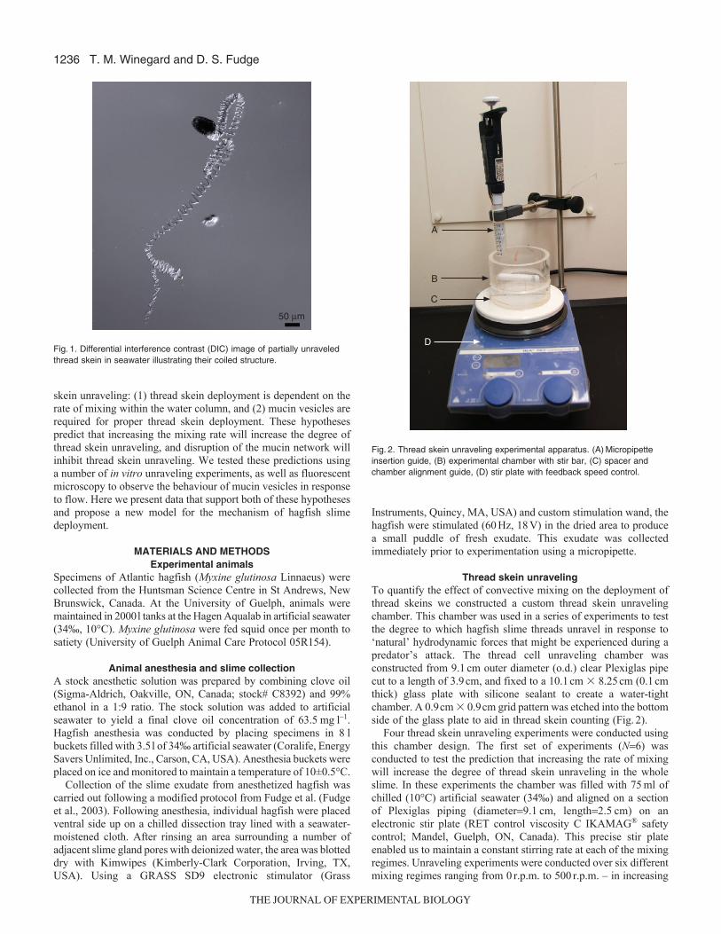
Thread skein unravelingTo quantify the effect of convective mixing on the deployment ofthread skeins we constructed a custom thread skein unravelingchamber. This chamber was used in a series of experiments to testthe degree to which hagfish slime threads unravel in response to‘natural’ hydrodynamic forces that might be experienced during apredator’s attack. The thread cell unraveling chamber wasconstructed from 9.1 cm outer diameter (o.d.) clear Plexiglas pipecut to a length of 3.9 cm, and fixed to a 10.1 cm � 8.25 cm (0.1 cmthick) glass plate with silicone sealant to create a water-tightchamber. A 0.9cm � 0.9cm grid pattern was etched into the bottomside of the glass plate to aid in thread skein counting (Fig. 2).
Four thread skein unraveling experiments were conducted usingthis chamber design. The first set of experiments (N=6) wasconducted to test the prediction that increasing the rate of mixingwill increase the degree of thread skein unraveling in the wholeslime. In these experiments the chamber was filled with 75 ml ofchilled (10°C) artificial seawater (34‰) and aligned on a sectionof Plexiglas piping (diameter=9.1 cm, length=2.5 cm) on anelectronic stir plate (RET control viscosity C IKAMAG® safetycontrol; Mandel, Guelph, ON, Canada). This precise stir plateenabled us to maintain a constant stirring rate at each of the mixingregimes. Unraveling experiments were conducted over six differentmixing regimes ranging from 0 r.p.m. to 500 r.p.m. – in increasing
T. M. Winegard and D. S. Fudge
50 µm
Fig. 1. Differential interference contrast (DIC) image of partially unraveledthread skein in seawater illustrating their coiled structure.
A
B
C
D
Fig. 2. Thread skein unraveling experimental apparatus. (A) Micropipetteinsertion guide, (B) experimental chamber with stir bar, (C) spacer andchamber alignment guide, (D) stir plate with feedback speed control.
THE JOURNAL OF EXPERIMENTAL BIOLOGY

1237Hagfish slime thread skein deployment
increments of 100 r.p.m.. A 5.1 cm magnetic stir bar was used tocreate the desired mixing forces in the chamber. Each trial wasconducted using 0.5ml of fresh slime exudate, which was collectedfrom the anesthetized hagfish following stimulation with amicropipette. The exudate was then introduced into the chamber,following removal of the stir bar, by placing the micropipette intoa custom insertion guide and ejecting the exudate into the seawater.The guide ensured consistent placement of the slime into thechamber (1.5 cm from inner wall of the chamber, 1 mm deep).Preliminary trials showed that the stir bar interacted with the slimeexudate in a way that made it difficult to measure the number ofskeins that failed to deploy. This was primarily a result of physicalinteraction of the stir bar with the exudate in the form ofentanglement of threads around it as well as shearing of pelletedskeins as the stir bar continued to rotate on the bottom of thechamber. Thus, while removing the stir bar was somewhat disruptiveto the flow in the chamber, we are confident that the exudate wassubjected to real differences in the level of hydrodynamic forcesamong the six mixing rates used. After water movement had ceased,the chamber was moved to a microscope stage (Nikon SMZ 1500,Nikon Canada, Mississauga, ON, Canada) that was chilled to 10°Cusing a water recirculation/chiller unit. Chilling was necessary tomaintain a consistent environment for the formed slime, as well asto prevent condensation and mitigate convective currents caused byheat given off by the microscope lamp. The condensed thread skeinsthat pelleted out of solution after each mixing regime were countedusing a monochrome digital camera (Q-imaging Retiga 1300,Surrey, BC, Canada) and Open Lap 3.5.1 software (Improvision,Waltham, MA, USA) on a computer. Counting the number ofcondensed thread skeins on the bottom provided an inverse measureof the degree of thread bundle unraveling in the whole slime. Datawere converted to the percentage of the number of skeins thatpelleted at 0 r.p.m..
The second set of experiments (N=6) involved a similarexperimental protocol; however, 75ml of 10mmoll–1 DTT solution(Sigma-Aldrich; CAT. #BP172-25), prepared in 34‰ artificialseawater at 10°C, was used. This experiment was aimed ataddressing the prediction that an intact mucin network is necessaryfor thread skein unraveling. The third set of stir-plate experiments(N=6) were conducted using 75 ml of 200 mmol l–1 b-mercaptoethanol solution (MP Biomedicals, Solon, OH, USA; CAT.#194705) prepared in 34‰ artificial seawater at 10°C. Thisexperiment was performed at the 0 r.p.m. and 400 r.p.m. mixingregimes to test whether increasing the rate of mixing couldcompensate for the complete disruption of the mucin network infacilitating thread skein unraveling.
The fourth experiment was designed to test whether a viscoussolution could rescue skein unraveling in the presence of 200mmoll–1 b-mercaptoethanol. These unraveling trials were conducted in aviscous 2% polyethylene oxide (MW 900,000 Da) in seawatersolution with a viscosity approximately 150� higher than that ofseawater. Quantification of skein unraveling could not be carriedout in these trials because the condensed skeins did not pellet outin the viscous solution but the results were clear nonetheless.
Visualization of mucin vesicles under shear flowPreliminary experiments focused on optimizing the fluorescenttagging of ruptured mucin vesicles by testing a variety of fluorescein-labeled lectin dyes (Vector Laboratories Lectin Kit; VectorLaboratories, Inc.; Burlingame, CA, USA; CAT. #FLK-2100).Visualization of the ruptured mucins was superlative using a lectinsoybean agglutinin dye diluted 500� in 10°C artificial seawater
(34‰). Using this dye solution we tested the response of mucinvesicles to shear flow conditions in order to gain insight into therole of mucins during thread skein deployment. Experiments wereconducted using microscope slide flow-through chambersconstructed from 3 mm � 1 mm � 1 mm glass slides and 18 mm� 18 mm � 0.17 mm cover glass (Fisher Scientific, Ottawa, ON,Canada). By focusing on mucin vesicles that adhered to the glassslide we could image the mucin vesicles and visualize their responseto shear flow. Hagfish slime used in this experiment was collected,as detailed above, but placed into a 10°C stabilization solution(0.9 moll–1 sodium citrate, 0.1moll–1 PIPES) modified from Spitzeret al. (Spitzer et al., 1988).
In preparation for experimentation, the chamber was first loadedwith the stabilization solution by adding a small 50 ml drop nearone opening to the chamber. After allowing capillary action to pullthis drop into and fill the chamber, a small volume (50ml) of mucinvesicle suspension was placed near the same opening to thechamber. Using small pre-cut filter paper (Whatman #1, Maidstone,Kent, UK), the sample was pulled into the chamber by drawingfrom the opposite opening. A 50 ml drop of artificial seawatercontaining the lectin dye was placed adjacent to the flow-throughopening. The dye solution was drawn through the chamber toobserve the response of the mucin vesicles to flow. Images werecaptured using a Nikon 90i Eclipse epifluorescent microscope witha FITC filter cube, Nikon Intensilight (C-HGFIE) and a cooledmonochrome Q-Imaging EXi 12-bit camera. NIS Elements ARsoftware (Nikon Instruments, Inc., Melville, NY, USA) was usedfor image capture and analysis.
Visualization of thread skein unravelingIn order to visualize the mechanism of thread skein deployment, asmall 5ml chamber (6cm � 4cm) was made on a 10.1cm � 8.25 cm(0.1cm thick) glass plate using a Liquid Blocker super PAP pen (DaidoSangyo Co. Ltd., Tokyo, Japan). This chamber was filled with 10°Cartificial seawater and situated on a chilled (10°C) microscope stage.A syringe pump (Harvard 33; Holliston, MA, USA) with a 10ml B-DGlass syringe and 0.6 mm inner diameter (i.d.) flexible tubing witha 0.2mm � 2.0mm (i.d.) Vitrotube tip (Fiber Optic Center, Inc; CAT.#3520) were used to create suction flow within the chamber.Experiments were conducted using 0.1ml of fresh exudate collectedusing a micropipette and placed on the glass surface within theseawater chamber. A flow rate of 200ml min–1 and monochrome Q-Imaging EXi 12-bit camera were used to record the exudates’response to flow at a rate of 21 frames per second.
Visualization of whole slime networkIn order to visualize the formed slime network, 0.2ml of fresh slimeexudate was placed into 2 ml of artificial seawater containing thefluorescein-labeled lectin soybean agglutinin dye (500:1). The slimewas established by agitating the 2 ml microcentrifuge tube up anddown five consecutive times. The whole slime was then poured ontoa glass slide in preparation for visualization. A Nikon 90i Eclipsemicroscope with a Nikon Intensilight (C-HGFIE) and Q-Imagingmonochrome 12-bit camera were again used to visualize the networkin both fluorescence and differential interference contrast (DIC).
Statistical analysisA two-way analysis of variance (ANOVA) analysis was conductedon the 100r.p.m. to 500r.p.m. data to determine whether the degreeof thread skein unraveling was significantly affected by mixing rate,and whether the use of DTT significantly affected the degree towhich the thread skeins unraveled. The 0r.p.m. percentage data were
THE JOURNAL OF EXPERIMENTAL BIOLOGY

1238
not used in this analysis because the values for each individualhagfish were all 100% by definition. A two-tailed t-test assumingequal variance was conducted to determine whether a significantdifference existed between the two mixing regimes (0 r.p.m. and400 r.p.m.) in the presence of b-mercaptoethanol.
RESULTSThread skein unraveling
Two-way ANOVA analysis revealed significant main effects ofmixing rate (P<0.001) and the presence of DTT (P<0.001) but nosignificant interactive effect (P=0.169). The results are consistentwith the prediction that increasing the mixing rate significantlyincreases the degree of thread skein unraveling in the whole slime(Fig.3). As such, this implicates convective mixing as a necessaryrequirement for thread skein unraveling and ultimately whole slimeformation. The number of condensed thread skeins as a proportionof the total number of skeins at 0r.p.m. appears to level off between400 r.p.m. (5.08±2.07%) and 500 r.p.m. (8.31±3.39%) in the noDTT treatment (Fig. 3). This suggests that a certain fraction ofskeins are resistant to unraveling, at least in exudates expressedfrom anesthetized hagfish. The data also support the hypothesisthat thread skein unraveling is significantly affected by thepresence of the mucin network during slime maturation.Furthermore, our attempts to completely disrupt the mucin networkwith high concentrations of b-mercaptoethanol revealed nosignificant differences (P=0.697) between the number ofcondensed threads at 0 r.p.m. (497±79) and 400 r.p.m. (459±51).The lack of unraveling in the presence of high concentrations ofb-mercaptoethanol, even at high mixing rates suggests thatconvective mixing is necessary but not sufficient to affect threadskein unraveling, at least at the mixing rates we investigated. Thesedata also underscore the importance of mucin vesicles to theunraveling of thread skeins in hagfish slime. We carried out furtherunraveling trials in the presence of 200mmoll–1 b-mercaptoethanoland a viscous 2% polyethylene oxide solution in seawater to testwhether the mucins aid in unraveling by increasing the localviscosity around thread skeins. At a mixing rate of 400 r.p.m., wesaw no evidence of skein unraveling in this solution, but we could
not quantify our results due to the fact that the condensed skeinswere not able to pellet out of the highly viscous polyethylene oxidesolution. We conducted a similar trial in the absence of 200 mmoll–1 b-mercaptoethanol and also found no evidence of unraveling,which suggests that increased viscosity actually inhibits threadskein unraveling.
Mucin strand formation, thread cell unraveling and wholeslime structure
Fluorescent-labeling of ruptured mucin vesicles demonstrated thatthe vesicles readily elongate into strands when subjected to flow inthe experimental chamber. Fig. 4 shows the formation of mucinstrands from swollen mucin vesicles within the flow-throughchamber. Exposure of the condensed slime exudates to flow createdby the syringe pump apparatus revealed three importantobservations: (1) swollen mucin vesicles elongate in response toflow to form mucin strands, (2) the elongated mucin strands attachto the thread skeins, and (3) the mucin strands transducehydrodynamic forces directly to the thread skeins and initiateunraveling by pulling them apart (Fig. 5) (see Movie 1 insupplementary material). Fluorescent imaging of the whole slimenetwork provided a first-ever view of the interaction between mucinstrands and threads in slime produced from un-stabilized exudate.Images taken with differential interference contrast (DIC) depictthe whole slime network (Fig. 6A), while the same image viewedin fluorescence highlights the complexity of mucin and threadinteractions in the whole slime (Fig. 6B).
DISCUSSIONThread skein unraveling
The results from these experiments provide strong evidence for theimportance of vigorous mixing in facilitating thread skein unravelingin hagfish slime. These results are consistent with previous findingsthat demonstrated the effects of mixing on whole slime formation(Fudge et al., 2005; Lim et al., 2006). These studies demonstratedthat hagfish slime exudate released into still seawater does notexplosively mature into the whole slime and requires significantmixing forces. Our results presented here indicate that the importance
T. M. Winegard and D. S. Fudge
0 100 200 300 400 5000
20
40
60
80
100
r.p.m.
Pel
lete
d th
read
ske
ins
(% o
f num
ber
at 0
r.p.
m.)
DTT
No DTT
Fig. 3. Mean (±s.e.m.) number of condensed thread skeins as a function ofmixing rate expressed as the percentage of the number of skeins at 0r.p.m.. These data provide an inverse measure of the degree of threadskein unraveling in the whole slime. Solid line indicates data for trials inwhich mucins were disrupted with 10 mmol l–1 dithiothreitol.
50 µm
Fig. 4. Fluorescent tagging of mucins using a fluorescein-labeled lectin dyeillustrates the formation of mucin strands from elongated mucin vesiclesexposed to shear in a flow-through chamber. Arrow indicates anaggregation of ruptured mucin vesicles that have begun elongating to formstrands.
THE JOURNAL OF EXPERIMENTAL BIOLOGY

1239Hagfish slime thread skein deployment
of mixing on whole slime formation is at least in part due to theunraveling of individual thread skeins.
Previous studies found that the disulfide cleaving compound DTThas two main effects on hagfish slime maturation – it reduces themass of whole slime formed in vitro, and it delays the time requiredfor slime maturation (Koch et al., 1991b; Fudge et al., 2005). Ourdata indicate that high concentrations of disulfide cleaving compoundscompletely abolish thread skein unraveling and even lowconcentrations of these compounds significantly reduce the degreeof thread skein unraveling in vitro. These results suggest that mucinsare absolutely crucial for skein unraveling and furthermore suggestthat DTT effects on whole slime are mediated at least in part by theireffects at the level of thread skein unraveling. The data in Fig.3 suggestthat higher rates of mixing can compensate for disruption of the mucinnetwork with DTT; however, the concentration used (10mmoll–1) inthese experiments was probably not sufficient to completely disruptthe mucin networks (Koch et al., 1991b), especially over the timescaleof unraveling. The use of high concentrations (200 mmol l–1) of the
more highly diffusible b-mercaptoethanol, however, abolishedunraveling completely, even at high mixing rates. These data stronglysuggest that an intact mucin network is absolutely crucial for threadskein deployment.
A new model of hagfish slime deployment and structureAn early model of hagfish slime deployment proposed that threadskeins are packaged under pressure within the GTCs (Newby, 1946).According to this hypothesis, contact with seawater causes anosmotic swelling of the GTC and rupture of the membrane, followedby a rapid expansion and unraveling of the thread driven by a releaseof internal pressure. Newby also proposed that the role of the mucinsis simply to act as a fluid vehicle for the thread skeins. The discoverythat the GTCs in hagfish slime are liberated of their plasmamembranes during release of slime from the gland (Fernholm, 1981)cast doubt on Newby’s hypothesis. More recent models of hagfishslime deployment propose that the initiation of thread skeinunraveling is dependent on seawater-induced swelling of threadskeins as well as physical perturbation of the skeins themselves(Fernholm, 1981; Koch et al., 1991a). The model that we put forthhere differs from these previous models in that it directly implicatesthe mucin vesicles in the process of thread skein unraveling anddemonstrates their crucial importance in slime deployment. We alsosaw no evidence that thread skein swelling precedes unraveling overthe time course of natural slime maturation.
Visualization of mucins with fluorescently labeled lectins revealedthat mucin vesicles exposed to shear stresses in a flow chamberelongate into high aspect ratio mucin strands that readily attach tothreads and thread skeins. Previous models suggested that the mucins
A B
C D
E F
G
Fig. 5. Slime exudate exposed to flow created by a syringe pump.(A) Condensed exudate puddle prior to flow. Note the condensed threadskeins on the far right and the mucin vesicle boundary layer to the left.(B,C) Mucin strands and chains begin to form as flow is initiated.(D,E) Mucin strand attachment to thread skeins and movement of threadskeins. (F,G) Unraveling of thread skeins initiated. Arrowheads indicate anunraveling thread skein, arrows indicate aggregations of mucin strands.
A
B
Fig. 6. Whole slime formed in seawater containing the fluorescent lectin dye(A) Differential interference contrast (DIC) image of whole slime networkdepicting unraveled threads and mucin strand network. Arrowheadindicates mucin strands connecting slime threads, and the arrow indicatesa slime thread. (B) Same image viewed in fluorescence highlights thecomplexity of the mucin network.
THE JOURNAL OF EXPERIMENTAL BIOLOGY

1240
remain as coherent structures after they swell and rupture (Fudge etal., 2005; Koch et al., 1991b). Based on these observations, as wellas direct observations of fresh slime exudates mixed with seawaterunder the microscope, we propose a new mechanism of hagfish slimedeployment in which elongated mucin strands transmit mixing forcesto thread skeins to initiate their unraveling. According to this ‘mucintransmission hypothesis,’ hagfish slime deployment occurs via thefollowing sequence of events: (1) expulsion of the slime exudate intoconvectively mixing seawater results in the swelling and subsequentelongation of mucin vesicles to form mucin strands, (2) theseelongated mucin strands attach to the thread skeins, (3) the mucinstrands transmit the hydrodynamic forces of mixing to the threadskeins, thereby initiating unraveling, and (4) entanglement of thethreads and mucin strands results in the complete unraveling of threadskeins. The whole slime is therefore a highly complex network ofmucin strands, slime threads and seawater.
The results of this study also provide a new model for whole slimestructure in which thin mucin strands interact with unraveled slimethreads to yield a complex network capable of the viscous entrainmentof water described by Fudge et al. (Fudge et al., 2005) (Fig. 6A,B).Previous studies depicted swollen mucin vesicles decorating the slimethreads (Fudge et al., 2005; Koch et al., 1991a). According to thesemodels, the viscous entrainment of seawater within fully formed slimewas accomplished by confining water to channels between entangledslime threads covered with swollen mucin vesicles. The findings ofthis study expand our understanding of how the mucins and threadsslow the flow of water by forming a complex network of mucins andthreads in which water can be entrained.
Our results raise interesting questions about how exactly the mucinvesicles bring about thread skein unraveling. Videomicroscopy(Fig. 5) (see Movie 1 in supplementary material) provided compellingevidence that the mucin strands are capable of exerting elastic tensileforces on thread skeins but another possibility is that mucins facilitatethe transfer of shear stresses to the skeins by ‘viscosifying’ theseawater around them. To test this idea, we carried out an experimentto see if high viscosity solutions can rescue the abolition of skeinunraveling by b-mercaptoethanol. Not only did a viscous solutionof high molecular weight polyethylene oxide in seawater not rescueskein unraveling at high mixing rates (400r.p.m.), it had an inhibitoryeffect, as unraveling did not occur in a subsequent trial in which b-mercaptoethanol was not present.
A deeper understanding of how mucins transmit hydrodynamicforces to the thread skeins will require knowledge of the mechanicalproperties of swollen mucin vesicles. While little is known abouthagfish slime mucin material properties, chemical analyses suggestthat they may have distinct physical properties. Typical vertebratemucins contain up to 85% carbohydrate by dry mass but hagfishmucins contain only 12% (Salo et al., 1983). It is possible that theproperties of the mucins have been shaped by selection to optimizetheir ability to transmit hydrodynamic forces to thread skeins. Forexample, mucins must be compliant enough to elongate into strandswhen exposed to shear or elongational flow (in spite of beingconsiderably smaller than thread skeins), and yet the strands thatform must be robust enough to exert tensile forces on the skeinsthat can initiate their unraveling.
These results suggest that viscous transfer of shear forces to threadskeins is not sufficient to effect their unraveling. Furthermore,viscous solutions are likely to be inhibitory for skein unravelingbecause of their ability to dissipate turbulent energy before it canreach the microscale inhabited by the skeins. By contrast, soft,swollen mucin vesicles are susceptible to shear and extensional flow,which can elongate the vesicles into high aspect ratio elastic strands
that can attach to the skeins and increase their effective size. Wepropose that these mucin strands are capable of transmittinghydrodynamic forces to the skeins and exerting tensile forces onthem that effect their unraveling. More generally, the mechanismdescribed above may be valuable for industrial processes in whichhydrodynamic mixing forces are transmitted to suspended particlesthat are typically immune to such forces due to their small size.
CONCLUSIONSIn this study, we provide evidence that convective mixing and intactmucin networks are required for proper deployment of thread skeinsin hagfish slime. We also provide evidence for a new mechanismof hagfish slime deployment in which elongated mucin vesiclestransmit hydrodynamic forces to the skeins and facilitate theirunraveling. Our work provides a new model for hagfish slimestructure and also provides novel insights into the evolution of theslime, suggesting that the evolution of thread skeins could not havepreceded the appearance of the mucin vesicles in hagfish ancestors.The mechanisms described here may be of use for industrialprocesses in which hydrodynamic forces are transmitted tomicroscale particles that are typically immune to mixing forces.
LIST OF ABBREVIATIONSDIC differential interference contrastDTT dithiothreitolGMCs gland mucous cellsGTCs gland thread cells
ACKNOWLEDGEMENTSWe would like to thank several students who worked on this project and helped usdevelop some of the techniques used: Jessica Martino, Eyal Ellenbogen,Stephanie Kotschwar and Clare Armstrong. We also would like to thank GarethMcKinley and Randy Ewoldt for providing feedback on the manuscript. Thanksalso to Bob Frank for the capture and transport of the hagfish used in the study.This work was supported by a Natural Sciences and Engineering ResearchCouncil of Canada Discovery grant to D.S.F.
REFERENCESDowning, S. W., Spitzer, R. H., Salo, W. L., Downing, J. S., Saidel, L. J. and Koch,
E. A. (1981). Threads in the hagfish slime gland thread cells-organization,biochemical features, and length. Science 212, 326-328.
Fernholm, B. (1981). Thread cells from the slime glands of hagfish (Myxinidae). ActaZool. 62, 137-145.
Fudge, D. S., Gardner, K. H., Forsyth, T., Riekel, C. and Gosline, J. M. (2003). Themechanical role of intermediate filaments in cells: insights from hagfish slimethreads. Biophys. J. 85, 2015-2027.
Fudge, D. S., Levy, N., Chiu, S. and Gosline, J. M. (2005). Composition, morphologyand mechanics of hagfish slime. J. Exp. Biol. 208, 4613-4625.
Fudge, D. S., Winegard, T., Ewoldt, R. H., Beriault, D., Szewciw, L. and McKinley,G. H. (2009). From ultra-soft slime to hard a-keratins: the many lives of intermediatefilaments. J. Integr. Comp. Biol. 43, 32-39.
Koch, E. A., Spitzer, R. H. and Pithawalla, R. B. (1991a). Structural forms andpossible roles of aligned cytoskeletal biopolymers in hagfish (slime eel) mucus. J.Struct. Biol. 106, 205-210.
Koch, E. A., Spitzer, R. H., Pithawalla, R. B. and Downing, S. W. (1991b). Keratin-like components of gland thread cells modulate the properties of mucus from hagfish(Eptatretus stouti). Cell Tissue Res. 264, 79-86.
Koch, E. A., Spitzer, R. H., Pithawalla, R. B. and Parry, D. A. D. (1994). An unusualintermediate filament subunit from the cytoskeletal biopolymer released extracellularlyinto seawater by the primitive hagfish (Eptatretus stouti). J. Cell Sci. 107, 3133-3144.
Leppi, T. J. (1968). Morphochemical analysis of mucous cells in the skin and slimeglands of hagfishes. Histochemie 15, 68-78.
Lim, J., Fudge, D. S., Levy, N. and Gosline, J. M. (2006). Hagfish slimeecomechanics: testing the gill-clogging hypothesis. J. Exp. Biol. 209, 702-710.
Newby, W. W. (1946). The slime glands and thread cells of hagfish, Polistrotremastouti. J. Morphol. 78, 397-409.
Salo, W. L., Downing, S. W., Lidinsky, W. A., Gallagher, W. H., Spitzer, R. H. andKoch, E. A. (1983). Fractionation of hagfish slime gland secretions: partialcharacterization of the mucous vesicle fraction. Prep. Biochem. 13, 103-135.
Spitzer, R. H., Downing, S. W., Koch, E. A., Salo, W. L. and Saidel, L. J. (1984).Hagfish slime gland thread cells. 2. Isolation and characterization of intermediatefilament components associated with the thread. J. Cell Biol. 98, 670-677.
Spitzer, R. H., Koch, E. A. and Downing, S. W. (1988). Maturation of hagfish glandthread cells – composition and characterization of intermediate filament polypeptides.Cell Motil. Cytoskeleton 11, 31-45.
Verdugo, P. (1991). Mucin exocytosis. Am. Rev. Respir. Dis. 144, S33-S37.
T. M. Winegard and D. S. Fudge
THE JOURNAL OF EXPERIMENTAL BIOLOGY