depletion of collagen ii-reactive t cells and blocking of ... · age of 7 wk old and boosted at the...
TRANSCRIPT

of February 1, 2019.This information is current as
Collagen II-Induced Arthritis in DBA/1j MiceBlocking of B Cell Activation Prevents Depletion of Collagen II-Reactive T Cells and
and John D. MountzHsuLiu, Liang Xiu, Tong Zhou, Yongming Wang, Hui-Chen
Huang-Ge Zhang, PingAr Yang, Jinfu Xie, Zhongyu Liu, Di
http://www.jimmunol.org/content/168/8/4164doi: 10.4049/jimmunol.168.8.4164
2002; 168:4164-4172; ;J Immunol
Referenceshttp://www.jimmunol.org/content/168/8/4164.full#ref-list-1
, 19 of which you can access for free at: cites 45 articlesThis article
average*
4 weeks from acceptance to publicationFast Publication! •
Every submission reviewed by practicing scientistsNo Triage! •
from submission to initial decisionRapid Reviews! 30 days* •
Submit online. ?The JIWhy
Subscriptionhttp://jimmunol.org/subscription
is online at: The Journal of ImmunologyInformation about subscribing to
Permissionshttp://www.aai.org/About/Publications/JI/copyright.htmlSubmit copyright permission requests at:
Email Alertshttp://jimmunol.org/alertsReceive free email-alerts when new articles cite this article. Sign up at:
Print ISSN: 0022-1767 Online ISSN: 1550-6606. Immunologists All rights reserved.Copyright © 2002 by The American Association of1451 Rockville Pike, Suite 650, Rockville, MD 20852The American Association of Immunologists, Inc.,
is published twice each month byThe Journal of Immunology
by guest on February 1, 2019http://w
ww
.jimm
unol.org/D
ownloaded from
by guest on February 1, 2019
http://ww
w.jim
munol.org/
Dow
nloaded from

Depletion of Collagen II-Reactive T Cells and Blocking of BCell Activation Prevents Collagen II-Induced Arthritis inDBA/1j Mice
Huang-Ge Zhang,1*† PingAr Yang,* Jinfu Xie,* Zhongyu Liu,* Di Liu,* Liang Xiu,*Tong Zhou,* Yongming Wang,* Hui-Chen Hsu,* and John D. Mountz*†
Collagen II (CII)-induced arthritis in DBA/1j mice is mediated by both CII-reactive T cells and anti-CII Ab-producing B cells. Todetermine the relative role of these processes in the development of arthritis, we specifically eliminated CII-reactive T cells bytreating the mice with CII-pulsed syngeneic macrophages that had been transfected with a binary adenovirus system. Thesemacrophages express murine Fas ligand in a doxycycline-inducible manner with autocrine suicide inhibited by concomitantexpression of p35. The mice were treated i.v. with four doses of CII-APC-AdFasLp35Tet or a single dose of AdCMVsTACI (5�109 PFU), or both simultaneously, beginning 2 wk after priming with CII in CFA. Treatment with CII-APC-AdFasLp35Tet aloneor in combination with a single dose of AdCMVsTACI prevented the development of CII-induced arthritis and T cell infiltrationin the joint. The elimination of T cells was specific in that a normal T cell response was observed on stimulation with OVA aftertreatment with CII-APC-AdFasLp35Tet. Treatment with AdCMVsTACI alone prevented production of detectable levels of cir-culating anti-CII autoantibodies and reduced the severity of arthritis but did not prevent its development. These results indicatethat the CII-reactive T cells play a crucial role in the development of CII-induced arthritis and that the anti-CII Abs act to enhancethe development of CII-induced arthritis. The Journal of Immunology, 2002, 168: 4164–4172.
Rheumatoid arthritis is a chronic, progressive joint diseasethat is characterized by lymphocytic invasion of the sy-novial lining and hyperplasia of the resident synovio-
cytes (1). The overproduction of cytokines along with other factorsresults in cartilage destruction, bone erosion, and remodeling ofjoint structures (2–5). Typically, patients suffering from rheuma-toid arthritis exhibit systemic manifestations, most notably the pro-duction of autoantibodies (6, 7). An understanding of the relativecontributions of the T and B cell compartments of the immunesystem in the initiation and perpetuation of the disease process inrheumatoid arthritis is necessary for the design of effective thera-peutic strategies.
Collagen II (CII)-induced arthritis in DBA/1j mice is an auto-immune model of human rheumatoid arthritis (8). During the ini-tial stages of this disease, severe joint inflammation is associatedwith infiltration of leukocytes and production of proinflammatorycytokines. A chronic disease state, characterized by synovial cellhyperplasia and destruction of cartilage and bone, is then estab-lished. CII arthritis is associated with production of murine CIIautoantibodies that have been proposed to contribute to diseaseprogression (9–12). CII-induced arthritis can be transferred using
CD4-positive T cells, indicating that T cells can initiate the diseaseand play an important role in the early stages of development ofthis disease (13, 14). CII arthritis is associated with generation ofT cells specific for CII epitopes presented by APCs since it hasbeen possible to transfer disease using a CII-specific T cell clonein SCID mice after boosting with CII (15–19).
A central objective in the development of the new generation oftherapies for the treatment of rheumatoid arthritis is specific inhi-bition of the inflammatory disease processes in the absence ofgeneralized immunosuppression (1–4). As APCs play a centralrole in defining Ag specificity, they provide an access point forspecific manipulation of the immune system. It is well establishedthat APCs, such as macrophages, express processed Ags that spe-cifically stimulate those T cells that recognize processed Ag in thecontext of the MHC. We have developed techniques for modifyingAPCs such that they express specific Ags along with Fas ligand(FasL) and have demonstrated that these modified APCs deleteonly those T cells that recognize the specific Ag and do not incurgeneral immunosuppression or decreased B cell function (20–25).The interaction of APCs, including macrophages, with B cells is me-diated by the expression by the APCs of B lymphocyte stimulator(BlyS), A proliferation-inducing ligand, and B cell-activating factor ofthe TNF family that interact with the transmembrane activator andcalcium modulator and cyclophilin ligand interactor (TACI), B cellmaturation Ag, and BAFF receptor on the B cells (26–29). The in-teraction of the APCs and the B cells can be blocked by either solubleTACI (sTACI) or soluble B cell maturation Ag (29). CII arthritis wasprevented by administration of high-dose sTACI (100�g i.v.) admin-istered 3 days a week for 6 wk, which at this level blocks both T celland B cell activation (30).
In this study, we examined the relative contribution of Ag-spe-cific T cells and autoantibodies by eliminating the Ag-specific Tcells and blocking autoantibody production. The results show thattreatment with CII-APC-AdFasLp35Tet eliminates CII-reactive T
*University of Alabama, Birmingham, AL 35294; and†Birmingham Veterans Ad-ministration Medical Center, Birmingham, AL 35233
Received for publication September 7, 2001. Accepted for publication February13, 2002.
The costs of publication of this article were defrayed in part by the payment of pagecharges. This article must therefore be hereby markedadvertisement in accordancewith 18 U.S.C. Section 1734 solely to indicate this fact.1 Address correspondence and reprint requests to Dr. Huang-Ge Zhang, University ofAlabama, 701 South 19th Street, LHRB 473, Birmingham, AL 35294-0007. E-mailaddress: [email protected] Abbreviations used in this paper: CII, collagen II; BlyS, B lymphocyte stimulator;rtTA, reverse tetracycline transactivator; FasL, Fas ligand; TRE, tetracycline responseelement; TACI, transmembrane activator and calcium modulator and cyclophilin li-gand interactor; sTACI, soluble TACI; cps, counts per second; Dox, deoxycycline.
Copyright © 2002 by The American Association of Immunologists 0022-1767/02/$02.00
by guest on February 1, 2019http://w
ww
.jimm
unol.org/D
ownloaded from

cells and inhibits development of CII-induced arthritis but does notsignificantly diminish the production of anti-CII Abs. Treatmentwith AdCMVsTACI alone abrogates the production of anti-CIIAbs, but at the levels used here, it did not decrease CII Ag-specificT cell responses and did not greatly diminish the development ofCII-induced arthritis. Combined treatment with CII-APC-AdFasLp35Tet plus AdCMVsTACI prevents both the develop-ment of CII-reactive T cells and the production of anti-CII Abs,and effectively blocks the development of CII-induced arthritis.
Materials and MethodsMice
Female homozygous DBA/1j mice (7 wk old) were obtained from TheJackson Laboratory (Bar Harbor, ME). All mice were kept in a roomequipped with an air filtering system. The cages, bedding, water, and foodwere sterilized, and the mice were handled with sterile gloves.
Induction of arthritis
DBA/1j female mice were immunized at 7 wk of age at the base of the tailwith 200 �g of bovine CII dissolved in 100 �l of 0.05 M acetic acid andmixed with an equal volume (100 �l) of CFA (Chondrex, Redmond, WA).Four weeks later, the animals were reimmunized with 200 �g of bovine CIIin IFA.
Treatment protocols using CII-APC-AdFasLp35Tet andAdCMVsTACI
To differentiate the roles of T cells and B cells in CII-induced arthritis, theeffects of five treatment protocols were compared as follows: 1) CII-APC-AdFasLp35Tet alone with or without a doxycycline (Dox) inducer; 2) AdCMVsTACI alone; 3) CII-APC-AdFasLp35Tet in combination with AdCMVsTACI; 4) AdGFP; and 5) CII-APC-AdGFP. To delete the CII-reactive T cells,peritoneal macrophages from DBA/1j mice were pulsed with T cell prolifer-ation grade Arthrogen-CIA type II collagen (Chondrex) as described by themanufacturer, then transfected with AdTREFasLp35 plus AdCMVrtTA (CII-APC-AdFasLp35Tet) as described above. Treatment of the DBA/1j mice withthese APCs was commenced 2 wk after the mice had been immunized with CIIin CFA, with 106 CII-APC-AdFasLp35Tet administered twice per week for 2wk. As a control, other groups of mice were administered CII-pulsed APCsthat had been transfected with AdGFP (CII-APC-AdGFP) i.p. Induction ofFasL on these APCs was accomplished by addition of Dox to the drinkingwater (from 1 to 8 mg/ml in 2-fold increments) with 4% sucrose for 6 wkstarting at the time of administration of CII-APC-AdFasLp35Tet therapy. Totest whether the treatment of CII-APC-AdFasLp35Tet resulted in CII-specificT cell deletion without impairing host immune response to an irrelevant Ag, allCII-primed mice were cochallenged with OVA simultaneously. In brief, allfive groups of mice were s.c. immunized with 10 �g of OVA in CFA at theage of 7 wk old and boosted at the same dose in IFA 4 wk later.
To block the CII-reactive B cell activation, a single dose (5 � 109 PFU)of recombinant adenovirus expressing soluble sTACI (AdCMVsTACI)was injected i.v. 2 wk after immunization of the mice with CII in CFAeither alone or simultaneously with the initial CII-APC-AdFasLp35Tettreatment. Control mice were injected i.v. with CII-APC-AdGFP (5 � 109
PFU) or both.
Construction of inducible FasL adenovirus expression vector
An adenovirus coexpressing inducible FasL and p35 was constructed asdescribed previously (23–25). Briefly, the p35 anti-apoptosis gene (gener-ously supplied by Dr. Jiang Ling, University of Chicago, IL) was direc-tionally cloned into the BamHI and XbaI site of the pCA14 vector (Mi-crobix, Ontario, Canada). The p35 expression cassette, including a CMVpromoter and the SV40 poly(A) tail, was then excised with BglII and sub-sequently inserted into the BglII site of the pShuttle vector as described byHe et al. (31), resulting in the production of a pShuttleCMVp35. The full-length FasL was first cloned into the BamHI polylinker site of a pTREvector (Clontech Laboratories, Palo Alto, CA) site. Then, the TRE-regu-lated fas ligand fragment, including the bovine growth hormone poly(A)tail, was excised with XhoI and HindIII, followed by insertion into theKlenow-filled NotI site of the pShuttleCMVp35, leading to the productionof pShuttlep35TREFasL. The recombinant adenovirus AdTREFasLp35was produced by in vitro recombination of pShuttlep35TREFasL withpAdeasy1 as described previously (31). AdTREFasLp35 was produced in293 cells as described elsewhere (32). Recombinant AdCMVrtTA was con-structed as previously described (33) using the rtTA construct generously
provided by Dr. J. B. Uney (University of Bristol, Bristol, U. K.) (34) toallow expression of the reverse tetracycline transactivator (rtTA), therebyenabling Dox-inducible expression of FasL.
Regulation of FasL expression by Dox
Expression of functional FasL on the APC-AdFasLp35Tet or APC-AdGFPcells was evaluated by coincubation with FasL-sensitive A20 cells. Peri-toneal macrophages derived from DBA/1j mice were used as the APC. Themacrophages were transfected with AdTREFasLp35 plus AdCMVrtTA(35, 36) at 50 PFU/cell of each virus for 1 h, followed by washing, andincubation for an additional 18 h. The transfected macrophages were thenincubated with different concentrations of Dox (Sigma-Aldrich, St. Louis,MO) for 18 h. The in vitro activity of the macrophages was estimated usinga cytotoxicity assay in which they were mixed with 51Cr-labeled A20 targetcells at different E:T ratios. The cell cytotoxicity was determined by mea-suring the radioactivity in the cell culture supernatants at 8 h after cocul-ture [(cytotoxicity � (cpmexperimental � cpmspontaneous)/(cpmmaximal �cpmspontanous) � 100)].
Prevention of autocrine apoptosis of Fas-positive engineeredAPC derived from macrophages obtained from DBA/1j mice
The ability of p35 to inhibit apoptosis of the Fas-positive peritoneal mac-rophages from DBA/1j mice was determined after transfection with eitherAdLoxpFasL with AxCANCre (21, 25) or AdTREFasLp35 with AdCMVrtTA (AdTREFasLp35Tet) as described above and incubated withDox at different concentrations for 24 h. The percentage of apoptotic cellsafter Dox induction of the FasL on the DBA/1j macrophages was evaluatedusing the ATP-lite assay (25). ATP is determined as light units measuredas counts per second (cps) in a luminescence counter (Packard Instrument,Meriden, CT). The percentage of apoptotic cells was expressed as (1 � cpssample/cps control) � 100.
Construction of adenoviruses that produce sTACI and BlyS
An adenovirus expressing sTACI and murine BlyS was constructed ac-cording to a standard protocol (31). Briefly, the extracellular portion ofTACI (aa 1–165) was PCR amplified using cDNA synthesized from totalRNAs extracted from the Raji cell line purchased from American TypeCulture Collection (Manassas, VA). After sequence confirmation, thesTACI was ligated directionally into the BglII and SalI sites of thepAdTRACKCMV vector (31) and the human IgG Fc was fused intothe downstream SalI site of sTACI in the pAdTRACKCMV as describedby Cheng et al. (37). Full-length murine BlyS was PCR amplified usingcDNA synthesized from total RNAs extracted from the DBA/1j macro-phages. The sequence of BlyS was confirmed, and BlyS was directionallyligated into the Bglll and EcoRV sites of the PAdTRACKCMV vector (31).The production of recombinant AdCMVBlyS was accomplished as de-scribed elsewhere (33). The production of recombinant AdCMVsTACIwas accomplished as described in the protocol above. sTACI protein wasproduced in 293 cells, purified using a protein G column, and stored at�20°C in 100-�l aliquots until used.
Evaluation of development of arthritis
A loop calibrator was used to determine the diameter of each paw of eachmouse every day. Paw swelling was determined as the increase in diametercompared with the diameter at the initiation of the experiment. The severityof arthritis was graded according to the following scale, 0, normal with noswelling and erythema and no increase in joint diameter; 1, slight swellingand erythema with 0.1–0.3 mm increase in joint diameter; 2, swelling anderythema and 0.3–0.6 mm increase in joint diameter; 3, extensive swellingand erythema with 0.6–0.9 mm in joint diameter; and 4, pronounced swell-ing and erythema with joint thickness of 0.9–1.2 mm increase or obviousjoint destruction associated with visible joint deformity or ankylosis. Eachlimb was graded, resulting in a maximum clinical score of 16 per animaland expressed as the mean score on a given day.
After sacrifice, the joints (knee, elbow, ankle, and wrist) were harvested,fixed in 10% formaldehyde/PBS for at least 24 h, decalcified using EDTAfor 3 wk, sectioned at 4-�m thickness, deparaffinized, and stained withH&E (CMS, Houston, TX).
Evaluation of T cell or B cell infiltration in synovial joints
The phenotype of the infiltrating cells in the joints was determined byimmunoperoxidase staining of the tissue sections with an anti-CD3 Ab toidentify T cells or an anti-B220 Ab to identify B cells. Quenching of en-dogenous peroxidase was accomplished by incubating tissue sections with3% H2O2 for 10 min at room temperature in a humidified chamber. After
4165The Journal of Immunology
by guest on February 1, 2019http://w
ww
.jimm
unol.org/D
ownloaded from
washing with PBS, tissue sections were incubated with 0.1% trypsin at37°C for 10 min to reveal fixed Ag epitopes. Tissue sections were treatedwith the denaturing solution for 30 min at room temperature and blockingsolution for 10 min at room temperature. They were then incubated withHRP-conjugated anti-CD3 or B220 (DAKO, Carpinteria, CA). A DABstaining kit (DAKO) was used for visualization of Ab binding and theslides were counterstained with methyl green. At least five areas werechosen randomly for assessment of the percentage of CD3-positive cells orB220-positive cells in each specimen.
Analysis of TACI-BlyS interactions in the presence ofAdCMVsTACI
DBA/1j macrophages were transfected with AdCMVBlyS and gamma ir-radiated before use as stimulator cells. These stimulator cells were culturedat different ratios with A20 B cells, which express TACI, in the presenceof different concentrations of sTACI. Proliferation was evaluated using the[3H]thymidine uptake method.
Analysis of Ag-specific T cell response after CII-APC-AdFasLtreatment.
The CII-specific proliferative response of draining lymph node T cells wasevaluated by measuring the [3H]thymidine uptake of T cells coculturedwith gamma irradiated syngeneic APCs pulsed with or without bovine CIIand quantification of IL-2 production as described previously (24). In brief,1 �Ci of [3H]thymidine was added daily after coculture, the cells wereharvested 16 h later, and the incorporation of [3H]thymidine was deter-mined using a scintillation counter.
To determine whether the CII-APC-AdFasL treatment is Ag specific,the gamma-irradiated syngeneic APCs were also pulsed with OVA (1 �g/ml) and subsequently cocultured with the T cells from each group oftreated mice (APC:T cells � 1:10) for 5 days. The supernatants were col-lected at 72 and 96 h and production of IL-2 (IL-2) was quantified using anELISA kit as described (BioSource International, Camarillo, CA). The Tcell proliferation assay was also performed by [3H]thymidine incorporationassay as described above.
ELISA quantification of sTACI induction and autoantibodyproduction
The concentration of sTACI in the circulating blood was quantified usingan ELISA at different time points up to 50 days after administration ofAdCMVsTACI. In brief, the concentrations of the sTACI-Fc protein weredetermined using a rabbit anti-human Fc polyclonal Ab-coated plate tocapture the human Fc component of the recombinant protein, and the cap-tured protein was quantified using a rabbit anti-human Fc polyclonal Ab
conjugated to HRP. Sample dilutions were compared with standard curvesof human Fc (Sigma-Aldrich) to determine the concentrations of sTACI-Fc. The serum levels of anti-mouse CII IgG were assayed using a CIIELISA kit (Chondrex) before treatment and on day 50 after induction ofCII arthritis. The standard curve was produced using an anti-CII Ab pro-vided with the ELISA kit.
Statistical analysis
The results are expressed as the mean � SEM. The two-tailed Student’s ttest was used for statistical analysis. A p � 0.05 was considered to bestatistically significant.
ResultsAdTREFasLp35 plus AdCMVrtTA confers Dox-inducibleexpression of FasL in the absence of autocrine apoptosis
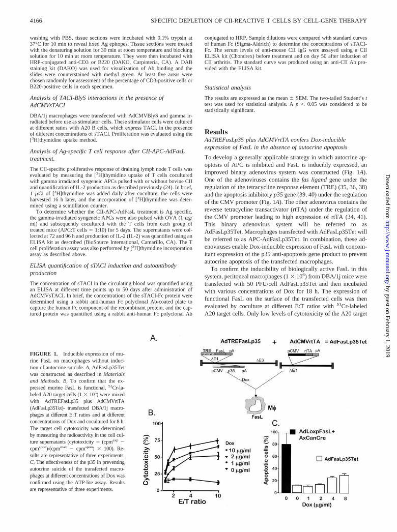
To develop a generally applicable strategy in which autocrine ap-optosis of APC is inhibited and FasL is inducibly expressed, animproved binary adenovirus system was constructed (Fig. 1A).One of the adenoviruses contains the fas ligand gene under theregulation of the tetracycline response element (TRE) (35, 36, 38)and the apoptosis inhibitory p35 gene (39, 40) under the regulationof the CMV promoter (Fig. 1A). The other adenovirus contains thereverse tetracycline transactivator (rtTA) under the regulation ofthe CMV promoter leading to high expression of rtTA (34, 41).This binary adenovirus system will be referred to asAdFasLp35Tet. Macrophages transfected with AdFasLp35Tet willbe referred to as APC-AdFasLp35Tet. In combination, these ad-enoviruses enable Dox-inducible expression of FasL with concom-itant expression of the p35 anti-apoptosis gene product to preventautocrine apoptosis of the transfected macrophages.
To confirm the inducibility of biologically active FasL in thissystem, peritoneal macrophages (1 � 106) from DBA/1j mice weretransfected with 50 PFU/cell AdFasLp35Tet and then incubatedwith various concentrations of Dox for 18 h. The expression offunctional FasL on the surface of the transfected cells was thenevaluated by coculture at different E:T ratios with 51Cr-labeledA20 target cells. Only low levels of cytotoxicity of the A20 target
FIGURE 1. Inducible expression of mu-rine FasL on macrophages without induc-tion of autocrine suicide. A, AdFasLp35Tetwas constructed as described in Materialsand Methods. B, To confirm that the ex-pressed murine FasL is functional, 51Cr-la-beled A20 target cells (1 � 105) were mixedwith AdTREFasLp35 plus AdCMVrtTA(AdFasLp35Tet)- transfected DBA/1j macro-phages at different E:T ratios and at differentconcentrations of Dox and cocultured for 8 h.The target cell cytotoxicity was determinedby measuring the radioactivity in the cell cul-ture supernatants (cytotoxicity � (cpmexp �cpmspon)/(cpmmax � cpmspon) � 100). Re-sults are representative of three experiments.C, The effectiveness of the p35 in preventingautocrine suicide of the transfected macro-phages at different concentrations of Dox wasconfirmed using the ATP-lite assay. Resultsare representative of three experiments.
4166 SPECIFIC DEPLETION OF CII-REACTIVE T CELLS BY CELL-GENE THERAPY
by guest on February 1, 2019http://w
ww
.jimm
unol.org/D
ownloaded from

cells were observed on incubation with the transfected macro-phages in the absence of Dox (Fig. 1B). The addition of Dox re-sulted in a Dox dose-dependent increase in the death of the A20target cells at 18 h, which was evident at doses of Dox that rangedfrom 1 to 10 �g/ml (Fig. 1B).
To test whether the AdFasLp35Tet construct effectively pre-vents autocrine apoptosis, the viability of the DBA/1j macrophagestransfected with AdFasLp35Tet was evaluated after incubationwith 0.0 to 8.0 �g/ml Dox for 18 h, as determined in an ATP-liteassay. As a control, DBA/1j macrophages were transfected withthe AdLoxpFasL plus AxCANCre, which results in expression ofFasL independently of Dox induction and in the absence of theanti-apoptosis gene p35 (39, 40). As anticipated, the control mac-rophages transfected with AdLoxPFasL plus AxCANCre under-went 80% of the apoptosis (Fig. 1C, column 1). In contrast, themacrophages transfected with AdFasLp35Tet exhibited onlylow levels of apoptosis even in the presence of high levels ofDox (Fig. 1C).
Decreased CII arthritis after treatment with CII-APC-AdFasLp35Tet in vivo
To demonstrate whether the treatment of CII-APC-AdFasLp35Tetcan prevent CII-induced arthritis, the macrophages were pulsedwith bovine CII and then transfected with AdFasLp35Tet. Thesemacrophages were then used to treat DBA/1j mice commencing 2wk after the mice had been immunized with CII in CFA at 9 wk ofage, as shown in Fig. 2A. Mice received a total of four doses ofmacrophages (106 cells/dose) over a 2-wk time period. At the sametime, the mice (10 mice/group) received 1.5 mg/ml Dox adminis-tered in the drinking water with 4% sucrose or 0.3% ethanol waterwith 4% sucrose as a control. The mice were next immunized withCII in IFA 4 wk after the first immunization at 11 wk of age, andthe development of arthritis was assessed weekly up to 20 wk ofage (Fig. 2A). The administration of Dox alone in the range of1.0–8.0 mg/ml confirmed that the administration of Dox alone hadno effect on the development of arthritis (data not shown). Asanticipated, the control groups of mice treated with either CII-APCor CII-APC-AdGFP developed severe arthritis 9 wk after the sec-ond immunization with CII in IFA. Histological examination of thejoints of the mice sacrificed at 9 wk after CII-APC-AdFasLp35Tetor control treatment confirmed that the control groups of mice thatwere treated with either CII-APC-AdGFP or CII-APC-AdFasLp35Tet/No Dox exhibited histological changes indicativeof severe arthritis, with nearly all of the joints showing pronouncedsynovial hyperplasia, cartilage erosion, and ankylosis (Fig. 2B).These histological features were significantly less apparent in thegroup of mice treated with CII-APC-AdFasLp35Tet plus Dox (Fig.2B). The severity of arthritis in the groups of mice treated withCII-APC-AdFasLp35Tet plus Dox was significantly lower thanthat of the control groups (��, p � 0.01; Fig. 2C). Interestingly,mice were treated with CII-APC-AdFasLp35Tet without Dox in-ducer also showed less severe arthritis at the first 3 wk after thetreatment but not afterward (Fig. 2C), indicating that low-levelexpression of FasL may occur in the absence of Dox and is suf-ficient to decrease the initial severity of CII-induced arthritis.
CII pulse is required for elimination of CII responsive T cellsusing APC-AdFasLp35Tet treatment
To test whether loading of APC-AdFasLp35Tet with CII is nec-essary for inhibition of arthritis and deletion of CII-specific T cells,APC-AdFasLp35Tet treatment with or without CII was conducted.CD3 T cell staining showed that there was extensive T cell infil-tration of the joints in the group of mice treated with APC-AdFasLp35Tet without CII (Fig. 3A). B220-positive B cell infil-
tration of the joints was minimal regardless of the treatments at 9wk after the CII boost (data not shown). The requirement of CII-pulsed APC-AdFasLp35Tet treatment was further demonstratedby an in vitro T cell proliferation assay and IL-2 induction. T cellproliferation was determined at different times after stimulation bypulsing with [3H]thymidine 18 h before harvest of the supernatantsat 48 and 72 h after stimulation (Fig. 3, B and C). There was asignificant decrease in T cell proliferation as indicated by de-creased [3H]thymidine uptake and a significant decrease in IL-2production at 48 and 72 h in the group of mice treated with CII-APC-AdFasLp35Tet compared with APC-AdFasLp35Tet-treatedmice. There was no T cell proliferation or IL-2 induction when thestimulator APC were not pulsed with CII, indicating that CII wasrequired (Fig. 3B). Thus, the results indicated that CII-loadedAPC-AdFasLp35Tet treatment is necessary to achieve high spec-ificity deletion of CII-reactive T cells since both [3H]thymidineuptake and IL-2 induction were much higher in the group of micetreated with APC-AdFasLp35Tet (Fig. 3, B and C).
FIGURE 2. CII-APC-AdFasLp35Tet prevents CII arthritis. A, Treat-ment schedule. DBA/1j mice were immunized with bovine CII or OVAplus CFA at 7 wk of age and boosted with CII or OVA plus IFA 4 wk later.Four doses of CII-APC-AdFasLp35Tet, or control treatments of CII-APCand CII-APC-AdGFP (1 � 106 APCs/dose), were administered betweenweeks 9 and 11 with drinking water containing either Dox (1.5 mg/ml) or0.3% ethanol as a control. The mice were analyzed at different time pointsand then sacrificed at 20 wk of age. B, DBA/1j mice immunized with CIIand treated with either CII-APC-AdGFP or CII-APC-AdFasLp35Tet with-out or with Dox were sacrificed at 20 wk of age, sectioned, and stained withH&E. C, DBA/1j mice immunized with CII and treated with either CII-APC, CII-APC-AdGFP, or CII-APC-AdFasLp35Tet with or without Dox,and arthritis severity was determined at different times up to 20 wk of age.Each bar represents the mean severity score from five mice in each treat-ment group.
4167The Journal of Immunology
by guest on February 1, 2019http://w
ww
.jimm
unol.org/D
ownloaded from

To determine whether the treatment of CII-APC-AdFasLp35Tetimpairs an irrelevant T cell-dependent Ag response, DBA/1j micewere immunized with both CII and OVA at 7 and 11 wk of age andtreated with CII-APC-AdFasLp35Tet plus Dox from 9 to 11 wk ofage as described above. Draining lymph node T cells isolated fromthese mice were cocultured with OVA-pulsed APCs. Both T cellproliferation and induction of IL-2 in response to OVA were notimpaired after treatment with CII-APC-AdFasLp35Tet plus Dox,indicating that the decreased of these responses for CII is specificfor CII (Fig. 3, D and E).
FIGURE 4. AdCMVsTACI produces functional sTACI. A, sTACI wasproduced by transfection of 293 cells with an AdCMVsTACI construct asdescribed in Materials and Methods, and the sTACI was purified and con-centrated to 1 �g/ml. DBA/1j macrophages were transfected (500 PFU/cell) with AdCMVBlyS followed by irradiation and culture with TACI-positive A20 B cells. After 18 h, the AdBlyS-transfected and gamma-irradiated APC were incubated with A20 B cells in the presence of mediaalone or with decreasing concentrations of sTACI. After 48 h, the prolif-eration was determined by an 18-h pulse of [3H]thymidine. The countswere determined using a scintillation counter. The data points represent themean � SEM of three experiments. B, Mice were immunized with CII asdescribed in Fig. 2A and treated at 7 wk of age with a single dose ofAdCMVsTACI (5 � 109 PFU/mouse). Serum levels of sTACI were eval-uated up to 50 days after treatment with AdCMVsTACI by ELISA. Theconcentrations of the sTACI-Fc protein were determined using a rabbitanti-human Fc polyclonal Ab to capture the human Fc component of therecombinant protein, and the captured protein was quantified using a rabbitanti-human Fc polyclonal Ab conjugated to HRP. Sample dilutions werecompared with standard curves of human Fc to determine the concentra-tions of sTACI-Fc. The data points represent the mean � SEM of at leastfive mice at each time point.
FIGURE 3. CII-pulsed APC-AdFasLp35Tet is required for induction ofCII-specific deletion of T cells. DBA/1j mice were immunized with CII orOVA and treated as described in Fig. 2A. The mice were sacrificed at 20wk of age. A, Anti-CD3 Ab staining of the joints of mice treated withAPC-AdFasLp35Tet pulsed with or without CII. The photographs weretaken using a DigiSpot camera system. Original magnification, �20. B,Draining lymph node T cells were isolated from mice at the time of sac-rifice (20 wk of age) and were stimulated with irradiated APCs fromDBA/1j mice that were either pulsed with CII or control unpulsed APCs(stimulator:responder ratio, 1:10). The proliferation of the T cells frommice treated with CII-APC, CII-APC-AdGFP, or APC-AdFasLp35Tetpulsed with or without CII was determined at different times after an 18-hpulse of [3H]thymidine. The counts were determined using a scintillationcounter. C, IL-2 was determined in the supernatant at 48 and 72 h afterculture. The results represent the mean � SEM of duplicate cultures of fivemice per group analyzed separately. ��, p � 0.01. Draining lymph node Tcells proliferation (D) and IL-2 induction (E) after OVA stimulation invitro were conducted as described in B and C.
4168 SPECIFIC DEPLETION OF CII-REACTIVE T CELLS BY CELL-GENE THERAPY
by guest on February 1, 2019http://w
ww
.jimm
unol.org/D
ownloaded from

AdCMVsTACI produces functional sTACI
To confirm first that sTACI can block the B cell proliferative re-sponse to APC expressing high levels of BlyS, DBA/1j macro-phages were transfected with AdCMVBlyS followed by irradiationand culture with A20 B cells that express TACI (26, 28, 29). Pro-liferation of the A20 B cells was evaluated using the [3H]thymi-dine uptake method. The strong proliferative response of theTACI-positive A20 B cells to the AdCMVBlyS-transfectedDBA/1j macrophages (Fig. 4A, column 1) was inhibited com-pletely by the presence of sTACI at a concentration of 10 ng/ml(Fig. 4A, column 4). To confirm that transfection in vivo withAdCMVsTACI results in effective therapeutic levels of sTACI inthe serum, AdCMVsTACI (5 � 109 PFU/mouse) was injected i.v.into DBA/1j mice and the sera were sampled at various timepoints. The levels of sTACI in the sera were then evaluated usingan ELISA (26). The sera levels of sTACI reached a peak level of330 ng/ml at day 7 postinjection, then gradually declined until day50, at which time the levels of sTACI in the sera were 78 ng/ml(Fig. 4B).
CII-APC-AdFasLp35Tet plus AdCMVsTACI treatment blockscartilage degradation and T cell infiltration
sTACI, at high dose, has recently been shown to inhibit develop-ment of CII arthritis (30). To determine the effect of sTACI de-livered by gene therapy (AdCMVsTACI) either alone or in com-bination with treatment with CII-APCFasLp35Tet, mice wereinjected with AdCMVsTACI alone or AdCMVsTACI plus CII-APC-AdFasLp35Tet. As a control, mice were injected with eitherAdGFP or CII-APCAdGFP plus AdGFP. Control-treated mice ex-hibited extensive synovial hyperplasia, cartilage erosion, and bonyankylosis at 20 wk of age (Fig. 5). There was a significant decreasein the number of CD3-positive T cells infiltrating the synovium ofmice treated with combined treatment of AdCMVsTACI plus CII-APC-AdFasLp35Tet (Fig. 5).
Decreased anti-CII Ab production after combined treatment withCII-APC-AdFasLp35Tet and AdCMVsTACI
There was a significant decrease in these parameters of arthritis inmice treated with AdCMVsTACI compared with treatment withAdGFP ( p � 0.05, Fig. 6A). There was also a significant decreasein these parameters of arthritis in mice treated with AdCMVsTACIcombined with CII-APC-AdFasLp35Tet compared with treatmentwith AdGFP plus CII-APC-AdGFP ( p � 0.01). CII arthritis wassignificantly decreased using this combined therapy comparedwith treatment with CII-APC-AdFasLp35Tet alone (Fig. 6A, p �0.05). These results indicate that using AdCMVsTACI gene ther-apy augments the efficacy of CII arthritis reduction compared withtreatment with CII-APC-AdFasLp35Tet; but at gene therapy dosesachieved here, it was unable to eliminate CII arthritis by treatmentwith AdCMVsTACI alone.
To determine whether enhancement of CII-APC-AdFasLp35Tettreatment combined with AdCMVsTACI is associated with reduc-tion of CII Ab production, sera were collected at the time of sac-rifice (20 wk of age) and analyzed for the anti-CII Ab by anELISA. The anti-CII IgG Ab levels in the sera of mice treated withAdCMVsTACI alone were significantly lower than those in con-trol mice (Fig. 6B). There was almost complete elimination ofanti-CII Abs in the sera of mice treated with both CII-APC-AdFasLp35Tet plus AdCMVsTACI.
DiscussionIn this study, we show that primary macrophages isolated fromDBA/1j mice can be engineered successfully such that they ex-press high levels of FasL in a Dox dose-dependent manner. TheseFasL-expressing macrophages (APC-AdFasLp35Tet) were capa-ble of killing Fas-positive A20 cells. Treatment with CII-APC-AdTREFasLp35Tet effectively prevents CII-primed DBA/1j mice
FIGURE 5. CII-APC-AdFasLp35Tet plus AdCMVsTACI treatment blocks cartilage degradation and T cell infiltration. DBA/1j mice were immunizedwith CII and treated with AdCMVsTACI or with CII-APC-AdFasLp35Tet plus AdCMVsTACI. Control DBA/1j mice were immunized with CII and treatedwith AdGFP or CII-APC-AdGFP plus AdGFP. The mice were sacrificed at 20 wk of age and the joints sectioned and stained with H&E and anti-CD3 Ab.The photographs were taken using a DigiSpot camera system. Original magnification, �20.
4169The Journal of Immunology
by guest on February 1, 2019http://w
ww
.jimm
unol.org/D
ownloaded from

from developing arthritis without impairing the host immune re-sponse to an irrelevant Ag OVA. Furthermore, we show that CII-APC-AdFasLp35Tet treatment in combination with blocking ofthe B cell costimulation TACI pathway is the most effective ap-proach to blocking CII-induced arthritis in DBA/1j mice. Blockingof the TACI pathway alone resulted in decreased production of theanti-CII autoantibodies but failed to inhibit the development of thedisease completely.
This is a significant advancement of our previous result usingFasL-transfected macrophage cell lines capable of inducing Ag-specific T cell deletion, since this novel approach is not limited tothe use of APCs with defective Fas expression (20, 21, 23–25). Toexpand the applicability of this system, we have coexpressed FasLwith the anti-apoptosis molecule p35 to prevent autocrine celldeath of the APCs (39, 40). In addition, the FasL has been placedunder the regulation of an inducible Tet-On system (34–36, 38).Here, we have evaluated the use of this strategy in the treatment ofan autoimmune disease, CII-induced arthritis, and to determine therole of T cells in the development and progression of the diseaseprocess. This strategy can be further applied to dissect the roles ofother T cell-mediated diseases and, in particular, autoimmunediseases.
Both B cells (9–14) and T cells (15–19) are considered to playa pathogenic role in collagen-induced arthritis, but the question ofwhich cell type acts as the initiator of the arthritis disease process
remains controversial. Both cell types have been implicatedthrough the observations that collagen-induced arthritis can be at-tenuated by treatment with mAbs to CD4 or the TCR, and thatrecipient mice develop arthritis after adoptive transfer of collagen-specific T cell lines or administration of anti-collagen Abs (17).Moreover, it has been shown that arthritis can be initiated inDBA/1j mice crossed with Rag-1 nullizgous mice, which lackfunctional mature T and B lymphocytes, resulting in a milder formof disease (18). Clearly, the inconsistencies in the available datamay be due to several factors including differences in the geneticbackground of the mice and the lack of methods that permit inhi-bition of one component of the immune system that is activated byCII without impairing the rest of the immune system.
Our recent results and those of others have shown that a FasL-transfected macrophage cell line is capable of inducing Ag-specificT cell depletion without affecting the host immune response toirrelevant Ags. This conclusion was further supported by two ob-servations in this article. First, CII- and OVA-primed DBA/1j micetreated with CII-APC-AdFasLp35Tet leads to T cell unresponsive-ness to CII but not OVA. Second, the therapeutic effects of pre-vention of CII-induced arthritis was significantly reduced in themice treated with APC-AdFasLp35Tet without preincubation ofthe APCs with CII. Notably, APC-AdFasLp35Tet treatment inwhich the APCs were not pulsed with CII resulted in a marginalreduction of T cell infiltration in the joints and decreased T cells
FIGURE 6. Effect of AdCMVsTACI and CII-APC-AdFasL plus AdCMVsTACI on arthritis andproduction of anti-CII Abs. DBA/1j mice were im-munized with CII and treated with AdCMVsTACI,CII-APC-AdFasLp35Tet, or combined treatmentwith CII-APC-AdFasLp35Tet plus AdCMVsTACI. Control DBA/1j mice were immunizedwith CII and treated with AdGFP or CII-APC-AdGFP plus AdGFP. A, The severity of arthritisdevelopment was scored from 0 to 4 as described inthe text. Each bar represents the scores from fivemice of each treatment. B, Sera were collected atthe time of sacrifice (20 wk of age) and analyzedfor anti-CII Ab by an ELISA using an IgG. Theresults represent the mean � SEM of two individ-ual determinations on five mice per group deter-mined separately.
4170 SPECIFIC DEPLETION OF CII-REACTIVE T CELLS BY CELL-GENE THERAPY
by guest on February 1, 2019http://w
ww
.jimm
unol.org/D
ownloaded from

compared with CII-pulsed APCs. The nonspecific effects due toAPC-AdFasLp35Tet treatment was not sufficient to suppress theOVA response. Therefore, CII-pulsed-APC AdFasLp35Tet specif-ically reduced CII-reactive T cells and CII arthritis. These resultsare consistent with our previous observation using an APC-FasLgene therapy protocol to eliminate the postinfectious arthritis thatresults after the administration of mycoplasma pulmonas (42). Inthese experiments, there were only a slight decrease in the totalnumber of CD3�, CD4�, and CD8� T cells, indicating that thispostinfectious arthritis can be greatly reduced with only minimalreduction of the total T cell population.
In this report, by using this novel strategy, we show that T cellscan play a dominant pathogenic role in CII- induced arthritis in-dependent of the anti-CII autoantibody production. Our immuno-histological analyses further indicate that both deletion of CII-ac-tivated T cells and blocking of B cell activation are necessary tocompletely prevent the development of CII-induced arthritis andfurther support the concept that the B cell compartment plays asignificant, although secondary, role in the development of CII-induced arthritis. Our data show that a wide range of Dox dosescan be used to prevent the development of CII-induced arthritis,implying that CII-specific T cells in both the early and late statesof activation are susceptible to apoptosis triggered by CII-APC-AdFasLp35Tet treatment. It will be of interest to further charac-terize the susceptibility of the CII-activated T cells at differentstages in the development of arthritis. The information will providea scientific basis for the initial starting point of CII-APC-AdFasLp35Tet treatment to achieve maximum deletion of Ag-ac-tivated T cells without impairment of host immune response toirrelevant Ags.
Our treatment initiated at wk 2 after CII immunization wasbased on our previous observation that spleen T cells reached max-imum response to CII 2 wk after immunization (data not shown).Therefore, we predict that these T cells may be highly susceptibleto Fas-mediated apoptosis. This is supported by our previouslypublished data that the APC-AdFasL cell-gene therapy first mi-grates to the spleen where the APCs come into contact with acti-vated T cells, resulting in apoptosis of these T cells. We are cur-rently analyzing the initial migration and survival of CII-reactive Tcells using T cells that express a receptor with high affinity for anuclear imaging reagent (43, 44).
BlyS/TALL-1 is a potent B cell costimulatory factor which actsby direct binding and activation of its cell surface receptor on Bcells (26, 28, 29). Transgenic mice that overexpress TALL-1 de-velop severe B cell hyperplasia and hypergammaglobulinemia(45). These mice also develop an autoimmune, lupus-like diseasecharacterized by the presence of autoantibodies and immune com-plex deposits in the kidney. Wang et al. (30) have shown thatadministration of high-dose sTACI can inhibit CII arthritis. Ourfindings clearly demonstrate that TACI is one of the major mole-cules involved in the stimulation of anti-CII Ab production, asadministration of recombinant adenovirus expressing solubleTACI can completely block CII Ab production, but this is associ-ated with only a minor reduction in the severity of the diseasewhen used separately. The different results may be due to differentmethods for administration of sTACI which may result in dif-ferent sera levels of sTACI. Treatment with AdCMVsTACIlikely resulted in significantly lower serum levels of sTACI(maximum level of 330 ng/ml) 1 wk after AdCMVsTACI com-pared with mice receiving 100 �g i.v. administered three timesa week for 6 wk (30).
Interestingly, CII-APC-AdFasLp35Tet treatment alone did notcompletely block anti-CII Ab production, and the similar resultswere obtained previously (24). This implies either that there was
incomplete deletion of CII-reactive T cells or survival of the sub-population of helper T cells that promote B cell production ofanti-CII Abs. Alternatively, the T-B cell interaction could occurrelatively early, perhaps at week 8 before CII-APC-AdFasLp35Tettreatment was initiated or at early time points after the treatment.
AcknowledgmentsWe thank Linda Flurry for excellent secretarial work, Dr. Fiona Hunter foreditorial assistance, Dr. Uney generously provided AdrtTA construct, andDeidre Downs for critical review of this manuscript.
References1. Koopman, W. J. 2001. Prospects for autoimmune disease: research advances in
rheumatoid arthritis. JAMA 285:648.2. Arend, W. P. 2001. Physiology of cytokine pathways in rheumatoid arthritis.
Arthritis Rheum. 45:101.3. Kotzin, B. L., and J. Kappler. 1998. Targeting the T cell receptor in rheumatoid
arthritis. Arthritis Rheum. 41:1906.4. Yamamura, Y., R. Gupta, Y. Morita, X. He, R. Pai, J. Endres, A. Freiberg,
K. Chung, and D. A. Fox. 2001. Effector function of resting T cells: activation ofsynovial fibroblasts. J. Immunol. 166:2270.
5. Feldmann, M., F. M. Brennan, and R. N. Maini. 1996. Role of cytokines inrheumatoid arthritis. Annu. Rev. Immunol. 14:397.
6. Mustila, A., L. Paimela, M. Leirisalo-Repo, H. Huhtala, and A. Miettinen. 2000.Antineutrophil cytoplasmic Abs in patients with early rheumatoid arthritis: anearly marker of progressive erosive disease. Arthritis Rheum. 43:1371.
7. Goronzy, J. J., and C. M. Weyand. 1995. T and B cell-dependent pathways inrheumatoid arthritis. Curr. Opin. Rheumatol. 7:214.
8. Stuart, J. M., A. S. Townes, and A. H. Kang. 1982. Nature and specificity of theimmune response to collagen in type II collagen-induced arthritis in mice. J. Clin.Invest. 69:673.
9. Holmdahl, R., C. Bailey, I. Enander, R. Mayer, L. Klareskog, T. Moran, andC. Bona. 1989. Origin of the autoreactive anti-type II collagen response.II. Specificities, Ab isotypes and usage of V gene families of anti- type II collagenB cells. J. Immunol. 142:1881.
10. Svensson, L., J. Jirholt, R. Holmdahl, and L. Jansson. 1998. B cell-deficient micedo not develop type II collagen-induced arthritis (CIA). Clin. Exp. Immunol.111:521.
11. Burkhardt, H., R. Holmdahl, R. Deutzmann, H. Wiedemann, H. von der Mark,S. Goodman, and K. von der Mark. 1991. Identification of a major antigenicepitope on CNBr-fragment 11 of type II collagen recognized by murine autore-active B cells. Eur. J. Immunol. 21:49.
12. Terato, K., K. A. Hasty, R. A. Reife, M. A. Cremer, A. H. Kang, and J. M. Stuart.1992. Induction of arthritis with monoclonal antibodies to collagen. J. Immunol.148:2103.
13. Gumanovskaya, M. L., L. K. Myers, E. F. Rosloniec, J. M. Stuart, andA. H. Kang. 1999. Intravenous tolerization with type II collagen induces inter-leukin-4- and interleukin-10-producing CD4� T cells. Immunology 97:466.
14. Plows, D., G. Kontogeorgos, and G. Kollias. 1999. Mice lacking mature T and Blymphocytes develop arthritic lesions after immunization with type II collagen.J. Immunol. 162:1018.
15. Hom, J. T., J. M. Stuart, J. Tovey, and J. M. Chiller. 1986. Murine T cells reactiveto type II collagen. II. Functional characterization. J. Immunol. 136:776.
16. Myers, L. K., K. Terato, J. M. Seyer, J. M. Stuart, and A. H. Kang. 1992. Char-acterization of a tolerogenic T cell epitope of type II collagen and its relevanceto collagen-induced arthritis. J. Immunol. 149:1439.
17. Nakajima, A., C. M. Seroogy, M. R. Sandora, I. H. Tarner, G. L. Costa,C. Taylor-Edwards, M. H. Bachmann, C. H. Contag, and C. G. Fathman. 2001.Antigen-specific T cell-mediated gene therapy in collagen-induced arthritis.J. Clin. Invest. 107:1293.
18. Kadowaki, K. M., H. Matsuno, H. Tsuji, and I. Tunru. 1994. CD4� T cells fromcollagen-induced arthritic mice are essential to transfer arthritis into severe com-bined immunodeficient mice. Clin. Exp. Immunol. 97:212.
19. Krco, C. J., S. Watanabe, J. Harders, M. M. Griffths, H. Luthra, and C. S. David.1999. Identification of T cell determinants on human type II collagen recognizedby HLA-DQ8 and HLA-DQ6 transgenic mice. J. Immunol. 163:1661.
20. Zhang, H. G., D. Liu, Y. Heike, P. Yang, Z. Wang, X. Wang, D. T. Curiel,T. Zhou, and J. D. Mountz. 1998. Induction of specific T-cell tolerance by ade-novirus-transfected, Fas ligand-producing antigen presenting cells. Nat. Biotech-nol. 16:1045.
21. Zhang, H. G., G. Bilbao, T. Zhou, J. L. Contreras, J. Gomez-Navarro, M. Feng,I. Saito, J. D. Mountz, and D. T. Curiel. 1998. Application of a Fas ligandencoding a recombinant adenovirus vector for prolongation of transgene expres-sion. J. Virol. 72:2483.
22. Fleck, M., H. G. Zhang, E. R. Kern, H. C. Hsu, U. Muller-Ladner, andJ. D. Mountz. 2001. Treatment of chronic sialadenitis in a murine model ofSjogren’s syndrome by local fasL gene transfer. Arthritis Rheum. 44:964.
23. Mountz, J. D., H. Hsu, Y. Matsuki, and H. Zhang. 2001. Apoptosis and rheu-matoid arthritis: past, present, and future directions. Curr. Rheumatol. Rep. 3:70.
24. Zhang, H. G., M. Fleck, E. R. Kern, D. Liu, Y. Wang, H. C. Hsu, P. Yang,Z. Wang, D. T. Curiel, T. Zhou, and J. D. Mountz. 2000. Antigen presenting cellsexpressing Fas ligand down-modulate chronic inflammatory disease in Fas li-gand-deficient mice. J. Clin. Invest. 105:813.
4171The Journal of Immunology
by guest on February 1, 2019http://w
ww
.jimm
unol.org/D
ownloaded from

25. Zhang, H. G., X. Su, D. Liu, W. Liu, P. Yang, Z. Wang, C. K. Edwards,H. Bluethmann, J. D. Mountz, and T. Zhou. 1999. Induction of specific T celltolerance by Fas ligand-expressing antigen-presenting cells. J. Immunol. 162:1423.
26. Yu, G., T. Boone, J. Delaney, N. Hawkins, M. Kelley, M. Ramakrishnan,S. McCabe, W. R. Qiu, M. Kornuc, X. Z. Xia, et al. 2000. APRIL and TALL-Iand receptors BCMA and TACI: system for regulating humoral immunity. Nat.Immunol. 1:252.
27. Thompson, J. S., S. A. Bixler, F. Qian, K. Vora, M. L. Scott, T. G. Cachero,C. Hession, P. Schneider, I. D. Sizing, C. Mullen, et al. 2001. BAFF-R, a newlyidentified TNF receptor that specifically interacts with BAFF. Science 293:2108.
28. Moore, P. A., O. Belvedere, A. Orr, K. Pieri, D. W. LaFleur, P. Feng, D. Soppet,M. Charters, R. Gentz, D. Parmelee, et al. 1999. BLyS: member of the tumornecrosis factor family and B lymphocyte stimulator. Science 285:260.
29. Marsters, S. A., M. Yan, R. M. Pitti, P. E. Haas, V. M. Dixit, and A. Ashkenazi.2000. Interaction of the TNF homologues BLyS and APRIL with the TNF re-ceptor homologues BCMA and TACI. Curr. Biol. 10:785.
30. Wang, H., S. A. Marsters, T. Baker, B. Chan, W. P. Lee, L. Fu, D. Tumas,M. Yan, V. M. Dixit, A. Ashkenazi, and I. S. Grewal. 2001. TACI-ligand inter-actions are required for T cell activation and collagen-induced arthritis in mice.Nat. Immunol. 2:632.
31. He, T. C., S. Zhou, L. T. da Costa, J. Yu, K. W. Kinzler, and B. Vogelstein. 1998.A simplified system for generating recombinant adenoviruses. Proc. Natl. Acad.Sci. USA 95:2509.
32. Zhang, H. G., J. Xie, P. Yang, Y. Wang, L. Xu, D. Liu, H. C. Hsu, T. Zhou,C. K. Edwards, and J. D. Mountz. 2000. Adeno-associated virus production ofsoluble tumor necrosis factor receptor neutralizes tumor necrosis factor � andreduces arthritis. Hum. Gene Ther. 11:2431.
33. Zhang, H. G., N. Huang, D. Liu, L. Bilbao, X. Zhang, P. Yang, T. Zhou,D. T. Curiel, and J. D. Mountz. 2000. Gene therapy that inhibits nuclear trans-location of nuclear factor �B results in tumor necrosis factor �-induced apoptosisof human synovial fibroblasts. Arthritis Rheum. 43:1094.
34. Harding, T. C., B. J. Geddes, D. Murphy, D. Knight, and J. B. Uney. 1998.Switching transgene expression in the brain using an adenoviral tetracycline-regulatable system. Nat. Biotechnol. 16:553.
35. Baron, U., and H. Bujard. 2000. Tet repressor-based system for regulated geneexpression in eukaryotic cells: principles and advances. Methods Enzymol. 327:401.
36. Gossen, M., and H. Bujard. 1992. Tight control of gene expression in mammaliancells by tetracycline-responsive promoters. Proc. Natl. Acad. Sci. USA 89:5547.
37. Cheng, J., T. Zhou, C. Liu, J. P. Shapiro, M. J. Brauer, M. C. Kiefer, P. J. Barr,and J. D. Mountz. 1994. Protection from Fas-mediated apoptosis by a solubleform of the Fas molecule. Science 263:1759.
38. Gossen, M., S. Freundlieb, G. Bender, G. Muller, W. Hillen, and H. Bujard. 1995.Transcriptional activation by tetracyclines in mammalian cells. Science268:1766.
39. Beidler, D. R., M. Tewari, P. D. Friesen, G. Poirier, and V. M. Dixit. 1995. Thebaculovirus p35 protein inhibits Fas- and tumor necrosis factor-induced apopto-sis. J. Biol. Chem. 270:16526.
40. Seshagiri, S., D. Vucic, J. Lee, and V. M. Dixit. 1999. Baculovirus-based geneticscreen for antiapoptotic genes identifies a novel IAP. J. Biol. Chem. 274:36769.
41. Bohl, D., A. Salvetti, P. Moullier, and J. M. Heard. 1998. Control of erythropoi-etin delivery by doxycycline in mice after intramuscular injection of adeno-as-sociated vector. Blood 92:1512.
42. Hsu, H. C., H. G. Zhang, G. G. Song, J. Xie, D. Liu, P. A. Yang, M. Fleck,W. Wintersberger, T. Zhou, C. K. Edwards III, and J. D. Mountz. 2001. DefectiveFas ligand-mediated apoptosis predisposes to development of a chronic erosivearthritis subsequent to Mycoplasma pulmonis infection. Arthritis Rheum.44:2146.
43. Chaudhuri, T. R., J. M. Mountz, B. E. Rogers, E. E. Partridge, and K. R. Zinn.2001. Light-based imaging of green fluorescent protein-positive ovarian cancerxenografts during therapy. Gynecol. Oncol. 82:581.
44. Dodd, C. H., H. C. Hsu, W. J. Chu, P. Yang, H. G. Zhang, J. D. Mountz, Jr.,K. Zinn, J. Forder, L. Josephson, R. Weissleder, et al. 2001. Normal T-cell re-sponse and in vivo magnetic resonance imaging of T cells loaded with HIVtransactivator-peptide-derived superparamagnetic nanoparticles. J. Immunol.Methods 256:89.
45. Khare, S. D., I. Sarosi, X. Z. Xia, S. McCabe, K. Miner, I. Solovyev, N. Hawkins,M. Kelley, D. Chang, G. Van, et al. 2000. Severe B cell hyperplasia and auto-immune disease in TALL-1 transgenic mice. Proc. Natl. Acad. Sci. USA 97:3370.
4172 SPECIFIC DEPLETION OF CII-REACTIVE T CELLS BY CELL-GENE THERAPY
by guest on February 1, 2019http://w
ww
.jimm
unol.org/D
ownloaded from