department of veterans affairs office of inspector … patient developed pneumonia twice during his...
TRANSCRIPT
Department of Veterans Affairs Office of Inspector General
Healthcare Inspection
Alleged Inappropriate Treatment and Patient Abuse
Edward Hines, Jr. VA Hospital Hines, Illinois
Report No. 06-03175-208 September 26, 2007VA Office of Inspector General
Washington, DC 20420
To Report Suspected Wrongdoing in VA Programs and Operations Call the OIG Hotline – (800) 488-8244

Alleged Inappropriate Treatment and Patient Abuse, Edward Hines, Jr., VA Hospital, Hines, Illinois
VA Office of Inspector General i
Executive Summary The purpose of this inspection was to determine the validity of multiple allegations made by a patient’s mother regarding abuse and inappropriate medical and nursing care on the Spinal Cord Injury (SCI) Unit and Intensive Care Unit (ICU) at the Edward Hines, Jr. VA Hospital (Hines VA Hospital).
We substantiated that a healthcare technician verbally abused the patient.
We substantiated that a nurse entered the patient’s room and displayed inappropriate behavior.
We did not substantiate that quality of care was compromised during the patient’s stay in the ICU following a surgical procedure.
We substantiated that in December 2006 the SCI Service Chief told the patient and family members that he believed the treatment plan should be conservative, allowing the patient’s wound to heal on its own, while another Hines physician recommended further surgical intervention.
The patient reported that the SCI unit seemed geared toward older veterans. It was the patient's opinion that the needs and adaptive styles relevant to younger veterans had not been anticipated. We found the patient’s opinion seemed reasonable given the long-term nature of the SCI unit, the ambulatory limitations experienced by SCI patients, and the younger age of many returning Operation Iraqi Freedom/Operation Enduring Freedom (OIF/OEF) veterans.
We made recommendations that management take actions to:
• Develop a mechanism to identify complex cases that are not improving as expected and establish a process by which these cases would be reviewed for consideration of a wider source of options for medical care.
• Ensure staff complies with local policy regarding allegations of patient abuse.
• Ensure the SCI unit maintains an environment that addresses the unique needs of younger seriously injured OIF/OEF veterans and their families.
DEPARTMENT OF VETERANS AFFAIRS Office of Inspector General
Washington, DC 20420
TO: Director, Veterans Integrated Service Network 12 (10N12)
SUBJECT: Healthcare Inspection – Alleged Inappropriate Treatment and Patient Abuse, Edward Hines, Jr. VA Hospital, Hines, Illinois
1. Purpose
The purpose of the inspection was to determine the validity of allegations made by a patient’s mother regarding abuse and inappropriate medical and nursing care of a patient on the Spinal Cord Injury (SCI) Unit at the Edward Hines, Jr. VA Hospital (Hines VA Hospital). The patient’s mother contacted the VA Office of Inspector General (OIG) Hotline regarding allegations of abuse by a nursing employee, inappropriate behavior by another nursing employee, and other quality of care issues at the Hines VA Hospital.
2. Background
The Edward Hines, Jr. VA Hospital, is located 12 miles west of downtown Chicago and offers primary, extended, and specialty care to veteran patients. Specialized clinical programs at Hines VA Hospital include Blind Rehabilitation, Spinal Cord Injury, Neurosurgery, Radiation Therapy, and Cardiovascular Surgery. It currently operates 472 inpatient hospital beds and 7 community based outpatient clinics. Hines VA Hospital is institutionally affiliated with Loyola University Chicago Stritch School of Medicine and programmatically affiliated with the University of Illinois College of Medicine, and Chicago Medical School at the Rosalind Franklin University of Medicine and Science. On July 31, 2006, the patient’s mother sent the OIG a letter, in which she alleged that:
• On December 26, 2005, a healthcare technician (HCT) came into the room to take the patient’s blood pressure. The HCT tried to move the patient’s arm. The patient complained of pain. The HCT persisted in trying to move the arm to apply the blood pressure cuff. A verbal confrontation followed during which the HCT reportedly cursed at the patient and made a threatening comment.
• In a subsequent incident, a nurse reportedly entered the patient’s room and displayed inappropriate behavior.
VA Office of Inspector General 1
Alleged Inappropriate Treatment and Patient Abuse, Edward Hines, Jr., VA Hospital, Hines, Illinois
• The family was led to believe that both the HCT and the nurse had been fired because of these incidents.
• When family visited the patient in the intensive care unit following surgery in November 2005, none of the incisions were wrapped with gauze and one of the incisions was leaking blood which got onto the patient’s hair and pillow. The foul smelling pillow case was not removed for 24 hours.
• The SCI Service Chief stated that he would tell the patient’s Ear, Nose, and Throat (ENT) surgeon not to do any more surgery on the patient and to allow the hole to heal on its own and to drain out of the incision in the patient’s neck, a process that would take a long time. The SCI Service Chief also stated that the patient could eat food which would drain out of the open incision and staff could use a water pick to clean it out.
• The patient had multiple surgeries for a perforated pharynx which the doctors had been unable to resolve.
3. Scope and Methodology
We reviewed the patient’s medical records of his care at the Hines VA Hospital. We reviewed quality management (QM) and administrative records. We also reviewed the hospital policies on patient abuse, patient health education, disciplinary procedures under Title 38, patient safety, ethical conduct, and incident reports.
We interviewed the patient’s mother and wife by telephone. We also received additional documentation from the patient’s mother via electronic mail and by telephone. We made an initial site visit to the Hines VA Hospital on August 31, 2006, met with management officials, then proceeded to interview the patient and staff members. Additional staff members were interviewed on September 1 and on September 22. The patient’s family was interviewed by telephone on September 4.
With the patient’s consent, we obtained and reviewed selected medical records from the non-VA medical center where he had been treated after transfer from the Hines VA. In addition, we interviewed the ENT surgeon who had treated the patient at the non-VA facility. De-identified CT scans of the neck and video swallow studies from the Hines VA Hospital and de-identified radiology reports of similar tests from the non-VA medical center were reviewed by a Radiologist consultant with whom we discussed relevant issues. We reviewed and discussed at length the patient’s surgical treatment at the Hines VA Hospital with an ENT consultant.
VA Office of Inspector General 2

Alleged Inappropriate Treatment and Patient Abuse, Edward Hines, Jr., VA Hospital, Hines, Illinois
This inspection was performed in accordance with the Quality Standards for Inspections published by the President’s Council on Integrity and Efficiency.
In order to understand the nature of this patient’s injuries, the complex medical issues involved, and the care which he received, we present a detailed case history.
4. Inspection Results
Case History:
While on deployment in January 2005, the patient, a 22-year-old active duty male Army Ranger with 4 1/2 years of military service including tours in Afghanistan and Iraq, fractured his left patella (knee) requiring surgery. He returned stateside and was assigned to other duties. In early May, the patient purchased a new vehicle. On May 7, he was driving toward his fiancée’s home at 2:30 a.m. when he lost control of his vehicle. The vehicle flipped over 5 times, and the patient was ejected into a cornfield. The patient reportedly had not been wearing a seatbelt at the time of the accident and was found 30 feet from his vehicle. The patient’s fiancée, who was following behind in her car, immediately sought help. The patient was intubated and transported to a trauma center at a non-VA university medical center.
On evaluation at the university medical center, he was found to have a comminuted fracture at the level between the 6th and 7th cervical vertebrae (C6–7) with retropulsion of bony fragments into the spinal cord/canal. In addition, he sustained 15 vertebral pedicle fractures, 13 left sided vertebral transverse process fractures, a left sided hemo and pneumothorax, splenic and liver contusions, a left scapular fracture, fractures of the 6th and 7th ribs on the right side and 2nd through 8th ribs on the left, and a right non-displaced scaphoid fracture. On May 7, the patient underwent vertebral corpectomy and anterior spinal fusion from the 5th cervical to the 1st thoracic vertebrae (C5–T1) with placement of a bone graft and steel cage transfixed with anterior metallic plate and screws in order to stabilize his spine. As a result of the accident he had been placed on a ventilator and subsequently underwent a tracheostomy. A bronchoscopy on May 9 noted an erythematous glottic area with moderate swelling of the subglottic area and vocal cords. The patient developed pneumonia twice during his university hospital admission for which he was treated with the antibiotic medications vancomycin and Zosyn® (a piperacillin/tazobactam combination antibiotic medication). In addition, he underwent multiple bronchoscopies to evacuate secretions and a mucous plug. A gastrostomy tube (G-tube) was placed for provision of nutrition. An episode of cardiac arrest secondary to respiratory distress was noted during his hospital course. While in the intensive care unit (ICU) he married his fiancée.
On June 10, the patient was transferred to the SCI unit at the Hines VA Hospital for comprehensive rehabilitation and continued management of his acute medical issues.
VA Office of Inspector General 3
Alleged Inappropriate Treatment and Patient Abuse, Edward Hines, Jr., VA Hospital, Hines, Illinois
The patient was on a ventilator and in a SOMI1 brace at the time of transfer. Initial treatment plans included attempting to gradually wean the patient off the ventilator; initiation of anti-anxiety medication to aid in the weaning process; use of a mechanical device (theravest) to help mobilize secretions; vocal cord evaluation by an ENT specialist; continuation of pain medication; assessment and monitoring of skin integrity; orthopedic consultation regarding management of a wrist fracture; consultation with neurosurgery for follow-up of his spinal surgery; obtaining blood work to better evaluate his nutritional status; psychiatric consultation to evaluate for post-traumatic stress disorder; prophylactic lovenox to decrease the risk of deep venous thrombosis formation; initiation of the antibiotic flagyl and lactobacillus for empiric treatment of clostridium difficile related diarrhea, and initiation of physical, occupational, speech, and recreational therapies. The patient demonstrated some use of both upper extremities at the time of transfer, and it was felt that he could ultimately be able to use a wheelchair and perform self care.
On June 13, the patient was evaluated by a speech pathologist who noted that swallow evaluation would be deferred since the patient was ventilator dependent with a fully inflated tracheostomy cuff. The speech pathologist noted that if the patient could be weaned from the ventilator and cuff could be completely deflated, a Passy-Muir speaking valve would be an option and would offer improved speech and swallow functions. She stated that a swallow study would not be advisable with a completely inflated tracheostomy cuff due to possible complications documented in the dysphagia literature. An ENT physician reviewed the patient’s chart and felt that his glottic/subglottic edema was likely related to gastroesophageal reflux and recommended starting the patient on a proton pump inhibitor (a medication to treat reflux).
On June 14, the patient was evaluated by a neurosurgeon who recommended additional x-rays and continuation of the SOMI brace for at least 2–3 months. The patient had developed a fever and elevated white blood cell count. He was seen by an Infectious Disease (ID) specialist who recommended re-starting the antibiotics vancomycin and Zosyn® for possible pneumonia, checking a chest and abdominal computerized tomography (CT) scan, and obtaining the results of a blood culture that had already been drawn. The ID specialist raised the possibility that the site of the cage could harbor an infection in the area where the bone grafts were placed. This possibility would be further explored if the patient did not show subsequent clinical response to the antibiotic treatment. An ENT physician used a scope to re-evaluate the patient’s vocal cords and noted some mild edema and posterior notching of the vocal cords, and minimal subglottic edema.
1 A SOMI brace holds the neck in a straight line with the spine and prevents movement, in order to protect the neck from injury as it heals.
VA Office of Inspector General 4

Alleged Inappropriate Treatment and Patient Abuse, Edward Hines, Jr., VA Hospital, Hines, Illinois
By June 16, the patient was afebrile and his white blood cell count was decreasing. Based on blood/sputum culture results and clinical response, the ID service recommended continuing antibiotics for 10–14 days. The patient had been able to tolerate deflation of his tracheostomy cuff and the patient was able to participate in a bedside-swallow study. The patient demonstrated good double-swallow technique and no choking or coughs with half teaspoonfuls of pudding and sherbet.
On June 17, the speech pathologist further evaluated the patient’s swallow with the use of food coloring; no overt signs of aspiration were evident. Oral intake of a regular select diet with thin liquids was recommended while the patient was sitting upright with the tracheostomy cuff deflated.
On June 21, the patient experienced an episode of oxygen desaturation felt to be secondary to mucous plugging, which was relieved with aggressive suctioning. He was evaluated by a Pulmonary Service consultant who suggested frequent suctioning, finishing a course of antibiotics, and a future attempt to wean off the ventilator. On June 24, the patient developed a fever and was transferred to the ICU for bronchoscopy with bronchoalveolar lavage (BAL) to evaluate for the etiology of a ventilator assisted pneumonia. The antibiotic azithromicin was added to his regimen to provide antibiotic coverage for legionella pending antigen test results. Cultures from the BAL grew Zosyn® resistant pseudomonas, and the patient’s antibiotic regimen was changed to imipenam, to which the antibiotic ciprofloxacin was subsequently added.
As the patient clinically improved during his ICU stay, the intensive care physicians attempted to wean him off the ventilator. The patient had recurrence of low-grade temperatures and weaning was put on hold. The ID service was consulted and considered possible sources of infection including a new lung infection, possible osteomyelitis (a bone infection) from his neck hardware, and/or worsening of a stage 2 decubitus skin ulcer that had developed. A repeat CT scan of the cervical spine was recommended. The ID service noted that if the CT were to show any changes suspicious for osteomyelitis, then a 6-12 week course of the antibiotic vancomycin would be indicated. The CT scan reportedly showed no evidence of infection at the site of the cervical spine fracture. Persistent lower lobe lung infiltrates were seen on CT scan and the ID service recommended discontinuing the ciprofloxacin and adding aerosolized amikacin to the patient’s antibiotic regimen. In addition, Zosyn® was later re-started. The patient’s skin decubitus ulcer was felt to be related to maceration and some shear and not just pressure. The SCI attending physician suggested a lower air loss surface and recommended daily treatment with a particular wound care ointment.
The ventilator assisted multi-drug resistant pneumonia improved and the patient was transferred from the ICU back to the SCI unit on July 13, 2005. It was noted that the C7 level of SCI might be incomplete because sensation had increased below the injury level. The Pulmonary Service was re-consulted to assist in weaning the patient from the ventilator. The prior attempt was felt to have been unsuccessful due to the pneumonia
VA Office of Inspector General 5
Alleged Inappropriate Treatment and Patient Abuse, Edward Hines, Jr., VA Hospital, Hines, Illinois
and patient anxiety. Given the patient’s continued respiratory failure, the SCI Service planned to transfer the patient to an outside non-VA specialty facility for aggressive ventilator weaning, since weaning would greatly enhance his rehabilitation potential.
On August 3, the patient was transferred to the outside facility for aggressive ventilator weaning. On August 10, he was weaned off the ventilator. On August 19, the SCI physician asked for a physician from the outside facility to contact the Neurosurgery and ENT service at the Hines VA Hospital because the patient was experiencing difficulty with swallowing. A video swallow study at the outside facility was suggestive of a retropharyngeal mass which was elevating the esophagus and compressing the airway. The patient was transferred to the ICU at the Hines VA Hospital for an emergent CT scan and ENT evaluation. The CT scan showed a large retropharyngeal abscess. The patient did not appear septic. The neurosurgery note indicates that the CT scan was without evidence to support a diagnosis of osteomyelitis. He was taken to the operating room (OR) by ENT Service and Neurosurgery Service physicians on August 20 for incision and drainage of the retropharyngeal abscess which extended from the level of the C4 vertebra to the level of the T4 vertebra. The abscess was evacuated and two drains were placed. Antibiotics were started pending abscess and blood culture results. Blood cultures were negative. The neck abscess culture grew Candida Albicans, and lactobacillus. The patient was transferred from the ICU to the SCI unit on August 20.
Initially, the patient was doing well post-operatively. On August 25 his white blood cell count became elevated and the antibiotic vancoymycin was added to his regimen. A repeat CT scan on August 26 showed resolution of the retropharyngeal fluid collection. An ID specialist re-evaluated the patient and recommended discontinuing the vancomycin and starting the anti-fungal agent, fluconazole. The consultant also noted that infection of the hardware and/or the bone cannot be ruled out and the patient would need follow-up imaging. A surgical drain was removed the last day in August since it was putting out only scant drainage. On September 1 the patient developed an acute right sided neck mass and complained of pain with swallowing. A CT scan was obtained and according to the medical record quoted below it showed:
• Retropharyngeal phlegmon or abscess, beginning at level just above hyoid bone/C3 vertebral body, is 5 mm AP x 4 cm transv and extends down to level of pyriform sinuses.
• Prevertebral space 3 x 5 fluid collection begins at level of pyriform sinus and extends from C4-T3. The collection also extends anteriorly to Rt and at level of and 4 cm lateral to site of tracheostomy skin entry image 75, which is site of a previous drain.
• Large amount of air could be due to gas forming infection of fistula with skin, esophagus or trachea.
VA Office of Inspector General 6
Alleged Inappropriate Treatment and Patient Abuse, Edward Hines, Jr., VA Hospital, Hines, Illinois
• Air bubble in Rt anterior epidural space at C5-6 level may represent another site of infection. Evaluation of canal limited by artifact from shoulders and hardware. No spinal stenosis or epidural lesions above this level.
The patient was taken to the operating room for incision and drainage of the mass. The ENT surgeon noted copious amounts of pus admixed with air. The ENT surgeon questioned the possibility of a gas forming organism and doubted breach of the aerodigestive tract. Fiberoptic exam of the trachea showed only some granulation tissue on the left trachea. The ENT surgeon recommended pursuing a barium swallow for further evaluation when the patient was stable.
Postoperatively the patient was transferred to the ICU for monitoring and management in case of possible sepsis. Vancomycin had been re-started prior to taking the patient to the OR. The following day ciprofloxacin was added to the vancomycin, fluconazole, Zosyn® regimen to provide double coverage for the organism pseudomonas. An ID consultant noted concern that the etiology of the retropharyngeal abscess could be contiguous extension from the vertebral rod. The patient’s neurosurgeon noted that the cervical hardware is most likely contaminated. “It cannot be removed because this would totally destabilize his neck. There is no alternative; there must be hardware in his neck to keep it stable, either this hardware or fresh hardware inserted into a dirty environment which would then be contaminated. The net gain for this last maneuver is not apparent. For this reason, I am not in favor [of] hardware removal at this time.”
On September 3, the patient was transferred back to the SCI unit from the ICU. The patient was continued on tube feedings. Cultures from the abscess grew Strep mitis, Escherichia Coli, and few yeast. A gastrograffin2 swallow study was arranged to look for evidence of esophageal compromise. On the study, performed on September 12, no puncture and no other pathology of the pharynx was identified. The patient was started on an oral clear liquid diet and continued on intravenous antibiotics. He was evaluated by speech pathology and on September 15 his diet was upgraded to regular solids and thin liquids. The ENT surgeon noted continued evidence of purulence from the neck wound, no evidence of injury to the upper aerodigestive tract and noted that the source of infection appeared to be the hardware and/or bone graft from the cervical fusion. On September 20, the ID attending physician recommended switching from the antifungal agent fluconzole to the antifungal agent Abelcet® and repeating neck wound cultures. The patient was without fever. A CT scan was suspicious for osteomyelitis in the T2 vertebra and possibly C6–7. The ID physician recommended continuing the antibiotic regimen for at least 4–6 weeks.
2 Gastrograffin (or gastrografin) is a liquid radiocontrast agent, sometimes used as an alternative to barium for medical imaging of the gastrointestinal tract. It is indicated for use in patients who are allergic to barium or in cases where the barium might leak into the abdominal cavity.
VA Office of Inspector General 7
Alleged Inappropriate Treatment and Patient Abuse, Edward Hines, Jr., VA Hospital, Hines, Illinois
On September 27, the patient was evaluated secondary to concern that green drainage was coming out of the lateral neck drain after the patient had been eating green jello and sherbet. An ENT Service physician discussed the case with an attending radiologist. A gastrograffin swallow study was ordered to evaluate the possibility of an esophageal fistula. The plan was to repeat a CT scan immediately after, if the gastrograffin study were negative. Both tests were reportedly negative for an esophageal fistula. The patient resumed his oral diet. The SCI treatment team planned to add blue food coloring to help discern whether the green drainage had been related to intake of green colored food which had subsequently leaked into the neck wound area or if the coloration of the wound drainage itself was green. It was hoped that this would help clarify whether a connection existed between the pharynx and the abscessed area. Extravasation of blue dye from around the right neck drains was subsequently noted. The ENT service planned to continue antibiotics, place the patient on NPO (nothing per os or nothing by mouth) status, keep the drains on high wall suction, and repeat the blue dye test in a few weeks with the hope that the problem would resolve with a conservative approach.
By the first week in October, the drainage from the neck wound was clear. The neurosurgeon discussed the case at a regularly scheduled Spine Conference at a local affiliated university hospital where he was also on faculty. Consensus at the conference was that the vertebral cage and plate should be replaced. Since the drainage appeared to be clearing and the fistula closing, the neurosurgeon planned to let this healing process continue prior to attempting to change the hardware. The ENT surgeon noted that the injury to the pharynx may have occurred at the time of the original trauma but it would be impossible to determine at that point. The ENT surgeon planned to continue non-operative management for a total of 4 weeks (2 more weeks) and then repeat a trial of blue dyed soft food. The operative report from the original surgery at the outside university hospital (which had not been provided at the time of original transfer to the Hines VA Hospital), was requested and the ENT surgeon recommended consultation with the GI Service to perform an esophagogastroduodenoscopy (EGD) to assess the hypopharynx/cervical esophagus. The EGD was scheduled so that the ENT surgeon could also be present during the procedure.
The EGD was performed using a pediatric endoscope on October 26. The GI physician documented that there appeared to be a fistulous tract above the upper esophageal sphincter, however they were unable to visualize any further due to patient movement. The vocal cords were reportedly normal with normal motion and the exam showed no obvious site of esophageal disruption. The ENT surgeon spoke with the neurosurgeon and the plan was for the neurosurgeon to remove the hardware. At that time the ENT surgeon planned to use the pectoralis (chest) muscle to place a myofascial pedicled flap between the spine and the pharynx/ cervical esophagus. The combined procedure was discussed with the patient and planned for mid-November. The patient was to be continued on NPO status until the procedure and for 10–14 days following the surgery in order to decrease the risk of another fluid collection and/or infection. Options were
VA Office of Inspector General 8
Alleged Inappropriate Treatment and Patient Abuse, Edward Hines, Jr., VA Hospital, Hines, Illinois
presented to the patient including (1) observation with continued medical therapy; (2) endoscopy to try to identify the rent, neck exploration with closure of the pharyngeal rent, and pectoralis major flap placement; (3) replacement of the current hardware and bone graft followed by the neck exploration and flap placement and/or removal of the current hardware and placement of an anterior bone graft followed by neck exploration and flap placement. The last option would require placing the patient in a Halo3 device followed by posterior vertebral fusion surgery at a later date.
On November 17, the ENT surgeon performed direct laryngoscopy, right neck exploration, closure of a pharyngo-cutaneous fistula with placement of a right pectoralis major myofacial flap in the retropharyngeal space to separate the hypopharynx from the spine hardware and bone graft. A 4–5 mm rent in the right hypopharynx was found at approximately the level of the cricoid cartilage. Findings at surgery included a loosened anterior cervical plate (inferiorly) and titanium mesh cage with soft fibrinous material inside that was not consistent with solid bony fusion. The Neurosurgeon noted “at surgery that there was little palpable bone within the cage and some motion of the plate. It was not possible to remove hardware without placing Halo device due to concerns about stability. Will discuss further stabilization with patient.” The neurosurgeon reported that prior to surgery the patient had refused placement of a Halo device. The neurosurgeon therefore performed an exploration but not removal of the infected cervical instrumentation. Post-operatively, the patient was maintained on NPO status and antibiotic coverage.
The patient initially was doing well in the weeks after surgery with only serous (pale, transparent, and non-purulent) drainage noted from the drains. Cultures obtained at the start of surgery grew Streptococcus Mitis and scant coagulase negative Staphylococcus. A modified barium swallow was scheduled for December 1. If the swallow study was negative, a blue food dye trial would be undertaken the next day, initially limiting intake to water as this consistency would be most likely to pass through a small fistula tract if one was still present. The patient tolerated the blue dye study without extravasation of dye into the drain. His diet was advanced and the patient was reportedly elated about being able to eat and drink. By mid-December, his diet was to be further advanced and attempts were made to cap the tracheostomy tube with plans for tracheostomy tube decanulation later in the week.
On December 12, the patient developed neck pain and swelling. A CT scan was obtained and showed return of an abscess in the neck tracking to the level of hardware in the spine. The abscess was drained concurrently by the ENT surgeon and the neurosurgeon. The neurosurgeon planned to return to the OR within the next few days to place the patient in 3 A Halo device allows for rigid fixation of the cervical spine. It consists of a titanium ring (or Halo) around the patient's head which is held in place by four pins in the skull. The ring is attached by four bars to a vest worn on the trunk to anchor the device and hold the neck in place. The Halo is worn at all times until the spine heals. Some patients find chewing and talking less awkward with this device because it does not restrict chin movement.
VA Office of Inspector General 9

Alleged Inappropriate Treatment and Patient Abuse, Edward Hines, Jr., VA Hospital, Hines, Illinois
a Halo device and remove the anterior hardware. A posterior fusion would then be performed at a later date. Purulent material was present under the pectoralis flap in the prevertebral space. The purulent material was found tracking down to the spinal hardware with another extension towards the skin with breakdown but with marked thinning. The ID Service recommended continuing vancomycin and fluconazole, stopping ciprofloxacin and adding Zosyn.®
On December 15, the patient returned to the OR. The ENT surgeon explored the neck and no perforation was noted after “thorough exam of the hypopharynx including B pyriforms, postcricoid region and post hypopharyngeal wall.” The endolarynx was also normal and the pectoralis major flap was adhered to the area of previous repair. Blue dyed saline was instilled intraoperatively and no evidence of leak into the neck was found. With the patient’s consent, the neurosurgeon placed the patient in a Halo device and removed the anterior cervical cage hardware, plate and screws. Findings included purulent material within the cage and prevertebral phlegmon. The neurosurgeon planned to possibly return to the OR the following week in order to place posterior cervical hardware for stabilization. The ID Service documented that the patient’s most recent CT scan did not show definite evidence for osteomyelitis, but the ID attending physician suspected there might be a focus of osteomyelitis present. He recommended deferring posterior stabilization surgery until the status of a bone infection could be better assessed. He also recommended continuing the present antibiotic regimen for several months. The patient’s regimen was noted to cover all organisms grown from intraoperative cultures.
Given the nature of the bacterial isolates from the intraoperative bone and abscess cultures, the ID attending physician wondered about the presence of a residual pinpoint leak in the esophagus. In the later part of December, the ENT surgeon tested drainage from the patient’s neck for amylase (an indicator of the presence of saliva leaked from the pharynx/esophagus) and the amylase level of the drainage was zero. However, a swallow test was performed using red dye and the dye was seen coming out of the drain. The patient’s diet which had been advanced to ice chips and sips of water was returned to NPO status.
The GI Service was re-consulted and performed an EGD on January 4, 2006, to evaluate for the presence of a pharyngo-esophageal fistula. The ENT surgeon arranged to be present during the procedure. In the upper esophagus, just proximal to the upper esophageal sphincter, there was evidence of a fistula in the right pyriformis area. No obvious abscess or pus was seen. The mucosa looked normal in the concerning area. The EGD confirmed reappearance of a pharyngeal perforation in the same area as the previous perforation. The ENT surgeon was concerned that the soft tissue area where the hardware had been might still be infected and inflamed. The ENT surgeon believed that the patient’s cricopharyngeus muscle might be hypertonic and the combination of these two factors might have led to re-perforation at the same area. Since the hardware had been removed, the ENT surgeon suggested to the patient a repeat neck exploration to
VA Office of Inspector General 10

Alleged Inappropriate Treatment and Patient Abuse, Edward Hines, Jr., VA Hospital, Hines, Illinois
close the pharyngeal fistula and a cricopharyngeal myotomy with patch in order to decrease the ability to generate a level of pressure that would put stress on the area of closure. The ENT surgeon also planned for a subsequent EGD with therapeutic injection of botulinum toxin (botox) to reduce upper esophageal sphincter tone (UES).
On January 23, 2006, the patient underwent pharyngocutaneous fistula repair. Intraoperatively, a pharyngocutaneous fistula tracking from the right pyriform sinus was found. The pharyngeal perforation was closed, a dermal graft from the right thigh was placed over the closure, and the previously constructed pectoralis major flap was released from adhesions to lie between the pharynx and spine. Significant granulation tissue (perfused fibrous connective tissue seen in healing wounds) was noted in the retropharyngeal space surrounding the area from where the hardware and bone graft had previously been removed.
The ENT surgeon planned UES injection of botox for February 2006, with a modified barium swallow and CT of the neck to follow the injection. If there was no evidence of leakage then a blue dye test and testing of neck wound drainage for amylase would be pursued. An EGD was performed on February 13 with no definite fistula noted, and the left side of the UES was injected twice with botox. A videofluroscopic swallow study was performed on February 16; it revealed bilateral flow of material through the pharynx, questionable evidence of very trace barium flow into the lateral neck but it was difficult for the speech pathologist to discern if this was of significance. In addition, the study noted severe oropharyngeal dysphagia with marked aspiration without reflexive cough. The speech pathologist recommended NPO status and optimization of oral hygiene. She called the floor nurse later that day to check to see if any barium was present in the neck drain. The floor nurse indicated that no barium was present.
In light of the questionable leak into the lateral neck, a CT scan of the cervical spine was repeated. The patient was reluctant to sit up in the Halo device since it pushed on his chest and abdomen when he did. The SCI attending physician noted the patient’s frustration with the lack of progress in healing and swallowing. The neurosurgeon asked the ID Service to comment on criteria for allowing neurosurgery to do the posterior cervical fusion in an attempt to remove the Halo device. The ID physician indicated that the current CT scan showed the abscess was getting smaller but there was still a loculated (divided into small membrane bound cavities) pocket of fluid and small fragments of bone in the soft tissue. He suggested adopting a watchful waiting approach and keeping the patient on present antibiotic therapy for several more weeks. The ID physician commented “in my limited experience these infections often take months to close.” He met with the patient, the patient’s wife, and the SCI attending. He recommended obtaining a bone scan to help rule out a bone infection, if negative, repeat CT scan in 1 month. Although he acknowledged the discomfort of the Halo device, the ID attending physician recommended deferring posterior stabilization of the spine and placement of new hardware.
VA Office of Inspector General 11

Alleged Inappropriate Treatment and Patient Abuse, Edward Hines, Jr., VA Hospital, Hines, Illinois
On February 22, the ENT surgeon reviewed the CT scan’s of February 7 and 20, and the videofluoroscopic study of February 16. The ENT surgeon indicated that there was no obvious leak on the swallow study and noted the presence of a continued relatively stable pocket of air adjacent to the spine on CT and no increase in the contrast adjacent to the larynx after the swallow study. The ENT surgeon was concerned about the source of this air and was not convinced that it arose from the aerodigestive tract. The persistent air pocket anterior to the vertebrae did not seem to be tracking from the pharyngeal area and the ENT surgeon believed that it may have represented persistent infection at the vertebrae. The ENT surgeon recommended continuation of NPO status until the surrounding paraspinal soft tissues were without air or evidence of infection.
A bone scan was obtained and was negative essentially ruling out osteomyelitis. The ID attending physician noted that C-Reactive Protein and Erythrocyte Sedimentation Rate (ESR) levels were checked and were normal (markers of ongoing inflammation and/or infection that are often significantly elevated in the setting of osteomyelitis). The ID attending physician favored continuing with the current regimen possibly for several more weeks until the fistula healed and then proceeding with posterior stabilization. Despite the normal bone scan the ENT surgeon remained concerned about the presence of air in the prevertebral area.
During the second week in March, the patient experienced re-accumulation of the abscess. A March 8 CT scan showed fluid and air collection in the lower right neck tracking down to air in the prevertebral space in the area of the previous cage. The ENT surgeon noted that the upper neck at the level of the larynx and pyriforms was without air or fluid collection. The ENT surgeon documented that there was no evidence of salivary contamination or pharyngeal perforation as the upper neck was normal on CT and not in continuity with the current collection of pus. The situation was discussed with the neurosurgeon, the patient, and the patient’s wife. The following day the patient was taken to the OR for incision and drainage of the right neck abscess and neck exploration, and debridement by the neurosurgeon of the tissue in the region from where the hardware had previously been removed.
On March 10, the patient’s wife expressed concern about the nature and quality of the drainage from the wound drains placed at surgery. Fluid was sent for amylase and the value was high indicating the fluid contained saliva and the possible presence of a fistula. The following day the ENT surgeon spoke at length with the patient’s family who expressed their extreme frustration at the course of events. The ENT surgeon reviewed the patient’s course of treatment up to that point and the collaboration with the ID, Neurosurgery, and GI Services. The ENT surgeon planned to contact a mentor at a university hospital in another state, and would discuss the case with members of the ENT department at a local university hospital in Chicago where the ENT surgeon was also on staff.
VA Office of Inspector General 12

Alleged Inappropriate Treatment and Patient Abuse, Edward Hines, Jr., VA Hospital, Hines, Illinois
On March 13, the ENT surgeon spoke to the mentor who agreed with the surgical management up to that point, and suggested no further surgery until review of the case again by the ID service. In addition, the mentor felt that if surgery were to be considered in the future, a limited partial pharyngectomy just around the perforation could be performed with vascularized tissue transposed to bolster the closure and that hyperbaric oxygen treatment of the wound could be a post operative consideration.
The cultures from the abscess grew pseudomonas. The ID service indicated that the patient had a persistent osteomyelitis and soft tissue infection which would require a minimum of 8 weeks of intravenous antibiotics but possibly much longer. The ENT surgeon and SCI Acute Rehab Director continued to monitor the patient’s nutritional status. A pre-albumin level remained normal, the albumen to total protein ratio was borderline normal. The SCI Acute Rehab Director queried the Dietary Service about changing the tube feeds to a higher concentration, if feasible, in order to optimize nutritional status.
The ENT surgeon met with the patient and his wife on March 14, to discuss further plans. The amylase results indicated the presence of salivary contamination of the abscess cavity. The ENT surgeon discussed optimizing medical management before considering further surgical management. The ENT surgeon recommended checking thyroid function tests, increasing drug therapy for gastroesophageal reflux, maximizing nutrition, and pursuing ID recommendations regarding the length of double drug coverage to clear the pseudomonas infection, and considering hyperbaric oxygen therapy if logistically possible. The hepatitis and thyroid function tests were normal.
In late March, the ENT surgeon documented having discussed the case, at the request of the patient’s family, with colleagues at a local university hospital, a mentor at an out of state university hospital, and with an ENT surgeon whom the patient’s family had contacted at a prominent, private, medical school affiliated, tertiary care medical center (Medical Center 2). The ENT surgeon noted that the consensus of opinion was to continue the present course and maximize the patient’s medical condition, and if further surgery were done in the future consideration should be given to local resection of the area of the pharyngeal perforation and transfer of more vascularized tissue to further cushion the pharynx from the cervical spine. The ENT surgeon documented being happy “to discuss the case with any other physicians that may be able to add to this pt’s care.” The ENT surgeon expressed the belief that the pharyngeal perforation had not closed in a timelier manner, such as seen with head and neck cancer patients, because of the initial presence of the plate and bone graft as foreign bodies, and persisting infected tissue adjacent to the area from where the hardware and bone graft had been removed.
The ENT surgeon suggested a gallium scan to look for ongoing signs of infection and for use as a baseline for comparison with future scans in order to further assess effectiveness of treatment. The ID attending physician reviewed the gallium scan with a Nuclear Medicine physician and noted there was no active uptake in the area of the cervical spine
VA Office of Inspector General 13

Alleged Inappropriate Treatment and Patient Abuse, Edward Hines, Jr., VA Hospital, Hines, Illinois
that would suggest continuation of an infectious process. From an ID perspective, the gallium scan result indicated that the Neurosurgery Service should be clear to proceed with posterior stabilization of the cervical spine.
However, on April 12, purulence was noted around the neck drain. A CT scan with contrast was obtained and showed fluid and air collection anterior to the spine in the area of previous debridement, along the site of the drain. In light of the conflicting results between the CT scan and the gallium scan from the previous week, the ID Service suggested continuing the present antibiotics pending new cultures and putting off a cervical stabilization procedure until all parameters appeared normal. The patient was taken to the OR for incision and drainage of a right neck abscess. The ENT surgeon found air and salivary collection in the right neck adjacent to the site of the removed vertebral hardware. The neurosurgeon was present and debrided the area. The ENT surgeon planned to pack the wound open and allow it to heal by secondary intention (allowing a wound to close on its own as opposed to surgical closure) in order to prevent further accumulation of saliva and pus in the area. Prior to the procedure, the ENT surgeon, patient, and his wife had also discussed the possibility that a future procedure might be necessary to close the pharyngeal perforation and this future procedure might entail use of a left pectoralis major muscle flap.
Cultures from the incision and drainage again grew pseudomonas aeruginosa. Vancomycin and amikacin were found to be at therapeutic levels. The patient was continued on NPO with ongoing nutrition provided by tube feedings through the gastrostomy tube. Through mid-April, the SCI Rehab Director continued a workup to determine if systemic factors might be causing poor healing. Rheumatoid factor, serum protein electrophoresis, and antinuclear antibody tests (tests for underlying autoimmune disorders) were within normal limits. Repeat C-Reactive Protein test was normal and repeat ESR was 30 which was only slightly elevated.
In late April, the ENT surgeon described the packed neck wound area as very clean with a healthy bed of granulation tissue and without signs of purulence or gross salivary collection. The ENT surgeon planned to gradually decrease the amount of packing to allow the wound to granulate in from the deepest aspects toward the superficial aspects and to monitor over time to see if the salivary fistula would seal itself off as the wound granulated.
In early and mid May 2006, the neck abscess cavity appeared to be healing well by secondary intention. The patient began sitting up. He continued on an antibiotic regimen of amikacin, piperacillin, tazobactam, and vancomycin. The ENT surgeon discussed with the patient and his wife the possibility that a pharyngeal perforation might still be present and arranged for a barium swallow study with a trial of blue colored liquid in order to evaluate this possibility. On May 25, a videofluroscopic swallow study showed mild to moderate oropharyngeal dysphagia, very trace aspiration of straw sips of thin liquids, and very trace penetration of nectar thick liquids. The speech pathologist asked the patient’s
VA Office of Inspector General 14
Alleged Inappropriate Treatment and Patient Abuse, Edward Hines, Jr., VA Hospital, Hines, Illinois
nurse to call and report whether blue dye was present or absent at the time of suctioning the patient’s airway later in the day. The night nurse reported that no blue dye was noted.
The ENT surgeon reviewed the swallow study with the speech pathologist and noted that there was no obvious leak, but the surgeon indicated that there had never been an obvious opening on previous swallow studies either. A repeat swallow study and CT scan inclusive of views of the spine were ordered. On June 6, 2006, the ENT surgeon met with the patient and his wife and reviewed options. The lower neck wound appeared clean, the hardware was gone, and the wound was closing. One option would be a trial of oral intake if the repeat CT scan and swallow study did not show evidence of a pharyngeal leak. Though the tracts of salivary contamination appeared closed, it was possible that the site of pharyngeal perforation still persisted. If this were the case and a trial of oral intake were to fail, then drainage of the neck would again be required, followed by waiting again, and then a repeat attempt at closure. For this reason, the ENT surgeon did not recommend this option.
A second option would be a neck exploration of the area of the previous perforation at the base of the right pyriform. If a persistent hole was found, then the ENT surgeon would perform a limited resection and closure of the pharynx and cover the area with a dermal graft and either the existing right pectoralis major flap or a new right sternocleidomastoid muscle flap. Since there did not appear to be evidence of deep tissue infection at that point, the ENT surgeon recommended this option and hoped that this would definitively seal the perforation. A trial of oral intake would begin a few weeks later. The third option was neck exploration and placement of a left pectoralis major flap. Since the infection appeared to have been responsive to removal of the foreign bodies, antibiotics, and debridement, the ENT surgeon was less concerned about the need for distant tissue to bolster a closure. The ENT surgeon did not feel that using the bulk of the left pectoralis muscle would be needed to bolster closure, but pursuing this option would also provide coverage of the right lower neck wound, which would eliminate the need to continue packing this wound.
The patient and his wife opted to pursue a trial of oral intake pending results of the upcoming CT scan and swallow study. Although this was not the option that had been recommended, the ENT surgeon felt that this was reasonable since a possible risk of the neck exploration itself would be inadvertently opening up an area of perforation that might be closed. The SCI Rehab Director continued to follow the patient’s C-Reactive Protein and ESR levels. His antibiotic regimen was simplified. Repeat CT scan and swallow study were obtained. By the end of June 2006, the tract from the April 8 surgery had healed extensively but needed to be kept open to avoid loculation of the narrow canal. On CT and swallow study, the ENT surgeon noted a small pocket of contrast at the level of the previous hardware that appeared to be extraluminal (outside of the esophagus or trachea), though there was no obvious source for a leak.
VA Office of Inspector General 15

Alleged Inappropriate Treatment and Patient Abuse, Edward Hines, Jr., VA Hospital, Hines, Illinois
The ENT surgeon discussed the findings with the patient. The patient reportedly stated that he had surreptitiously been eating without difficulty for the past week, including a cheeseburger, French fries, and chicken helper casserole. In light of this information, the ENT surgeon felt it would be reasonable to have speech pathology re-evaluate the patient’s ability to clear foods with the hope of advancing his diet to oral intake with a regular food tray. Nursing had not observed the patient eating; but they believed they had seen food particles in his mouth when doing oral care and that they might have seen food wrappers.
The patient was evaluated by speech pathology. He reported no difficulties swallowing the foods he had been eating. He was observed eating and did not show signs or symptoms of aspirating. The speech pathologist suggested a regular food diet. The patient was instructed to sit up while eating, limit each swallow to small bites and sips, and to use a breath hold when swallowing sips of liquid. He was advised of the risk of re-injuring the fistula and possible aspiration of thin liquids. He was begun on a regular diet and tube feeds were discontinued.
A repeat EGD was requested on July 6, 2006 secondary to new evidence of a fistulous tract in the neck. The ENT surgeon was concerned regarding the possibility of a persistent pharyngeal perforation with contamination tracking down to the level of the low (external) neck wound. An EGD, on the other hand, would help rule out a cervical esophageal abnormality or a non-healing track between the esophagus and neck tissues. Examination revealed a clean, non-inflamed opening that appeared just above the upper esophageal sphincter with no drainage noted. In light of the persistent pharyngeal perforation tracking down to the level of the low neck wound, a neck exploration and closure of the perforation with a limited dermal graft were planned for July 10. Intraoperatively, the ENT surgeon found a substantial amount of purulence from the lower right neck wound compared to what had been appreciated on bedside examination while the patient was awake. The ENT surgeon spoke with the patient’s wife and at that point recommended irrigation and debridement of the wound with healing by secondary intention because attempting to close the perforation in the presence of a grossly infected field would likely have a high risk of failure.
On the evening of July 10, the patient reportedly pulled the (emergency) code alarm because of frustration with his nursing care. The SCI Disciplinary Review Committee subsequently met with the patient to discuss the incident. The team discussed episodes of the patient refusing care during hospitalization. He was told that he must participate as part of the team, cooperate with staff, and notify the SCI Chief or Nurse Manager of concerns rather than acting inappropriately toward staff. He was also asked to work with psychology to deal with his anger and feelings of frustration.
In mid-July, the ENT surgeon anticipated returning to the OR in 3 weeks for closure of the perforation, depending on how the wound looked at that time. The Neurosurgeon consulted the ID Service regarding clearance for posterior stabilization. The ID attending
VA Office of Inspector General 16
Alleged Inappropriate Treatment and Patient Abuse, Edward Hines, Jr., VA Hospital, Hines, Illinois
felt that it would be appropriate to stabilize the cervical spine to allow the patient to have the Halo device removed. The ID attending indicated that the area of the procedure would now be in a different tissue plane than the area of the fistula at this point, though a slightly greater than normal risk of infection could not be ruled out. In terms of the fistula tract, the ID attending physician stated there were no easy options for the management of this very complex patient and advocated continuing antibiotics until the fistula healed spontaneously. The ID attending indicated that this might entail several more months of waiting.
On July 17, the SCI Service Chief held a meeting at the request of the patient’s parents. The parents expressed their concerns over the recent Disciplinary Committee meeting and expressed understanding of the patient’s ongoing frustration with his physical circumstance and hospital course. In addition, family voiced dissatisfaction regarding incidents involving select nursing service employees, and their concerns regarding the outcome of his surgical procedures. The neurosurgeon spoke with the parents about the Halo device and posterior stabilization surgery. The ID attending physician and SCI Service Chief advocated continuing antibiotics and wound care and letting the pharyngeal fistula slowly heal by itself. The ENT surgeon noted being unable to attend due to commitments in the OR.
On July 18, 2006, the ENT surgeon discussed further treatment options with the patient, including a repeated attempt at non-operative medical management and neck exploration with closure of the perforation to be performed after the posterior spinal fusion. The ENT surgeon felt that non-operative management had essentially been tried. The surgeon noted that after debridement in April, the open wound ultimately became clean by late May. However a trial of oral intake in late June led to gross purulence,o suggesting a persistent perforation. Though it was possible, the ENT surgeon believed that based on the post-feeding result seen in June, spontaneous closure of the perforation would be an unlikely outcome. The ENT surgeon and the patient favored neck exploration and closure of the perforation which was tentatively planned for mid-August. The ENT surgeon informed the patient that this would likely be the first attempt at closure in a non-infected wound (the deep soft tissue pseudomonas infection appeared to be resolved) but there was still a chance of failure.
Later that week, the ENT surgeon, neurosurgeon, patient and his wife discussed the pros and cons of whether to do the posterior fixation before or after closure of the pharyngeal perforation. With earlier spinal fusion there was a risk of blood borne seeding of the fixation hardware by the bacteria Serratia from a pharyngeal source. By waiting until after the closure, the source of salivary contamination would be closed. In addition, a longer course of antibiotics would have been received prior to the posterior spinal fixation surgery. The main downside to doing the fixation after the closure would be the need for the patient to remain in the Halo device several weeks longer until the stabilization surgery. After the meeting, the plan was to perform the neck exploration on
VA Office of Inspector General 17

Alleged Inappropriate Treatment and Patient Abuse, Edward Hines, Jr., VA Hospital, Hines, Illinois
August 14 and the posterior spinal stabilization on August 16. In the meantime, the wound would continue to be treated with dressing changes and intravenous antibiotics. The ENT surgeon asked the ID service if it would be best to treat Serratia with double antibiotic coverage. Based on the most recent wound culture isolate results, the ID service recommended an antibiotic regimen of cefepime for Serratia and vancomycin for coverage of gram positive organisms. The patient had a history of high frequency hearing loss which argued against addition of an aminoglycoside antibiotic. The Serratia isolate had only intermediate sensitivity to quinolone antibiotics, the cefipime had excellent activity versus Serratia, and there did not appear to be a benefit to adding a second beta lactam (family of antibiotic medications of which cefipime is a member) antibiotic.
In early August, the patient and his wife were reportedly excited about his upcoming surgeries, were optimistic that he would get better and “return home to a somewhat normal life.” The ENT surgeon spoke with the patient to review the surgical plan for the upcoming procedure, which consisted of direct laryngoscopy/esophagoscopy followed by neck exploration, possible limited pharyngeal resection, primary closure, possible dermal graft, and/or possible “STS” graft to the right neck wound. The ENT surgeon planned to leave the lower neck cutaneous wound open to close by secondary intention in order to allow monitoring for recurrence of the fistula. The patient would remain NPO for 2 weeks post-operatively and then a modified barium swallow study would be reviewed prior to initiating oral feedings.
On August 14, the patient underwent a right neck surgery. Intra-operatively no fistula was identified. Neither endoscopy of the hypopharynx and cervical esophagus nor exploration of the neck simultaneously with endoscopy revealed evidence of perforation. Methylene blue colored saline was injected into the pharynx and did not leak into the neck. The vertebrectomy defect was becoming covered with fibrinous material, some purulence was seen, and the gelatinous debris was debrided by the neurosurgeon from the colonized cavity. The neurosurgeon wondered if further stabilization of the cervical spine was actually needed at this point and planned to discuss the question later that week at regularly scheduled Spine Conference at a local university hospital.
The ENT surgeon planned to test for any unseen pharyngeal leak with repeated blue dye swallow studies over the next 3–4 weeks, starting with thin liquid and advancing to thicker liquids. If at any time there was evidence of a leak, the neck would be re-explored surgically. If repeated tests were negative, the patient would be placed on a liquid diet which would be further advanced as indicated.
On August 15, 2006, the neurosurgeon discussed the case with the ENT surgeon, the patient, the patient’s mother, and a nurse wound specialist. The wound specialist felt that the wound was behaving as a critical colonization and suggested packing the wound with a broad spectrum antibiotic packing (Aquacel), which was ordered. The wound specialist felt that this would help the wound to heal if there was no residual underlying
VA Office of Inspector General 18

Alleged Inappropriate Treatment and Patient Abuse, Edward Hines, Jr., VA Hospital, Hines, Illinois
osteomyelitis or persistent fistula present. Although osteomyelitis had not been present on recent prior workup, the neurosurgeon suggested a bone scan to confirm that it was still not present. The neurosurgeon spoke with a hyperbaric oxygen specialist at an outside hospital who had treated similar persistent wound infections. He viewed hyperbaric oxygen treatment as an option if there was inadequate response to the wound packing strategy. In addition, the neurosurgeon reviewed the case with another neurosurgeon who suggested removal of the Halo without need for further fusion at that point. The neurosurgeon planned to discuss the neck stability issue at Spine Conference to obtain consensus on the best course of action. The following day the neurosurgeon showed the patient’s imaging studies to the patient in order to discuss stabilization options.
On August 21, the ID Service reviewed the most recent surgical cultures from the area of the neck fistula and fistula track which had grown Serratia, Psuedomonas, and Candida. The ID Service recommended continuation of the antibiotics vancomycin and imipenam, and suggested adding oral fluconazole for long term treatment. The patient underwent a videofluoroscopic swallow study which showed a normal oral stage of the swallow, mild oropharyngeal dysphagia for thin liquids with penetration but no aspiration. The following afternoon the patient was fed thin liquids with blue dye at a 70-degree angle, the neck area was draped to prevent spillage, a sample was left at bedside for ENT review, and intake was charted on a bedside clipboard. The patient was able to take the liquid without apparent difficulty. The process was to be repeated for several days, and if no dye was found, the patient’s diet would be slowly advanced. On the evening of August 24, 2006, blue dye was noted on a dressing change.
The ENT surgeon informed the patient that despite the negative neck exploration during the August 14 surgery, he still had a pharyngeal perforation. In the OR the ENT surgeon had infused dye into the pharynx without leakage into the neck; however the ENT surgeon speculated that it was possible that manual infusion in the OR did not reach a level of pharyngeal pressure to mimic a true swallow. They discussed obtaining a repeat EGD and swallow study and scheduling a repeat neck exploration for the following week. The neurosurgeon reported that participants at the Spine Conference recommended removing the Halo device, placing the patient in a hard cervical collar, and repeating spine imaging in 2–3 weeks. The following day, the neurosurgeon met with the patient, the patient’s wife, and the patient’s parents. The plan was to flex/extend the patient under live fluoroscopy while in the Halo device and if the neck was stable, remove the Halo device.
The ENT surgeon, Chief of Staff, and Facility Director discussed transfer to another institution and/or offering an outside second opinion. On August 24, the patient reported that he did not desire further neck surgery at Hines. The ENT surgeon offered the opportunity to have an outside ENT come and render a second opinion. The patient reported that he desired transfer to Medical Center 2 which his family had previously
VA Office of Inspector General 19

Alleged Inappropriate Treatment and Patient Abuse, Edward Hines, Jr., VA Hospital, Hines, Illinois
contacted, if his insurance company would approve. The ENT surgeon told the patient that if he were transferred, a complete summary of his care would be provided to his new physicians. The ENT surgeon suggested an examination of the neck wound with debridement under anesthesia during the upcoming Halo device removal, but the patient declined. The ENT surgeon requested further input and advice from the chairman of the Thoracic Surgery Service and Chairman of the ENT Department at a local university hospital. On August 30, because the patient was refusing further surgery, the ENT surgeon discussed possible transfer to Walter Reed Medical Center or a similar level tertiary care institution.
Later that day, the Halo device was removed and the patient reported feeling more comfortable and happy to be out of the Halo device. On September 1, repeat fluoroscopic cervical imaging showed good cervical spine stability and his ability to sit in a wheelchair was evaluated. By the end of the first week in September, an SCI attending physician noted that the patient was happy about progress in therapy and was working on upper extremity strengthening. The patient’s family reported plans to have the patient moved to Medical Center 2. The SCI attending physician indicated that the hospital administration was working on getting Tricare (medical insurance coverage for most active duty military personnel) approval for the transfer. Documentation indicates that, at the families’ request, the ENT surgeon had previously spoken about the patient’s hospital course with an ENT surgeon at Medical Center 2.
On September 11, the patient developed fever and chills and a chest radiograph showed a left lower lobe pneumonia for which the antibiotic ciprofloxacin was added. At request of the patient’s family and the hospital administration, the ENT surgeon again contacted the ENT surgeon at Medical Center 2 with whom the ENT surgeon had previously spoken. The Hines administration and the patient’s family continued to pursue approval for transfer.
On October 2, the patient was transferred to Medical Center 2 for further management. At the private facility, the patient underwent a Magnetic Resonance Imaging (MRI) study of the cervical spine and a GI-Esophagus Video Study (swallow study). At the Hines VA Hospital, the patient had previously declined MRI’s due to his concern that he might have residual metal from shrapnel in his body which would be a contraindication to placing a patient in an MRI machine. The Medical Center 2 radiologist compared the MRI to a CT scan done at Hines VA Hospital on August 11, 2006. A fistulous tract again was seen extending from the anterior neck along the right thyroid lobe to the vertebral corpectomy site. Along the posterior margin there was likely involvement of the “retropharyngeal, danger, and perivertebral spaces.” The fistulous tract abutted the cervical esophagus at the level of the thyroid gland but a direct communication was not visualized. The tract also extended along the posterior margin of the right pyriform sinus but no direct communication was identified. Marked enhancement indicative of chronic inflammation
VA Office of Inspector General 20
Alleged Inappropriate Treatment and Patient Abuse, Edward Hines, Jr., VA Hospital, Hines, Illinois
versus infection was noted along the margins of the tract. Enhancement was also noted within the C6, C7, and T1 vertebrae and osteomyelitis could not be excluded.
The video swallow study demonstrated a fistula arising from the cervical esophagus at the level of C7 directed anteriorly and laterally to the right. The tract was noted to be a short, irregular cavity likely representing the abscess noted clinically. The cricopharyngeus was normal. On October 10, the patient was taken to the OR by ENT and orthopedic surgeons and underwent neck exploration, direct laryngoscopy, and esophagoscopy. This showed an opening into the posterior esophagus at the lower border of cricoid, approximately 1 centimeter in diameter, associated with a slight outpouching of the hypopharynx/upper esophagus. In addition, identification and primary repair of the esophageal fistula, revision and insetting of the revised pectoralis major myocutaneous flap, and debridement of the cervical spine with revision of C6, C7 corpectomies were performed. Post operative diagnosis included vertebral osteomyelitis and hypopharyngeal/cervical esophageal cutaneous fistula.
Post-operatively, the ID Service was consulted for fevers, left lower lobe infiltrate, and chronic osteomyelitis. The patient was transferred to the inpatient rehabilitation unit on October 18, 2006. Repeat video swallow studies showed no evidence of residual or recurrent fistula that had been present on the October 4 study. The patient participated in rehabilitation and reportedly progressed well with therapies. He was continued on an antibiotic regimen of imipenam, ciprofloxacin, and fluconazole and received a short course of vancomycin. A repeat MRI dated November 21 reportedly showed resolution of the fistula and abscess. His tracheostomy tube was removed in mid-November. Initially, he was kept NPO for several weeks. After repeated swallow studies his diet was slowly advanced to a regular diet. The gastrostomy tube was removed in late November. The patient was discharged home on December 1 with plans to continue physical and occupational therapies as an outpatient.
Issue 1: A Healthcare Technician Verbally Abused the Patient.
We substantiated that a HCT verbally abused the patient. The Hines VA Hospital managers conducted an Administrative Board of Investigation (ABI) and also substantiated the allegation of patient abuse. The patient alleged that on December 26, 2005, a HCT grabbed his arm to take his blood pressure and would not release it despite the patient’s request and cursed at the patient. The patient’s brother was present in the room during this time. However, the HCT reported that he entered the patient’s room to take his vital signs and found the patient and his brother both sleeping. The HCT informed us he entered the room and touched the bed, the patient immediately awakened and started yelling, cursing, and calling him names so he left the room and reported the incident to the charge nurse.
The charge nurse told us that when the HCT reported the incident to her she immediately went into the patient’s room and talked with the patient about the incident. The patient
VA Office of Inspector General 21

Alleged Inappropriate Treatment and Patient Abuse, Edward Hines, Jr., VA Hospital, Hines, Illinois
told the charge nurse that he was asleep and the HCT scared him and hurt his arm. The patient told the charge nurse that he then cursed at the HCT. The charge nurse went back to the desk and asked the HCT to write down exactly what happened in the room, and the HCT completed a Report of Contact form.
Later, the patient’s parents approached the nurse’s desk with a complaint. The patient’s mother stated it was unacceptable for her son to lie in bed and be threatened by an employee. She presented the charge nurse with a written statement describing the incident signed by the patient. The charge nurse then alerted the nursing coordinator of the complaint. The nursing coordinator re-assigned the HCT to other patients on the unit and documented the allegation in the patient’s medical record. An incident report was not completed until 5 months later.
On returning from a holiday vacation, the Associate Chief Nurse for the SCI unit became aware of the incident. The SCI Associate Chief Nurse and the nurse manager discussed the incident and recommended continuing the HCT’s duties on the unit, yet always having the HCT assigned to other patients. The patient’s physician reportedly discussed this with the patient, who at the time was reportedly agreeable with this arrangement. However, the incident was not reported to the SCI Service Chief, Chief of Staff, or Director.
Prior to the time of our inspection, the HCT had been reassigned to another work-duty station. This reassignment occurred 6 months after the incident. Local policy memorandum 578-02-011 states it is a fundamental policy of the VA that an employee, whether or not provoked, shall not physically, verbally, or psychologically abuse patients. Employees who become aware of an alleged patient abuse are directed to first assist the patient, contact their supervisor, and initiate a patient incident form within 24 hours of the event. The supervisor for the staff member involved is directed to contact the patient’s physician and notify the Service Chief/Service Line Manager. The Service Chief or Service Line Manager will then review the information, make recommendations, and contact the Chief of Staff and Director through the Performance Improvement Office/Risk Management. The Chief of Staff will then conduct a preliminary review and if patient abuse is suspected, appoint when indicated an individual or board to investigate the incident.
The SCI Service Chief told us that he first became aware of the incident in June 2006. He then requested that supervisory staff immediately re-assign the HCT to another unit. On July 17, the patient’s family met with the SCI Service Chief and other staff. The family told us that those present at the meeting alluded to the fact that the HCT had been fired. The family reported that a source at the hospital told them that the HCT was still employed at the facility, but moved to the main hospital and placed in a position that did not involve patient care duties. The SCI Service Chief did not recall anyone telling the patient’s family that the HCT had been fired.
VA Office of Inspector General 22
Alleged Inappropriate Treatment and Patient Abuse, Edward Hines, Jr., VA Hospital, Hines, Illinois
In early August, an ABI was convened. The ABI substantiated the allegation of patient abuse and appropriate action was taken.
Issue 2: A Nurse Entered the Patient’s Room and Displayed Inappropriate Behavior.
We did substantiate that a nurse entered the patient’s room displaying inappropriate behavior. According to the nurse manager, on June 17, 2006, the nurse reported to work and did not appear to be physically well. The nurse was asked by the nurse manager to go to the emergency room (ER) for evaluation; however, the nurse refused. The nurse was then escorted out of the building by another nursing employee to obtain public transportation to go to a private physician or home. Later, the patient was heard by a resident physician arguing with the same nurse. The patient stated that he yelled for the nurse to leave his room as he believed the nurse to be intoxicated. In addition, the patient reported that the nurse had verbally offered street drugs to the patient. When staff members entered the room they found the nurse standing at the patient’s bedside. Staff members had not witnessed the nurse re-entering the building. The nurse was then escorted out of the patient’s room by another nursing employee. The nurse verbalized feeling ill and wanting to go to the hospital. The nurse manager offered the nurse the opportunity to go to Hines VA Hospital ER for evaluation; however, the nurse refused. The nurse manager instructed another nursing employee to drive the nurse to another hospital and stay with the nurse until the nurse was admitted into the ER. After being escorted from the unit on June 17, the nurse was in telephone contact with his supervisors at the hospital and subsequently did not return to work at the hospital.
Issue 3: Nursing Staff Provided Poor Care to the Patient While in the Intensive Care Unit.
We did not substantiate that the quality of care was compromised during the patient’s stay in the ICU following a surgical procedure.
The allegation states that following a surgical procedure on November 17, 2005, the patient was allowed to lie on blood stained linen for over 24 hours. It was further alleged that the incision in his neck was not dressed and blood seeped onto his pillow and into his hair while nursing staff made no attempts to change the linen. The patient’s family reported changing the linen after the drainage began to smell.
We interviewed the nurse manager for the ICU regarding this allegation. The nurse manager informed us that at no time was this patient allowed to lie on blood stained linen for an extended period of time. The nurse manager could not recall specifically if the patient had a dressing covering his wound at that time of the family visit. The nursing documentation during this time period indicates that patient had four drains that were intact and connected to wall suction. The nurse manager could not recall any complaints from the patient’s family during his stay in the ICU.
VA Office of Inspector General 23

Alleged Inappropriate Treatment and Patient Abuse, Edward Hines, Jr., VA Hospital, Hines, Illinois
The patient’s family and the nurse manager provided divergent accounts. Linen changes are often not documented. If the wound was leaking then, shortly after a changing, the linen could re-stain. Absent additional corroborative data, we were therefore unable to substantiate the allegation.
Issue 4: The Patient and Family Were Told the Patient’s Treatment Would be Conservative.
We substantiated this allegation. The SCI Service Chief held a meeting with the patient’s parents on July 17. The ENT surgeon was not present due to the operating room schedule that day. The SCI Service Chief documented that the pharyngeal cutaneous fistula was discussed in general terms along with the ID attending physician. The ID physician’s opinion was that the fistula should be left to heal on its own. The patient’s family asked whether he would be able to eat. The SCI Service Chief informed the patient and family that the patient would be able to eat anything he wanted. When asked by the family about the hole in the patient’s neck, the SCI Service Chief stated that food would drain out and the wound would have to be cleaned out after each meal with a water-pik instrument. The SCI Service Chief told us that this was presented only as an option, and that he informed the patient and family that the final decision would be made by the ENT Service physicians.
The SCI Service Chief told us that in his experience pharyngeal fistulas are often hard to fix. He estimated a 50 percent chance of healing with primary closure in the absence of infection. He reported that over the many years of his practice, he had seen cases resulting from trauma and cancer that ultimately did resolve when allowed to heal on their own, but often the process would take 1–2 if not 3 years.
The ENT surgeon met with the patient the following day. The ENT surgeon acknowledged that it was possible that the perforation would close spontaneously but felt that this was unlikely because a previous attempt at non-surgical management had been unsuccessful. The ENT surgeon favored a surgical approach and indicated that the patient should remain NPO until that time.
Documentation in response to a telephone call from the patient’s mother on July 21, indicates that at the time of the family meeting the SCI Service Chief was under the assumption that the ENT Service would not be able to close the fistula and was answering the family’s questions from this perspective. Though unintended, there seems to have been miscommunication regarding further plans for fistula management.
Issue 5: The Patient Had Multiple Surgeries for a Perforated Pharynx Which the Doctors Had Been Unable To Resolve.
As detailed in the case review, the patient did undergo multiple surgeries and extensive medical and procedural interventions in an attempt to drain, heal, and repair recurrent
VA Office of Inspector General 24

Alleged Inappropriate Treatment and Patient Abuse, Edward Hines, Jr., VA Hospital, Hines, Illinois
retropharyngeal abscesses associated with a pharyngocutaneous fistula, and pharyngeal perforation. The patient’s hospital course was also complicated by the initial presence of infected vertebral stabilization hardware, vertebral osteomyelitis, and later by an infected soft tissue bed adjacent to the area from which the stabilization hardware had been removed. Unfortunately, at the time of transfer to Medical Center 2, medical and surgical intervention had not resulted in a favorable outcome.
The patient presented with a highly complex set of medical and surgical issues. Timely resolution and/or repair of pharyngeal fistulas and/or perforation in the polytrauma or head and neck oncology patient can be clinically challenging. Review of the contemporaneous documentation indicates attentive, ongoing involvement in the patient’s care by the ENT surgeon, neurosurgeon, and ID attending physician in an attempt to address these issues. Throughout the care of this patient, the ENT surgeon documented a methodical, stepwise approach to management of the problem, and consideration and analysis of available surgical and non-surgical options. In addition, discussions with the patient and/or his wife of treatment options, risks and benefits of the options, and the ENT surgeon’s rationale for recommending particular interventions are documented throughout the patient’s stay.
The contemporaneous notes indicate that the ENT surgeon would request scheduling of certain non-ENT procedures such as the EGDs in order to be present to view the findings first hand. The medical record indicates ongoing collaboration and discussion among the ENT surgeon, neurosurgeon, ID attending physician, the Speech Pathology Service, and SCI attending physicians. Neurosurgical and ENT surgical interventions were largely coordinated so that both the ENT surgeon and neurosurgeon would be present in the OR. The neurosurgeon repeatedly consulted with the ID physician regarding appropriate timing for placement of posterior vertebral stabilization hardware. Cultures were repeatedly obtained and monitored, with ongoing adjustment of the patient’s antibiotic regimen by the ID Service.
The neurosurgeon sought external analysis and input from discussion of the case with other neurosurgeons and orthopedists at a local university hospital’s Spine Conference. In March 2006, in response to the patient’s family’s frustration with the course of events, the ENT surgeon sought external input from a mentor under whom the ENT had trained at an outside university hospital, and from members of the ENT department at a nearby university hospital in Chicago. Prior to discussion of a transfer of the patient, his family contacted an ENT surgeon at Medical Center 2. The family asked the ENT surgeon at the Hines VA Hospital to contact the ENT surgeon at Medical Center 2 and the VA ENT surgeon noted having complied with this request.
A video swallow study and MRI performed at Medical Center 2 to which the patient was transferred indicated the presence of a fistula. The fistula was seen in the same location as visualized on EGD at the Hines VA hospital. The ENT surgeon at Medical Center 2 was able to isolate and repair the fistula after extensive dissection of the area. The
VA Office of Inspector General 25
Alleged Inappropriate Treatment and Patient Abuse, Edward Hines, Jr., VA Hospital, Hines, Illinois
perforation was found in the same area of the hypopharynx adjacent to the cricoarytenoid joint, as seen on EGD and prior attempts at closure at the Hines VA Hospital.
We sent de-identified radiology reports from Medical Center 2 and de-identified images of CT scans and Video Swallow Studies from the Hines VA Hospital to a radiologist consultant for review. On review of the films, the consultant concurred with the readings by the Hines VA Hospital radiology staff. The radiologist consultant and the ENT surgeon at Medical Center 2 indicated that pharyngeal and/or esophageal tears cannot always be fully visualized on imaging studies.
On review of this patient’s care, the following questions remain:
(a) We were unable to determine if a pharyngeal tear may have occurred at the time of his original trauma, during intubation in the field, during spine stabilization and placement of hardware at the non-VA university hospital, or at a subsequent point in time. On August 19, 2005, the patient developed a retropharyngeal abscess; the next day, on August 20, he was taken to the operating room at the Hines VA Hospital for incision and drainage. The abscess re-accumulated on September 1, one day after removal of the surgical drain, and a repeat incision and drainage was performed. In light of the history of extensive trauma and spine surgery, and the CT scan on September 1, indicating “a large amount of air could be due to gas forming infection or fistula with skin, esophagus, or trachea,” it is unclear why the ENT surgeon did not hold a higher index of suspicion and pursue an EGD to look for a perforation at the time of the incision and drainage surgeries on August 20 and September 1.
In late September, the patient was evaluated regarding green drainage. A CT scan and gastrograffin swallow study did not reveal the presence of the fistula. Oral intake of blue dye colored food revealed extravasation of dye around the right neck drains. The plan was to continue antibiotics and repeat a trial of blue dyed soft food in a few weeks. The presence of blue dye around the drains suggested the presence of a possible hole. However, an opportunity to surgically look for a perforation at this point was deferred as the ENT surgeon hoped that problem would resolve with a conservative approach. The fistulous tract was later visualized and confirmed on EGD in late October 2005.
(b) During the course of the patient’s hospitalization a modified swallow was obtained on several occasions using water soluble gastrograffin as the contrast media, and a fistula or perforation was not demonstrated. After no leak or aspiration was identified with the gastrograffin, it was unclear why a swallow study using barium (barium follow through) was not pursued. Barium provides improved resolution and detection of a perforation and may have more likely demonstrated the presence of a perforation in this area. However, barium causes a greater inflammatory response in soft tissues than gastrograffin, which is why it is often used as a follow through only after a negative gastrograffin study. This measure avoids unnecessary extravasation of barium in cases where there is an obvious perforation, while improving the detection of a perforation.
VA Office of Inspector General 26
Alleged Inappropriate Treatment and Patient Abuse, Edward Hines, Jr., VA Hospital, Hines, Illinois
(c) In mid-November 2005, the patient was taken to the operating room for a right neck exploration and closure of a pharyngo-cutaneous fistula. This and subsequent operative notes do not indicate whether or not the ENT surgeon “freshened up” the raw wound edges prior to sewing the perforation. Surgeons will frequently cut away a small piece from the wound edges, thereby stripping the mucosal lining from the wound edges, to aid wound closure.
(d) In March 2006, amylase was detected in the fluid from the neck drain indicating the presence of saliva and the continued or recurrent presence of a fistula. Immunologic, nutrition, and infectious disease issues related to the infected spinal hardware and infected surrounding areas were considered as contributing factors to the failure of the perforation to close. The perforation/fistula seen on EGD in October 2005, January 2006, and July 2006; during surgeries in November 2005 and January 2006 at the Hines VA hospital; and during surgery at Medical Center 2 was in the same location. On August 14, no neck fistula was identified during neck exploration with simultaneous EGD and with intra-operative injection of methylene blue into the pharynx. The procedure later performed at the Medical Center 2 on October 2006 was similar in nature to the procedure followed on August 14 at Hines VA Hospital. It is unclear why the perforation was visible at the time of surgery in October 2006 at Medical Center 2 and remained closed after that surgery. We were unable to fully assess the contribution of associated technical variables including those pertaining to detection and/or repair of the fistula/perforation.
This was a difficult case requiring the input of several specialty services. The patient had sustained a multiplicity of complex injuries as a result of a devastating trauma. The patient had a long, complicated hospital course and underwent multiple medical and surgical interventions at the Hines VA Hospital. Although the overall outcome of these procedures was not definitive, the patient’s VA medical and surgical treatment met the standard of care.
Other Relevant Findings:
1. SCI Unit Milieu:
The patient reported that the SCI unit seemed geared toward older veterans. It was his opinion that the unit had not anticipated the needs and adaptive styles relevant to younger veterans. For example, the patient reported that the unit’s video player was not readily accessible and the available videos were more in line with the preferences of an older adult population. The patient likened the physical layout to an airplane hanger and did not feel that the unit was accommodating to younger patients with visiting young families.
On a recent visit to a polytrauma center at another VA hospital, we did tour a visitor’s room that had been set up with amenities and toys to accommodate visiting families with
VA Office of Inspector General 27

Alleged Inappropriate Treatment and Patient Abuse, Edward Hines, Jr., VA Hospital, Hines, Illinois
young children. We did not find a similarly designed visitor’s room in the Hines SCI unit. Although similar amenity-focused expectations might be impractical for mobile patients on shorter, acute care units, the patient’s opinion seemed reasonable given the long-term nature of the SCI unit, the ambulatory limitations experienced by SCI patients, and the younger age of many returning Operation Iraqi Freedom/Operation Enduring Freedom (OIF/OEF) veterans.
2. Communication Issues:
At the time of our inspection, the Halo device had recently been removed, as a result, the patient reported feeling more comfortable and optimistic. He expressed frustration at having gone from an active, vital, and vigorous career soldier to a patient confined by the limitations of his spinal cord injury and ongoing associated medical issues. He expressed frustration regarding not being able to eat and the discomfort related to his injuries and their treatment.
The patient was an active duty soldier with multiple treatment venue options. At the time of our visit to Hines VA Hospital, the patient was pursuing transfer to Medical Center 2 with which his family had been in contact. While it seemed reasonable that further workup and treatment by a new set of eyes might have been beneficial at that point in time (either through in-house visitation of an outside consultant or transfer), we were equally concerned that the relationship between the patient and his care providers appeared to have deteriorated to a point of standstill.
The patient’s physicians appeared to communicate treatment options, rationale, plans, and caveats to the patient. The patient acknowledged that he could get frustrated, demanding, and could act out at times. The SCI treatment team offered and recommended supportive behavioral health involvement in the patient’s care in which the patient sporadically engaged. The SCI Service Chief arranged a Disciplinary Committee meeting with the patient in mid-July 2006, after he pulled the (emergency) code bell. Given the patient’s understandably frustrating hospital course, complex pharyngeal/neurosurgical issues, younger age profile compared to other patients on the unit, history of intermittent challenging behavior, and the previously described encounters with nursing staff employees discussed in Issue 1 and Issue 2, we were surprised that we could not find documentation indicating that SCI unit senior leadership had previously met with the patient and his family to facilitate bilateral communication of shared goals, frustrations, expectations, and understanding.
VA Office of Inspector General 28
Alleged Inappropriate Treatment and Patient Abuse, Edward Hines, Jr., VA Hospital, Hines, Illinois
5. Conclusions
• The Hines VA Hospital conducted an ABI and substantiated the allegation of patient abuse. The HCT remained on the SCI Unit from the time of the incident in December 2005 until being reassigned in June 2006 to a non-patient care area. After the reassignment, the hospital conducted an internal investigation and appropriate action was subsequently taken.
• A nurse entered the patient’s room and displayed inappropriate behavior. The nurse did not return to work at the hospital.
• We could not substantiate or refute that the quality of nursing care was compromised while the patient was in the ICU.
• As detailed in the case review, the patient did undergo multiple surgeries and extensive medical interventions in an attempt to drain, heal, and repair retropharyngeal abscesses associated with a pharyngocutaneous fistula, and pharyngeal perforation initially complicated by the presence of infected spine stabilization hardware and surrounding tissue. On October 2, 2006, the patient was transferred to Medical Center 2 for further workup and management of his pharyngocutananeous fistula, follow-up of his orthopedic and cervical spine issues, and physical rehabilitation. Unfortunately, at the time of transfer to Medical Center 2, medical and surgical intervention had not resulted in a favorable outcome.
• Review of the contemporaneous documentation indicates attentive and ongoing involvement in the patient’s care by the ENT surgeon, neurosurgeon, and ID attending physician in an attempt to address the patient’s issues.
• On review of this patient’s care, when the patient first developed a retropharyngeal abscess, it is unclear why the ENT surgeon did not hold a higher index of suspicion and pursue an EGD to look for a perforation at the time of the incision and drainage surgeries on August 20 and September 1. The fistulous tract was later visualized on an EGD performed in late October. In September 2005, an opportunity to look for a perforation was deferred, as it was hoped that the problem would resolve with conservative management. The fistulous tract was later visualized and confirmed on EGD in late October 2005.
• During the course of the patient’s hospitalization a modified barium swallow was obtained on several occasions using water soluble gastrograffin as the contrast media, and a fistula or perforation was not demonstrated. Barium provides improved resolution and detection of a perforation and may have more likely demonstrated the presence of a perforation in this area. However, in the presence
VA Office of Inspector General 29

Alleged Inappropriate Treatment and Patient Abuse, Edward Hines, Jr., VA Hospital, Hines, Illinois
of a leak, barium may cause a greater inflammatory response in soft tissues than gastrograffin.
• On August 14, no neck fistula was identified during neck exploration with simultaneous EGD and with intra-operative injection of methylene blue into the pharynx. The procedure later performed at Medical Center 2 was similar in nature to the procedure followed on August 14 at the Hines VA Hospital. It is unclear why the perforation was visible at the time of surgery at Medical Center 2 and remained closed after surgery. We were unable to fully assess the contribution of associated technical variables including those pertaining to detection and/or repair of the fistula/perforation.
• This was a difficult case requiring the input of multiple specialty services. The patient had sustained multiple complex injuries as a result of a motor vehicle accident. The patient had a long, complicated hospital course and underwent multiple procedures at the Hines VA Hospital. Although the overall outcome of these procedures was not definitive, the patient’s VA medical and surgical treatment met the standard of care.
• At the time of our site visit, the relationship between the patient and his care providers appeared to have deteriorated to a point of standstill. On July 17, the SCI Service Chief had met with the patient’s family. Given the patient’s understandably frustrating hospital course, complex pharyngeal/neurosurgical issues, younger age profile compared to other patients on the unit, history of intermittent challenging behavior, and the previously described encounters with nursing staff employees discussed in Issue 1 and Issue 2, we were surprised that we could not find documentation indicating that SCI senior leadership had previously met with the patient and his family to facilitate bilateral communication of shared goals, frustrations, expectations, and understanding.
6. Recommendations
Recommendation 1 . The VISN Director ensures that the Hospital Director develops a mechanism to identify complex cases that are not improving as expected and establishes a process by which these cases would be reviewed for consideration of a wider source of options for medical care.
Recommendation 2 . The Hospital Director ensures staff compliance with local policy regarding allegations of patient abuse.
Recommendation 3. The Hospital Director ensures that the SCI unit maintains an environment that addresses the unique needs of younger seriously injured OIF/OEF veterans and their families.
VA Office of Inspector General 30

Alleged Inappropriate Treatment and Patient Abuse, Edward Hines, Jr., VA Hospital, Hines, Illinois
7. Comments
The VISN and Medical Center Directors agreed with our findings and recommendations and provided acceptable improvement plans. (See Appendixes A and B, pages 34–38 for the full text of their comments.) We will follow up on the planned actions until they are completed.
(original signed by:) JOHN D. DAIGH JR., M.D.
Assistant Inspector General for Healthcare Inspections
VA Office of Inspector General 31
Alleged Inappropriate Treatment and Patient Abuse, Edward Hines, Jr., VA Hospital, Hines, Illinois
Appendix A
VISN Director Comments
VA Office of Inspector General 32
Department of Veterans Affairs Memorandum
Date: August 27, 2007
From: VISN Director (10N12)
Subject: Healthcare Inspection: Alleged Inappropriate Treatment and
Patient Abuse at the Edward Hines, Jr. VA Hospital
Attached please find responses from Hines VA Hospital to
all recommendations. I have reviewed and concur with the
responses and action plans. Thank you.
James W. Roseborough, FACHE
Alleged Inappropriate Treatment and Patient Abuse, Edward Hines, Jr., VA Hospital, Hines, Illinois
Appendix B
Hospital Director Comments
VA Office of Inspector General 33
Department of Veterans Affairs Memorandum
Date: August 24, 2007
From: Hospital Director (578/00)
Subject: Healthcare Inspection: Alleged Inappropriate Treatment and Patient Abuse at the Edward Hines, Jr. VA Hospital
To: Director, Chicago Office of Healthcare Inspections (54CH)
1. This is to acknowledge receipt and thorough review of the findings and recommendations from the review of the Alleged Inappropriate Treatment.. Hines VAH concurs with the IG findings and the recommendations and appreciates the opportunity to review the draft report. Attached is our response.
2. As noted, this was a complex case and the assistance provided by the Office of the Medical Inspector in gaining approval from Tricare to have the patient transferred was appreciated.
(original signed by:)Nathan L. Geraths Director
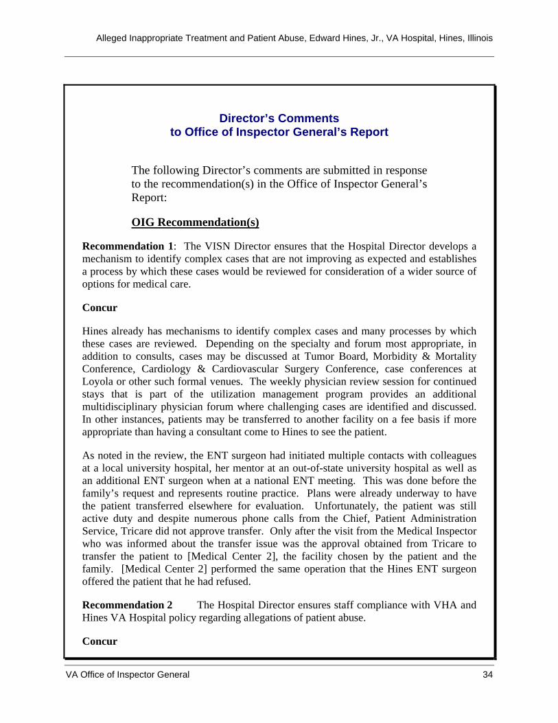
Alleged Inappropriate Treatment and Patient Abuse, Edward Hines, Jr., VA Hospital, Hines, Illinois
VA Office of Inspector General 34
Director’s Comments to Office of Inspector General’s Report
The following Director’s comments are submitted in response to the recommendation(s) in the Office of Inspector General’s Report:
OIG Recommendation(s)
Recommendation 1: The VISN Director ensures that the Hospital Director develops a mechanism to identify complex cases that are not improving as expected and establishes a process by which these cases would be reviewed for consideration of a wider source of options for medical care.
Concur
Hines already has mechanisms to identify complex cases and many processes by which these cases are reviewed. Depending on the specialty and forum most appropriate, in addition to consults, cases may be discussed at Tumor Board, Morbidity & Mortality Conference, Cardiology & Cardiovascular Surgery Conference, case conferences at Loyola or other such formal venues. The weekly physician review session for continued stays that is part of the utilization management program provides an additional multidisciplinary physician forum where challenging cases are identified and discussed. In other instances, patients may be transferred to another facility on a fee basis if more appropriate than having a consultant come to Hines to see the patient.
As noted in the review, the ENT surgeon had initiated multiple contacts with colleagues at a local university hospital, her mentor at an out-of-state university hospital as well as an additional ENT surgeon when at a national ENT meeting. This was done before the family’s request and represents routine practice. Plans were already underway to have the patient transferred elsewhere for evaluation. Unfortunately, the patient was still active duty and despite numerous phone calls from the Chief, Patient Administration Service, Tricare did not approve transfer. Only after the visit from the Medical Inspector who was informed about the transfer issue was the approval obtained from Tricare to transfer the patient to [Medical Center 2], the facility chosen by the patient and the family. [Medical Center 2] performed the same operation that the Hines ENT surgeon offered the patient that he had refused.
Recommendation 2 The Hospital Director ensures staff compliance with VHA and Hines VA Hospital policy regarding allegations of patient abuse.
Concur
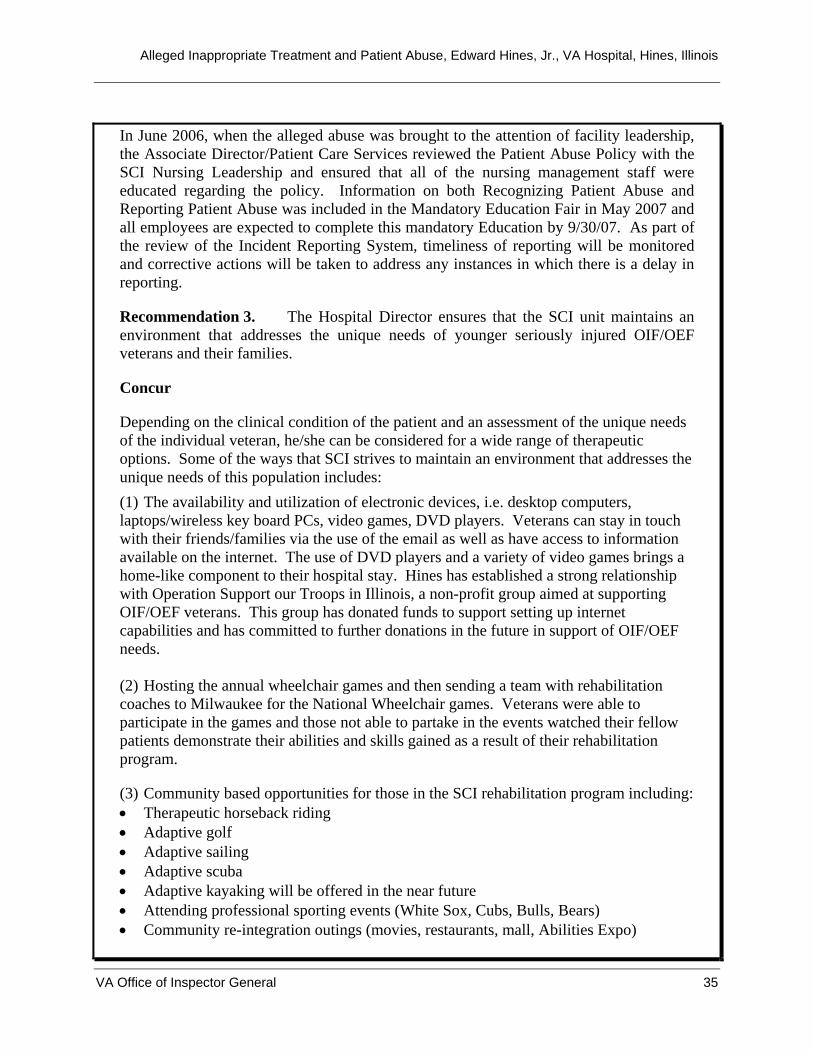
Alleged Inappropriate Treatment and Patient Abuse, Edward Hines, Jr., VA Hospital, Hines, Illinois
VA Office of Inspector General 35
In June 2006, when the alleged abuse was brought to the attention of facility leadership, the Associate Director/Patient Care Services reviewed the Patient Abuse Policy with the SCI Nursing Leadership and ensured that all of the nursing management staff were educated regarding the policy. Information on both Recognizing Patient Abuse and Reporting Patient Abuse was included in the Mandatory Education Fair in May 2007 and all employees are expected to complete this mandatory Education by 9/30/07. As part of the review of the Incident Reporting System, timeliness of reporting will be monitored and corrective actions will be taken to address any instances in which there is a delay in reporting.
Recommendation 3. The Hospital Director ensures that the SCI unit maintains an environment that addresses the unique needs of younger seriously injured OIF/OEF veterans and their families.
Concur
Depending on the clinical condition of the patient and an assessment of the unique needs of the individual veteran, he/she can be considered for a wide range of therapeutic options. Some of the ways that SCI strives to maintain an environment that addresses the unique needs of this population includes: (1) The availability and utilization of electronic devices, i.e. desktop computers, laptops/wireless key board PCs, video games, DVD players. Veterans can stay in touch with their friends/families via the use of the email as well as have access to information available on the internet. The use of DVD players and a variety of video games brings a home-like component to their hospital stay. Hines has established a strong relationship with Operation Support our Troops in Illinois, a non-profit group aimed at supporting OIF/OEF veterans. This group has donated funds to support setting up internet capabilities and has committed to further donations in the future in support of OIF/OEF needs. (2) Hosting the annual wheelchair games and then sending a team with rehabilitation coaches to Milwaukee for the National Wheelchair games. Veterans were able to participate in the games and those not able to partake in the events watched their fellow patients demonstrate their abilities and skills gained as a result of their rehabilitation program. (3) Community based opportunities for those in the SCI rehabilitation program including: • Therapeutic horseback riding • Adaptive golf • Adaptive sailing • Adaptive scuba • Adaptive kayaking will be offered in the near future • Attending professional sporting events (White Sox, Cubs, Bulls, Bears) • Community re-integration outings (movies, restaurants, mall, Abilities Expo)

Alleged Inappropriate Treatment and Patient Abuse, Edward Hines, Jr., VA Hospital, Hines, Illinois
VA Office of Inspector General 36
(4) Availability of an educational therapy program where patients are seen bedside or clinic for assessment/therapy depending on their current status. An individualized program is then designed for veterans based on their individual needs. Educational therapists identify, provide and train patients on adaptive equipment and specialized computer software including Dragon Dictate is available and designed especially for each veteran’s limitations. (5) Hines has conducted focus group interviews with younger, Polytrauma veterans in order to solicit feedback on how to best meet their unique needs. Polytrauma staff also held a support group/dinner focused on the family members of these younger veterans. Both of these initiatives resulted in opening new lines of communication between the Hines staff and this specific population of veterans. Additional focus group interviews are scheduled on an ongoing basis. (6)Hines has established a special OIF/OEF Intake Center. The OIF/OEF Program Manager, as well as two newly-hired Seamless Transition Coordinators are in the Intake Center full-time. The Seamless Transition Coordinators are dedicated case managers specifically for OIF/OEF veterans. (7))Planning for the opening of a Fisher House on the grounds which will provide housing support for families of OIF/OEF veterans receiving treatment at Hines VA Hospital (8)In recognition of younger patients and their connection to the internet, in the very near future Hines will be offering CAREPAGES to our patients. This service will allow patients to establish a website in which to post updates to their conditions and a place for their friends and families to email them and receive regular updates.

Alleged Inappropriate Treatment and Patient Abuse, Edward Hines, Jr., VA Hospital, Hines, Illinois
Appendix C
VA Office of Inspector General 37
OIG Contact and Staff Acknowledgments
OIG Contact Verena Briley-Hudson, MN, RN
Director, Chicago Office of Healthcare Inspections (708) 202-2672
Acknowledgments Wachita Haywood, RN, Associate Director Gregg Hirsteen Michael Shepherd, MD
Alleged Inappropriate Treatment and Patient Abuse, Edward Hines, Jr., VA Hospital, Hines, Illinois
Appendix D
VA Office of Inspector General 38
Report Distribution VA Distribution Office of the Secretary Veterans Health Administration Assistant Secretaries General Counsel Director, Veterans Integrated Service Network (10N12) Director, Edward Hines, Jr. VA Hospital (578/00) Non-VA Distribution House Committee on Veterans’ Affairs House Appropriations Subcommittee on Military Construction, Veterans Affairs, and Related Agencies House Committee on Oversight and Government Reform Senate Committee on Veterans’ Affairs Senate Appropriations Subcommittee on Military Construction, Veterans Affairs, and Related Agencies Senate Committee on Homeland Security and Governmental Affairs National Veterans Service Organizations Government Accountability Office Office of Management and Budget U.S. Senate: Richard Durbin and Barack Obama U.S. House of Representatives: Ike Skelton and Danny K. Davis
This report is available at http://www.va.gov/oig/publications/reports-list.asp.