dental microbiology #211 immunology 2006 lecture 4 the antibodies and the complement system
TRANSCRIPT

Dental Microbiology #211IMMUNOLOGY2006 Lecture 4
The Antibodies and the Complement System
Antibodies are also called Immunoglobulins

Topics
• The structure of Immunoglobulins
• Classes of Immunoglobulins
• Biological properties of Immunoglobulins
• The Complement system and its functions

The Antibodies
Antibodies are synthesized by B lymphocytes in 2 forms: soluble and cell bound. The cell-bound form is the BCR. The soluble form is released into the circulation as antibodies
Each B cell produces Ab of a single specificity and expresses on the cell surface only one BCR specificity
The specificity of the Ab produced by a B cell is the same as of its BCR

The Antibodies Fig 1

Antibodies are also called Immunoglobulins
Immunoglobulins (Ig) are divided into 5 classes:IgGIgMIgAIgDIgE
Ig are glycoproteinsThey differ in size, amount of CHO and biologic functions
Ig structure Fig 2
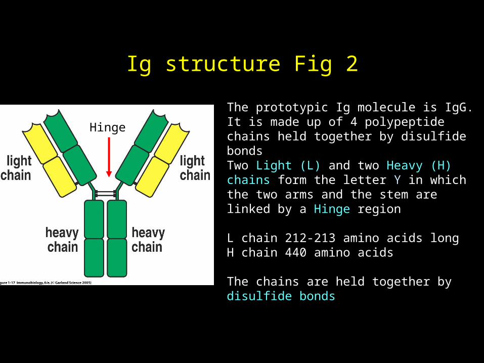
The prototypic Ig molecule is IgG.It is made up of 4 polypeptide chains held together by disulfide bondsTwo Light (L) and two Heavy (H) chains form the letter Y in which the two arms and the stem are linked by a Hinge region
L chain 212-213 amino acids longH chain 440 amino acids
The chains are held together by disulfide bonds
S-SHinge

Variable and Constant regions Fig 3
VL
CL
CH
CH
The first 110 amino acids in each chain are variable (V regions) blue and purpleThe remaining 110 aa in the L chain and the remaining 330 aa in the H chain are constant (C regions) (green and yellow) The V regions contact the antigen. The C regions are involved in biological functions
V V
C
CC
C

Ig Domains
Each segment of about 110 amino acids is tightly packed and forms a domain.
Each Ab molecule has therefore 4 V domains (one in each H and one in each L chain), one C domain in each L chain and either 3 or 4 C domains in each H chain. Each IgG H chain has 3 C domains but IgM and IgE H chains each has 4 C domains.

Ig Fragments Fig 4
The IgG molecule can be dissected by proteolytic enzymes:Papain 2 Fab and 1 Fc fragment.
Pepsin 1 large F(ab)2 and several small fragments from the Fc segment

The function of the Ig segments
The distinct Ig segments are involved in different functions:
The Fab binds AgThe Fc is involved in:Transplacental passage of AbEnhanced phagocytosis by macrophages and PMN (opsonization)Activation of Complement:Kills foreign cells, triggers inflammatory reactions. Enhances phagocytosis

Ag-binding function Fig 5
Antibodies bind Ag in pockets or grooves defined by the V regions of the H and L chains.

Ig Classes Structure Fig 6

Ig classes and their functions
All Ig classes bind Ag in the same way via their Variable regions
Distinct classes have different Fc segments and thus perform distinct biologic functions:IgG:•Neutralizes toxins and prevents viral entry into cells•Crosses the placenta from mother to fetus. •Activates the Complement cascade. •Enhances phagocytosis (opsonization)

IgM: Neutralizes toxins and virusesActivates the Complement cascadeIgA: Crosses secretory epithelia. Neutralizes toxins and virusesClears pathogens in secretions: saliva, nasal, bronchial secretions, seminal fluid, etc.IgE:Releases pharmacologic mediators from basophils and mast cells and triggers allergic reactions (Hay fever) IgD: Function unknown

Polymeric Ig (Fig. 7)
In order to form the polymers, both IgM and IgA have an additional polypeptide chain, called the J chain, that helps to hold the subunits together
All immunoglobulins are constructed from a basic unit of two H and two L chains, but IgM and IgA form polymers

sIgA Fig 8

The Complement System
The complement system (Abbr. C): • A group of proteins present in the plasma of
all individuals. Part of the innate immune system.• A major biological effector system of both
the innate and the adaptive immune responses. • The C components are present in the plasma
in an inactive state. • Upon activation most C components become proteolytic enzymes and act in sequence to
cleave the next C component into active fragments.

There are 9 complement proteins (C1 to C9)
The C facilitates and amplifies inflammatory responses in several ways:
1. Increases vascular permeability
2. Destroys cell membranes of pathogens and thus induce cell death through lysis
3. Enhances phagocytosis (opsonization).

The three C activation pathways Fig 9
Adaptive Innate Innate

In the adaptive immune response, the classical C pathway becomes activated when either IgM or IgG Ab binds to specific Ag.
The binding of Ab to Ag exposes a site on the Fc segment of the Ig H chain which in turn binds to and triggers the activation of the first C component (C1).

Activation of the C cascade Fig 10

Activation of the C cascade. Fig 11

The Membrane Attack Complex
The activation of the first C component called C1 starts the sequential enzymatic activity that becomes progressively amplified (C cascade).
Each component splits the next one into a large and a small fragment.
The large fragments become attached to the target cell membrane and end up by forming the Membrane Attack Complex (MAC).
MAC Fig 12
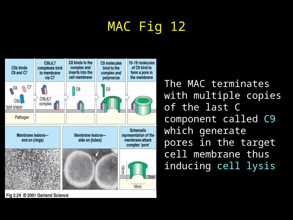
The MAC terminates with multiple copies of the last C component called C9 which generate pores in the target cell membrane thus inducing cell lysis

Role of the small C fragments Fig 13
The small fragments called anaphylatoxins induce powerful inflammatory reactions, by increasing vascular permeability and thus allowing passage of inflammatory cells from the blood vessels into the surrounding tissue (diapedesis).

END